Publisher’s version / Version de l'éditeur:
Journal of Thermal Spray Technology, 15, 4, pp. 628-633, 2006-12-01
READ THESE TERMS AND CONDITIONS CAREFULLY BEFORE USING THIS WEBSITE. https://nrc-publications.canada.ca/eng/copyright
Vous avez des questions? Nous pouvons vous aider. Pour communiquer directement avec un auteur, consultez la première page de la revue dans laquelle son article a été publié afin de trouver ses coordonnées. Si vous n’arrivez pas à les repérer, communiquez avec nous à PublicationsArchive-ArchivesPublications@nrc-cnrc.gc.ca.
Questions? Contact the NRC Publications Archive team at
PublicationsArchive-ArchivesPublications@nrc-cnrc.gc.ca. If you wish to email the authors directly, please see the first page of the publication for their contact information.
NRC Publications Archive
Archives des publications du CNRC
This publication could be one of several versions: author’s original, accepted manuscript or the publisher’s version. / La version de cette publication peut être l’une des suivantes : la version prépublication de l’auteur, la version acceptée du manuscrit ou la version de l’éditeur.
For the publisher’s version, please access the DOI link below./ Pour consulter la version de l’éditeur, utilisez le lien DOI ci-dessous.
https://doi.org/10.1361/105996306X146893
Access and use of this website and the material on it are subject to the Terms and Conditions set forth at
Development of osteoblast colonies on new bioactive coatings
Legoux, J. G.; Chellat, F.; Lima, R. S.; Marple, B. R.; Bureau, M. N.; Shen,
H.; Candeliere, G. A.
https://publications-cnrc.canada.ca/fra/droits
L’accès à ce site Web et l’utilisation de son contenu sont assujettis aux conditions présentées dans le site LISEZ CES CONDITIONS ATTENTIVEMENT AVANT D’UTILISER CE SITE WEB.
NRC Publications Record / Notice d'Archives des publications de CNRC:
https://nrc-publications.canada.ca/eng/view/object/?id=a3d6efcb-bbd8-449e-a417-0314eb8c0543 https://publications-cnrc.canada.ca/fra/voir/objet/?id=a3d6efcb-bbd8-449e-a417-0314eb8c0543Development of Osteoblast
Colonies on New Bioactive Coatings
J.-G. Legoux, F. Chellat, R.S. Lima, B.R. Marple, M.N. Bureau, H. Shen, and G.A. Candeliere(Submitted February 21, 2006; in revised form April 28, 2006)
The aging baby boomer population coupled with an increase in life expectancy is leading to a rising number of active elderly persons in occidental countries. As a result, the orthopedic implant industry is facing nu-merous challenges such as the need to extend implant life, reduce the incidence of revision surgery, and improve implant performance. This paper reports results of an investigation on the bioperformance of newly developed coating-substrate systems. Hydroxyapatite (HA) and nano-titania (nano-TiO2) coatings were pro-duced on Ti-6Al-4V and fiber reinforced polymer composite substrates. In vitro studies were conducted to determine the capacity of bioactive coatings developed to sustain osteoblast cells (fetal rat calvaria) adher-ence, growth, and differentiation.
As revealed by scanning electron microscopy (SEM) observations and alkaline phosphatase activity, cell adhesion and proliferation demonstrated that HA coatings over a polymer composite are at least as good as HA coatings made over Ti-6Al-4V substrate in terms of osteoblast cell activity. Nano-TiO2coatings produced by high-velocity oxyfuel (HVOF) spraying led to different results. For short-term cell culture (4.5 and 24 h), the osteoblasts appeared more flattened when grown on nano-TiO2than on HA. The surface cell coverage after seven days of incubation was also more complete on nano-TiO2than HA. Preliminary results indicate that osteoblast activity after 15 days of incubation on nano-TiO2is equivalent to or greater than that observed on HA.
Keywords bioactive coatings, hydroxyapatite, nano titania, os-teoblast adhesion, polymer composite substrate, Ti-6Al-4V substrate
1. Introduction
One of the largest successes of modern medicine is the total hip replacement surgery. Presently, this procedure has one of the highest success rates among surgical interventions, only second to the appendix removal procedure. However, the lifetime of the prosthesis itself is still limited to 10 to 20 years, which means that for numerous patients a revision surgery will become man-datory. This need for replacement arises for a number of reasons including aseptic loosening of the prosthesis. This can be caused by the formation of wear particles at articular joints and by the difference in stiffness between the bone and the metallic pros-thesis, leading to the phenomena called stress shielding and im-plant migration. To overcome this problem, two solutions can be envisioned. The first implies design of more biomimetic pros-theses, with bone-matching stiffness. A novel design based on polymer composite materials of total hip replacement prosthesis is under development (Ref 1). One of the key characteristics of
this biomimetic prosthesis is its hydroxyapatite (HA) coating, which permits osseointegration (integration with the bone). Thermally sprayed HA coatings are already used successfully for metallic implants (Ref 2), but such coatings have yet to be developed for polymer composites due to quite challenging heat management and adhesion concerns. The second approach is re-lated to the development of new coating systems. Regarding this aspect, nanostructured titania (TiO2) coatings have
demon-strated interesting potential. The goal of this paper is to evaluate both coating systems with regard to their response to osteoblast (fetal rat calvaria) cells. Other properties for the nano-TiO2
coat-ing and its optimization are described in more detail in a com-panion paper (Ref 3).
2. Materials and Methods
2.1 Substrate Materials
Coatings were produced on two types of substrates, a tita-nium alloy (Ti-6Al-4V) that is widely used for hip prostheses and a polyamide 12/carbon fiber (PA12/CF) composite used for a novel design of hip prostheses (patent pending). To improve the coating adhesion and heat resistance, a 100 µm layer made of twin-screw-extruder compounded PA12/HA was overmolded onto composite substrates (patent pending).
2.2 Bioactive Coatings
Two types of coatings over two different substrates were pro-duced: a plasma sprayed HA coating and a high-velocity oxyfuel (HVOF) nano-TiO2coating on both polymer composite and
Ti-based substrates.
2.2.1 HA Coating. A bioactive HA powder (Captal 30,
Plasma Biotal Ltd, Tideswell, UK) was used for depositing HA
This article was originally published in Building on 100 Years of
Suc-cess, Proceedings of the 2006 International Thermal Spray Conference
(Seattle, WA), May 15-18, 2006, B.R. Marple, M.M. Hyland, Y.-Ch. Lau, R.S. Lima, and J. Voyer, Ed., ASM International, Materials Park, OH, 2006.
J.-G. Legoux, F. Chellat, R.S. Lima, B.R. Marple, and M.N. Bureau,
Industrial Materials Institute, National Research Council Canada, Boucherville, QC, Canada; and H. Shen and G.A. Candeliere, Institute for Biological Sciences, National Research Council Canada, Ottawa, ON, Canada. Contact e-mail: Jean-Gabriel.Legoux@cnrc-nrc.gc.ca. JTTEE5 15:628-633
DOI: 10.1361/105996306X146893 1059-9630/$19.00 © ASM International
Peer
coatings. Granulometry testing on the initial HA powder (LS Particle Size Analyzer, Beckham Coulter, Fullerton, CA) indi-cated a number-average diameter (sum of particle diameters/ number of particles) of 33 µm. The HA coatings were produced using atmospheric plasma spray.
2.2.2 Nano-TiO2 Coating. Titania feedstock used in this
work (VHP-DCS, Altair Nanomaterials Inc., Reno, NV) exhib-ited a nominal particle size range from 5 to 20 µm. Each feed-stock particle was formed via the agglomeration of individual nanostructured TiO2particles smaller than 100 nm. The
feed-stock powder was thermally sprayed via the HVOF technique using an oxypropylene-based torch (Diamond Jet 2700-hybrid, Sulzer Metco, Westbury, NY).
The coatings were sprayed on grit-blasted substrates to roughen the surface prior to spraying. During the spraying pro-cess on the substrates a cooling system (air jets) was applied to reduce the coating temperature, which was monitored using a pyrometer. The maximum surface temperature was approxi-mately 240 °C for the Ti-6Al-4V substrates and 130 °C for the PA12/CF substrates. Detailed information about coating depo-sition, microstructural characterization, and mechanical prop-erty evaluation of this biomedical coating can be found in a com-panion paper contained in this journal (Ref 3).
2.3 Osteoblast Isolation and Seeding
Osteoblasts were isolated from the calvariae of 21-day-old Spargue Dawley rat fetuses by sequential collagenase digestion as described by Bellows et al. (Ref 4). The cells were then plated in T-75 flasks in a Dulbecco’s modified Eagle medium contain-ing 10% of fetal bovine serum. After 24 h, the adhered cells were washed with phosphate buffer saline to remove dead cells and other debris, then detached using 0.01% trypsin in phosphate buffer saline. The resuspended cells were counted and seeded on the different disc-shaped material surfaces previously placed in the six-well culture plates at 2 × 10⁴cells/well in an osteogenic
medium (growth medium containing 50 mg/mL of ascorbic acid, 10 mM Na-b-glycerophosphate, and 1% antibiotics). The cells were incubated at 37 °C in a humidified atmosphere con-sisting of 95% O2and 5% CO2and allowed to grow for 4.5 h, 1,
7, and 15 days. For these periods, the medium was changed three times per week.
2.4 Osteoblast Adherence, Growth, and Differentiation on Materials
Scanning electron microscopy (SEM) observation was used to determine the adherence, morphology, and growth of the os-teoblasts on the different coatings after 4.5 h, 1, 7, and 15 days. At the end of each incubation period, the cells were rinsed in phosphate buffer pH 7.2, fixed in a 0.089 M phosphate buffer solution containing 2.5% glutaraldehyde and 2.5 mM magne-sium chloride, pH 7.2 for 3 h. The samples were then rinsed in 0.1 M phosphate buffer, postfixed in 1% osmium tetroxide for 1 h, washed in distilled water three times, and then dehydrated in a graded series of ethanol solutions (70 through 100% dry etha-nol). Specimens were then treated with mixtures consisting of 75:25, 50:50, 25:75, and 0:100 ethanol:amyl acetate. The samples were dried by the critical-point drying method, sputter-coated by gold/palladium and observed using a scanning
elec-tron microscope (Hitachi, Model S-4700, manufacturer, Hitachi Science Systems, Ibaraki, Japan).
2.5 Alkaline Phosphatase Activity
The osteoblast phenotype of cells cultured on different sur-faces was determined by enzymatic alkaline phosphatase activ-ity test after 15 days. Before staining, coated samples (with at-tached cells) were rinsed once with cold PBS, then the cells were fixed in 10% cold neutral buffered formalin for 15 min, rinsed with distilled water, and then left in distilled water for 15 min. A fresh mixture constituted of 10 mg Naphthol AS MX-PO4 in 400 µL N,N-dimethylformamide, 50 mL distilled water, 50 mL of 0.2 M Tris-HCl pH 8.3, 60 mg red violet LB salt was used for alkaline phosphatase staining. The alkaline phosphatase staining mixture was placed on the coated samples covered with the fixed cells and incubated for 1 h at room temperature. All the chemi-cals were purchased from Sigma-Aldrich Chemical Company (Oakville, Canada). The different material discs were then re-moved from the wells, rinsed in tap water, drained and air dried, and then photographed. The alkaline phosphatase positive signal was quantified with Imagine J software. For normalization, the background color was subtracted by setting a threshold.
3. Results and Discussion
3.1 Coating Surfaces Characterization by SEM Prior to cell culture experiments, HA and nano-TiO2coatings
are quite different, as shown in Fig. 1. Both coating surfaces are constituted of smooth zones formed from the solidification of a liquid and rougher areas constituted either of unmelted or “over-sprayed” material. The HA coating is considerably rougher than the nano-TiO2 as can be expected when comparing a HVOF
coating made with fast and small particles with plasma sprayed coatings made from relatively large and slow particles. 3.2 Cell Adhesion and Growth
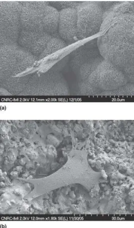
After 4.5 h of incubation the coating surfaces were examined by SEM. Typical cell morphology is presented in Fig. 2 for cells attached to HA and nano-TiO2coatings. It can be noted that the
two coating topologies are quite different, the plasma sprayed HA coating being rougher than the HVOF nano-TiO2that
ex-hibits a smoother aspect caused by flattening of semimolten par-ticles impinging on the surface at high velocity. Osteoblasts found on the nano-TiO2(Fig. 2b) are well flattened on the
sur-face and have started to spread, whereas osteoblast cells do not appear to follow the contour of the HA coating surface (Fig. 2a). Similar elongated osteoblast cells not in close contact with the HA surface have been reported (Ref 5). Also, osteoblast cells were more difficult to locate on the HA surface, which might be related to a slower initial adhesion (Ref 6), but also to the diffi-culty of locating cells on a rougher surface (e.g., cells at bottom of valleys). It should also be noted that the HA coating surface was modified during its immersion in the culture media. Similar phenomena of dissolution and reprecipitation of apatite on coat-ing surfaces have been reported (Ref 7, 8).
Images of cells after one day of incubation are presented in Fig. 3. As observed previously for 4.5 h, cell morphology on HA
Peer
and nano-TiO2coatings is quite different. Cells remained with
an elongated shape on the HA coating; they had a close to cir-cular shape on nano-TiO2. Interestingly, osteoblast cells
at-tached to the HA surface appeared partly covered by mineral concretion, probably some HA precipitated from the culture me-dia. Indeed, it has been widely demonstrated that while im-mersed in body fluid, plasma sprayed HA coatings undergo sev-eral modifications, with some phases dissolved while a layer is formed on the surface (Ref 7). It is possible in this case that the precipitation process took place as well on the cell surface. With time, cells become incorporated into the reprecipitated HA matrix. After 7 days, cells spread to the complete substrate surfaces (Fig. 4). However, while they remain elongated and penetrate the HA coating structure (Fig. 4a), they cover the entire surface on TiO2coatings (Fig. 4b). Also apparent from Fig. 4(a) is the
smoother HA surface after 7 days of incubation in culture media when compared with the shorter time (Fig. 2a and 3a).
After 15 days, osteoblast cells are also covering the entire surface of HA coatings (Fig. 5). It has been reported that solu-bilization of calcium phosphate phases might decrease cell ad-hesion early after the initial cell exposition, but this was com-pensated by a noticeable acceleration later in the process (Ref 9). 3.3 Alkaline Phosphatase Activity and
Cell Differentiation
After 15 days of incubation the number of cells was charac-terized by the staining of the alkaline phosphatase, which
pro-duce a red color. Figure 6 presents the substrate after staining. All surfaces were almost completely covered by red stained cells. Osteoblast morphology remains more elongated on the HA coatings (Fig. 5) compared with that developed on nano-TiO2coatings. It is difficult to evaluate the difference between
white HA coatings and dark gray nano-TiO2coatings mainly
due to the difference in contrast. To quantify the coating re-sponse, color image analysis was performed on samples after proper normalization to take into account the divergence in con-trast caused by the substrate color. Results are presented by Fig. 7. The nano-TiO2 coating exhibits the highest intensity,
fol-lowed closely by HA coating on the polymer composite sub-strate, and finally HA coatings over Ti-6Al-4V substrate. These results indicate that even though no morphological difference was seen between the different substrates, the cell activity as defined by the alkaline phosphatase staining shows a difference. The reason for this difference is not explained at this point. How-ever, it should be highlighted that the staining for alkaline phos-phatase activity, as performed in this study, does not allow for a precise quantification. Also the large variation in the data espe-cially for the HA coating over the Ti-6Al-4V substrate would justify further study. The quantification of alkaline phosphatase activity is very important especially for the rougher HA coating where only the stained surface is visible, whereas the cells em-bedded in the valleys or in the reprecipitated apatite are not shown. A more detailed and a quantifiable means of measure-ment will be required to investigate quantitatively the alkaline phosphatase activity in the future.
Fig. 1 Typical surface morphology for (a) HA coatings and (b) nano-TiO2coatings
Fig. 2 Cell attachment after 4.5 h: (a) HA on PA12/CF substrate; (b) nano-TiO2on PA12C substrate
Peer
The interpretation of the observed differences of the osteo-blast initial adhesion and proliferation for the two coatings can be quite complex. Differences in coating chemistry may play a role; it has been shown that for titanium alloys, chemistry (i.e., pure Ti, Ti-6Al-4V, TiNb13Zr13, TiNb30) (Ref 10) induces
dif-ferent responses. Also titanium oxide formed by laser heating might have a different effect than native oxide. However, other studies show no effect of the surface chemistry (Ref 6, 11). An-other important factor linked to the cell adhesion is the wettabil-ity of the surface, which can have either a direct effect on adhe-sion by promoting the cell contact with the surface or an indirect one by promoting the protein unfolding at the surface (Ref 12-14). The effect of protein type on cell adhesion is certainly very important. For example, it has been shown that vitronectin, fi-bronectin, or osteopontine have an important effect on cell ad-hesion (Ref 10, 15). Transmembranous integrins might play a role in the signal transduction from the environmental milieu up to the cell nucleus leading to an appropriate cell response such as proliferation rates or morphology. This signal seems to be the consequence of the interaction of various molecules and growth factors (Ref 16). Regarding the effect of surface roughness, it has been shown that osteogenicity was enhanced by increasing surface roughness (Ref 17). However, it was shown that for samples with the highest roughness, cells were less adhesive, which was attributed to an effect of confinement of cells at the bottom of deep holes, leading to early decease and detachment. This might be related only to in vitro experiments. Other studies have related this apparent negative effect of high surface
rough-ness mainly to the fact that at the cell level the surface appears to be flat (Ref 18).
It is beyond the scope of this paper to determine what exact biomechanisms predominate in this study. Aside from their chemistry, three major differences can be identified between the HA and nano-TiO2coatings studied here:
• Surface roughness: the HA coatings are considerably
rougher than the nano-TiO2coatings.
• Crystallite size: this peculiar microstructure of the
nano-TiO2coatings contains well dispersed “nano-zones.”
Fig. 3 Osteoblast morphology after 1 day: (a) HA on Ti-6Al-4V; (b) nano-TiO2on Ti-6Al-4V
Fig. 4 Osteoblast surface after 7 days: (a) HA on PA12C; (b) nano-TiO2on PA12C
Fig. 5 Surface of the HA coating on PA12C substrate after 15 days of incubation
Peer
• Chemical stability: higher stability of the nano-TiO2
coat-ings compared with the dissolution and reprecipitation be-havior of the HA coating.
The early stage of cell adhesion was characterized by direct observation of the coating surfaces, which can be more difficult on rough surfaces. The presence of nanostructured surfaces on the nano-TiO2coatings may help the unfolding of adhesion
pro-teins, which results in very good growth and alkaline phospha-tase activity values. At the same time, as cell adhesion involves surface adsorption phenomena, it is reasonable to think that these phenomena would be slower or even inhibited on a surface that is undergoing major transformation (both dissolving and re-forming itself). This may explain why after a longer exposure time (7 days), when the surface became more stable, cell prolif-eration accelerated, reaching both a high degree of covering and of alkaline phosphatase activity.
4. Conclusions
The current study demonstrates that nano-TiO2as well as HA
coatings support the attachment, growth, and expression of the osteoblastic phenotype of the cells as assessed by the alkaline phosphatase activity assay.
Results on cell adhesion and proliferation have demonstrated that hydroxyapatite coatings on a polymer composite are at least as good as HA coatings deposited on a Ti-6Al-4V substrate, in terms of osteoblast cell activity.
Nano-TiO2coatings produced by the HVOF technique
be-have differently when compared with HA coatings. Preliminary osteoblast cell culture revealed that the activity of the cells after 15 days of incubation is equivalent or superior to that observed on hydroxyapatite coatings.
The intrinsic structural and chemical differences of each type of coating led to some important differences in the attachment behavior of the cells and their morphological appearance (elon-gated versus flattened). Further planned studies may allow relat-ing the effect of the coatrelat-ing structure and composition on the rate of osteoblast adherence and growth, as well as the differentiation stage as a function of time.
References
1. M. Campbell, H.A. Bougherara, M.N. Bureau, J. Denault, and L’H. Ya-hia, Biomimetic Polymer Composites for Orthopedic Implants, ac-cepted for publication in the Proc. Materials and Processes for Medical
Devices 2005, Nov 14-16, 2005 (Boston, MA), ASM International
2. R. Vedantam and C. Ruddlesdin, The Fully Hydroxyapatite-Coated To-tal Hip Implant, J. Arthroplasty, 1996, 11(5), p 534-542
3. R.S. Lima, H. Li, K.A. Khor, and B.R. Marple, Biocompatible Nano-structured High Velocity Oxy-Fuel Sprayed Titania Coating: Deposi-tion, Characterization and Mechanical Properties, J. Therm. Spray
Technol., 2006, 15(4), p 623-627
4. C.G. Bellows, J.E. Aubin, J.N. Heersche, and M.E. Antosz, Mineralized Bone Nodules Formed in vitro from Enzymatically Released Rat Cal-varia Cell Populations, Calcif. Tissue Int., 1986, 38, p 143-154 5. L.C. Baxter, V. Frauchiger, M. Textor, and I.Ap Gwynn, and R.G.
Ri-chards, Fibroblast and Osteoblast Adhesion and Morphology on Cal-cium Phosphate Surfaces, Eur. Cells Mater., 2002, l4, p 1-17 6. D.D. Deligianni, D.N. Katsala, P.G. Koutsoukos, and Y.F. Missirlis,
Effect of Surface Roughness of Hydroxyapatite on Human Bone
Mar-Fig. 6 Alkaline phosphatase stained sample surfaces after 15 days of growth. (a) HA coating on PA12C. (b) HA coating on Ti-6Al-4V. (c) Nano-TiO2coating on PA12C (only two replicate coatings tested). (d) Nano-TiO2coating on Ti-6Al-4V
Fig. 7 Intensity of the alkaline phosphatase staining for different coat-ings
Peer
row Cell Adhesion, Proliferation, Differentiation and Detachment Strength, Biomaterials, 2001, 22, p 87-96
7. C. Auclair-Daigle, M.N. Bureau, J.-G. Legoux, and L’H. Yahia, Bioac-tive Hydroxyapatite Coatings on Polymer Composites for Orthopedic Implants, J. Biomed. Mater. Res. A, 2005, 73, p 398-408
8. A. John, H.K. Varma, and T.V. Kumari, Surface Reactivity of Calcium Phosphate Based Ceramics in a Cell Culture System, J. Biomater. Appl., 2003, 18, p 63-78
9. K. Ogata, S. Imazato, A. Ehara, S. Ebisu, Y. Kinomot, T. Nakano, and Y. Umakoshi, Comparison of Osteoblast Responses to Hydroxyapatite and Hydroxyapatite/Soluble Calcium Phosphate Composites, J.
Biomed. Mater. Res. A, 2005, 72, p 127-135
10. T.K. Monsees, K. Barth, S. Tippelt, K. Heidel, A. Gorbunov, W. Pompe, and R.H.W. Funk, Effects of Different Titanium Alloys and Nanosize Surface Patterning on Adhesion, Differentiation, and Orientation of Os-teoblast-Like Cells, Cell. Tis. Org., 2005, 180(2), p 81-95
11. T.J. Webster, R.W. Siegel, and R. Bizios, Osteoblast Adhesion on Nanophase Ceramics, Biomaterials, 1999, 20, p 1221-1227
12. X. Zhu, J. Chen, L. Scheideler, R. Reichl, and J. Geis-Gerstorfer, Effects of Topography and Composition of Titanium Surface Oxides on Osteo-blast Responses, Biomaterials, 2004, 25, p 4087-4103
13. M. Karlsson, E.P. Palsgard, P.R. Wilshaw, and L. Di Silvio, Initial in
vitro Interaction of Osteoblasts with Nano-Porous Alumina,
Biomateri-als, 2003, 24, p 3039-3046
14. L. Hao, J. Lawrence, and K.S. Chian, Osteoblast Cell Adhesion on a Laser Modified Zirconia Based Bioceramic, J. Mater. Sci.: Mater.
Med., 2005, 16, p 719-726
15. S. Spriano, M. Bosetti, M. Bronzoni, E. Vernè, G. Maina, V. Bergo, and M. Cannas, Surface Properties and Cell Response of Low Metal Ion Release Ti-6Al-7Nb Alloy after Multi-Step Chemical and Thermal Treatments, Biomaterials, 2005, 26, p 1219-1229
16. P. Linez-Bataillon, F. Monchau, M. Bigerelle, and H.F. Hildebrand, In vitro MC3T3 Osteoblast Adhesion with Respect to Surface Roughness of Ti6Al4V Substrates, Biomol. Eng., 2002, 19, p 133-141
17. H. Kawahara, Y. Soeda, K. Niwa, M. Takahashi, D. Kawahara, and N. Araki, In vitro Study on Bone Formation and Surface Topography from the Standpoint of Biomechanics, J. Mater. Sci.: Mater. Med., 2004, 15, p 1297-1307
18. V. Borsari, G. Giavaresi, M. Fini, P. Torrielli, A. Salito, R. Chiesa, L. Chiusoli, A. Volpert, L. Rimondini, and R. Giardino, Physical Charac-terization of Different-Roughness Titanium Surfaces, with and without Hydroxyapatite Coating, and Their Effect on Human Osteoblast-Like Cells, J. Biomed. Mater. Res. B Appl. Biomater., 2005, 75(2), p 359-368