HAL Id: hal-02417835
https://hal-amu.archives-ouvertes.fr/hal-02417835
Submitted on 26 Mar 2020
HAL is a multi-disciplinary open access
archive for the deposit and dissemination of
sci-entific research documents, whether they are
pub-lished or not. The documents may come from
teaching and research institutions in France or
abroad, or from public or private research centers.
L’archive ouverte pluridisciplinaire HAL, est
destinée au dépôt et à la diffusion de documents
scientifiques de niveau recherche, publiés ou non,
émanant des établissements d’enseignement et de
recherche français ou étrangers, des laboratoires
publics ou privés.
Bacteria Infections Can Favor Bacterial Invasion and
Transmigration, Dysregulation of the Immune Response
and Cancer Induction in Humans
Christian Devaux, Soraya Mezouar, Jean-Louis Mege
To cite this version:
Christian Devaux, Soraya Mezouar, Jean-Louis Mege.
The E-Cadherin Cleavage Associated to
Pathogenic Bacteria Infections Can Favor Bacterial Invasion and Transmigration, Dysregulation of
the Immune Response and Cancer Induction in Humans. Frontiers in Microbiology, Frontiers Media,
2019, 10, �10.3389/fmicb.2019.02598�. �hal-02417835�
Edited by:
Christoph Hölscher, Research Center Borstel (LG), Germany
Reviewed by:
Manja Boehm, University of Erlangen Nuremberg, Germany Mónica Hebe Vazquez-Levin, National Council for Scientific and Technical Research (CONICET), Argentina *Correspondence: Christian A. Devaux christian.devaux@mediterranee-infection.com Specialty section:
This article was submitted to Microbial Immunology, a section of the journal Frontiers in Microbiology
Received: 19 June 2019 Accepted: 25 October 2019 Published: 08 November 2019 Citation:
Devaux CA, Mezouar S and Mege J-L (2019) The E-Cadherin Cleavage Associated to Pathogenic Bacteria Infections Can Favor Bacterial Invasion and Transmigration, Dysregulation of the Immune Response and Cancer Induction in Humans. Front. Microbiol. 10:2598. doi: 10.3389/fmicb.2019.02598
The E-Cadherin Cleavage Associated
to Pathogenic Bacteria Infections
Can Favor Bacterial Invasion and
Transmigration, Dysregulation of the
Immune Response and Cancer
Induction in Humans
Christian A. Devaux
1,2,3*, Soraya Mezouar
1,3and Jean-Louis Mege
1,3,41 IRD, MEPHI, APHM, Aix-Marseille University, Marseille, France, 2 CNRS, Institute of Biological Science (INSB), Marseille,
France, 3 Institut Hospitalo-Universitaire (IHU)-Mediterranee Infection, Marseille, France, 4 APHM, UF Immunology
Department, Marseille, France
Once bound to the epithelium, pathogenic bacteria have to cross epithelial barriers to invade
their human host. In order to achieve this goal, they have to destroy the adherens junctions
insured by cell adhesion molecules (CAM), such as E-cadherin (E-cad). The invasive bacteria
use more or less sophisticated mechanisms aimed to deregulate CAM genes expression or
to modulate the cell-surface expression of CAM proteins, which are otherwise rigorously
regulated by a molecular crosstalk essential for homeostasis. Apart from the repression of
CAM genes, a drastic decrease in adhesion molecules on human epithelial cells can be obtained
by induction of eukaryotic endoproteases named sheddases or through synthesis of their
own (prokaryotic) sheddases. Cleavage of CAM by sheddases results in the release of soluble
forms of CAM. The overexpression of soluble CAM in body fluids can trigger inflammation
and pro-carcinogenic programming leading to tumor induction and metastasis. In addition,
the reduction of the surface expression of E-cad on epithelia could be accompanied by an
alteration of the anti-bacterial and anti-tumoral immune responses. This immune response
dysfunction is likely to occur through the deregulation of immune cells homing, which is
controlled at the level of E-cad interaction by surface molecules
α
Eintegrin (CD103) and lectin
receptor KLRG1. In this review, we highlight the central role of CAM cell-surface expression
during pathogenic microbial invasion, with a particular focus on bacterial-induced cleavage
of E-cad. We revisit herein the rapidly growing body of evidence indicating that high levels of
soluble E-cad (sE-cad) in patients’ sera could serve as biomarker of bacterial-induced diseases.
Keywords: bacterial invasion, bacteria-inducing cancer, pathophysiology, E-cadherin, sheddases
EPITHELIAL CADHERIN IN CELL-TO-CELL ADHESION AND
CELL ACTIVATION
Cadherins (cad) belong to the superfamily of cell-adhesion molecules (CAMs) (
Takeichi,
1977;
Nollet et al., 2000;
Angst et al., 2001;
Hulpiau and van Roy, 2009
). Characterized by
their adhesion properties mediated through repeated extracellular cad domains (ECs) under
Ca
2+control, cad play a key role in cell-to-cell interactions (
Hyafil et al., 1981
). Several
subtypes of cad are encoded in the human genome (
Oda and
Takeichi, 2011
). These molecules were classified according to
their tissue distribution: for example, the P prefix is used in
P-cad (encoded by CDH3) to define placental cad, N-cad
(encoded by CDH2 and CDH12) for neural cad, VE-cad
(encoded by CDH5) for vascular endothelial cad, and E-cadherin
(E-cad) (encoded by CDH1) for epithelial cad. The CDH1
gene, located on chromosome 16q22.1, comprises 16 exons
and 15 introns (
Berx et al., 1995
), and it is transcribed into
a 4.5Kb pre-mRNA that is spliced to generate the E-cad mRNA.
Transcriptional repression of CDH1 gene is achieved by a
range of transcriptional repressors that bind its promoter,
including members of the SNAIL and ZEB gene families of
zinc-finger transcription factors (
Cano et al., 2000;
Bolós et al.,
2003;
Cadigan and Waterman, 2012
). Repression of CDH1
gene can also be the result of CpG-island hypermethylation
of its promoter, loss of heterozygosis at 16q22.1, and inactivating
mutations (
Berx et al., 1998;
Lombaerts et al., 2006
).
Initially described as liver cell adhesion molecule (L-CAM)
and uvomorulin (
Gallin et al., 1983;
Schuh et al., 1986
), E-cad
is a single-pass type I transmembrane glycoprotein of 120 kDa
that plays a major role in cell polarity, intercellular adhesion,
and tissue integrity (
Ogou et al., 1983;
Niessen et al., 2011;
van Roy, 2014
). It possesses five EC repeats with binding sites
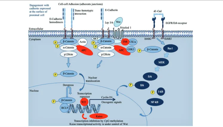
FIGURE 1 | Schematic representation of the E-cadherin (E-cad) interactions and signaling pathway. Newly synthesized E-cad are transported from the Golgi
apparatus to the cell surface where they are available to engagement in intercellular interactions. The model presented reflects evidence that E-cad homodimers are involved in adherens’ junctions. Loss of E-cad expression in epithelia results in loosening of intercellular contacts. E-cad regulates the intracytoplasmic pool of α-cat and β-cat acts as a signal transducer molecule in response to upstream Wnt pathway (Fagotto, 2013). Briefly, the Wnt pathway is initiated by the binding of an extracellular Wnt ligand to a surface receptor composed of Frizzled, a seven transmembrane (7TM) molecule and low-density lipoprotein receptor-related protein 5 or 6 (Lrp5/6). As a result of the Wnt pathway activation that mobilizes several intracytoplasmic molecules (including disheveled, adenomatous polyposis coli – APC binds axin and β-cat and inhibits glycogen synthase kinase 3β – and ΑΚΤ kinase) (Fang et al., 2007;MacDonald et al., 2009;Wu et al., 2009), free cytoplasmic β-cat destruction is inhibited and β-cat translocates to the nucleus. Once in the nucleus, β-cat activates expression of genes such as cyclin D1 or c-MYC, otherwise repressed by the T cell factor/lymphoid enhancer factor (TCF/LEF) (Gumbiner, 1995). To achieve trans-activation, β-cat recruits a range of nuclear co-factors including CBP/p300, Brg-1, and the adaptor protein BCL9 (Hecht et al., 2000;Barker et al., 2001;Kramps et al., 2002;de la Roche et al., 2008). Cad-free p120ctn
can also trigger the nuclear translocation of β-cat through its association with the Vav2 small GTPase and activation of JNK kinase (Wu et al., 2009;Valls et al., 2012). It is worth noting that TCF/LEF-binding sites have been identified in the CDH1 gene promoter, which may permit a feedback control loop where β-cat might activate E-cad mRNA production (Huber et al., 1996). E-cad expression can also be regulated by microRNAs (not shown), such as miR200, which favor E-cad mRNA expression (Gregory et al., 2008). Cells with greater E-cad abundance can sequester and thereby inhibit the ability of β-catenin to translocate to the cell nucleus to derepress the activity of its DNA-binding factor TCF/LEF. When unable to engage in interactions, E-cad enters an endocytic uptake and is directed to early sorting endosomes from which they can either be recycled back to the cell surface by recycling endosomes or routed from late endosomes to proteasome where they undergo degradation (for details see the reviews by (Niessen et al., 2011;Nava et al., 2013;McCrea et al., 2015). During infection with bacteria, the pathogen can either regulate the expression of the CDH1 gene and thereby the cell-surface abundance of E-cad or trigger the catalytic cleavage of E-cad. This can result in the release of a cytosolic pool of β-catenin acting as a downstream effector in the Wnt signaling pathway and induction of oncogenic signals. The sE-cad can associate with intact E-cad present on other cells to alter Cad-dependent cellular behavior. It can also interact with the EGFR molecule to activate the ERK signaling pathway.
for Ca
2+(
Shapiro et al., 1995
). These predominantly homophilic
E-cad dimerize in cis at the cell’s surface and the homodimer
can then interact in trans with an adjacent E-cad homodimer
on a neighboring epithelial cell to form adherens junctions
(
Boggon, 2002
). However, E-cad can also exhibit heterophilic
interactions in trans with the α
Eβ
7integrin, also called CD103
antigen of T-lymphocytes, which generally lacks E-cad cell
surface expression (
Cepek et al., 1994;
Sheridan and Lefrançois,
2011
) as well as it can bind the killer cell lectin receptor G1
(KLRG1) expressed on T-lymphocytes and natural killer (NK)
cells (
Kilshaw, 1999;
Ito et al., 2006
). Over-expression of E-cad
can delay the rate of cell migration (
Hermiston et al., 1996
).
Loss of E-cad can reduce CD103
+T-cell antitumor activity
(
Shields et al., 2019
). Under physiological conditions, E-cad
interacts with p120-ctn and
β-catenin (β-cat) via its
intracytoplasmic tail (
Nagafuchi and Takeichi, 1988;
McCrea
and Gumbiner, 1991;
Kourtidis et al., 2013
). The cytoplasmic
tail of E-cad consists of the juxta membrane domain (JMD),
which allows the clustering of cad and contributes to the
adhesive strength via p120-ctn, and the cat-binding domain
(CBD), which interacts with β-cat and γ-cat (
Kemler, 1993;
Yap et al., 1998
). The α-cat links the bound β-cat and the
actin cytoskeleton. Signaling through E-cad cytoplasmic tail
is a complex process which involves multiple contacts with
intracytoplasmic partners, whose diversity is just beginning to
be elucidated by the characterization of the E-cad interactome
(
Guo et al., 2014
). E-cad is a tumor suppressor acting through
intracytoplasmic retention of β-catenin stocks and suppresses
inflammatory signaling pathways (Figure 1).
E-CADHERIN AND OTHER CADHERIN/
CELL ADHESION MOLECULES USED
AS TARGET RECEPTORS FOR
BACTERIA
Fusobacterium nucleatum, a pathogen associated with oral
plaque formation and colorectal cancers, binds E-cad through
its FadA adhesin (
Rubinstein et al., 2013
). This interaction
up-regulates the Annexin A1 and activates β-cat signaling
(
Zhou et al., 2018;
Rubinstein et al., 2019
). The F. nucleatum
was reported to be associated with a specific epigenetic pattern
of tumor cells characterized by hypermethylation of CpG
islands, high MSI and MLH1 hypermethylation (epigenetic
silencing), and up-regulation of microRNA-21 (
Tahara et al.,
2014;
Yang et al., 2017
). Listeria monocytogenes, the causative
agent of severe food poisoning, which sometimes lead to
meningitis, internalizes when internalin A (InlA) and InlB
bind to E-cad and the hepatocyte growth factor receptor on
the basolateral surface of epithelial cells (
Cossart and Sansonetti,
2004;
Barbuddhe and Chakraborty, 2009;
Ortega et al., 2017
).
Streptococcus pneumoniae can cause pneumonia, meningitis,
and bacteremia. The flamingo cadherin was reported to serve
as receptor for the S. pneumoniae fructose bisphosphate aldolase
(
Blau et al., 2007
), and E-cad was found to act as adherence
receptor for the pneumococcal surface adhesin A (PsaA) of
S. pneumoniae during the colonization of nasopharyngeal
epithelial cells (
Anderton et al., 2007
). Helicobacter pylori, a
bacterium responsible for severe gastric disease, adhere to
target cells through interaction with CEACAM cell-surface
receptor via its HopQ adhesin (
Javaheri et al., 2016
). Then,
the bacterial HtrA sheddase cleaves the gastric epithelial
cell-to-cell junctions through endoproteolysis of E-cad, occludin,
and claudin-8 (
Tegtmeyer et al., 2017
). After transmigration
of H. pylori to the basolateral membrane of gastric epithelial
cells, the T4SS pilus is activated and injects the CagA
cytotoxin into the target cell where the release of β-cat is
stimulated (
Suzuki et al., 2005;
Murata-Kamiya et al., 2007
)
(
Zhao et al., 2018;
Tegtmeyer et al., 2019
).
In the world of bacteria, other cadherin and CAM play a
role during the phase of attachment and invasion (Table 1).
The attachment of Leptospira interrogans to host cells was found
to be mediated through its interaction with VE-cad, which
triggers the process leading to different symptoms, including
liver dysfunction, kidney failure, myocarditis, and sometimes
the pulmonary hemorrhagic manifestations of leptospirosis
(
Evangelista et al., 2014
). Human host colonization by
Haemophilus influenzae began with the binding of the bacteria
to I-CAM1 on the surface of the respiratory tract epithelial
cells through its Type IV pilus (Tfp), a process leading to
respiratory diseases such as cystic fibrosis or chronic obstructive
pulmonary disease (
Novotny and Bakaletz, 2016
). H. influenzae
invasion is made even more effective as humans carry adenovirus
or respiratory syncytial virus, which are known to increase
cell-surface expression of I-CAM1. H. influenzae can also bind
CEACAM through outer membrane protein OMP-1 (
Tchoupa
et al., 2015
). CEACAM was also reported to serve as a receptor
for the Opa protein of Neisseria gonorrhoeae during the
colonization of urogenital mucosal surfaces in humans. It can
progress toward acute urethritis with purulent urethral discharge
in men, while the infection can remain asymptomatic in women
or evolve toward an inflammation of the endocervix or an
infection of fallopian tubes (
Sintsova et al., 2015
).
Besides bacteria, many human pathogens also use E-cad
and/or other CAM during the human host colonization,
indicating the ubiquitous nature of this process. For example,
a fungus, Aspergillus fumigatus, which is responsible for the
majority of invasive mold infections in patients undergoing
chemotherapy or organ transplantation, was found to bind
E-cad and to use it as a receptor for adhesion and endocytosis
of blastopores in epithelial cells (
Xu et al., 2012;
Yan et al.,
2015
), and Candida albicans, the causative agent of
hematogenously disseminated and oropharyngeal candidiasis,
internalizes through direct interactions between its surface
adhesin Als3 and E-cad on the target cell (
Egusa et al., 2005;
Phan et al., 2007
). Regarding viruses, it has been reported
that E-cad, together with claudin 1 and occludin, plays a role
in the hepatitis C virus entry into hepatocytes (
Colpitts et al.,
2016;
Li et al., 2016
). Several CAM, such as I-CAM, V-CAM,
and N-CAM have also been identified as viral receptors for
viruses, such as coxsackie A virus; rhinovirus, Enterovirus
D68, encephalomyocarditis virus, and rabies virus, respectively
(
Thoulouze et al., 1998;
Bhella, 2015;
Wei et al., 2016
).
E-CADHERIN DEGRADATION INDUCED
BY BACTERIA DURING TISSUES
INVASION AND TRANSMIGRATION
Usually, the commensal microbiota, which supplies the host
with molecules essential to life and shapes gene expression in
eukaryotic cells (
Devaux and Raoult, 2018
), together with
epithelial cell barriers and appropriate immune responses,
efficiently protects the internal body against pathogenic microbial
invasion (
Abt and Pamer, 2014
). Pathogenic bacteria have
engineered different strategies to get around these natural
defenses by the transcellular route, by acting on cell-to-cell
junctions, or by taking advantage of damaged tissues.
Clostridium perfringens, which is the causative agent of gas
gangrene and food poisoning, produces a pore-forming delta
toxin, which was found capable of reducing cell surface expression
of E-cad by enhancing ADAM-10 sheddase activity (Figure 2;
Seike et al., 2018
). Similarly, the alpha toxin of Staphylococcus
aureus binds to and up-regulates ADAM-10 metalloprotease
activity in alveolar epithelial cells. This activity results in the
cleavage of E-cad and contributes to the pathogenesis of lethal
pneumonia (
Inoshima et al., 2011
). Clostridium botulinum
produces the botulinum neurotoxin (BoNT), which provokes
flaccid paralysis known as botulism, by inhibiting neurotransmitter
release at the neuro-muscular junctions (
Carter and Peck, 2015
).
To allow BoNT complex to pass through the epithelial barrier
of the intestinal tract and act on the neurotransmission process,
one compound of the BoNT complex termed hemagglutinin
(HA), binds E-cad and disrupts the tight junctions (
Sugawara
and Fujinaga, 2011
). The prokaryotic high temperature
requirement A (HtrA) protease-mediated cleavage of E-cad that
precedes the process of transmigration has been described for
gastrointestinal pathogens, such as enteropathogenic Escherichia
coli, Shigella flexneri, and Campylobacter jejuni (
Boehm et al.,
2012;
Hoy et al., 2012;
Elmi et al., 2016
). The HtrA sheddase
of Helicobacter pylori was found to open adherens junctions
by cleaving E-cad and claudin-8 occludin (
Tegtmeyer et al.,
2017
). HtrA sheddase has been found in most of the bacterial
genomes studied to date and is associated with pathogenicity.
An opportunistic pathogen like Serratia marcescens produces
a pore-forming toxin (ShlA) responsible for the tissues damage
required to cross cellular barriers (
Hertle and Schwarz, 2004
).
Another opportunistic bacterium, Pseudomonas aeruginosa, which
causes aggressive infections in patients compromised by respiratory
diseases such as cystic fibrosis, also encodes a pore-forming
toxin (exolysin A) that induces major injuries of tissues (
Reboud
et al., 2017
). Both SHlA and ExlA influence ADAM-10 activation
triggering E-cad and VE-cad cleavage in epithelial and endothelial
cells, respectively, as well as soluble CAMs shedding, and
intercellular junction rupture (
Reboud et al., 2017
). Leptospira
interrogans, which crosses host tissue barriers and causes
leptospirosis, secretes a protein named LIC10831 that binds
E-cad and VE-cad and plays a role during bacterial invasion
(
Eshghi et al., 2019
). The genome of Porphyromonas gingivalis,
a bacterium associated with adult periodontitis (
Katz et al.,
2000
), encodes three cysteine proteases named Gingipains
(HRgpA, RgpB, and Kgp). The Kgp protease was found capable
to disrupt adherens junction by cleavage of E-cad (
Katz, 2002
).
P. gingivalis also cleaves N- and VE-cads (
Sheets et al., 2005
).
With the L. monocytogenes, the infectious process starts with
the interaction between the invasion proteins internalin and
InlB and their cellular receptor E-cad and hepatocyte growth
factor receptor (HGF-R)/Met (
Seveau et al., 2004
). E-cad also
constitutes a target for L. monocytogenes in order to disrupt
the blood brain barrier and facilitate the invasion of the brain
(
Al-Obaidi and Desa, 2018
). In Chlamydia trachomatis infections,
a bacterium responsible for acute salpingitis and cervicitis, which
can also induce scarring disease of the ocular mucosa, a DNA
methylation of the CDH1 promoter and downregulation of
E-cad expression, was reported (
Rajić et al., 2017
). It can
be hypothesized that the list of pathogenic bacteria shown to
cleave the E-cad during the invasion process will increase rapidly.
For other pathogens, such a requirement to achieve
transmigration for colonizing their host can also be illustrated
with a few examples. Tissues invasion by C. albicans is associated
with degradation of E-cad mediated by the fungus aspartyl
proteinase Sap5p under the control of the transcription factor
TABLE 1 | Function of E-cad and other CAM molecules in bacteria-mediatedinfectious diseases.
Bacteria Receptor(s) Interaction receptor/ pathogen
References
Listeria monocytogenes
E-cadherin Entry receptor Bonazzi et al. (2009)
Helicobacter pylori CEA-CAM (E-cadherin) Induce E-cad cleavage, β-cat signaling Murata-Kamiya et al. (2007);Hoy et al. (2010);Tegtmeyer et al. (2019) Fusobacterium nucleatum
E-cadherin Bacteria receptor, β-cat signaling
Rubinstein et al. (2013, 2019);Ma et al. (2018)
Bacteroides fragilis E-cadherin Bacteria receptor induce E-cad cleavage Chambers et al. (1997);Obiso et al. (1997) Campylobacter jejuni
E-cadherin Induce E-cad cleavage, transmigration Boehm et al. (2015) Streptococcus pneumoniae E-cadherin, flamingo-CAM
Bacteria adhesion Anderton et al. (2007)
Leptospira interrogans
VE-cadherin Bacteria adhesion Evangelista et al. (2014)
Haemophilus influenza
ICAM-1, CEA-CAM
Bacteria adhesion Bookwalter et al. (2008)
Haemophilus influenza
ICAM-1, CEA-CAM
Bacteria adhesion Bookwalter et al. (2008)
Neisseria meningitidis
CEA-CAM Entry receptor Griffiths et al. (2007)
Yersinia
pseudotuberculosis β1-integrin
Bacteria adhesion and internalization
Isberg and Leong (1990)
CAM, cell adhesion molecule; V-CAM, vascular cell adhesion molecule; ICAM, intercellular adhesion molecule; NCAM, neuronal cell adhesion molecule; CEA-CAM, carcinoembryonic antigen-related cell adhesion molecule.
Rim101p (
Villar et al., 2007
). C. albicans was also reported
capable of reducing E-cad mRNA expression (
Rouabhia et al.,
2012
). More recently, it was reported that C. albicans synergized
with Streptococcus oralis to increase the proteolytic degradation
of E-cad by
μ-calpain, which facilitates fungal invasion (
Xu
et al., 2016
). It illustrates the fact that the modulation of
cell-surface expression of E-cad is not limited to bacteria, but is
rather a general mechanism used by infectious pathogens for
invasion and transmigration (
Grabowska and Day, 2012
).
PROTEOLYTIC CLEAVAGE OF
E-CADHERIN BY EUKARYOTIC
SHEDDASES
As described above in this paper, pathogenic bacteria such as
H. pylori, Pseudomonas aeruginosa, Serratia marcescens,
Clostridium perfringens, and S. aureus have developed takeover
stratagems to use eukaryotic sheddases of the human host
(e.g., ADAM-10) in order to modulate the host cell surface
expression of E-cad.
The cleavage of adhesion molecules is far from being limited
to the pathogens' invasion processes. Apart dysbiosis, proteolysis
is a common physiological mechanism of post-translational
regulation that affects 2–4% of the proteins expressed on the
surface of cells (
Pandiella et al., 1992;
Arribas and
Merlos-Suárez, 2003
). E-cad is one of the molecules that can undergo
proteolytic cleavage (both intracellular and extracellular),
providing an alternative regulatory mechanism to reduce its
cell surface expression (
Noë et al., 2001;
Marambaud, 2002;
van Roy and Berx, 2008
). It is essential to regulate the balance
between adhesion and migration of cells. The human genome
encodes almost 600 proteases, which control a wide range of
processes essential to life (
Turk et al., 2012
). Proteases can
be organized into five main classes, including cysteine proteases,
serine proteases, metalloproteases, threonine proteases, and
aspartic proteases, with approximately one half being extracellular
and the other half intracellular. Quantitative cell surface expression
of E-cad is therefore determined by the balance between
biosynthesis, trafficking, transfer to cell-surface, intracellular and
extracellular proteolytic cleavage, and intracellular degradation,
and these processes are considered crucial determinants for
cell behavior (
Godt and Tepass, 1998
). Endoproteases (which
cleave internal peptides bonds), which are capable of extracellular
E-cad cleavage, belong to the large family of sheddases (
Grabowska
and Day, 2012
). The human sheddases (Figure 3) include
zinc-dependent matrix metalloproteases (matrilysin/MMP-2, 3, 7, 9,
and 14) (
Lee et al., 2007;
Symowicz et al., 2007;
Klein and
Bischoff, 2011
), members of the disintegrin metalloproteases
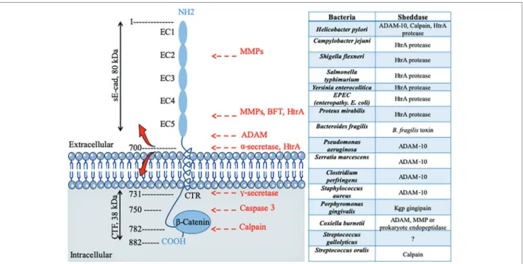
FIGURE 2 | Schematic diagram of E-cad and its cleavage sites by proteases after pathogenic bacteria infection. E-cad is a transmembrane protein containingfive extracellular repeated domains (EC1 to EC5), a transmembrane region, and an intracytoplasmic C-terminal region (CTR). The extracellular portion of E-cad forms junction with CAM on proximal cell (see Figure 1), whereas the CTR binds β-catenin and other signaling molecules. Left panel, catalytic enzymes have the capacity to cleave E-cad at specific sites indicated by red arrows. Cleavages occur either in the intracellular CTR of the molecule (e.g., caspase 3 or calpain) generating polypeptides that are capable of triggering signals, or in the extracellular portion of the molecule (e.g., the eukaryotic sheddases MMP and ADAM or the prokaryotic sheddases: BTF or HtrA), leading to soluble extracellular E-cad fragment release. For instance, the eukaryotic ADAM sheddases catalyze a cleavage of E-cad that results in the release of the 80-kDa soluble ectodomain form sE-cad and a 38-kDa C-terminal fragment (CTF). The prokaryotic HtrA serine protease can also cleave E-cad at different extracellular sites. Right panel bacteria reported to trigger E-cad cleavage (and release of sE-cad) and identification of sheddases involved in this process.
family (adamalysin/ADAM-10 and -15) (
Maretzky et al., 2008;
Najy et al., 2008;
Giebeler and Zigrino, 2016
), cysteine cathepsins
(B, L, S) (
Jordans et al., 2009
), serine protease kallikrein (KLK-6
and -7) (
Johnson et al., 2007;
Klucky et al., 2007
), plasmin
serine protease (
Ryniers et al., 2002
), and the membrane-bound
aspartic proteinases BACE-1 and BACE-2 (
Wakabayashi and
De Strooper, 2008;
Grabowska and Day, 2012
). Sheddases are
secreted as proenzymes and become mature after processing
of the propeptide; for example, the matrix metalloprotease 3
is synthesized as a zymogen (proMMP-3) which is converted
to full activity (two active forms of 45-kDa and 25-kDa) by
limited proteolysis mediated by elastase and cathepsin G (
Okada
and Nakanishi, 1989;
Becker et al., 1995
). Similarly, the MMP-9
is synthesized as a 92-kDa proMMP-9, and then the active
MMP-3 initially cleaves proMMP-9 to generate a 86-kDa
intermediate ultimately cleaved to convert the intermediate form
into a 82-kDa catalytic form (
Ogata et al., 1992
). The catalytic
domains of MMP-9 alone can hydrolyze non-collagenous proteins
and synthetic substrates, but cannot cleave triple helix collagens
without the hemopexin domain (
Nagase et al., 2006
). Its
fibronectin type II domains are important to cleave type IV
collagen, elastin, and gelatins. The hemopexin domain is required
for collagenolytic activity of the collagenase. The catalytic activity
of sheddases triggers the extracellular release of a soluble E-cad
(sE-cad) fragment of about 80-kDa from the cell surface. This
process is accompanied by the simultaneous delivery of free
β-cat into the cell cytosol, which then translocates into the
cell nucleus where it contributes to the modulation of gene
expression. It is worth noting that sE-cad might also behave
as a signaling molecule through ErbB receptor activation (
Najy
et al., 2008
). As already mentioned above, some pathogenic
bacteria can enslave eukaryotic sheddase to get E-cad cleaved.
INACTIVATION OF THE CDH1 GENE BY
METHYLATION IS ASSOCIATED WITH
PRE-CARCINOGENIC PROGRAMMING
DURING PATHOGENIC BACTERIA
INVASION
Several pathogenic bacteria were found to control the CDH1
gene expression at the chromatin level through activation of
signaling cascade leading to modulation of DNA-binding proteins
or directly in the nucleus through epigenetic modifications
(
Bierne et al., 2012
). Inactivation of the CDH1 gene leads to
the down-modulates of E-cad protein expression.
It was reported that methylation of the CDH1 gene promoter
is a frequent event in samples from H. pylori infected patients
with chronic gastritis, suggesting that CDH1 inactivation is
an early step toward gastric tumorigenesis (
Kague et al., 2010
).
Methylation of the CDH1 gene promoter was also reported
in about 30–40% of patients with H. pylori-associated gastric
carcinoma (
Bahnassy et al., 2018
). The CDH1 promoter
methylation was reduced after H. pylori eradication (
Perri et al.,
2007
). Chlamydia trachomatis infection inducing scarring disease
of the ocular mucosa was found to be associated with CDH1
promoter DNA methylation and down-regulation of E-cad
(
Rajić et al., 2017
). Acinetobacter baumannii is an opportunistic
pathogen causing severe diseases in patients with mechanical
ventilation. The nuclear targeting of Acinetobacter baumannii
transposase (Tnp) induces DNA methylation of CpG regions
in the promoter of the CDH1 gene resulting in down-regulation
of gene expression (
Moon et al., 2012
). To date, the CDH1
gene inactivation has not been systematically explored for
pathogenic bacteria and should be questioned as part of the
exploration of the molecular mechanisms by which pathogenic
bacteria deregulate E-cad expression to colonize the human host.
FIGURE 3 | Schematic representation of the structural organization of somehuman ADAM and MMP pro-enzyme sheddases. The ADAM molecules (ADAM-10 and ADAM-15) are transmembrane proteins, whereas the MMP (MMP-3, MMP-7, and MMP-9) are soluble proteins. It should be noted (not shown) that several MMP are anchored at the cell-surface by a
transmembrane domain (e.g., MMP-14 or MMP-15), by a
glycosylphosphatidylinositol anchor (e.g., MMP-17 or MMP-25) or by an amino-terminal signal anchor (e.g., MMP-23). From the NH2-terminal
extremity to the COOH-terminal extremity, ADAM-10 (748 a.a. residues) and ADAM-15 (772 a.a. to 863 a.a. residues depending the isoform) contain a signal peptide, a pro-peptide, a metalloprotease domain with catalytic activity, a disintegrin-like domain, a cysteine-rich domain, a transmembrane domain, and a cytoplasmic tail. ADAM-15 is characterized by the presence of an EGF-like domain taking place between the cysteine-rich domain and the transmembrane polypeptide. MMP-3 (or stromelysin-1, a 477 a.a. residues pro-enzyme protein of 51-kDa which can be activated in the 43-kDa mature catalytic form by removal of the pro-domain) has a basic MMP structure with a signal peptide, a pro-peptide, a catalytic domain, a hinge region, and an hemopexin domain. MMP9 (or gelatinase B, type IV collagenase, a 707 a.a. residues pro-enzyme protein of 92-kDa which can be activated as a 83-kDa mature enzyme following removal of the pro-domain), contains a signal peptide, a pro-peptide, a catalytic domain, a fibronectin type II domain, a second catalytic domain, an hinge region, and an hemopexin domain.
The epigenetic modulation of the CDH1 gene during
pathogenic bacteria infection mimics processes well-described
in the studies regarding the embryologic development. Repression
of the CDH1 gene and cad-switching from E-cad to N-cad
were considered essential for transitioning away from
pluripotency (
Sauka-Spengler and Bronner-Fraser, 2008;
Li
et al., 2010;
Pieters and van Roy, 2014
). Such cad-switching
(
Thiery, 2002
) as well as reduced expression of E-cad have
also been observed in cancer cases (including breast cancer,
gastric cancer, colorectal cancer, and hepatocellular carcinomas),
indicating that inhibition of E-cad surface expression can play
a central role in the progression toward cancer (
Jeanes et al.,
2008;
Gall and Frampton, 2013
). However, in lung cancer cells,
E-cad was found induced by WNT7a (
Ohira et al., 2003
), and
E-cad expression is increased in both ovarian cancer malignant
effusions and solid metastases (
Davidson et al., 2000;
Elloul
et al., 2006
). E-cad is generally considered a tumor suppressor
through inhibition of β-cat nuclear translocation (
Gottardi
et al., 2001
) and can act as oncogene through binding to
EGFR/Erb receptor that triggers ERK and AKT signaling
pathways providing advantage for cancer development and
metastasis (
Wells et al., 2008;
Rodriguez et al., 2012
).
With regard to viruses as examples of other pathogens
modulating CDH1 gene expression, it has been reported that
hepatitis B virus (HBV) represses E-cad at the transcriptional
level by hypermethylation of the CDH1 promoter on CpG Island
1 with possible consequences on hepatocellular carcinogenesis
by promoting detachment of surrounding cells and their migration
to the primary tumor site (
Lee et al., 2005
). Alternatively, loss
of E-cad in HBV infected cells can also be regulated at a
post-translational level by proteases and SUMOylation (
Ha et al.,
2016
). Hypermethylation that triggers CDH-1 gene repression
was also found with the human papillomavirus (HPV) that
increases cellular methyltransferase 1 (Dnmt1) activity via its
viral E7 protein (
Laurson et al., 2010
).
ABERRANT SPLICING OF THE
E-CADHERIN TRANSCRIPT
Among the gene silencing molecular mechanisms underlying
E-cad loss, one involves the expression of nonfunctional truncated
CDH1 gene transcript. E-cadherin mRNA with premature
termination codon mutation was reported in chronic lymphocytic
leukemia cells (
Sharma and Lichtenstein, 2009
). This transcript
(which lacks the exon 11) plays a role in silencing the production
of E-cad. The amounts of wild-type E-cad mRNA inversely
correlated with the amounts of aberrant transcript resulting
in the up-regulation of the Wnt-β catenin pathway. The production
of E-cad mRNA variant by alternative splicing has also been
linked to a decrease in cell-cell adhesion and an increase in
cell migration (
Matos et al., 2017
). The novel E-cadherin variant,
a truncated soluble form of E-cad resulting from a deletion
of the first 34 nt in the exon 14 of the CDH1 mRNA, induces
changes characteristic of the Epithelial to Mesenchymal Transition
(EMT) process, a key event in tumor progression (
Matos et al.,
2017
). Moreover, the over-expression of this novel E-cad truncated
form in transfected cells resulted in downregulation of
wild-type E-cad expression. This truncated CDH1 gene transcript
was recently found in breast cancer cells (
Rosso et al., 2019
).
Aberrant splicing of CDH1 gene transcript (exon 8 or exon
11 skipped aberrant transcripts) was also reported in gastric
carcinoma (
Garziera et al., 2013;
Li et al., 2015
). Although
this is a possibility, it has not yet been demonstrated that the
expression of a CDH1 aberrant transcript in gastric cancers
is controlled by H. pylori. However, it is known that aberrant
splicing of cellular gene transcripts can occur during bacterial
invasion (
Sun, 2017
). Indeed, massive alterations in the pattern
of cellular mRNA splicing were reported upon infection with
bacteria such as Anaplasma phagocytophilum, the etiologic agent
of the human granulocytic anaplasmosis (
Dumler et al., 2018
)
and Mycobacterium tuberculosis the etiologic agent of tuberculosis
(
Kalam et al., 2018
). This may be consistent with the
observation that Mycobacterium tuberculosis induced an epithelial
mesenchymal transition in a pulmonary adenocarcinoma
epithelial cell line (
Gupta et al., 2016
), a phenomenon orchestrated
at the level of E-cad cell-surface expression. It was also reported
that the EMT of mesothelial cells occurred in Mycobacterium
tuberculosis-associated pleurisy, together with a reduction in
E-cad expression (
Kim et al., 2011
). Moreover, expression level
of E-cad was reported to differ between pulmonary tuberculosis
patients and latent tuberculosis individuals (
Sun et al., 2018
).
RELEASE OF SOLUBLE E-CADHERIN
AFTER BACTERIAL INFECTION AS
AN EARLY EVENT TOWARD
CARCINOGENESIS
Aside from the transcriptional repression of the CDH1 gene,
another interesting mechanism, which could interfere with the
anti-bacterial defenses of the host, is the release of sE-cad in
body fluids following a sheddase-mediated cleavage of E-cad.
The combination of sE-cad release together with other
pro-inflammatory factors was highlighted as a triggering signal
that promotes gastric adenocarcinoma or colorectal tumors in
patients infected with H. pylori, B. fragilis, or Streptococcus
gallolyticus (
Wu et al., 2003;
O’Connor et al., 2011;
Kumar
et al., 2017;
Chung et al., 2018
).
H. pylori, a bacterium that colonizes the gastric mucosa, is
known as a risk factor for the development of chronic atrophic
gastritis, gastroduodenal ulcers, and adenocarcinoma. An in vitro
experimental model of cell transfection has demonstrated that
H. pylori triggers
β-cat activation through the interaction of
virulence factor CagA with E-cad (
Murata-Kamiya et al., 2007
).
O’Connor et al. (2011)
reported that H. pylori induced the
activation of host protease calpain via the toll-like receptor 2
(TLR2) and disruption of gastric epithelium. Another report
suggests that ADAM-10 is induced in H. pylori infection and
contributes to the shedding of E-cad (
Schirrmeister et al., 2009
).
It was also reported that a serine protease HtrA from H. pylori
mediate direct cleavage of E-cad (
Hoy et al., 2010;
Schmidt et al.,
2016
). Bacterium S. gallolyticus has a strong association with
colorectal cancer with increased levels of β-cat and c-Myc (
Kumar
et al., 2017
). The genome of another bacterium, Bacteroides fragilis,
encodes a sheddase, the B. fragilis toxin (BFT) also termed fragilis
(FRA), which cleaves E-cad and is associated with C-Myc expression
and cellular proliferation (
Wu et al., 1998, 2003;
Remacle et al.,
2014;
Chung et al., 2018
). Chung and co-workers uncovered a
complex mechanism whereby the B. fragilis toxin (BFT)-mediated
cleavage of E-cad initiates a multi-step inflammatory cascade
requiring
β-cat, IL-17R, NF-B, and Stat3 signaling in colonic
epithelial cells. IL-17 dependent NF-κB activation in colic epithelial
cells induces a mucosal gradient of C-X-C chemokines that
initiates pro-tumoral myeloid cell infiltration to the distal colon
and colon cancer. All these cancers are solid tumors. However,
a role for sE-cad in the initiation of a pro-carcinogenic process
might also be considered. Over the past 3 years, we have reported
results suggesting that the bacterium Coxiella burnetii, responsible
of Q fever, was associated with a higher frequency of Non-Hodgkin
Lymphoma (NHL) in C. burnetii-infected patients compared to
the general population (
Melenotte et al., 2016
). Recently,
we investigated the transcriptional signature that could be associated
with the development of NHL in Q fever patients and found
an over-expression of genes involved in anti-apoptotic process
and a repression of pro-apoptotic genes (
Melenotte et al., 2019
).
Since cell surface expression of E-cad and release of sE-cad have
been associated to various pathogenic bacteria known for inducing
solid tumors, we have also investigated the levels of expression
of these molecules in Q fever patients and observed a significant
release of sE-cad in their sera and a down-regulation of E-cad
mRNA expression. The sE-cad levels were found increased in
the sera of acute and persistent Q fever patients, whereas they
remained at the baseline in the control groups of healthy donors,
people cured of Q fever, patients suffering from unrelated
inflammatory diseases, and past Q fever patients who developed
NHL (Table 2). Consequently, sE-cad could be considered a
new biomarker of C. burnetii infection rather than a marker of
NHL-associated to Q fever (
Mezouar et al., 2019
). We do not
yet know which eukaryotic or prokaryotic sheddase could
be responsible for the cleavage of E-cad in patients infected with
C. burnetii. Preliminary studies on peripheral blood mononuclear
cells exposed to heat-inactivated C. burnetii suggested variations
in ADAM-10 and MMP-9 expression. Microarray performed on
samples from macrophages and dendritic cells (DC) infected in
vitro by C. burnetii revealed an over-expression of MMP-3 in
C. burnetii-infected DC. Preliminary investigations in search of
sheddase have also been conducted. We tried to blast the protein
sequences of more than 20 sheddases known to catalyze the
cleavage of E-cad against the hypothetical proteins of four strains
of C. burnetii. We found that three eukaryotic sheddases (MMP-3,
MMP-9, and ADAM-15) presented sequence similarities with
bacterial proteins. Experiments are under progress to identify
whether an ADAM or MMP eukaryotic protease or a prokaryotic
protease encoded by C. burnetii could be responsible for sE-cad
release in Q fever patients.
Care must be taken not to overinterpret the significance of
sE-cad increase in body fluids during bacterial infections. It is
possible that sE-cad release in body fluids corresponds to a
ubiquitous phenomenon induced during the process of
colonization of the host by a pathogenic bacterium. We already
know that it is not limited to bacteria since there is an apparent
correlation between the levels of sE-cad in human
immunodeficiency virus (HIV)-positive patients and their viral
titers (
Streeck et al., 2011
). Moreover, abnormal concentrations
of sE-cad in patients’ sera has been observed in several metabolic
and inflammatory diseases (
Pittard et al., 1996;
Jiang et al.,
2009;
Shirahata et al., 2018;
Sato et al., 2019
). High concentration
of sE-cad has also been found associated with cancer progression
(
Grabowska and Day, 2012;
Salama et al., 2013;
Repetto et al.,
2014
). For example, high concentration of sE-cad was described
in prostate cancer patients and was associated with an
over-expression of MMP-2 and MMP-9 (
Kuefer et al., 2005;
Biswas
et al., 2010;
Tsaur et al., 2015
). Similarly, increased expression
of sE-cad was reported in gastric cancer that was associated
with the over-expression of MMP-7 (
Lee et al., 2006, 2007
).
Serum levels of sE-cad were increased in patients with ovarian
carcinoma the cleavage of E-cad being mediated by MMP-9
(
Gadducci et al., 1999;
Symowicz et al., 2007
). Altogether, these
data suggest that high sE-cad concentration in body fluids could
simply behave as a factor that predisposes to inflammation and
development of cancers, as described for H. pylori-infected patients.
DYSFUNCTION OF THE IMMUNE
RESPONSE ORCHESTRATED BY
BACTERIA-INDUCED E-CADHERIN
CLEAVAGE
The immune system naturally provides an anti-infectious
surveillance at the epithelium level via cells that express
cell-surface molecules, such as CD103 and KLRG1, able to bind
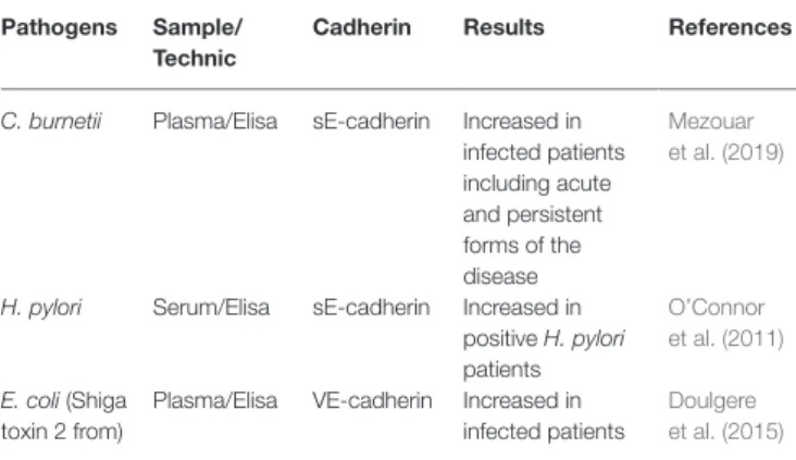
TABLE 2 | Soluble Cad molecules released in body fluids duringbacteria-associated infectious diseases.
Pathogens Sample/ Technic
Cadherin Results References
C. burnetii Plasma/Elisa sE-cadherin Increased in infected patients including acute and persistent forms of the disease Mezouar et al. (2019)
H. pylori Serum/Elisa sE-cadherin Increased in positive H. pylori patients O’Connor et al. (2011) E. coli (Shiga toxin 2 from)
Plasma/Elisa VE-cadherin Increased in infected patients
Doulgere et al. (2015)
If the general trend is to increase the soluble forms of cadherin molecules in body fluids of patients infected with pathogenic bacteria, caution should be exercised when comparing results obtained with different diagnostic kits, in different laboratories and with serum or plasma samples. Regarding the results reported in the papers mentioned in this table, the concentrations of soluble Cad measured in the samples were two times higher in the infected patients than in healthy controls. For example, in the plasma of C. burnetii-infected patients, the sE-cad concentration was about 170–450 ng/ml in acute Q fever (median 272 ng/ml), 260–530 ng/ml in persistent Q fever (median 333 ng/ml), while it was 80–320 ng/ml (median 164 ng/ml) in healthy controls.
the E-cad found at the surface of epithelial cells. These immune
cells are not heavily engaged, unless the other defenses have
failed. For example, under normal conditions, the intestinal
epithelium is protected by a mucus layer that acts as host
defense against microbial attachment (
Kim and Ho, 2010;
Desai
et al., 2016
). Upon infection, the specialized intestinal Paneth
cells secretes antimicrobial proteins and the commensal intestinal
microbiota competes with the infectious pathogens, thereby
acting as first line of innate defense to fight against pathogenic
bacteria (
Cash et al., 2006;
Mason and Huffnagle, 2009;
Vazeille
et al., 2011;
Bel et al., 2017
). The epithelium integrity controlled
by the homotypic interaction of E-cad in trans represents a
second barrier protecting the host against intruder transmigration
(
Kim et al., 2010
). When pathogenic bacteria had evaded
epithelial cell autophagic clearance and dead cells renewal
(
Yoshikawa et al., 2009;
Benjamin et al., 2013
), the host’s
immune system represents the last rampart before the pathogens
can breach the epithelium and disseminate deeper.
The intestinal villous microfold cells (M cells) are specialized
epithelial cells of the gut-associated lymphoid tissues (GALT)
that deliver luminal antigens to the underlying immune system
after their transport to the basolateral membrane of M cells
(
Miller et al., 2007
). An efficient immune response against a
microbial attack requires the migration of cells of the host
immune system in the microenvironment where infection occurs
and the sequential detection of stress signals, tissue damages
and conserved bacterial molecules termed pathogen-associated
molecular patterns (PAMPs) (
Broz and Monack, 2013
). PAMPS
include molecules as diverse as lipopolysaccharide, flagellin,
peptidoglycan, lipoproteins, and unique bacterial nucleic acid
structures. Upon detection of bacterial invasion, host cell receptors
such as toll-like receptors and C-type lectin receptors, activate
signaling pathways that govern the production of inflammatory
cytokines including the IL-1
β and IL-18 that can restrict bacterial
replication. Causative agents of infectious diseases are therefore
characterized by their capacity to elaborate mechanisms aimed
to damage the protective cellular barriers and/or to modulate
immune responses of the host to achieve invasion.
We can question the place of the E-cad in this process of
mobilization of immunity cells via trans homotypic (e.g., E-cad)
or trans heterotypic (e.g., CD103 and KLRG1) interactions under
normal and pathological conditions (Figure 4). Although E-cad
is expressed by immature CD4
+CD8
+thymocytes (
Munro et al.,
1996;
Müller et al., 1997
), after the thymocytes have left the
thymus, E-cad is generally absent from most mature lymphocytes
(
Lee et al., 1994
). Yet, under certain pathological conditions,
cell-surface expression of E-cad was reported on mature
T-lymphocytes (CD3
+) subsets, as well as B cells (CD19
+), NK
cells (DX5
+), and monocyte/macrophages (CD11b
+) subpopulations
(
Esch et al., 2000;
Sakai et al., 2008
). E-cad expression was also
confirmed for subpopulations of epithelial γδ T-cells (
Lee et al.,
1994
) and memory CD8
+T-cells in intestinal mucosa (
Hofmann
and Pircher, 2011
). Moreover, we have recently observed that
30% of CD16
+monocytes expressed E-cad after C. burnetii
infection (
Mezouar et al., 2019
). In the intestinal epithelium, it
is generally accepted that the immune host defense is mainly
mediated by effector cells that express the α
Eintegrin (CD103),
an E-cad ligand (
Banh and Brossay, 2009;
Van den Bossche
et al., 2012
). CD103 is expressed on 40–50% of CD4
+T-lymphocytes
and 90% of CD8
+T-lymphocytes that reside in the intestinal
mucosa as well as on the surface of intraepithelial lymphocytes
(
Cerf-Bensussan et al., 1987;
Kilshaw and Murant, 1990;
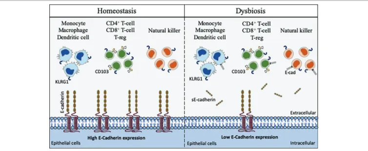
FIGURE 4 | Schematic model of interaction between cells that express CD103, KLRG1, and E-cad. E-cad expressed on epithelial cells (as well as dendritic cells,
Langerhans cells, and macrophages) can potentially interact with CD103, KLRG1 or both molecules expressed on the surface of immune cells. In the absence of bacterial infection, the epithelial cells express E-cad at high level, allowing immune cells to ensure the immune surveillance of epithelia (left panel). After pathogenic bacteria invasion, the cell-surface expression of E-cad is weak on epithelial cells due to the activation of sheddases and sE-cad is released in the epithelium microenvironment. The abnormal expression of E-cad on subpopulations of immune cells, the release of sE-cad, the weak expression of E-cad on epithelial cells likely contribute to immune system dysfunction at the bacterial invasion site (right panel).
Hadley et al., 1997;
Uchida et al., 2011
). It is also present on
T cells of the intestinal lamina propria (
Agace et al., 2000
) and
a subset of CD4
+CD25
+Foxp3
+Treg-cells (
Stephens et al., 2007
).
It was demonstrated that after epithelial damages, the intraepithelial
CD103
+γδ T-lymphocytes that reside on the surface of epithelium
promote mucosal repair through antibacterial factors (e.g., Reg3γ)
and immunomodulatory molecules (e.g., IL1β, CXCL9) (
Cepek
et al., 1994;
Ismail et al., 2009, 2011
). It is worth noting that
the intestinal CD103
+intraepithelial lymphocytes adherence to
epithelial cells is inhibited by antibodies against CD103 (
Cepek
et al., 1993
). Another cell-surface receptor named KLRG1 that
is encountered on subsets of immune cells including mature NK
cells, memory CD4
+T cells, effector CD8
+T cells, and FoxP3
+Treg cells, is known to bind E-cad (
Schwartzkopff et al., 2007;
Tessmer et al., 2007;
Banh and Brossay, 2009;
Van den Bossche
et al., 2012
). It has been reported that high levels of sE-cad
could be sufficient to inhibit CD8
+T-cell function in a
KLRG1-dependent manner (
Streeck et al., 2011
).
The E-cad induction on subpopulations of immune response
cells in pathological situations remain to be elucidated. It can
be hypothesized that the aberrant expression of E-cad under
pathological conditions reflects changes in the transmigration
and homing capacity of these cells (
Reyat et al., 2017
). Because
several pathogenic bacteria reduce the epithelium surface
expression of E-cad at the site of infection, it might be speculated
that the decreased expression or the lack of expression of
E-cad on epithelial cells is likely to trigger the rerouting of
immune cells far from the infection site. As previously shown
by
Streeck et al. (2011)
for the KLRG1
+CD8
+T-cells
subpopulation, the release of sE-cad might also serve as a
decoy for diverting from their function the immune cells
expressing E-cad, CD103 or KLRG1 after engagement of such
receptors with sE-cad. Modulation of E-cad expression on the
host epithelial cells and sE-cad release could therefore
be considered a very efficient stratagem to prevent the immune
system from behaving as a line of defense against invaders.
In addition, for bacteria that induce cancer, reducing the
expression of E-cad on certain tumor cells as previously reported
(
Shields et al., 2019
) and disrupting the migration and attachment
capabilities of immune survey cells could be a way of promoting
the development of bacteria-induced cancers.
CONCLUSION AND DISCUSSION
This review highlights how the E-cad can be diverted from
its function of maintenance of tissues integrity and prevention
of cell migration/differentiation during pathogenic bacterial
infections. Pathogenic bacteria can use E-cad for their attachment
to epithelial cells. Indeed, they can cleave E-cad to ensure
their transmigration and can modulate the responsiveness of
immune cells through modulation of cell-surface expression
of E-cad and sE-cad release in body fluids. Some bacteria use
E-cad to enter their target cells (e.g., F. nucleatum, L.
monocytogenes, S. pneumoniae). Several bacteria act on the
cell-surface expression of this molecule, either by modulating
the CDH1 gene transcription (e.g., C. trachomatis, H. pylori)
or by inducing the cleavage of the E-cad molecule (e.g., C.
perfringens, S. aureus, C. burnetii) via proteases (sheddases).
This process can favor achievement of the trans-epithelial host
invasion or modulate host-pathogen molecular crosstalk.
Currently, the best studied models are those that refer to
intestinal infections that can lead to cancer (Figure 5).
Proteases (cysteine proteases, serine proteases, aspartate
proteases, and metalloproteases) are ubiquitously encountered
in the microbial world and are essential for their survival and
replication cycle (
Häse and Finkelstein, 1993
). Proteases, such
as collagenase (
Bond and Van Wart, 1984
), elastase (
Bever
and Iglewski, 1988
), or metalloprotease (
Schiavo et al., 1992
)
(
Domann et al., 1992
) were associated with bacterial pathogenesis.
Some pathogenic bacteria can activate the production of
eukaryotic proteases such as ADAM-10 via signaling (e.g., this
was reported for P. aeruginosa, S. marcescens, C. perfringens,
S. aureus), whereas others use a portion of their genome to
encode their own sheddase, including the HtrA protease (encoded
by H. pylori, C. jejuni, S. flexneri, enteropathogenic E. coli)
or B. fragilis toxin (encoded by B. fragilis).
As described above in this paper, evidence emphasizing that
cleavage of E-cad by sheddases and release of sE-cad into the
body fluids are factors that contribute to the progression of
cancer (sometimes it was demonstrated). Within the group of
bacteria that modulate the expression of E-cad, the association
with carcinogenic processes has been investigated, in particular
with H. pylori, F. nucleatum, S. gallolyticus, and B. fragilis. H.
pylori is known to be a risk factor for the development of
gastric adenocarcinoma and the progression toward cancer is
likely related to E-cad cleavage and β-cat activation (
Murata-Kamiya et al., 2007
), F. nucleatum promotes colorectal cancer
by modulating the E-cad and Wnt/β-cat signaling via its FadA
adhesin and up-regulating annexin A1 (
Rubinstein et al., 2013,
2019
). S. gallolyticus is also associated with colorectal cancer
with known increased level of β-cat and c-Myc activation (
Kumar
et al., 2017
). B. fragilis toxin (BFT)-mediated cleavage of E-cad
initiates a multi-step inflammatory cascade requiring β-cat nuclear
translocation, activation of NF-κB and Stat3 signaling pathways
in colonic epithelial cells as early events leading to pro-tumoral
myeloid cell infiltration to the distal colon and colon cancer
(
Chung et al., 2018
). Our recent data indicate that C. burnetii,
reported as associated with occurrence of non-Hodgkin lymphoma,
is also capable of triggering cleavage of E-cad and release of
sE-cad in the sera of Q fever patients (Figure 6;
Mezouar
et al., 2019
). Yet, further experiments are required to formerly
demonstrate the association between sE-cad release in sera from
Q fever patients and the initiation of a pro-carcinogenic
inflammatory process leading to lymphoma development.
The accumulation of data showing that sE-cad is produced
in many pathological processes that can lead to cancer development
raises the question of what value can be attributed to this compound
as a biomarker of disease severity. At the moment, we do not
have enough information to conclude. The presence of sE-cad
in body fluids was considered a possible biomarker in H. pylori
(
O’Connor et al., 2011
) and C. burnetii (
Mezouar et al., 2019
)
infections, and soluble VE-cad was also regarded as possible
biomarker in E. coli infections (
Doulgere et al., 2015
). We are
FIGURE 5 | This cartoon illustrates how, by simply acting on the E-cad expression, an intestinal pathogenic bacterium can both bypass the physical defense
system represented by the epithelial barrier and confuse the cells of the immune system intended to defend the host. Step 1: under homeostasis, the epithelium (composed of epithelial cells) is protected by a mucus layer synthesized by goblet cells which secrete the mucins (e.g, MUC2 – mucin gel). The mucus layer prevents microbial attachment without interference with the transport of nutrients. The commensal intestinal microbiota is limited to the epithelium-distal mucus layer, while the epithelium-proximal mucus is largely devoid of bacteria. Step 2: upon infection, the commensal intestinal microbiota competes with the infectious pathogens and the Paneth cells produce antimicrobial proteins (e.g., C-type lectin REG3γ, β-defensins, cathelicidins, and lysozyme), to fight the invasion. A regulation of infection is also achieved by epithelial cell autophagic clearance and dead cells renewal. At the same time, the villous microfold cells (M cells) expressing TLR deliver luminal antigens to the underlying immune system to set up a whole arsenal of anti-bacterial actions. In case this response proves sufficient, the invader is destroyed, and the microenvironment returns to homeostasis (Step 1). If not, the conflict is prolonged. Step 3: an efficient immune response against the pathogens requires the migration of cells of the host immune system in the microenvironment where the infection occurs and the sequential detection of stress signals, tissue damages, and PAMPs (e.g., lipopolysaccharide, flagellin, peptidoglycan, lipoproteins, and unique bacterial nucleic acid structures). KLRG1+ dendritic
cells and monocytes/macrophages, CD103+ T-cells, KLRG1+ T-cells, and other immune cell subpopulations, colonize the lamina propria. Upon detection of bacterial
invasion, host cell receptors, such as TLR and C-type lectin receptors, activate signaling pathways that govern the production of inflammatory cytokines, including the IL-1β and IL-18 that can restrict bacterial replication. Step 4: the pathogenic bacteria reduce the epithelium surface expression of E-cad at the site of infection, resulting in the destruction of adherent’s junctions and allowing transmigration. Moreover, it might be speculated that the induction of E-cad on subpopulation of immune response cells (E-cad+ T-cells and CD16+/E-cad+ monocytes) redirects those cells far from the infection site in microenvironments where they have a higher
probability to interact with E-cad+ epithelial cells. The release of sE-cad might also serve as a decoy for diverting immune cells from their function through interaction
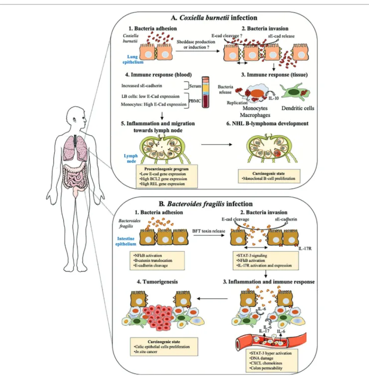
FIGURE 6 | Cellular host E-cad as target during both pulmonary and intestinal bacterial infections. This drawing summarizes the hypothetical models of
tumorigenesis associated with Coxiella burnetii and enterotoxigenic Bacteroides fragilis. (A) Coxiella burnetii. Although rare, the incidence of NHL B-lymphoma in
patients infected by C. burnetii in France was significantly higher (25-fold) than within the general population. In Q fever, the overproduction of IL-10 by infected monocytes was found critical in both sustaining replication of the C. burnetii and preventing the macrophages microbicidal activity. Moreover, specific genes involved in anti-apoptotic process are over-expressed, whereas pro-apoptotic genes are repressed. Recently, we found elevated concentrations of sE-cad in Q fever patients, along with an increase in cell-surface expression of E-cad in more than 30% of HLADR+/CD16+ monocytes and a decrease in E-cad expression that
concerned about 3% of the E-cad+/CD20+ B-cells subpopulation (LB cells). We speculate that the release of sE-cad might participate to the molecular crosstalk,
which takes place in the microenvironment of the lymph node during persistent Q fever and might possibly trigger a pro-carcinogenic program required for the initiation of NHL lymphoma. (B) Enterotoxigenic Bacteroides fragilis. The enterotoxigenic Bacteroides fragilis synthesizes a toxin, BFToxin, which damages the
protective intestinal epithelial barriers of the host by cleavage of E-cad to achieve invasion. This process leads to epithelial cells activation through nuclear translocation of β-cat and NF-κB, inducing the transcription of genes such as IL-17 receptor (IL17R). A pro-inflammatory immune response is initiated against the pathogen characterized by the in-situ production of IL-17. IL-17R positive cells are induced to produce STAT3 and a gradient of chemokines in the
microenvironment favors the recruitment of pro-tumoral myeloid cells that accumulate in the distal colon producing growth factors triggering the proliferation of colic epithelial cells. These cells progressively accumulate DNA damages and form a solid tumor in the colon.