HAL Id: hal-02629275
https://hal.inrae.fr/hal-02629275
Submitted on 27 May 2020
HAL is a multi-disciplinary open access
archive for the deposit and dissemination of
sci-entific research documents, whether they are
pub-lished or not. The documents may come from
teaching and research institutions in France or
abroad, or from public or private research centers.
L’archive ouverte pluridisciplinaire HAL, est
destinée au dépôt et à la diffusion de documents
scientifiques de niveau recherche, publiés ou non,
émanant des établissements d’enseignement et de
recherche français ou étrangers, des laboratoires
publics ou privés.
Distributed under a Creative Commons Attribution| 4.0 International License
Schymanski, A. J. Williams, M. M. L. Dingemans, R. Massei, W. Brack,
Xavier Cousin, et al.
To cite this version:
J. B. Legradi, C. Di Paolo, M. H. S. Kraak, H. G. van der Geest, E. L. Schymanski, et al.. An
ecotoxicological view on neurotoxicity assessment. European Journal of Environmental Sciences, 2018,
30, pp.1-34. �10.1186/s12302-018-0173-x�. �hal-02629275�
REVIEW
An ecotoxicological view on neurotoxicity
assessment
J. B. Legradi
1,2*, C. Di Paolo
1, M. H. S. Kraak
3, H. G. van der Geest
3, E. L. Schymanski
4, A. J. Williams
5,
M. M. L. Dingemans
6, R. Massei
7, W. Brack
7, X. Cousin
8,9, M.‑L. Begout
10, R. van der Oost
11, A. Carion
12,
V. Suarez‑Ulloa
12, F. Silvestre
12, B. I. Escher
13,14, M. Engwall
15, G. Nilén
15, S. H. Keiter
15, D. Pollet
16, P. Waldmann
16,
C. Kienle
17, I. Werner
17, A.‑C. Haigis
1, D. Knapen
18, L. Vergauwen
18, M. Spehr
19, W. Schulz
20, W. Busch
21,
D. Leuthold
21, S. Scholz
21, C. M. vom Berg
22, N. Basu
23, C. A. Murphy
24, A. Lampert
25, J. Kuckelkorn
26,
T. Grummt
26and H. Hollert
1*Abstract
The numbers of potential neurotoxicants in the environment are raising and pose a great risk for humans and the environment. Currently neurotoxicity assessment is mostly performed to predict and prevent harm to human popula‑ tions. Despite all the efforts invested in the last years in developing novel in vitro or in silico test systems, in vivo tests with rodents are still the only accepted test for neurotoxicity risk assessment in Europe. Despite an increasing num‑ ber of reports of species showing altered behaviour, neurotoxicity assessment for species in the environment is not required and therefore mostly not performed. Considering the increasing numbers of environmental contaminants with potential neurotoxic potential, eco‑neurotoxicity should be also considered in risk assessment. In order to do so novel test systems are needed that can cope with species differences within ecosystems. In the field, online‑biomoni‑ toring systems using behavioural information could be used to detect neurotoxic effects and effect‑directed analyses could be applied to identify the neurotoxicants causing the effect. Additionally, toxic pressure calculations in com‑ bination with mixture modelling could use environmental chemical monitoring data to predict adverse effects and prioritize pollutants for laboratory testing. Cheminformatics based on computational toxicological data from in vitro and in vivo studies could help to identify potential neurotoxicants. An array of in vitro assays covering different modes of action could be applied to screen compounds for neurotoxicity. The selection of in vitro assays could be guided by AOPs relevant for eco‑neurotoxicity. In order to be able to perform risk assessment for eco‑neurotoxicity, methods need to focus on the most sensitive species in an ecosystem. A test battery using species from different trophic levels might be the best approach. To implement eco‑neurotoxicity assessment into European risk assessment, cheminfor‑ matics and in vitro screening tests could be used as first approach to identify eco‑neurotoxic pollutants. In a second step, a small species test battery could be applied to assess the risks of ecosystems.
Keywords: Eco‑neurotoxicity, Neurotoxicity, EDA, REACH, AOP, Behaviour, Computational toxicity, Ecological, Species
© The Author(s) 2018. This article is distributed under the terms of the Creative Commons Attribution 4.0 International License (http://creat iveco mmons .org/licen ses/by/4.0/), which permits unrestricted use, distribution, and reproduction in any medium, provided you give appropriate credit to the original author(s) and the source, provide a link to the Creative Commons license, and indicate if changes were made.
Open Access
*Correspondence: [email protected]‑aachen.de; henner. [email protected]‑aachen.de
1 Institute for Environmental Research, Department of Ecosystem Analysis,
ABBt–Aachen Biology and Biotechnology, RWTH Aachen University, Worringerweg 1, 52074 Aachen, Germany
Background
Neurotoxic pollutants are an emerging issue beyond human health because neurotoxicants causes poten-tially serious threats to vertebrate and invertebrate pop-ulations and ecosystems in general. Indeed, neurotoxic chemicals are suspected to produce changes in organ-ism behaviour (e.g. mating behaviour, predator escape response and feeding behaviour), which can reduce an individual’s fitness, lead to population declines and
ultimately have severe impacts on ecosystems [1].
Dif-ferent classes of environmental contaminants, includ-ing metals and organic pollutants, were shown to affect the performance of complex behaviours in different
fish species [2] and in wildlife [3, 4]. In wildlife, as in
humans, early life stages are particularly sensitive to toxicant insults. Additionally, neurotoxic effects in early life stages might not be directly visible but lead to detrimental effects later in life. Besides, exposure to neurotoxic compounds can trigger epigenetic pathways which can underlie long-term effects as well as multi-
or transgenerational effects [5, 6].
Typically, several thousand compounds are detect-able in environmental samples, including synthetic and natural compounds and their transformation
prod-ucts [7]. However, knowledge regarding the neurotoxic
potential of environmental contaminants in ecosystems is very limited, since the assessment of neurotoxicity is currently mostly focused on human exposure to indi-vidual chemicals. Known human neurotoxic or neuro-active compounds, such as pesticides, pharmaceuticals, and heavy metals, occur in the environment together with thousands of chemicals with unknown neuro-toxic potential to different species and life stages. It has been estimated that up to 30% of all commercially used chemicals (~ 30,000 chemicals) may have neurotoxic
potential [8]. Additionally, in a recent literature study
looking at the known modes of action (MoA) of organic contaminants detected in freshwater monitoring stud-ies, neurotoxicity was identified as the MoA linked to
nearly 30% of all detected chemicals [9]. This shows the
relevance of detecting neurotoxic compounds in the environment, increasing the demand for bioanalytical tools capable of identifying and possibly quantifying neurotoxic effects in organisms inhabiting contami-nated ecosystems.
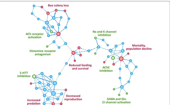
The aim of this article is to provide a critical over-view of the state of the art of hazard characterization, effects, bioassays and chemical approaches regarding neurotoxicity in organisms as well as for ecosystems. This review will contribute a scientific perspective on the needs and future directions in neurotoxicity
assess-ment for environassess-mental protection (cf. Fig. 1).
Environmental neurotoxicity versus eco‑neurotoxicity
Neurotoxicity can be defined as the capacity of agents (chemical, biological, or physical) to cause adverse
func-tional or structural changes in the nervous system [10].
Environmental neurotoxicity describes neurotoxicity caused by exposure to chemicals in the environment and commonly refers to human exposure and human
neuro-toxicity [10]. In contrast, we define ecological
neurotox-icity (eco-neurotoxneurotox-icity) as neurotoxneurotox-icity resulting from exposure to environmental chemicals in species other than humans (e.g. fish, birds, invertebrates). It is impor-tant to distinguish between human and non-human neurotoxicity as the effects of exposure to compounds, both in terms of levels and pathways, as well as the struc-ture and function of the nervous system itself, can differ widely between species.
Current role of eco‑neurotoxicology in risk assessment for regulation
REACH/EU general food law
Within the current European chemical regulation, neurotoxicity is only assessed using in vivo test
sys-tems [11]. The EU legislation for industrial chemicals
(REACH) assesses neurotoxicity only for compounds produced ≥ 10 tons/year. These compounds need to be tested with standard oral 28-day and 90-day toxicity studies in rodents. Clinical observations including motor activity, a functional observational battery and histo-pathological assessments of the spinal cord and sciatic nerve can be indicators of neurotoxicity. If these tests indicate neurotoxicity at levels below systemic toxicity, more detailed neurotoxicity tests are required (OECD technical guideline (TG) 424 to assess neurotoxicity and TG 426 to assess developmental neurotoxicity).
In terms of ecotoxicological impacts, current guide-lines for neurotoxicity assessment in vertebrates focus
on mammals and birds [12–16]. There is no regulatory
guideline available to identify neurotoxic risks to other vertebrates or invertebrate animals. Furthermore, thus far there is no European regulatory framework for eco-neurotoxicity assessment.
Within risk assessment and risk management of eco-neurotoxic substances, pesticides are a substance class of special interest. Some pesticides kill pests via neurotoxic mechanisms. Neurotoxic actions on non-target species have been determined for several species and pesticides
[17–22]. The European Food Safety Agency (EFSA) is
responsible for the registration of pesticides and all other substances that can contact or occur in food and are not assessed under REACH. Until now, the active com-pounds in pesticides need to be assessed for potential
neurotoxic effects in mammals using the same rodent studies as under REACH (TG 424 and TG 426) only if it is indicative from their intended MoA or other informa-tion, like chemical structure, that the substance could be
neurotoxic [23]. Neurotoxic effects on non-target species
in the environment are not assessed. Water Framework Directive
The European Water Framework Directive (WFD) aims to integrate biological and chemical information to obtain an overall insight into the quality of individual water bod-ies. According to the WFD, the chemical status of a water body is determined by analysing the concentrations of 45 priority substances, which are not selected based on their potential neurotoxicity. A good chemical status is defined by concentrations of all of these substances below the annual average and maximum allowable Environmen-tal Quality Standards (AA- and MAC-EQSs), which are defined to protect the environment and human health
[24].
As a result, regular chemical monitoring of the water quality is almost exclusively performed by targeted chemical analysis of a limited set of (indicator) com-pounds. There are, however, some serious limitations related to the use of target chemical analyses of large vol-ume samples for monitoring the overall chemical status of a water body. First, because only a limited number of target substances are analysed, the risk of other, non-priority and unknown substances in the aquatic
envi-ronment remains unknown [25]. At present (August
2018), more than 142,000,000 substances are registered in SciFinder with the Chemical Abstracts Service, while there are over 140,000 substances that are produced over 1 ton/year listed in REACH. Some of those compounds might eventually end up in the environment. Second, it is obvious that chemicals do not occur alone in the envi-ronment, but as complex mixtures. While concentrations of individual chemicals can be below the lowest observed effect concentrations (LOEC) or detection limits, the
entire mixture may still cause adverse effects [26].
More-over, transformation products of micropollutants formed in the environment or by biological metabolism are not always known or registered and may be more toxic and
persistent than the parent compounds [27]. These
limi-tations may thus result in an incomplete assessment of
chemical hazards and risks, e.g. [28], urging alternative
approaches to be explored [29].
German drinking water ordinance
There is an urgent need for quick assessments of sub-stances with unknown toxicological potential to prevent possible harm for consumers by water suppliers and pub-lic health departments, who supervise the process. At
this time, there is no explicit regulation for neurotoxicity in drinking water in countries like Germany. While the
German Drinking Water Ordinance (TrinkwV 2018) [30]
gives threshold values for some metals, e.g. lead,
cad-mium, arsenic, with a known neurotoxic potential [31],
no specific endpoints or proposals for a testing strategy are given for neurotoxicity.
The health-related indicator value (HRIV; in Ger-man: Gesundheitsbasierter Orientierungswert, GOW) concept provides a temporary value for toxicologically unknown single substances detected in drinking water systems. This hierarchically built concept is based on a precautionary in vitro approach with endpoints related to genotoxicity, neurotoxicity, endocrine disrupting
effects and (sub-)chronic effects [32]. In a first step,
sev-eral cell-based assays are used to detect effects of water concentrates or individual chemicals on basic parameters like apoptosis, necrosis and oxidative stress in HepG2 liver cells, Jurkat and U-937 blood cells. In a second step, organ-specific effects are compared between SH-SY5Y
nerve cells and HepG2 liver cells using RTCA™ and
Cas-pase assay. Finally, neurotoxic effects like neural differ-entiation of SH SY5Y cells are measured. Therefore, this concept can be used for high-throughput screening with the first and second test level and for determining neu-rotoxicity-effect concentrations in the third assay step. Furthermore, this approach can be applied to compare chemicals or exposure situations, although other neu-rotoxic mechanisms may remain obscured. The current approach could be extended also for eco-neurotoxicity assessment.
Developmental eco‑neurotoxicity
Developmental neurotoxicity (DNT) is particularly con-cerned with the effects of toxicants on the developing nervous system of organisms. The developing brain and nervous system is supposed to be more sensitive to toxic
effects than the mature brain and nervous system [33].
Such studies must consider the temporal and regional occurrence of critical developmental processes of the nervous system, and the fact that early life exposure can
lead to long-lasting or delayed neurotoxic effects [33].
Despite particular concern, the availability of informa-tion regarding developmental neurotoxicity of chemicals is very limited, even for humans. In a systematic literature review considering the neurotoxic potential of industrial chemicals to human populations, Grandjean and
Land-rigan identified 201 proven human neurotoxicants [34]
and, moreover, they estimate that there are over 1000 compounds which were neurotoxic in laboratory animals,
respectively [21]. Five of the 201 chemicals identified as
human neurotoxicants were also classified as develop-mental neurotoxicants, while the other compounds could
not be classified due to lack of experimental data [34]. Such low numbers demonstrate a clear lack of develop-mental neurotoxicity assessment studies. Additionally, a 2009 report indicated that only around 110 chemi-cals had been tested for potential human developmen-tal neurotoxicity following respective OECD or US-EPA
guidelines [35, 36]. As a consequence, there is a demand
for time and cost-efficient testing methods capable of evaluating large numbers of chemicals for developmen-tal neurotoxicity. Such methods may include in vitro and in silico tools as well as in vivo studies with alternative
model species such as zebrafish (Danio rerio) [36]. Based
on the 2009 reports [35, 36], an international
collabora-tion was started led by Prof. E. Fritsche (IUF) with the goal to assemble a developmental neurotoxicity (DNT) testing battery for regulatory purposes. The in vitro test-ing battery will cover a variety of neurodevelopmental key events, distinct brain cell types and will investigate
over 100 potential developmental neurotoxicants [37].
Developmental eco-neurotoxicity not only has to deal with similar challenges as for human neurotoxic investi-gations, such as complex temporal toxicity profiles due to different sensitivities of developmental stages combined with diverse target site susceptibility due to the complex-ity of the nervous system (depending on the species of interest), but in addition must consider the
ecotoxicolog-ical perspective [38]. As a result, eco-neurotoxicity
stud-ies must aim to focus on protecting the most sensitive organisms and respective developmental stages among the multitude of different species in the environment.
In this sense, it is of great advantage that developmen-tal eco-neurotoxicity can benefit from the knowledge obtained with model organisms such as the fish species medaka and zebrafish. Such fish models are used in both (developmental) neurotoxicity as well as in ecotoxicologi-cal studies, with investigations considering the involved MoAs and mechanisms of toxicity. For instance, changes in protein expression and whole mount antibody staining in medaka early life stages have been proposed as meth-odological approaches to characterize neurotoxic effects
and respective mechanisms involved [39]. Zebrafish
early life stages have been used as model organisms in a screening protocol to investigate environmental neuro-toxicants considering various nervous system endpoints
[40] and were proposed as systems toxicology models
to support the identification of pathways of
develop-mental neurotoxicity [41]. There are similar approaches
with invertebrates, for example the characterization of sea urchins, which use neurotransmitters as embryonic growth regulatory signals, as model organisms for
devel-opmental neurotoxicity testing [42, 43]. Behavioural
screening systems developed for zebrafish have also been
applied for invertebrates like flat- and roundworms [44].
The nematode, Caenorhabditis elegans, is already a com-monly used model organism for developmental biology and recently emerged as model organism for human
neurotoxicity studies [45]. Their size makes them
ide-ally suited for high-throughput behavioural screening approaches, whereas their well-known neurophysiology can be used to identify and study neurotoxic mecha-nisms. Although mostly used for human studies so far, flat- and roundworms could be easily used for environ-mental studies. There is also growing interest in using avian models, particularly the use of in ovo egg injection methods in which developmental exposures can be care-fully controlled and linked with a range of structural and functional outcomes in the hatchling and later life stages
[46, 47].
In the long run, developmental eco-neurotoxicity should integrate the outcomes of experimental investiga-tions utilizing such ecotoxicologically relevant organisms with data from in vitro and in silico predictive models, in
a similar way as proposed for humans [37]. In order to
be successful, predictive developmental eco-neurotoxic-ity should consider the diverse mechanisms and MoAs involved, as well as their variation across species and tox-icants, as already suggested for predictive ecotoxicology
in support of ecological risk assessment [48, 49].
Epigenetics in eco‑neurotoxicity
Epigenetics can be defined as the study of changes in gene expression that occur without changes in the DNA sequence, and which may be heritable. Inheritance is understood in two different ways; mitotic inheritance (i.e. from cell-to-cell through cell division) and meiotic inher-itance (i.e. from one organism to its offspring through
reproduction) [50]. Three main epigenetic mechanisms
are generally described: DNA methylation, histone
modifications and non-coding RNA [51, 52]. Cell-to-cell
inheritance involves the maintenance of epigenetic marks during the life of the individual, offering very interesting hypotheses for delayed effects of exposure to toxicants in early stages of life. On the other hand, transgenerational inheritance of epigenetic marks could explain how spe-cific traits that were induced by exposure to toxicants can be observed in offspring that itself is not directly exposed
[53].
DNA methylation is the most studied epigenetic modification and consists in the methylation of cytosine nucleotides in the genome by DNA methyltransferase (DNMTs). One particularity of DNA methylation is that it can be depleted and replaced again during epigenetic reprogramming events to set up cell- and tissue-specific
gene expression [52, 54]. More precisely, DNA
meth-ylation patterns are reprogrammed across the whole genome in early embryos and primordial germ cells. This
is well known in the case of mammals [55, 56], but it has also been observed in flowering plants and other animals
such as fish [57–59]. This process is essential for a
nor-mal development of the aninor-mal brain as it modulates the expression of neural genes during specific developmen-tal time periods, but it may also represent a particularly
vulnerable period for an exposure to toxicants [60, 61].
Consequently, an early life stage exposure to neurotoxi-cants may thus impact the later or adult phenotype by interfering with reprogramming, leading to negative consequences on the development of the central nervous
system (CNS) [6, 62].
The exact MoAs of neurotoxic compounds on the epi-genome are almost completely unknown. Neurotoxic effects of pollutants can be channelled by oxidative stress, mainly interfering with the ability of DNMTs to link and
interact with DNA [5, 6]. Similarly, transient exposure
to chemical compounds such as bisph enol A or valpr oic
acid in the womb can alter DNA methylation and histone
deacetylation processes, which has been linked to persis-tent consequences such as defective brain development
and memory loss in later stages of life [63]. Parallel work
in metals demonstrates that epigenetics may be a critical pathway for metal-induced neurotoxicity as a result of Fe,
As or Cd exposure [6]. Impairment of human and animal
behaviour following either pre- or post-natal exposure to neurotoxicants has been recorded and linked with
neu-rodegenerative diseases in adults [64–67]. However, the
role of epigenetics in the development of these degenera-tive processes requires further research.
Beyond the organism’s lifetime, transgenerational epi-genetic inheritance (TEI) may have critical implications for populations and species. The evaluation of a poten-tial transmission of environmentally induced epigenetic modifications has been highlighted as a necessary field of
research in ecological risk assessment [68]. Here, a clear
distinction between intergenerational and transgenera-tional inheritance is required. The first involves a direct exposure of the germ cells that will later constitute the next generation, and the latter involves an indirect trans-mission of non-genetic information from one generation
to another [69]. Evidence of TEI underpinning neurotoxic
effects in multiple generations is very rare. A recent study by Knecht et al. reported transgenerational behavioural
effects of benzo [a]pyren e on zebrafish exposed during
development (hyper locomotor activity, hyper-avoidance
behaviour) [70]. The same study also showed that global
DNA methylation was decreased, as well as DNMT expression. Similarly, Carvan et al. showed a correla-tion between developmental induced transgeneracorrela-tional inheritance of abnormal behaviour in zebrafish exposed to methy lmerc ury, and sperm epimutations in the second
filial (F2) generation [71]. In another example, exposure
of one generation of zebrafish to a complex mixture of PCBs and PBDEs simultaneously triggered changes in DNMTs expression and behaviour in larvae and/or adults
in up to four non-exposed offspring generations [72].
The current understanding of epigenetics is strongly biased towards the use of laboratory animals such as mice and rats, which limits its applicability to eco-rotoxicology. Nonetheless, the observed effects of neu-rotoxicity of some compounds in humans and laboratory animals can be transposed to wildlife: birds, terrestrial
mammals or marine and freshwater organisms [73– 77].
For example, in a comparative study DNMT activity and DNA methylation were measured in brain tissues from methylmercury-exposed mink (mammal), chicken (bird), and yellow perch (fish), thus showcasing how relevant epigenetic measures can be incorporated into
labora-tory-based studies on ecologically relevant species [78].
Altered DNA methylation has also been shown in
Daph-nia exposed to toxicants [79], highlighting that epigenetic
effects are not only occurring in vertebrates. Behaviour mediates the interaction between the organism and its environment, e.g. helping organisms adapt to new
envi-ronmental conditions [80]. Appropriate behaviour is
crucial for organisms to survive. Neurotoxicants in air, water and/or soil could affect the CNS and the behaviour of organisms and lead to changes in ecology, particu-larly if the consequences of exposure to neurotoxicants can be transmitted to following generations. Epigenetics research may hold the key to understand the mechanisms of transmission of such environmental information, potentially playing a role in processes of rapid
adapta-tion [81]. This aspect of eco-neurotoxicity requires
fur-ther investigation by the scientific community to improve the understanding of the molecular underpinnings of eco-neurotoxicity and its long-term consequences on ecosystems.
Endocrine eco‑neurotoxicity
Accurate spatial and temporal hormone signalling is required for correct neuronal development. Conse-quently, chemicals disrupting the hormone (endocrine) signalling during neurogenesis may cause severe,
irre-versible cognitive defects in exposed organisms [82]. For
example, perturbation of the thyroid system was associ-ated with motor and mental disorders in rats, apes and humans and the emergence of diseases such as
attention-deficit hyperactivity disorder/syndrome [83–86]. This
is of concern for regulatory authorities since endocrine active compounds are ubiquitous in the environment
[87, 88]. Hence, an increasing number of researchers
are investigating the link between endocrine disruption
and neurotoxicity [82, 85, 89–93]. Several studies,
chemicals (EDCs) on behaviour, which is thought to be
a representative endpoint for neurotoxicity [94– 99].
Evidence also exists for quails, tadpoles and a few inver-tebrate species, suggesting endocrine developmental neurotoxicity after exposure to EDCs. However, further
research is needed to reveal possible links [91, 100–103].
While mechanisms causing developmental
neuro-toxicity remain unknown [92, 104], endocrine
develop-mental neurotoxicity is of concern for the environment since hormone systems are conserved within animal taxa
[105]. Adverse effects observed in the laboratory may
thus occur in wildlife [82, 87]. To avoid effects on
eco-systems and perform reliable environmental risk assess-ment, research needs to understand the mechanisms
evoking neurotoxicity [93]. In addition, mixture effects,
spatial and temporal exposure scenarios and community
structures need to be considered [106–108].
Neurotransmitter system related modes of action of eco‑neurotoxicity
One of the MoA relevant for eco-neurotoxicity are dis-turbances in electric signal transduction and inhibition of chemical signal transduction, mainly through
inter-ference with the neurotransmitters [109]. Examples
include the inhibition of the degradation of acety lchol
ine by blocking the enzyme acethylcholinesterase (AChE)
in the excitatory synapses or by inhibition of the GABA
(g-aminobutyric acid) ρreceptor in the inhibitory
syn-apses [110].
Environmental pollutants such as DDT bind to open
sodium channels in neurons, which prevents closing of
the channels and leads to over-excitation [111].
Pyre-throids, such as perme thrin , increase the time of
open-ing of the sodium channels, leadopen-ing to similar symptoms [112]. Linda ne and cyclodiene insecticides block
GABA-mediated chloride channels [113]. Organophosphate
insecticides bind to AChE and hence prevent the deg-radation of acetylcholine, leading also to overexcitation and severe toxic symptoms, when over 50% of the AChE
receptors are blocked. Neonicotinoids (e.g. imida clopr id)
bind to the nicotinic acetylcholine receptors (nAChR), and their binding is irreversible but the potency is much higher on insect nAChR than on the
correspond-ing mammalian receptors [114]. However, they are
dan-gerous to non-target insects like bees and have been
associated with a decline in the bee population [115].
Phenyl-pyrazols such as fipro nil bind to GABA
recep-tors and the selectivity for insects over mammals is also
caused by a higher binding affinity [116].
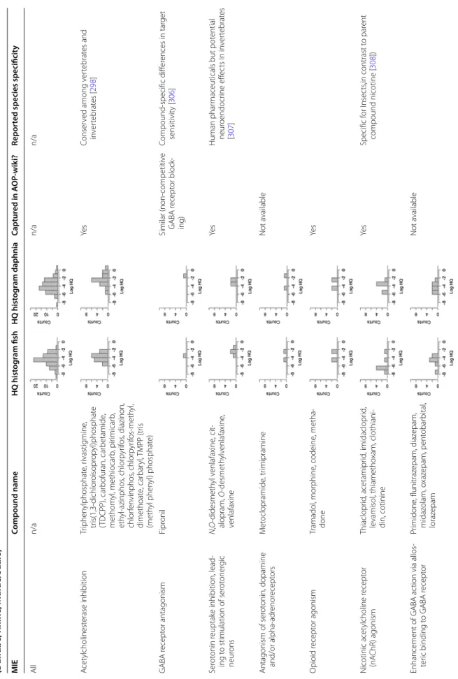
Most organophosphate insecticides are thio-phos-phoesters that require oxidation prior to causing
inhi-bition of AChE as is illustrated by diazi non in Fig. 2.
The oxidation catalyzed by cytochrome p450
monoox-ygenases transforms diazinon to diazo xon, which
binds to the esterase site on the AChE by releasing the pyrimidinol species as a leaving group (in this example 2-isopr opyl-6-methy l-4-pyrim idino l). The remaining AChE-phosphoester complex is then further hydro-lysed, leading to a so-called ageing (irreversible bind-ing) of the inhibitor-enzyme complex. AChE inhibitors that do not have a good secondary leaving group are reversible inhibitors as they do not age and can be
released again from the complex. In contrast the aged complex is fairly stable, and recovery is mainly due to new formation of AChE. In parallel to activation and inhibition, both diazinon and diazoxon can be detoxi-fied by carboxylesterases and the resulting pyrimidinol can be conjugated prior to elimination.
As detoxification is the dominant pathway in mam-mals and oxidation is the dominant pathway in inverte-brates, organophosphate insecticides are typically more
toxic to invertebrates [117] than to vertebrates [118].
Differences in species sensitivity have been explained by the interplay between activation by oxidation and detoxification. The fish carp was found to be less sen-sitive than other fish species (trout, guppy, zebrafish) despite a more sensitive AChE site because it had the
most active detoxifying enzymes [119]. Daphnia magna
was found to be more sensitive to diazinon than
Gam-marus pulex, partially due to a six times slower
detoxifi-cation by carboxylesterases, which compensated for the twice faster oxidative activation in G. pulex, but mainly due to toxicodynamic differences as observed by apply-ing a toxicokinetic–toxicodynamic (TK–TD) model to
survival data for G. pulex and D. magna [120]. TK–TD
models are especially suitable to investigate the time-dependent effects and complex mechanism of AChE inhibitors and allow estimation whether toxicokinetic or toxicodynamic parameters determine the overall
effect [121]. Interestingly, for some organophosphate
insecticides, the organism recovery is the rate-limiting
step of toxicity [121]. For an uncoupler such as penta
chlor ophen ol, the TK depuration and the TD organ-ism recovery in G. pulex are 2 and 3 days, respectively; hence, the organism has basically recovered when it has eliminated the chemicals. However, this is
differ-ent for carba ryl, chlor pyrif os and diazi non with a TK
depuration of 1.4–11 days but a much longer organism
recovery in the range of 15–28 days [121]. Hence, the
sequence of exposure matters when mixtures are
inves-tigated over a time period [122].
Such detailed studies about the uptake, metabolism and excretion of neurotoxicants are extremely impor-tant as they help to link exposure to effects which is necessary to properly assess eco-neurotoxicity for dif-ferent species.
In vitro eco‑neurotoxicity approaches
As mentioned above, the research field of neurotoxicology is mainly focused on the potential effects of chemicals on development, structure and function of the human nerv-ous system. There is general agreement within the field of toxicology that state-of-the-art toxicity testing includes tiered testing strategies that focus on the pathways that are critical for adequate functioning of cells, organs, and
organisms, using different testing strategies to collect information on exposure and toxicity. This includes an emphasis on non-animal models, including integrated genomic and proteomic analyses of chemical-induced effects. This principle is embraced in the neurotoxicology research field and many efforts are focusing on developing and optimizing non-animal test systems to model such
effects by increasing their sensitivity and specificity [123].
Non-animal test systems to test neurotoxicity include small intact organisms (zebrafish embryos, C. elegans), brain slices, cell lines, primary cell models and stem-cell derived models as well as assays assessing the tion of the bare enzyme (e.g. in the case of AChE
inhibi-tion assays) [124]. Different types of model systems have
their own advantages and disadvantages related to mul-ticellular complexity, ease of culture, variability between cultures, possibilities with regard to differentiation or genetic modification, species and costs. Test systems with
non-mammalian test organisms [125, 126] may include
ecotoxicologically relevant species, or methods to study the neurotoxicity endpoints may be modified to enable an application for other species.
Many neurotoxic mechanisms can be studied in vitro using biochemical and morphological endpoints that have the potential for medium-to-high-throughput test-ing. Parameters to investigate neuronal network func-tionality include network formation, action potential generation, calcium homeostasis, synaptic transmission, and synaptic plasticity. However, these assays were devel-oped with the aim of studying effects of chemicals on the human (mammalian) nervous system and not on wildlife. Nevertheless, if the goal is to investigate whether neuro-active chemicals or chemicals with neurodevelopmental toxicity potential are present in the aquatic environment, these in vitro cell systems may also be suitable for water quality monitoring. Both ecotoxicological model spe-cies and in vitro bioassays for molecular mechanisms are included in the Smart Integrated Monitoring (SIMONI)
framework for water quality monitoring [127].
Innovative experimental approaches are available to investigate effects on neuronal function, such as opti-cal and electrophysiologiopti-cal measurements of intra- and intercellular signalling (calcium signalling, neurotrans-mitter release and post-synaptic receptor function) in cell models to measurements of spontaneous activity or network activity in neuronal networks using
multi-elec-trode arrays (MEAs) [124]. Chemical-induced changes in
network function measured in a MEA system may be due to changes in electrical activity as well as in the release or reception of intercellular signals. MEA systems thus provide an integrated, but not pathway specific, measure for effects on neurotransmission. Efforts are ongoing to increase throughput by using multi-well MEA systems.
Primary (rodent) cell cultures can be used for routine
neurotoxicity testing [128], and efforts are ongoing to
increase throughput capacity of functional neuronal net-works of (human) embryonic stem cells and
neural/neu-ronal progenitor cells [129, 130]. The generation of large
amounts of data, either by testing many samples using high-throughput approaches or by generating high den-sity data (e.g. using MEAs or optical recordings), requires ample data storage. The resulting challenges associated with data analysis require the implementation of chem/ bioinformatics approaches.
If the aim of effect-based monitoring is to study the impact of neuroactive chemicals, or chemicals with neu-rodevelopmental toxicity potential, on ecologically rel-evant species, it is critical to consider (dis)similarities in brain development, structure and function between mammals and ecotoxicologically relevant species. Con-siderable underlying differences may exist between mam-mals and other taxa in sensitivity to neurotoxic chemicals
[131]. For example, it is well known that, due to
interspe-cies differences in kinetic parameters of AChE, insects are more sensitive to organophosphate insecticides than
mammals [132]. Moreover, sensitivity to
neurodevel-opmental effects resulting from exposure to chemicals depends critically on the phase of mammalian brain
development [33]. Specific sensitivity for neurotoxicity
dependence on exposure timing may need to be inves-tigated further using ecotoxicological model species. Additionally, neuronal network function depends
criti-cally on the presence of multiple cell types [133],
includ-ing neurons, oligodendrocytes, microglia and astrocytes. The importance of multiple cell types for eco-neurotoxi-cology may depend on the endpoint of interest. It is thus required to identify the most relevant neuronal cell types and the impact of absence or presence of other cell types in the in vitro test system under consideration.
Due to the complexity of neurotoxic and neurodevelop-mental mechanisms, and in view of potential differences between mammals and other species, it is recommended to develop a specific test set of chemicals for eco-neuro-toxicity (including, for example, water relevant chemicals and model chemicals that affect relevant mechanisms) for (interlaboratory) studies to test candidate in vitro
bio-assays (e.g. based partly Aschner et al. [134]). Emerging
techniques and innovations in neuroscience and neuro-toxicity should be closely followed and assessed for their potential and applicability in ecotoxicological water qual-ity monitoring.
Stem cells in eco‑neurotoxicity
In the area of neurotoxicity and developmental neuro-toxicity testing, the use of pluri- and multipotent stem
cells differentiating into diverse neural cell types as well as standardized methods for differentiation will lead to an improved understanding of chemically induced adverse reactions. In contrast to cell lines or primary cells, stem cells and their derivatives are neither geneti-cally transformed nor easily lose their tissue character-istics. However, differentiation conditions need to be strictly controlled to prevent differences in cell charac-teristics between cultures, which calls for appropriate control conditions to be included in toxicological testing procedures with stem cells. Neural differentiation occurs early in development and the formation of glial cells and
neurons can quite easily be mimicked in vitro [135–138].
Thus, stem cells facilitate high-throughput neurotoxic-ity testing on a wide range of neural cell types. In this context, human induced pluripotent stem cells (iPSCs) have the potential to play an important role in
predict-ing human-specific neurotoxicity and DNT [139]. Stem
cell derived neuronal and glial models allow MoA-based
DNT testing [37]. In recent years, the number of studies
using stem cells for neurotoxicity and DNT testing with a variety of endpoints increased considerably. Many rep-resentative studies investigating neurotoxicity or DNT of drugs using stem cells applied endpoints such as cell-specific cytotoxicity (apoptosis, necrosis), cell
migra-tion, intracellular Ca2+ levels, disordered differentiation,
neurite and dendrite outgrowth, neural network forma-tion and activity, as well as synaptogenesis and synaptic
activity [140–145]. Furthermore, 3D models such as
neu-rospheres are now available and provide an improved comparability to the in vivo situation. Progress on in vitro
3D brain models has also been achieved [146]. 3D brain
organoids derived from pluripotent stem cells are prom-ising experimental models for brain development, DNT testing and neurodegenerative disorders enabling
mecha-nistic pathway and stage-specific studies [147]. Despite
this good progress in neurotoxicity and DNT testing with stem cells, validated methods for several endpoints still have to be established.
Mechanisms employed in the in vitro stem cell neuro-toxicity and DNT testing are in general highly conserved in evolution. Results based on highly conserved mecha-nisms can be transferred to a variety of species, including aquatic organisms.
By now, many protocols for generating specific neu-ronal populations of different areas of the brain and peripheral nervous system using human iPSCs are
avail-able, and their efficiency is continuously increasing [148].
To date a general problem is that the protocols generate a heterogenous population comprised of the specific neu-rons they aimed for and also other cells, which may have a different identity. This increases the variability of poten-tial test readouts. Also, especially for peripheral neurons,
very long maturation times are needed to generate mature neurons, and tests performed at an earlier time point may be disturbed by the immature cellular answer of some of the cells. Additionally, some protocols suffer from a low reproducibility and relatively high variabil-ity. Thus, many repetitions are need, which renders the potential tests time and cost intensive. Current research is focusing on improving these shortcomings, and one solution is already in use: differentiating iPSCs into neu-ronal precursor cells offers the possibility to freeze and store larger amounts of cells of one differentiation, which then can be defrosted for the tests. This has the advan-tage that several tests can be run with the cells that were generated at the same time, at least until the precursor state. In most protocols for neuronal differentiation, many cells are lost during the first few days, thus making it hard to predict the cell number resulting from a sin-gle differentiation, even though a constant number of iPS cells were used. Now, one can defrost neuronal precursor cells and use a specific number of these cells for the last steps of the differentiation protocols, during which cell loss is almost negligible. This approach allows for a bet-ter control over the total number of generated neurons, thus increasing the comparability between different sets of tests.
Using gene-editing and other genetic methods progeni-tor cells can, e.g. be transduced to stably express Ngn2 under a Tet-On Advanced transactivator, allowing
differ-entiation to be switched on by adding tetracyclin [149].
Apart from mice and rats, which often serve as model systems for human diseases, iPSCs from other
mam-mals, such as farm animals [150], pets such as dogs [151]
or endangered wild animals such as felids or
orangu-tans [152, 153] are reported and offer a whole new set of
opportunities to study the impact of environmental tox-icity on their physiology.
A drawback of iPSCs is that the cells lose their epige-netic signature during reprogramming. To overcome this problem, somatic cells were used to transdifferentiate into neurons, bypassing the iPSC-state. Although to date the yield of this method is low and protocols show high variability, this allows to study age-dependent effects and may be a worthy option for eco-neurotoxicity testing in
the future (see, e.g. collection of papers here: [154]).
Cell‑free neurochemical methods
Cell-free neurochemical assays are simplified in vitro sys-tems that may help evaluate the effects of a test chemi-cal on neurobiochemichemi-cal processes. Cell-free assays are performed with cell lysates, tissue homogenates or with purified membranes (but not with living cells) and give information about direct biochemical interactions
between test molecule and biological targets like recep-tors. They form an important assay category within the US-EPA’s ToxCast program and show promise for use
in ecotoxicology as discussed by Arini et al. [155]. A
great advantage, for example, is that they are amenable for use from any species from which brain tissue can be obtained, with one study comparing responses across 20
species of fish, mammals and birds [156]. Besides
study-ing chemicals, cell-free assays have also been used to screen extracts from real-world samples including pulp
and paper mill effluents [157] and wastewater effluents
[158].
Sensory system tests in eco‑neurotoxicity
Neurotoxic effects on sensory systems are mostly studies in vertebrates. Little is known about effects in inverte-brates. Sensory structures receive information from the environment and transduce it into a signal recognizable to the nervous system. The information or stimuli can be of different modality including light, sound, smell, taste, pressure and temperature. Generally, receptive cells con-tain transmembrane receptors which undergo a confor-mational change upon stimulation. A signal transduction cascade leads to the opening of ion channels and con-comitant membrane potential changes, thereby creating an action potential. Many behaviours like feeding, mat-ing, predator avoidance, migration, social interaction and communication are crucially informed by sensory sys-tems. Thus, their impairment can have severe impact on fitness and survival of an animal.
Environmental contaminants such as pharmaceuticals, pesticides and heavy metals have been shown to interfere with the sensory structures of different species including humans and fish, thereby creating deficiencies in sensa-tion and behaviour. Behavioural output is an increasingly measured, ecologically relevant and very sensitive end-point. To localize specific sensory impairments within the nervous system using behaviour is, however, challenging because behaviour is the integrated output of multisen-sory, neuroendocrine and neuromuscular signals, and tests are often not specific enough (e.g. impaired feed-ing might result from motor deficits, impaired olfaction, impaired vision or a combination thereof).
Generally, four techniques listed in Table 1 are applied
to assess the different sensory systems like, e.g. olfaction, vision and mechanosensation (discussed below). While all of them have their advantages and drawbacks, there is no recommendation as to which one is the best. Rather, tests have to be tailored to the study purpose and a mul-tidisciplinary integrated approach is necessary to fully
Olfaction
In fish, the olfactory system is particularly vulnerable to neurotoxic contaminants because of the direct contact of olfactory sensory neurons with the surrounding water. Reduced or absent ability to smell (hyposmia or anosmia) have been shown to occur upon exposure to metals,
pes-ticides and other contaminants like, e.g. surfactants [159].
The classical method to assess olfactory impairment is by
electro-olfactography [160]. It assesses
electrophysiologi-cal changes in olfactory sensory neurons by extracellular recordings. Olfactory behavioural tests include either attraction to food extract, avoidance of skin extract or
attraction/avoidance of the chemical itself [159]. Notably,
while the zebrafish olfactory system offers several experi-mental advantages to study sensory neurobiology in gen-eral and olfactory neurotoxicity in particular, there are a number of profound differences that might render trans-lation of results from such studies to other species.
Among the major advantages of the zebrafish model are (i) identified ecologically relevant classes of natural
odours such as amino acids and bile acids [161, 162];
(ii) cultivation of the adult zebrafish head ex vivo with-out anaesthesia, allowing neurophysiological
measure-ments in the intact brain [163]; (iii) comparably small
brain size that provides access to larger fractions of
neurons by multiphoton microscopy [164]—a fact
par-ticularly true for the zebrafish olfactory system, which
contains relatively few neurons and glomeruli [165];
and (iv) large detailed data at both the single neuron and population level that allows realistic
mathemati-cal simulations of circuit function [166]. Moreover,
at first glance, the zebrafish olfactory system contains molecular and cellular constituents that appear similar in organization to the rodent olfactory system, thus, providing an attractive vertebrate model system to investigate the mechanisms underlying olfactory sys-tem development and function. However, the fish olfac-tory epithelium is of a “mixed” type, containing two major types of olfactory sensory neurons, i.e. ciliated and microvillous neurons. Both express distinct types of chemosensory receptors, project to different brain
regions and likely mediate different behaviours [167].
Moreover, the three canonical zebrafish chemosensory
gene families (or, taar and olfC/V2r) are somewhat unique and quite distinct in size and relative propor-tions to those of most tetrapods (indicative of the
diver-gence of both lineages ~ 430 million years ago [168].
The mouse has become the most widely used model system in olfactory research based on established proto-cols for genetic manipulation. An important distinction between the olfactory systems in fish and mice is stimu-lus delivery. While the fish olfactory organs are exposed to pollutants and xenobiotics that are dissolved in water, rodent noses constantly sample volatile air-borne chemi-cals at minute concentrations. Thus, the range of poten-tially hazardous chemicals that water-living species like fish are naturally exposed to will be dramatically different from the repertoire of potential harmful compounds that land-living species like mice encounter (and vice versa). The olfactory system’s remarkable capacity to renew upon perturbation also needs to be taken into account
[169]. The olfactory epithelium has extensive neurogenic
and regenerative capacity in both rodents and humans that persists throughout adult life and is unmatched
else-where in the nervous system [170]. Cells within the basal
epithelial layer function as neuronal precursors, multi-potent progenitors and/or stem cells. However, the niche signals that control the self-renewal and differentiation
of these basal cells are not well understood [170]. This
regenerative capacity will strongly impact eco-neurotox-icological assays that target the olfactory system. Accord-ingly, the system’s vulnerability will, at least to some extent, be compensated by adult neurogenesis.
Few neurotoxicological assays have been developed using mice. This is somewhat surprising given the large body of knowledge available, established animal care facilities, comparably short generation turnover and the large translational promise that rodent model systems offer. Thus, it appears likely that future eco-neurotoxi-cology assays will utilize a pipeline that spans cell-based in vitro experiments, high-throughput behavioural assays in zebrafish and other ecologically relevant species. Vision
About 3000 chemicals are toxic to the human eye and
visual system [171]. Retinotoxic effects for organic
Table 1 Techniques to test sensory system
* Depending on marker
Sensitivity Throughput Specificity Remarks
Electrophysiology +++ + +++ Link to behaviour often unclear, sophisticated preparations needed
Behaviour ++ +++ + Multisensory input; depends on proper locomotor function; high
ecological relevance
Anatomical changes + ++ +++ Only apparent when sensory function already impaired
solvents and metals have been described, not only for
humans [172] but also for fish [173–176], for which most
literature focuses on effects of methylmercury and etha-nol. Moreover, the fish retina was affected upon herbicide
[177, 178] and pesticide exposure [179] and was shown to
accumulate cocaine [180]. Electrophysiological
measure-ments of retinal function are called electroretinograms. They record the retinal sum field potential to a visual
stimulus [181] and are applied in many species including
fish [182]. Visual behaviour tests for fish are well
devel-oped, even to the point that different visual properties like motion detection, colour detection and discrimina-tion, object recognition and visual acuity can be tested
[183]. A popular assay is the measurement of optokinetic
reflex, in which the animal is presented a moving grating which it follows with accurate eye movements. Impaired visual function results in reduced or absent eye
move-ments [184, 185]. Although both techniques are widely
established in zebrafish models for human ocular
dis-eases [186], toxicant-induced impairments of visual
func-tion are only scarcely studied. Instead, retinotoxic effects are mostly assessed based on rather insensitive endpoints like histology or eye size (e.g. microphthalmia = smaller eyes).
Mechanosensation
Hair cells are sensory cells of the vertebrate inner ear and the lateral line system of aquatic vertebrates. They transduce pressure changes in the surrounding medium into a neuronal signal as a result of deflection of their cilia, which leads to the opening of ion channels, ena-bling the detection of acoustic stimuli and hydrodynamic flow. Many drugs such as aminoglycoside antibiotics, platinum-based anti-cancer drugs, anti-malarics or non-steroidal anti-inflammatory drugs are known to induce
ototoxicity in humans (see references in [187]), for
the most part irreversible. In fish, some of these drugs equally cause ototoxicity and damage to the lateral line
[188]. Moreover, metals such as copper, cadmium and
others have been shown to cause hair cell death and
deficits in behavioural responses in zebrafish [189, 190]
and other fish [188]. Behavioural responses to
acous-tic stimuli [191–193], responsiveness to water motion
[194] and rheotaxis (counter-flow swimming) [195, 196]
have been measured to assess hair cell function. Moreo-ver, vital dyes to stain hair cells have been widely used to assess the structure of lateral line hair cells in zebrafish
[197]. Effects on the lateral line hair cells are one of the
most promising sensory endpoints because of their great accessibility, amenability for staining’s and dyes and straightforward implication in rheotactic behaviour
[198]. Another method to assess hearing abilities in fish
is sound-evoked potential audiometry, which measures
field potentials in response to an auditory stimulus using
cutaneous electrodes [199].
In order to assess the full neurotoxic potential of envi-ronmental pollutants, a combination of tests and the assessment of multiple sensory systems are necessary to
precisely localize effects within the nervous system [2,
200]. Future studies should strive to increase our
mecha-nistic understanding of chemical neurotoxicity, which would help predicting eco-neurotoxicological effects. In this respect, model organisms such as the zebrafish are very helpful, because a large variety of genetic tools and genomic resources are available and many tests are already established for the analysis of human brain
dis-orders [98], but they are not yet fully adopted for
neu-rotoxicity testing. Additionally, emerging neuroscience techniques such as in vivo 2-photon calcium-imaging
of neuronal activity [201–203] or optogenetics [204]
might hold underexplored opportunities for the mecha-nistic dissection of complex neurotoxicological pro-cesses. Moreover, large-scale toxicity screenings using the zebrafish model has been implemented in the
frame-work of ToxCast and Tox21 [205–208], but more efforts
are needed to increase the specificity of tests for sensory neurotoxicity in larval zebrafish and implement them in a high-throughput manner in order to keep pace with toxicity testing of the vast number of newly registered chemicals.
Biomarkers of eco‑neurotoxicity
Biomarkers are defined as molecular, biochemical, cellu-lar and physiological changes, caused by external stress factors. The two mostly discussed groups of biomark-ers are: biomarkbiomark-ers of exposure that allow statements about the quality and/or quantity of exposure, whereas biomarkers of effect allow statements about effects and
the health status of exposed organisms [209].
Classi-cal examples for the first category are metallothioneins,
which indicate metal contamination [210]. A typical
bio-marker of effect is the induction of stress proteins (heat
shock proteins) [211] or a decrease of lysosomal stability
[212]. With the latter two one can tell that the organism
was exposed to environmental stressors, but it is not pos-sible to tell, which stressor or contaminant exactly caused
the observed effect [209]. Biomarkers can either be
meas-ured invasive/destructively, e.g. by determining enzyme inhibition in brain or whole-body homogenates or non-destructively, by determining the biomarkers of interest,
e.g. in blood, mucus or skin samples [213, 214].
Biomarkers of eco-neurotoxicity include parameters reacting specifically to neurotoxic chemicals. The most well-known biomarker of effect for neurotoxicity is the measurement of AChE inhibition. This is the primary mechanism of action of organophosphate and carbamate
insecticides. Enzyme activity is quantified in brain or whole-body homogenates of exposed organisms, and compared to reference inhibitors, as has been shown,
e.g. for zebrafish embryos [215], fish [216] and Daphnia
magna [217]. In addition, non-destructive measurement of cholinesterase is possible, e.g. by determining
butyryl-cholinesterase in blood serum [218, 219]. Alternatively,
commercially available isolated AChE can be used to test chemicals and complex environmental mixtures such as water samples. The tests applied are typically based on
the Ellman assay [220], which is a colorimetric assay that
detects the hydrolysis of the substrate acetylthiocholine. Despite its wide application in water quality assessment, the assay using isolated AChE should be used with cau-tion because concentracau-tions of organic matter as low as
2 mgC/L, when present in solid-phase extracts of typical
surface water, can act as non-specific inhibitor of AChE
[221]. This can lead to an overestimation of insecticidal
activity in ambient samples. The application of cholinest-erase biomarkers for environmental monitoring has been
reviewed by Mineau [222].
Other biomarkers of eco-neurotoxicity involve key neurotransmitter pathways and the measurement of corresponding enzyme inhibition or receptor activity, i.e. neurochemical biomarkers. Apart from AChE and the corresponding nicotinic and muscarinic receptors, activity of monoamine oxidase with dopamine or sero-tonin receptors, GABA transaminase with GABA(A) and GABA(B) receptors as well as glutamic acid decarboxy-lase and glutamine synthetase with the receptors NMDA, AMPA and Kainate can be determined. These biomark-ers have been applied in studies on a variety of organ-isms such as worms, bivalves, fish, mammalian wildlife
and birds (for review see [38]). For example, documented
measurements have included a decrease in serotonin lev-els and an increase in monoamine oxidase levlev-els in caged mussels in a river downstream of wastewater treatment
plants effluent [223]. If mussels were exposed to
pri-mary treated and ozonated effluents in a flow-through experiment, GABA levels as well as the activities of sev-eral neuroactive enzymes (glutamic acid decarboxylase, monoamine oxidase and AChE) were reduced and levels
of serotonin and dopamine increased [224]. With regard
to fish, the application of biomarkers of eco-neurotoxicity
has been reviewed in several publications [2, 225, 226].
In juvenile rainbow trout, altered brain neurotransmit-ter metabolism afneurotransmit-ter exposure to β-naphthoflavone and benzo(a)pyrene resulted in impaired availability of sero-tonin at short term (after 3 h) and increased neuronal metabolic utilization of serotonin and dopamine after 24
and 72 h [227].
For any study, it has to be taken into account that the flexibility (plasticity) of the nervous system, as well as
the age and developmental stage of the investigated organisms, may play an important role in the measured
responses [38].
Behavioural screening tests
For aquatic eco-neurotoxicological screenings, behav-ioural toxicity tests in small organisms may be of special interest. Behaviour is an understudied but sensitive and ecological relevant endpoint in ecotoxicity testing for all kinds of different species. Several studies reported effects on behaviour at concentrations orders of
magni-tudes below lethal concentrations [228, 229]. Behaviour
is the integrated response of the conditions to which an organism is exposed. A variety of activities are used as behavioural endpoints to screen for effects of
chemi-cals, for example avoidance, feeding and locomotion [1].
Some of these behavioural endpoints may be applicable to investigate rapid acute neurotoxic responses or effects of longer exposures with consequences that may have larger impact, such as neurodevelopmental effects. Such effects on behaviour may be caused by acute neurotoxic effects on neuronal functioning (inter- and intracellu-lar signalling and neuronal network function to receive, conduct, and transmit signals via chemical or electrical synapses and relay information between specific brain regions for information processing as well as learning and memory formation) or to mechanisms related to neurodevelopmental processes (proliferation, migration, differentiation, formation of axons, and dendrites, synap-togenesis, network formation and apoptosis). Automated behavioural analysis technologies allow medium-to-high-throughput assessments.
Part of behavioural disruptions resulting from chemi-cal exposure may rely on direct neurotoxicity or parental transfer, but beyond this direct relationship, behaviour is an interesting endpoint because it may also be an inte-grative indicator of several other physiological issues, e.g. metabolism, sensory organs, morphology or molecular pathways alteration.
Behavioural test with laboratory fish species
The fact that behavioural responses may be integra-tive indicators of many physiological issues explains the rapid expansion of behavioural studies in ecotoxicologi-cal research during the last decades, particularly those using fish early life stages (ELS). Indeed, fish ELS are very amenable to high-throughput monitoring evalua-tion in multi-well plates. With that system behaviour like basal activity, response to a light change or other stimuli (indicative of anxiety) can be assessed. For the photo-motor response test with zebrafish embryos, it has been even shown that different compound groups induce dif-ferent behavioural profiles which mean the test can be
used to identify neurotoxic MoAs of compounds [230]. In addition, the possibility of using behaviour as a com-plementary approach to common fish embryo toxicity (FET) tests has recently been explored, demonstrating a significantly higher sensitivity of behaviour EC50
com-pared to FET LC50 for some compounds [231]. ELS have,
however, a limited behavioural repertoire which is par-tially linked to a lower level of nervous system matura-tion compared to older stages. For example, it appears that learning associated with classical and operant con-ditioning is not efficient in zebrafish before 3 weeks of
age, which is roughly the end of the larval stage [232].
In addition, and partly related to this limitation, the pre-dictability of late outcomes from behavioural disruption
in ELS is not obvious. As an example diqua t, one of the
compounds tested by Kluver et al. showed no behavioural disruption in ELS while it induced hyperactivity in older
larvae [233].
Juveniles or adults have a much larger behavioural repertoire and/or cognitive abilities, which can easily be divided in behavioural units and evaluated. Behavioural disruptions resulting from exposure to organic
pollut-ants, including PAHs or PCBs have been characterised
using long-term dietary exposures to environmental mixtures. An increase in psychological stress is a com-mon trait observed relating to all examined pollutants
[234]. In case of PAHs, a decrease in the
neurotransmit-ter serotonin could be the driver for this behavioural
change [235]. More recently, behavioural changes in the
offspring of fish exposed to a mixture of PCBs and PBDEs
showed that even if exposed parents displayed no change in behaviour, two offspring generations showed a signifi-cant increase in anxiety as adults while behaviour of lar-vae was modified in up to four offspring generations. Behavioural tests with non‑fish species
Van der Geest et al. showed that changes in ventilation behaviour of fifth instar larvae of the caddisfly
Hyd-ropsyche angustipennis occurred at approximately 150
times lower copper concentrations than mortality of first
instar larvae [229]. Avoidance behaviour of the
amphi-pod Corophium volutator to contaminated sediments
was 1000 times more sensitive than survival [236].
Chev-alier et al. tested the effect of twelve compounds cover-ing different toxic MoA on the swimmcover-ing behaviour of daphnids and observed that most compounds induced an early and significant swimming speed increase at con-centrations near or below the 10% effective concentra-tion (48 h) of the acute immobilizaconcentra-tion test. A reducconcentra-tion in defence and orientation behaviour of rusty crayfish after exposure to nicotinoid pesticides below morpho-logical effect concentrations was observed by Sohn et al.
[237]. The clam avoidance behaviour (closing valve after
trigger) is suggested as a fast and easy screening tool
for neurotoxicants [238]. Diamesa zernyi larvae from
the wild also showed altered swimming behaviour after exposure to contaminants at low effect concentrations
[239]. These examples and numerous others all showed
that organisms may exhibit altered behaviour at rela-tively low and therefore often environmentally relevant
toxicant concentrations [240]. Behavioural responses to
toxicant exposure can also be very fast, allowing organ-isms to avoid further exposure and subsequent bioac-cumulation and toxicity. A wide array of such avoidance responses have been incorporated in ecotoxicity testing
[28, 241], including the avoidance of contaminated soil
by earthworms (Eisenia fetida) [242], feeding inhibition
of mussels (Corbicula fluminea) [238], aversive
swim-ming response to silver nanoparticles by the
unicellu-lar green alga Chlamydomonas reinhardtii [243] and by
daphnids to twelve compounds covering different toxic
MoA [240].
Field studies
Field studies are the most relevant approach to evalu-ate disruption resulting from chemical exposure but have drawbacks since it is (currently) almost impossible to disentangle the consequences of chemical exposure from other stressors such as food deprivation or envi-ronmental changes other than chemicals. On the other hand, laboratory experiments generally using optimal conditions except for the chemical exposure may lead to an underestimation of these exposure effects. They allow, however, the establishment of a potential direct link between exposure and physiological effects, includ-ing behavioural disruption. Indeed, behavioural abilities condition individual survival and fitness and hence have consequences at the population level. In the environ-ment, behaviour abilities are important, e.g. to find food, to escape predators, to find new territory or partners for mating. All these behaviours are complex and rely on simpler so-called behavioural units, which are of ultimate importance in behavioural ecology because they are what
can be measured [244].
Subtle changes in animal behaviour may affect trophic interactions and ecosystem functioning. Langer-Jaesrich et al. reported that midge larvae (Chironomus riparius)
exposed to chlor pyrif os, a neurotoxic insecticide, showed
a decrease in burrowing behaviour, resulting in an increase in the feeding rate of zebrafish preying on these
exposed chironomids [245]. However, when exposing
predators and prey simultaneously, no significant differ-ences in the feeding rate of zebrafish were observed, sug-gesting impairment in prey recognition of the exposed zebrafish. In a laboratory toxicity experiment Hunt-ing et al. (2013) observed that endpoints representHunt-ing

![Fig. 2 Mechanism of action of an AChE inhibitor on the example of the insecticide diazinon (simplified from [117])](https://thumb-eu.123doks.com/thumbv2/123doknet/13607754.424544/17.892.93.809.649.1027/mechanism-action-ache-inhibitor-example-insecticide-diazinon-simplified.webp)