HAL Id: inserm-02443557
https://www.hal.inserm.fr/inserm-02443557
Submitted on 17 Jan 2020
HAL is a multi-disciplinary open access
archive for the deposit and dissemination of
sci-entific research documents, whether they are
pub-lished or not. The documents may come from
teaching and research institutions in France or
abroad, or from public or private research centers.
L’archive ouverte pluridisciplinaire HAL, est
destinée au dépôt et à la diffusion de documents
scientifiques de niveau recherche, publiés ou non,
émanant des établissements d’enseignement et de
recherche français ou étrangers, des laboratoires
publics ou privés.
elimination and maturation
Anouar Khayachi, Carole Gwizdek, Gwénola Poupon, Damien Alcor, Magda
Chafai, Frédéric Cassé, Thomas Maurin, Alessandra Folci, Fabienne de
Graeve, Sara Castagnola, et al.
To cite this version:
Anouar Khayachi, Carole Gwizdek, Gwénola Poupon, Damien Alcor, Magda Chafai, et al..
Sumoyla-tion regulates FMRP-mediated dendritic spine eliminaSumoyla-tion and maturaSumoyla-tion. Nature CommunicaSumoyla-tions,
Nature Publishing Group, 2018, 9 (1), pp.757. �10.1038/s41467-018-03222-y�. �inserm-02443557�
Sumoylation regulates FMRP-mediated dendritic
spine elimination and maturation
Anouar Khayachi
1
, Carole Gwizdek
1
, Gwénola Poupon
1
, Damien Alcor
2
, Magda Chafai
1
, Frédéric Cassé
1
,
Thomas Maurin
1
, Marta Prieto
1
, Alessandra Folci
1
, Fabienne De Graeve
3
, Sara Castagnola
1
, Romain Gautier
1
,
Lenka Schorova
1
, Céline Loriol
1
, Marie Pronot
1
, Florence Besse
3
, Frédéric Brau
1
, Emmanuel Deval
1
,
Barbara Bardoni
4
& Stéphane Martin
4
Fragile X syndrome (FXS) is the most frequent inherited cause of intellectual disability and
the best-studied monogenic cause of autism. FXS results from the functional absence of the
fragile X mental retardation protein (FMRP) leading to abnormal pruning and consequently to
synaptic communication defects. Here we show that FMRP is a substrate of the small
ubiquitin-like modi
fier (SUMO) pathway in the brain and identify its active SUMO sites. We
unravel the functional consequences of FMRP sumoylation in neurons by combining
mole-cular replacement strategy, biochemical reconstitution assays with advanced live-cell
ima-ging. We
first demonstrate that FMRP sumoylation is promoted by activation of metabotropic
glutamate receptors. We then show that this increase in sumoylation controls the
homo-merization of FMRP within dendritic mRNA granules which, in turn, regulates spine
elim-ination and maturation. Altogether, our
findings reveal the sumoylation of FMRP as a critical
activity-dependent regulatory mechanism of FMRP-mediated neuronal function.
DOI: 10.1038/s41467-018-03222-y
OPEN
1Université Côte d’Azur, CNRS, IPMC, 06560 Valbonne, France.2Université Côte d’Azur, INSERM, C3M, 06200 Nice, France.3Université Côte d’Azur,
CNRS, INSERM, iBV, 06108 Nice, France.4Université Côte d’Azur, INSERM, CNRS, IPMC, 06560 Valbonne, France. These authors contributed equally:
Anouar Khayachi and Carole Gwizdek. Correspondence and requests for materials should be addressed to S.M. (email:[email protected])
123456789
I
n neurons, messenger RNA (mRNA) targeting to synapses and
local synthesis of synaptic proteins are tightly regulated.
Indeed, dysregulation of such processes leads to structural
synaptic abnormalities and consequently to neurological
dis-orders
1classified as synaptopathies
2. Among them, the fragile X
syndrome (FXS) is the most frequent form of inherited
intellec-tual disablility and a leading monogenic cause of autism with the
prevalence of 1:4000 males and 1:7000 females. FXS results from
mutations within the FMR1 gene causing the loss of function of
the RNA-binding protein FMRP. Localization studies revealed
that FMRP is highly expressed in the central nervous system.
FMRP binds a large subset of mRNAs in the mammalian brain
and is a key component of RNA granules. These granules
transport mRNA along axons and dendrites and are targeted to
the base of active synapses to regulate local translation in an
activity-dependent manner
3–5. Therefore, the transport and the
subsequent regulation of local translation are critical processes to
brain development as they play essential roles in stabilizing and
maturing synapses
3,4. According the role of FMRP in regulating
translation at synapses, the loss of FMRP function in FXS leads to
a patholological hyperabundance of long thin immature dendritic
protrusions called
filopodia
6,7. These structural defects result
from an abnormal post-synaptic maturation and/or a failure in
the synapse elimination process
8. An increased number of
immature spines associated with severe changes in synaptic
transmission and plasticity as well as in social and cognitive
behaviors have also been reported in Fmr1 knockout (Fmr1
−/y)
mouse models for FXS
4,9,10.
The majority of FMRP-containing mRNA granules localizes at
the base of dendritic spines
3,4. Neuronal activation leads to the
release of mRNAs from dendritic granules and their local
trans-lation at synapses (for a review, see ref.
5). Importantly, this
activity-dependent process requires a tight spatiotemporal
reg-ulation involving many protein–protein interactions. Such a
regulation is mainly governed by post-translational modifications
(PTMs). Previous reports have shown that FMRP function is
regulated by phosphorylation, which inhibits translation of its
associated mRNAs, whereas dephosphorylation of FMRP
pro-motes their translation
11,12. Activation of metabotropic glutamate
receptor 5 (mGlu5R) induces dephosphorylation of FMRP and its
subsequent ubiquitination, which ultimately leads to FMRP
degradation via the ubiquitin-proteasome pathway
13,14. Thus, a
deeper comprehension of the activity-dependent molecular
mechanisms controlling FMRP is absolutely criticial to
under-standing the functional regulation of FMRP-mediated mRNA
transport and local protein synthesis in physiological and
pathological conditions, including FXS.
Sumoylation is a PTM involved in many cellular signaling
pathways. It consists in the covalent enzymatic conjugation
of the small ubiquitin-like modifier (SUMO) protein to specific
lysine residues of substrate proteins
15,16. The sumoylation
process requires a dedicated enzymatic pathway
17–19. SUMO
paralogs (~100 amino acids; ~11 kDa) are conjugated to its
substrates via the action of the E2-conjugating enzyme Ubc9.
Sumoylation is a reversible process due to the activity of specific
desumoylation enzymes called Sentrin-proteases (SENPs
20). At
the molecular level, sumoylation can modulate the dynamics of
multi-protein complexes by preventing protein–protein
interac-tions and/or by providing new binding sites for novel
interactors
21,22.
Sumoylation regulates a wide range of neurodevelopmental
processes
18,19,23. For instance, our group has demonstrated the
spatiotemporal regulation of the SUMO system in the developing
rat brain
24and that sumoylation is regulated by neuronal
activ-ity
25and the activation of mGlu5R
26. Sumoylation also influences
various
aspects
of
the
neuronal
function
including
neurotransmitter release
27,28, spinogenesis
29,30, and synaptic
communication
31–33.
Here, we report that FMRP is a novel sumoylation substrate in
neurons. We demonstrate that FMRP sumoylation is absolutely
essential to maintaining the shape of mRNA granules in dendrites
and to controlling both the spine density and maturation. We
identify the active SUMO sites on FMRP and show that activation
of mGlu5R rapidly induces FMRP sumoylation triggering the
dissociation of FMRP from dendritic RNA granules to allow for
local translation. Altogether, our
findings shed light on
sumoy-lation as an essential activity-dependent mechanism that tunes
spine elimination and maturation in the mammalian brain.
Results
FMRP is sumoylated in vivo. Given the critical importance of
FMRP in brain development and maturation, it is of particular
interest to understand the molecular mechanisms regulating
FMRP function. Thus, we investigated whether FMRP is
sub-jected to sumoylation. To this end, we performed immunoblot
analyses and control assays using several commercial as well as
in-house anti-FMRP and anti-SUMO1 antibodies on rodent brain
homogenates (Fig.
1
; Supplementary Fig.
1
). We
first analyzed rat
brain homogenates in absence or presence of NEM (N-ethyl
maleimide), which protects proteins from desumoylation during
the lysis process
31(Fig.
1
a; Supplementary Fig.
1
f). FMRP is
detected as isoforms ranging from 70 to 90 kDa. Interestingly, we
found a higher molecular weight band at ~120 kDa that was
detected only in the presence of NEM (Fig.
1
a, total lane). The
densitometric analysis of the ratio between the sumoylated form
of FMRP and the total level of FMRP in NEM-treated input lanes
revealed that there is about 4% of sumoylated FMRP in all the
conditions tested (Supplementary Fig.
1
c). We confirmed the
upper band to be the sumoylated form of FMRP by
immuno-precipitation experiments with specific anti-FMRP antibodies and
anti-SUMO1 immunoblot (Fig.
1
b) or with the converse
experi-ment using anti-SUMO1 immunoprecipitation and anti-FMRP
immunoblot (Fig.
1
c). We also validated the sumoylation of
FMRP in wild-type (WT) mouse brain homogenates (Fig.
1
d).
Accordingly, we were also able to co-immunoprecipitate the sole
SUMO-conjugating enzyme Ubc9 from mouse brain
homo-genates using anti-FMRP antibodies (Fig.
1
e). We further
vali-dated the sumoylation of FMRP in vivo using several
combinations of FMRP/SUMO1 antibodies (Supplementary
Fig.
1
d, e, g–j) or in cultured neurons (Supplementary Fig.
1
k–n).
Immunolabeling experiments (Fig.
1
f) showed that FMRP
par-tially co-localizes with Ubc9 and SUMO1 in dendrites of mouse
hippocampal neurons, providing further evidence of the interplay
between FMRP and the SUMO pathway.
Sumoylation consists in the covalent binding of the SUMO
moiety to a lysine residue of the consensus sequence on the
substrate protein (ΨKxD/E, where Ψ is a large hydrophobic
residue, K is the target lysine, x can be any residue, and D/E are
aspartate or glutamate
34). To identify lysine residues in FMRP
potentially targeted by the sumoylation system, we used
SUMO-prediction softwares to analyze the primary sequence of FMRP
and then alignment tools to assess whether these potential sites
are evolutionary conserved across species (Fig.
1
g). We identified
three conserved residues, two proximal (K88, K130) and one
distal (K614) lysines as putative targets of the SUMO system. To
validate whether these lysine residues could be sumoylated, we
performed site-directed mutagenesis combined with bacterial
sumoylation assays
31,35(Fig.
1
h, i). We demonstrated that FMRP
sumoylation occurs at these residues (K88, K130, and K614) and
showed that their mutation into arginine residues (K-to-R
mutation) abolishes the sumoylation of FMRP (Fig.
1
h, i). We
confirmed these data using sumoylation assays in mammalian
COS7 cells in which the expression of the FMRP-K88,130,614R
mutant prevents the sumoylation of FMRP (Fig.
1
j). Consistent
with the sumoylation of FMRP in the brain and according to our
FMRP-SUMO1 models, which were computed using crystal
structures available for the N-terminal part of FMRP, both lysine
residues (K88 and K130) are clearly exposed and accessible to
sumoylation with a solvent accessible surface area (ASA) of ~70%
and ~45%, respectively (Fig.
1
k, l). We therefore conclude that
FMRP is a SUMO substrate in vivo and that its sumoylation can
occur at its N-terminal K88 and K130 and C-terminal K614
residues.
25 15 Mr (K) 25 – – – – – – – + + + + + + + + + + + + + + + + + + + + + + + + + + + + + + + Mr (K) 100 FMRP- mCherry-SUMO1 FMRP IB FMRP mCherry-SUMO1: mCherry-SUMO1ΔGG: Ubc9: – + + + + + + + + – – – – – – FMRP WT FMRP-K88, 130,614R 130 Mr (K) Human Chimpanzee Pig Dog Cat Mouse Rat XenopusSheep Duck Rabbit 139 610 614 130 88 Merge FMRP Ubc9f
Merge FMRP SUMO1 15 Input IB Ubc9 Mr (K) Ubc9 IgG IP IgG FMRPe
70FMRP Mander’s overlap coefficient 130 100 IB FMRP Mr (K) FMRP- SUMO1 IP IgG SUMO1
d
*
70 0.4 0.3 0.2 0.1 0 Ubc9 SUM01 130 100 IB FMRP Mr (K) FMRP- SUMO1 IP IgG SUMO1c
70 130 100 IP IB SUMO1 Mr (K) FMRP- SUMO1 IgG FMRPb
70a
Input NEM: – + 130 100 Mr (K) IB FMRP FMRP- SUMO1 FMRPg
IB FMRP IB SUMO1E1, E2, SUMO1: His-FMRP N-ter: FMRP- SUMO1 FMRP N-ter WT K130RK88R WT K88,130RWTWT K130RK88RK88,130R FMRP- SUMO1 FMRP C-ter IB FMRP IB SUMO1
E1, E2, SUMO1: His-FMRP C-ter: WT K614R WT WT WT K614R
i
h
j
k
l
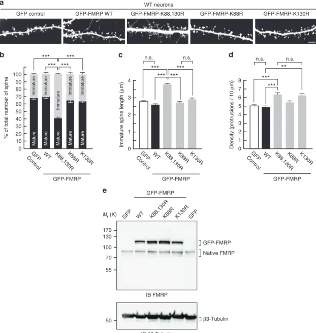
K88 SUMO1 SUMO1 35 K130FMRP sumoylation participates in dendritic spine regulation.
FMRP is essential to proper spine stabilization and maturation
3,4.
In FXS patients, the lack of functional FMRP leads to an
immature neuronal morphology with a characteristic excess of
abnormally long and thin
filopodia
36. Similar morphological
defects are also present in Fmr1
−/ymouse brains
37. Thus, we
hypothesized that FMRP sumoylation could be critical in
main-taining the density and the maturation of dendritic spines. To
address this point, we used attenuated Sindbis particles
38–40to
express either free green
fluorescent protein (GFP), the WT
GFP-FMRP, the N-terminal K88,130R, C-terminal K614R or
non-sumoylatable K88,130,614R GFP-FMRP mutants in cultured
Fmr1
−/yneurons at 17 days in vitro (17 DIV). We then analyzed
and compared the morphology of dendritic spines 24 h post
transduction (Fig.
2
a, b). In GFP-expressing Fmr1
−/yneurons,
~60% of protrusions showed an immature phenotype (see
Methods for the spine characterization; Fig.
2
a, b). Conversely,
the expression of either FMRP WT or the K614R GFP-FMRP
mutant, which behaves as the WT, promoted spine maturation
(Fig.
2
a, b). In stark contrast, expressing either the N-terminal
K88,130R or the non-sumoylatable K88,130,614R GFP-FMRP
mutant failed to promote spine maturation (Fig.
2
a, b).
The excess of dendritic protrusions in neurons is a hallmark of
FXS
6,7. Interestingly, the density of the protrusions was
considerably decreased upon the expression of the WT or
K614R mutant form of GFP-FMRP (Fig.
2
c; GFP control, 7.22 ±
0.16 protrusions per 10
μm; GFP-FMRP WT, 5.34 ± 0.13
protru-sions per 10
μm; GFP-FMRP-K614R, 5.39 ± 0.13 protrusions per
10
μm), whereas expressing either the N-terminal K130R, the
K88,130R FMRP mutants, or the non-sumoylatable
GFP-FMRP-K88,130,614R did not affect the spine density with
measured values almost identical to control neurons expressing
free GFP (Fig.
2
c). Furthermore, re-expressing WT GFP-FMRP in
Fmr1
−/yneurons not only affected the spine number but also
drastically reduced the mean length of immature spines from
~3.7
μm to <2.6 μm (Fig.
2
d).
To individually assess the role of the N-terminal lysine residue,
we quantified the morphological changes occuring in Fmr1
−/yneurons expressing GFP-FMRP with a single mutated lysine
residue (K88R or K130R; Supplementary Fig.
2
). While the
expression of both mutants promoted spine maturation similarly
to GFP-FMRP WT (Supplementary Fig.
2
b, d), the K130R mutant
failed to reduce the density of the protrusions (Supplementary
Fig.
2
c; GFP control, 7.22 ± 0.16 protrusions per 10
μm; WT, 5.34
± 0.13 protrusions per 10
μm; K130R, 6.48 ± 0.15 protrusions per
10
μm) indicating that the integrity of the K130 residue is
essential to maintain spine density. Altogether, the data above
indicate that the integrity of both N-terminal lysine residues is
critical for the regulation of spine density and maturation since
the expression of the K-to-R mutant forms failed to restore the
density and the maturity of dendritic spines in Fmr1
−/yneurons.
Our initial
findings therefore support the role of the N-terminal
sumoylation of FMRP in the regulation of spine elimination and
maturation events.
To start assessing the functional effect of FMRP sumoylation,
we compared synaptic transmission by measuring spontaneous
miniature excitatory post-synaptic currents (mEPSCs) in Fmr1
−/yneurons expressing either GFP-FMRP WT or its
non-sumoylatable K88,130,614R mutant (Supplementary Fig.
3
). The
comparison of cumulative distributions indicated that the
amplitude of mEPSCs (from 20 to 40 pA) was significantly
increased in neurons expressing the mutant form of GFP-FMRP
(Supplementary Fig.
3
a, b). Moreover, intervals between mEPSC
events (between 300 ms and 1 s) were slightly but significantly
increased upon expression of GFP-FMRP-K88,130,614R when
compared to GFP-FMRP WT indicating that the mEPSC
frequency is decreased in mutant-expressing cells (Supplementary
Fig.
3
a, c). Data comparing mEPSC properties in WT and Fmr1
−/ybrain slices have been described in the literature with either a
decrease, an increase or no changes in their amplitudes or
frequencies, depending on the brain area recorded, the age of the
animals, and/or the associated genetic background
41–43. To our
knowledge, there are no available data on mEPSCs recorded from
FMRP WT-expressing Fmr1
−/ycultured hippocampal neurons
and the results from Supplementary
fig.
3
indicate that restoring
the expression of FMRP in Fmr1
−/yneurons leads to changes in
basal synaptic transmission, occurring most probably via both
pre- and post-synaptic modifications. Additional experiments are
now needed to precisely define the associated mechanisms and to
address the electrophysiological consequences of FMRP
sumoyla-tion in synaptic plasticity in vivo.
Preventing FMRP sumoylation alters the size of mRNA
gran-ules. Since FMRP is an RNA-binding protein, we also examined
whether the mutation of the sumoylation sites interferes with the
RNA-binding capacity of FMRP by performing cross-linking and
immunoprecipitation (CLIP) assays (Fig.
2
e, f). FMRP-CLIPed
mRNAs from Fmr1
−/yneurons expressing either the WT or
K88,130,614R forms of GFP-FMRP were analyzed by quantitative
PCR to compare the abundance of some known FMRP
target mRNAs (Fig.
2
e). Our data showed that either forms of
GFP-FMRP are able to bind target RNAs to similar extent
(Fig.
2
f).
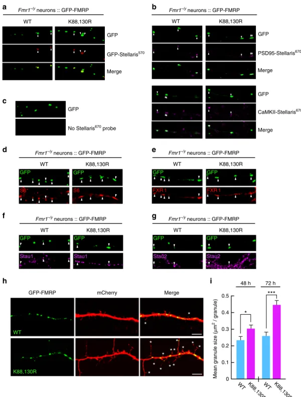
Fig. 1 FMRP is sumoylated in vivo in the rat and mouse brain and the SUMO system targets the conserved residues K88, 130, and 614 of FMRP. a Representative immunoblot anti-FMRP (Ab#056) of P7 post-nuclear rat brain extracts prepared or not in the presence of the cysteine protease inhibitor NEM to prevent desumoylation.b Immunoblot anti-SUMO1 of NEM-treated P7 post-nuclear rat brain extracts subjected to immunoprecipitation with FMRP (Ab#056) antibody or control IgG.c Converse immunoblot with anti-FMRP (Ab#056) antibody of NEM-treated P7 post-nuclear rat brain extracts subjected to immunoprecipitation with SUMO1 antibody or control IgG.d Immunoblot anti-SUMO1 of NEM-treated P1 post-nuclear mouse brain extracts subjected to immunoprecipitation with FMRP (Ab#056) antibody or control IgG. *Non-specific band. e Immunoblot of post-nuclear mouse brain extracts (input) subjected to immunoprecipitation with FMRP antibody or control IgG and probed with anti-Ubc9 antibody.f Co-localization assays performed on cultured mouse neurons (20 DIV) with antibodies directed against Ubc9, FMRP (Ab#4317), SUMO1. Bar, 2μm. Degree of co-localization (Manders’ coefficient) between FMRP and Ubc9 or SUMO1. N = 3 independent primary cultures with 60 dendrites analyzed for each condition. g Sequence alignments showing the evolutionary conservation of the potential SUMO-targeted lysine residues (stars) within the consensus sumoylation sites of FMRP. h, i Bacterial sumoylation assay. Representative immunoblots of purified fractions of N- and C-terminal WT or mutated parts of His-FMRP in a recombinant bacterial system and probed with anti-FMRP (h, Ab#1C3) or (i, #17722) and anti-SUMO1 antibodies as indicated. j COS7 sumoylation assay. Immunoblots with anti-FMRP (Ab#056) antibody of full-length WT or lysine-mutated FMRP expressed in COS7 cells with mcherry-SUMO1 WT or mutated (ΔGG) to prevent its conjugation.k Original X-ray structuresfitted of three human N-terminal FMRP (PDB: 4OVA in green, 4QVZ in light green, 4QW2 in dark green) shown in cartoon representation. K88 and K130 are shown in sphere representation in red and blue, respectively.l Original model of FMRP (PDB: 4OVA) and SUMO1 (PDB: 4WJQ) structural links in cartoon and surface representation (with transparency), respectively, in green and light blue. Lysine residues 88 and 130 of FMRP are shown in sphere representation in red and blue, respectively
Since preventing FMRP sumoylation with the K-to-R
muta-tions does not affect the ability of FMRP to interact with its target
RNAs, we hypothesized that FMRP sumoylation is involved in
the transport of mRNAs along dendrites. To this purpose, we
first
examined the FMRP-containing granules along dendrites. We
transfected Fmr1
−/yneurons to express either the WT or
K88,130R form of GFP-FMRP and performed smFISH
experi-ments using Stellaris probes complementary to three known
FMRP mRNA targets: GFP (for GFP-FMRP), PSD-95
44, and
CaMKII mRNAs (Fig.
3
a–c). Interestingly, the fluorescence of all
e
2 4 0 5 3 1 6 7 Density (protrusions / 10 μ m) GFP Control WT K88,130,614R GFP-FMRP 8 K88,130RK614R Immature Mature 20 40 0 50 30 10 70 90 80 60 100***
***
***
***
***
***
***
***
***
GFP Control WT K88,130,614R% of total number of spine
Immature Immature Mature Mature K88,130R Immature Mature GFP-FMRP
c
b
GFP control Fmr1–/y neurons GFP-FMRP WT GFP-FMRP-K88,130,614R GFP-FMRP-K88,130Ra
1 2 0 3Immature spine length (
μ m) GFP Control WT K88,130,614R 4 K88,130R GFP-FMRP K614R
d
GFP-FMRP-K614R K614R Mature Immature –2.5 0 –5 2.5 5 Fold enrichment (IP/input) 10 fmr1map1b camk2a sapap3 fxr1 kif3c psd95 tubb3 7.5 GFP-FMRP WT GFP-FMRP-K88,130,614R 100 GFP-FMRP Mr(K) GFP-FMRP Input WT K88,130,614R IB FMRP IP FMRP GFP GFP-FMRP WT K88,130,614R GFP
f
Fig. 2 The N-terminal sumoylation of FMRP is involved in the regulation of the spine density and maturation. a Representative confocal images of dendrites from transduced Fmr1−/yneurons expressing free GFP, the WT or the non-sumoylatable K88,130,614R, K88,130R, or K614R forms of GFP-FMRP for 24 h. Bar, 10μm. Enlargements of dendrites are also shown. Bar, 5 μm. Histograms show the relative proportion of mature and immature dendritic spines b and the density of the protrusionsc in GFP, in WT, and mutated GFP-FMRP-expressing cells as shown in a. d Histograms of immature spine length measured from Fmr1−/yneurons expressing the indicated constructs. Data shown inb–d are the mean ± s.e.m. and statistical significance determined by a one-way analysis of variance (ANOVA) with a Bonferonni post-test. N = ~4500 protrusions per condition from four independent experiments. ***p < 0.001. e, f CLIP analysis from transduced Fmr1−/ycortical neurons expressing the WT or the K88,130,614R form of GFP-FMRP revealed that they bind the same RNA repertoire.e Representative immunoblots anti-FMRP of the indicated neuronal extracts subjected or not (Input) to immunoprecipitation (IP) with FMRP antibodies. GFP-expressing Fmr1−/yneurons were used as a negative control.f Enrichment (CLIPed/Input) of a set of FMRP-target RNA fragments in the indicated conditions. Several known RNA targets of FMRP (fmr1, map1b, camk2a, sapap3, fxr1, kif3c, and psd95) as well as a non-targeted RNA (tubb3) were detected by quantitative PCR. Fold enrichment were calculated as described in the Methods section and did not show any statistical differences
Fmr1–/y neurons :: GFP-FMRP WT K88,130R Stau1 Stau1 GFP GFP WT K88,130R GFP-FMRP mCherry Merge
*
*
*
* *
*
*
*
*
*
*
*
* *
*
Mean granule size (
μ m 2 / granule) 0.2 0 0.1 0.3 0.5 0.4
*
***
WT K88,130RWT K88,130R 48 h 72 hi
h
Stau2 GFP Stau2 GFP Fmr1–/y neurons :: GFP-FMRP WT K88,130R Fmr1–/y neurons :: GFP-FMRP WT K88,130R GFP FXR 1 GFP FXR 1 Fmr1–/y neurons :: GFP-FMRP WT K88,130R S6 GFP S6 GFP PSD95-Stellaris670 GFP Merge Fmr1–/y neurons :: GFP-FMRP WT K88,130Rb
GFP-Stellaris570 GFP Merge Fmr1–/y neurons :: GFP-FMRP WT K88,130Ra
d
c
e
No Stellaris670 probe GFPg
f
CaMKII-Stellaris670 GFP MergeFig. 3 Preventing FMRP sumoylation drastically impacts on the size of dendritic FMRP-containing mRNA granules. a–c Representative images of WT and K88,130R GFP-FMRP-expressing Fmr1−/ydendrites were hybridized with GFPa, PSD-95 b, or CaMKII mRNA b, using Stellaris probes. Arrowheads show the co-localization between the indicated Stellaris signals and the GFP-FMRP granules.c GFP-FMRP-transfected neurons with no Stellaris probes were used as FISH controls.d–g Co-localization assays performed on WT and K88,130R GFP-FMRP-expressing Fmr1−/yneurons with antibodies directed against the S6 ribosomal proteind, FXR1 e, and the RNA-binding proteins Staufen 1 f and Staufen 2 g. Arrowheads indicate the co-localization with the GFP-FMRP positive mRNA granules.h Representative confocal images of dendrites from co-transfected Fmr1−/yneurons co-expressing free mCherry with either the WT or the K88,130R form of GFP-FMRP for 72 h. Bar, 5μm. i Histograms show the mean size of dendritic GFP-FMRP granules after 48 and 72 h of expression. N = 190–460 granules per condition from three to four separate experiments. Data shown in i are the mean ± s.e.m. and statistical significance was determined using unpaired t test. *p < 0.05; ***p < 0.0001
three probe sets was detectable in GFP-positive granules from
secondary dendrites containing either the WT or mutant
K88,130R form of GFP-FMRP (Fig.
3
a–c). Together with the
CLIP experiments (Fig.
2
e, f), this reveals that both WT and
K88,130R GFP-FMRP-containing granules can travel along
dendrites, carrying their mRNA cargoes.
We further characterized these mRNA granules using
co-localization assays to investigate whether known components of
such granules
45,46are also present in WT and
K88,130R-GFP-FMRP positive granules. As clearly depicted in Fig.
3
d–g, both the
WT and K88,130R GFP-FMRP granules co-localize with the
ribosomal protein S6 (Fig.
3
d) and the RNA-binding proteins
FXR1 (Fig.
3
e), Staufen 1 (Fig.
3
f), and Staufen 2 (Fig.
3
g),
indicating that these granules contain not only some of the target
mRNAs of FMRP (Fig.
3
a–c) but also several described
components of such dendritic mRNA granules
45,46.
We then measured the surface of dendritic
GFP-FMRP-positive mRNA granules at different time points post transfection
(Fig.
3
h, i). Interestingly, the expression of the K88,130R
GFP-FMRP for 48 h significantly increased the size of GFP-
FMRP-containing granules compared to the WT GFP-FMRP-positive
granules (Fig.
3
i; WT 48 h, 0.236 ± 0.017
μm
2; K88,130R 48 h,
0.305 ± 0.020
μm
2). The difference in granule size between the
WT and the K88,130R form of GFP-FMRP was further enhanced
after 72 h of transfection (Fig.
3
i; WT 72 h, 0.265 ± 0.020
μm
2;
K88,130R 72 h, 0.440 ± 0.030
μm
2). All these data reveal that the
expression of GFP-FMRP K88,130R results in larger
FMRP-containing dendritic mRNA granules suggesting that FMRP
WT K88,130R D+A D D+A D 2.0 1.8 1.9 2.1
***
**
Lifetime, Tau (ns) 1.7c
0.02 0.04 0 0.06 0.08 0.1 Lifetime, Tau (ns) D+A D 1.6 1.8 2.0 2.2 2.4 2.6 Frequency 0.02 0.04 0 0.06 0.08 Frequency 0.1 Lifetime, Tau (ns) D+A 1.6 1.8 2.0 2.2 2.4 2.6 Da
2.2 1.7 Tau (ns) Donor (GFP-FMRP WT) + Acceptor (mCherry-FMRP WT) Donor (GFP-FMRP-K88,130R) + Acceptor (mCherry-FMRP-K88,130R) 2.2 1.7 Tau (ns)b
Fig. 4 Preventing the N-terminal sumoylation of FMRP by the K88,130R mutation does not alter the homomeric FMRP–FMRP interaction within dendritic mRNA granules.a, b Analysis of GFP-FMRP/mCherry-FMRP interaction within dendritic mRNA granules byfluorescence lifetime imaging (FLIM). Representative confocal images showing the co-localization of the WTa or the K88,130R b forms of GFP-FMRP and mCherry-FMRP (left images) in dendritic granules; bar, 4μm. FLIM images of the same field are shown on the right images a, b where fluorescence lifetime is represented using a pseudo-color scale ranging from 1.7 to 2.2 ns. Insets show representative clusters for each condition; bar, 1μm. The third row represents the distribution histograms of GFP-FMRPfluorescence lifetime of the donor (D) alone in green and the donor + acceptor (D + A) in blue. FLIM images corresponding to the donor alone condition are displayed in Supplementary Fig.3b.c Box and whiskers plots show the variation of the lifetime determined from FLIM curves. This representation displays upper and lower quartiles, maximum and minimum values in addition to median. N = 114–189 granules per condition from three separate experiments. Statistical significance in c was determined by a non-parametric Mann–Whitney test. **p < 0.01; ***p < 0.0001
70 70 50 2 1.5 0.5 0 1 FMRP sumoylation (fold increase) 0 200
***
150 100 Half time (s) 26 WT WT + DHPG 27 50 0 300***
200 100 Half time (s) 27 29 WT K88,130R Dendra-FMRP WT Dendra-FMRP-K88,130R 0 Time, s 0.2 0.4 0.6 0.8 1.0 F/F 0 600 500 400 300 200 100d
a
Fmr1–/y neurons WT K88,130R Dendra2-FMRPc
e
f
Dendra2-FMRP photoconversion 50 100 200 600 –10 Time, s 0 WT K88,130Rb
Ctl DHPG 1′ 5′ 130 Mr (K) IP FMRP IgG FMRP- SUMO1 IP DHPG:*
– 1′ 5′+ + – 0 300 200 100 Half time (s) 29 K88,130R + DHPG K88,130R 29g
IB FMRP IB β3-Tubulin Input 5′ IB FMRP IP FMRP (10%) FitFig. 5 Activation of mGlu5 receptors promotes FMRP sumoylation and leads to the release of FMRP from dendritic mRNA granules. a Images of transfected Fmr1−/ydendrites expressing the WT or the non-sumoylatable K88,130R forms of GFP-FMRP before Dendra2-FMRP photoconversion are shown.b Time lapse series of confocal images of photoconverted Dendra2-FMRP redfluorescence in dendritic granules in basal unstimulated conditions. Enlargement of dendritic granules from the boxed area ina is also shown on the left. The decrease in red photoconverted Dendra2-FMRPfluorescence was then monitored over time. Scale bar, 1μm. c Representative sample recording traces of normalized fluorescence from photoconverted WT or mutated Dendra2-FMRP in individual granules in basal unstimulated conditions. The thin traces (black) represent the correspondingfits. d Histograms with scatter plots of computed half-time of photoconverted WT and K88,130R Dendra2-FMRPfluorescence diffusion in granules in basal conditions. The number of photoconverted granules is indicated on the bars.e Immunoprecipitation of FMRP (Ab#046) and immunoblotting for SUMO1. Control for the immunoprecipitated FMRP fractions is also depicted. Input lanes for FMRP andβ3-tubulin are also shown. Quantification for DHPG-induced endogenous FMRP sumoylation in neurons over time is also indicated. The data are from three separate experiments and show the mean ± s.e.m. *p = 0.0213. f Histograms with scatter plots of half-time of photoconverted Dendra2-FMRP WTfluorescence diffusion in granules from Fmr1−/yneurons stimulated with DHPG. The number of
photoconverted granules is indicated on the bars and the histogram/scatter plot in absence of stimulation is taken fromd. g Histograms with scatter plots of half-time of photoconverted Dendra2-FMRP-K88,130Rfluorescence diffusion in granules in basal and DHPG-stimulated conditions. The histogram/ scatter plot in absence of stimulation is taken fromd. The number of photoconverted granules is indicated on the bars. Data shown in d–f and g are the mean ± s.e.m. Statistical significance in d, f, and g was determined using a non-parametric Mann–Whitney test. Statistical significance in e was determined by an ANOVA with a Bonferroni post-test. *p < 0.05; ***p < 0.0001
sumoylation could participate in the regulation of FMRP
interactions within these granules.
FMRP has been reported to form homodimers via its
N-terminal 1–134 domain
47, where the sumoylatable K88 and K130
residues are localized. Thus, to assess whether the difference in
granule size measured in Fig.
3
i results from abnormal interaction
properties of FMRP homodimers directly inside dendritic
granules, we performed
fluorescence lifetime imaging microscopy
(FLIM) experiments on neurons co-expressing WT or K88,130R
GFP-FMRP with their respective WT or K88,130R
mCherry-tagged constructs (Fig.
4
; Supplementary Fig.
4
). We observed a
clear co-localization of the mCherry/GFP-FMRP constructs in
dendritic granules confirming the incorporation of the proteins
into granules (Fig.
4
a, b). The energy transfer known as
fluorescence resonance energy transfer from donor green
fluorescent protein (GFP) toward the acceptor mCherry is
quantified by the reduction of the donor fluorescence lifetime
(Fig.
4
c). We measured a significant reduction of the donor
GFP-FMRP
fluorescence lifetime in presence of mCherry-FMRP
indicating that FMRP/FMRP interaction occurs in dendritic
granules. Interestingly, we also found that this homomeric
interaction is not affected by the K88,130R mutations (Fig.
4
c).
Sumoylation triggers FMRP dissociation from mRNA
gran-ules. Our results so far indicate that preventing FMRP
sumoy-lation directly impacts on the morphology of mRNA granules in
dendrites (Fig.
3
h, i) without altering the intrinsic FMRP/FMRP
interacting properties within the granules (Fig.
4
). Therefore, we
investigated whether the absence of FMRP sumoylation affects
the dissociation of FMRP from dendritic granules. To assess the
diffusion properties of FMRP in dendritic granules, we performed
+ E1, E2 Pull-down GST 25 GST GST-FMRP(1–160) His-FMRP(1–160) – – + + 20 IB His Mr (K) His-FMRP(1–160) IB GST 40 GST-FMRP(1–160) Glutathione sepharose GST-FMRP GST-FMRP His-FMRP His-FMRP SUMO-dependent dissociation
Glut-sepharose beads Suprenatant
SUMO
SUMO
GST
GST-FMRP/His-FMRP dimers
55 E1, E2, SUMO1: – +
GST-FMRP(1–160)-SUMO1 GST-FMRP(1–160) In vitro sumoylation on immobilized GST-FMRP IB FMRP Mr (K) In vitro sumoylation on GST-FMRP/His-FMRP dimers Supernatant His-FMRP released by sumoylation – + – + Glut-sepharose beads His-FMRP remaining as GST-FMRP/His-FMRP dimers IB FMRP Beads His-FMRP (1–160) : E1, E2, SUMO1:
IB FMRP Supernatant 15 Mr (K) 15
b
a
c
d
Fig. 6 The N-terminal sumoylation of FMRP dissociates FMRP homomers. a GST pull-down of purified His-FMRP (1–160 aa) with the N-terminal (1–160 amino acids) domain of FMRP fused to the GST protein. Free GST is used as a negative control.b Schematic diagram of the SUMO-dependent dissociation assay showing the release into the supernatant of His-FMRP from the immobilized sumoylated GST-FMRP fraction.c In vitro sumoylation assay on immobilized GST-FMRP (1–160 aa). d In vitro sumoylation assay on GST-FMRP/His-FMRP dimers. Representative immunoblots anti-FMRP (Ab#2F5–1) following the SUMO-dependent dissociation of His-FMRP
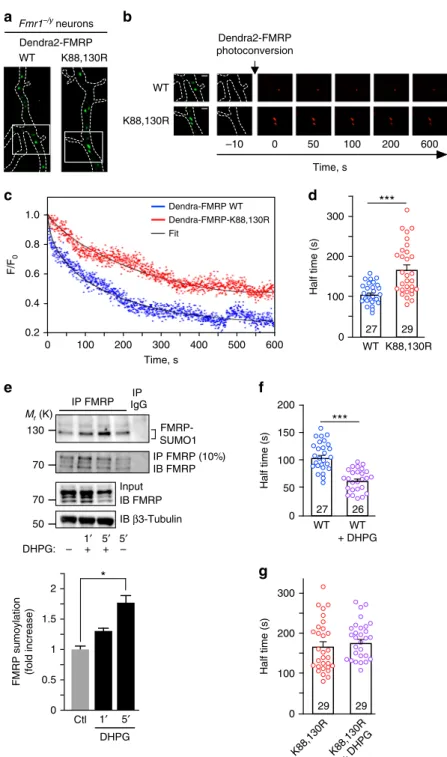
live-time restricted photoconversion experiments
48in Fmr1
−/yneurons expressing the photoswitchable WT or K88,130R
Dendra2-FMRP constructs (Fig.
5
a, b). Dendra2 is a green-to-red
photoactivatable
fluorescent protein that allows the real-time
tracking of a photoconverted protein
49,50. We measured and
compared the half-times of the decrease in red photoconverted
fluorescence, which corresponds to the real-time diffusion of WT
and K88,130R Dendra2-FMRP out of dendritic granules
(Fig.
5
b–d). In basal conditions, the mean half-time of
Dendra2-FMRP WT
fluorescence dissociation from dendritic granules was
significantly shorter than the value measured for the
Dendra2-FMRP K88,130R mutant (Fig.
5
d; half-time WT
= 101.8 ± 4.5 s vs
half-time K88,130R
= 165.3 ± 12.1 s) indicating that the
dis-sociation of WT FMRP from the granules is much faster than for
the K88,130R mutant. These data strongly support the
involve-ment of FMRP sumoylation in controlling the dissociation of the
protein from dendritic mRNA granules.
Activation of mGlu5R regulates FMRP-mediated mRNA
transport
51,52and also modulates its phosphorylation and
ubiquitination
13,14. Interestingly, we previously showed that
activation of these receptors also evokes sumoylation in cultured
neurons
26. This prompted us to assess whether the application of
the mGluR agonist DHPG triggers FMRP sumoylation in neurons
(Fig.
5
e). We
first confirmed that the activation of mGlu5R with
DHPG is effective in our neuronal cultures and evokes an
intracellular calcium increase (Supplementary Fig.
5
). Then,
FMRP-immunoprecipitates were probed with specific
anti-SUMO1 antibodies and revealed that the sumoylation of FMRP
50 55 170 100 70 130 Mr (K) GFP-FMRP IB FMRP WT GFP K88R GFP-FMRP GFP
e
K88,130R K130R Native FMRP IB β3-Tubulin β3-Tubulin Matureb
20 40 0 50 30 10 70 90 80 60 100***
***
***
***
**
***
***
*** ***
*** ***
GFP Control WT% of total number of spine
Immature Mature K88,130R Immature Mature 2 4 0 5 3 1 6 7 GFP Control WT 8
d
K88,130R n.s. Immaturea
WT neurons GFP control GFP-FMRP WT GFP-FMRP-K88,130R GFP-FMRP K88,130R 1 2 0 3Immature spine length (
μ m) GFP Control WT 4 n.s.
c
Density (protrusions / 10 μ m) K88R K130R Mature Mature Immature Immature GFP-FMRP-K88R GFP-FMRP-K130R K88R K130R n.s. K88R K130R n.s. GFP-FMRP GFP-FMRPFig. 7 Spine density and maturation processes are intrinsically linked to the ability of FMRP to be sumoylated. a Representative confocal images of dendrites from transduced WT neurons expressing free GFP, the WT, K88R, K130R, or K88,130R mutant forms of GFP-FMRP for 30 h. Bar, 5μm. Histograms show the relative proportion of mature and immature spinesb and the density of the protrusions d in the indicated conditions shown in a. c Histograms of immature spine length measured from WT neurons expressing the indicated constructs.e Relative protein expression levels of the WT and mutant forms of GFP-FMRP in WT transduced neurons as ina showing an approximate threefold increase in the levels of exogenous GFP-FMRP expression. Data shown inb–d are the mean ± s.e.m. Statistical significance in b–d was determined by a one-way analysis of variance (ANOVA) with a Bonferroni post-test. N = ~3000 spines per condition from four independent experiments. ***p < 0.001; n.s. not significant
is low in basal unstimulated conditions but rapidly increases after
1 and 5 min of DHPG treatment (DHPG 1 min, 1.28 ± 0.12 fold/
control; DHPG 5 min, 1.73 ± 0.2 fold/control; Fig.
5
e) indicating
that FMRP sumoylation is rapidly triggered by the mGlu5R
activation.
These results led us to hypothesize that the activity-dependent
sumoylation of FMRP controls FMRP dissociation from dendritic
mRNA granules. To address this point, we pharmacologically
stimulated mGlu5R in Fmr1
−/yneurons expressing either
Dendra2-FMRP WT or K88,130R and measured the dissociation
properties of FMRP from dendritic granules using the
photo-conversion assay (Fig.
5
f, g). Interestingly, mGlu5R stimulation
enhanced the exit rate of the red photoconverted Dendra2-FMRP
WT
fluorescence from granules by ~40% (Fig.
5
f). By contrast,
mGlu5R activation had no effect on the dissociation of
Dendra2-FMRP-K88,130R positive granules (Fig.
5
g). These
findings
strongly support that the mGlu5R-dependent sumoylation of
FMRP regulates the dissociation of FMRP from dendritic mRNA
granules.
Sumoylation regulates homomeric FMRP–FMRP interaction.
Our data demonstrate that FMRP sumoylation controls FMRP
release from dendritic granules. To further assess the role of
sumoylation in the regulation of FMRP–FMRP interaction, we
combined pull-down assays with in vitro SUMO reactions and
analyzed the impact of sumoylation on the dissociation of FMRP
homomers (Fig.
6
).
We purified GST- and His-tagged FMRP (1–160 aa) fusion
proteins and found that GST-FMRP (1–160) specifically interacts
with His-FMRP (1–160 aa) and forms N-terminal FMRP
homodimers in vitro (Fig.
6
a). We then performed an in vitro
sumoylation assay
31on purified FMRP (1–160 aa) dimers to
assess
whether
sumoylation
promotes
their
dissociation
(Fig.
6
b–d). First, we verified that the immobilization of
GST-FMRP (1–160 aa) on the glutathione matrix did not prevent the
in vitro sumoylation of the protein (Fig.
6
c). Incubation of
immobilized GST-FMRP (1–160 aa) with the sumoylation
reaction mix gave rise to higher molecular weight bands
corresponding to the sumoylated forms of GST-FMRP (1–160
aa). These bands were absent in control conditions (Fig.
6
c).
Next, we performed in vitro sumoylation assays on
immobi-lized GST-FMRP–His-FMRP dimers (Fig.
6
d). The pool of
His-FMRP (1–160 aa) released by sumoylation was separated from
the remaining immobilized dimers by centrifugation of the
glutathion beads. Proteins either in the supernatant or bound to
the beads were both analyzed by immunoblotting with
anti-FMRP antibodies. As seen in Fig.
6
d, the release of His-FMRP
(1–160 aa) from the immobilized dimers was only promoted
upon sumoylation with the concurrent decrease of the remaining
His-FMRP (1–160 aa) in the pelleted FMRP fraction. This
particular set of data demonstrates that sumoylation promotes the
dissociation of FMRP–FMRP dimers.
SUMO-de
ficient FMRP-expressing WT neurons show FXS
phenotype. Collectively, our data clearly demonstrate that
sumoylation of the N-terminal part of FMRP is essential to allow
for the dissociation of the protein from dendritic mRNA granules
and to promote spine elimination and maturation. To confirm
the key involvement of FMRP sumoylation in neuronal
matura-tion events, we hypothesized that the expression of the
non-sumoylatable FMRP mutant could reverse the spine density and
maturation of WT neurons. Thus, we expressed either the WT or
the K88,130R mutant form of GFP-FMRP into WT mouse
neu-rons (Fig.
7
). WT neurons expressing GFP-FMRP-K88,130R
resembled the GFP-expressing Fmr1
−/yneurons (Fig.
2
) with
>67% of protrusions characterized by an immature phenotype
(Fig.
7
a, b). Similarly, the length of dendritic spines in WT
neurons expressing GFP-FMRP-K88,130R was also significantly
increased (Fig.
7
c; K88,130R, 3.77 ± 0.08
μm) comparable to the
values measured in Fmr1
−/yneurons (Fig.
2
d).
Importantly, the density of dendritic spines was dramatically
increased upon the expression of the K88,130R mutant (Fig.
7
a, d;
GFP control, 5.03 ± 0.17 protrusions per 10
μm; K88,130R, 6.33 ±
0.24 protrusions per 10
μm), comparable to the values obtained in
Fmr1
−/yneurons (Fig.
2
c). Interestingly, the expression of the
single K130R mutant in WT mouse neurons also leads to a
significant increase in the density of the protrusions (Fig.
7
a, d;
GFP control, 5.03 ± 0.17 protrusions per 10
μm; K130R, 6.29 ±
0.41 protrusions per 10μm) without altering the maturity of
dendritic spines (Fig.
7
b, c). As expected, expressing the WT form
of GFP-FMRP in WT neurons did not affect any of the spine
characteristics confirming the essential role of FMRP sumoylation
in spine elimination and maturation processes.
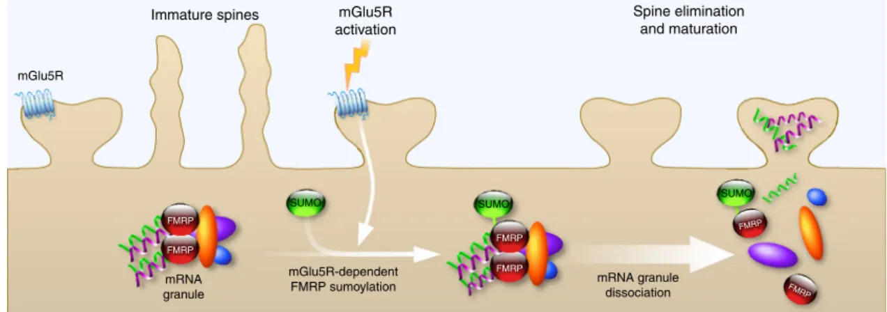
Discussion
Here, we report for the
first time that FMRP is a sumoylation
target in vivo. We identify three sumoylatable residues, two of
which lay within the N-terminal domain of FMRP and are the
active SUMO sites. We further
find that the activation of
meta-botropic mGlu5R promotes the sumoylation of FMRP and
rapidly leads to the dissociation of FMRP from dendritic mRNA
granules allowing for the regulation of spine elimination and
maturation (Fig.
8
). Thus, our work uncovers a novel
activity-mGlu5R FMRP FMRP FMRP FMRP FMRP FMRP mGlu5R-dependent FMRP sumoylation mRNA granule
SUMO SUMO SUMO
mRNA granule dissociation
Immature spines mGlu5R
activation
Spine elimination and maturation
Fig. 8 Schematic model for the mGlu5R-dependent regulation of FMRP function via the sumoylation process. The activity-dependent sumoylation of FMRP is a key step to dissociate FMRP from dendritic mRNA granules and consequently to regulate spine elimination and maturation
dependent role of sumoylation in the regulation of FMRP
neu-ronal function.
We provide the
first evidence that FMRP sumoylation is
required for spine elimination and proper maturation. The initial
step of spine formation is the emergence of immature long thin
protrusions, which are later on eliminated or matured with
enlargement of spine head
8. A tight balance between these
pro-cesses is thus required for the development of a functional
neu-ronal network. This is in line with our data showing a decrease in
the density of protrusions when expressing FMRP in Fmr1
−/yneurons, and an increased density in WT neurons expressing the
SUMO-deficient form of FMRP. Such compensatory and
dele-terious effects support the idea that immature spines are
over-produced and/or less efficiently eliminated when FMRP
sumoylation is perturbed.
In correlation with our
findings, the role of sumoylation at the
post-synaptic compartment has already been described for several
proteins
19. For instance, sumoylation of the scaffolding calcium/
calmodulin-dependent serine protein kinase (CASK) reduces
CASK interaction with protein 4.1, a protein that connects
spectrin to the actin cytoskeleton in dendritic spines. Mimicking
CASK sumoylation dramatically impairs spine formation
53.
According to the importance of sumoylation in the post-synaptic
formation and maturation, our
findings demonstrate a role of
sumoylation in spine elimination and maturation by tuning
FMRP dimerization within dendritic mRNA granules. Altogether,
these data shed light on the role of sumoylation as a critical
molecular regulator in neuronal development and maturation.
Interestingly, we demonstrate that the sumoylation of FMRP is
triggered upon mGlu5R activation. mGlu5R has been previously
reported to differentially regulate FMRP function depending on
its subcellular localization. For instance, a direct involvement of
FMRP was shown in targeting and transport of several mRNAs
from the soma along dendrites upon mGlu5R activation
52.
Fur-thermore, the repression of mRNA translation exerted by FMRP
in dendrites is counteracted by the activation of mGlu5R
51. Here,
we unravel a novel activity-dependent regulation of the FMRP
function. We show that mGlu5R-induced sumoylation of FMRP
drives its own dissociation from dendritic mRNA granules to
regulate both spine elimination and maturation.
It has been previously described that FMRP is a target of
mGluR-dependent PTMs
11,13,14,54,55. Activation of mGluRs in
neurons induces a rapid dephosphorylation of FMRP C-terminal
region as a result of an enhanced protein phosphatase 2A (PP2A)
activity
11. Conversely, mGluR activation that lasts longer than 5
min results in an mTOR-mediated PP2A suppression followed by
rapid rephosphorylation of FMRP C-terminus by the ribosomal
protein S6 kinase (S6K1)
11,55. Accordingly to the role of
phos-phorylation in controlling FMRP function, the lack of
S6K1-dependent FMRP phosphorylation mimics FMRP loss of function
and leads to an increased expression of the FMRP target mRNA
SAPAP3
55. In addition, Nalavadi et al.
14described a rapid
ubi-quitination of the C-terminal part of FMRP upon stimulation
with the mGlu5R agonist DHPG in rat cultured neurons. FMRP
ubiquitination promotes a proteasome-mediated FMRP
degra-dation, which in turn controls FMRP levels at the synapse.
Interestingly, these authors showed that FMRP ubiquitination
requires a prior FMRP-dephosphorylation carried by PP2A.
Taken together, these pieces of evidence suggest a crosstalk
between various PTMs in the regulation of FMRP function. Here,
we demonstrate that mGlu5R activation triggers a rapid
sumoy-lation of FMRP. This event promotes the release of FMRP from
transport mRNA granules. Thus, the present study adds another
level of complexity to the post-translational regulation of FMRP
and advances our understanding of the activity-dependent
con-trol of FMRP function in neurons. It will therefore be of future
interest to examine whether the interplay between these PTMs
could take place to orchestrate the mGlu5R-dependent regulation
of FMRP.
The present study shows that the activation of mGlu5R directly
promotes FMRP sumoylation, regulating its neuronal function in
spine elimination and maturation. Our work therefore raises the
intriguing possibility that the impairment of FMRP sumoylation
could contribute to FXS physiopathology. Recent publications
have reported missense point mutations within the FMR1 gene in
patients affected by FXS. Importantly, these mutations lead to
amino-acid changes close to the SUMO active sites of FMRP
(F126S
56and R138Q
57). Similarly to our data on the K88,130R
FMRP mutant, the FXS R138Q mutation does not modify the
expression of FMRP nor its RNA-binding properties, indicating
that the pathogenicity is caused independently of the FMRP
expression level and the ability of FMRP to bind mRNAs
58. To
date, no data have been reported regarding the functional
impairment due to the F126S mutation. Our data report that the
reintroduction of the FMRP WT but not the K88,130R mutant in
Fmr1
−/yneurons promotes spine maturation and elimination
demonstrating that FMRP sumoylation is critical for these
pro-cesses. Therefore, an interesting possible explanation could be
that the F126S and R138Q FXS mutations, which are very close to
the active K130-SUMO site, would directly impact on the
mGlu5R-dependent regulation of FMRP sumoylation and
con-sequently, on post-synaptic FMRP-driven regulatory events.
Future work will have to be performed aiming at understanding
the effect of these FXS mutations on FMRP sumoylation. These
next exciting steps will allow assessing whether FMRP
sumoyla-tion defects participate in the pathophysiology of FXS patients,
raising the possibility to identify new targets and potentially
develop novel therapeutic approaches.
Methods
Constructs. GFP-FMRP was obtained by subcloning the isoform 1 of the human FMR1 sequence into the EcoR1/Pst1 site of the mammalian expression vector pEGFP-C2 (Clontech). GFP-/Dendra2-/GST-/His-FMRP mutant constructs were all made by site-directed mutagenesis using the Quick-change mutagenesis solution (Agilent). pSinRep5 constructs used to produce Sindbis particles were generated using the Gateway recombination technology (Invitrogen). All constructs were then entirely sequenced.
Building model for FMRP-SUMO1. Three X-ray structures of human FMRP are available in Protein Data Bank (PDB,http://www.rcsb.org; PDB ID: 4OVA residues 1–209 at 3.0 Å resolution59, 4QVZ residues 1–213 at 3.2 Å resolution, and 4QW2 residues 1–213 with the mutation R138Q at 3.0 Å resolution60). The solvent ASA values for each residue have been calculated using Naccess tool61on all monomers of each PDBfiles (4 for 4OVA, 2 for 4QVZ, and 2 for 4QW2). We calculated the average values for K88 and K130 for each structure. The classical parameters used are 1.4 for the radius of the“solvent” sphere and 25% for the threshold that determines if a residue is considered as buried or exposed. We utilized the X-ray structures of human FMRP PDB ID: 4OVA residues 1–209 at 3.0 Å resolution59 and of human SUMO1 PDB ID: 4WJQ at 1.35 Å resolution62. To build models of FMRP modified with the SUMO1 protein, we first verified the shape compatibility and then used the Pymol software to manipulate the structures, make and visualize the FMRP-SUMO1 models.
Mouse lines and rat strain. All animals (3–10-month-old pregnant female Wistar rats from Janvier, St Berthevin, France; 3–10 month-old female C57BL/6 WT and Fmr1 knockout (Fmr1−/y) mice10) were handled in our facility in accordance with the European Council Guidelines for the Care and Use of Laboratory animals and approved by the Animal Care and Ethics Committee (Comité Institutionnel d’Ethique Pour l’Animal de Laboratoire N°28, Nice, France; project reference NCE/ 2012-63). All animals had free access to water and food. The light cycle was controlled as 12 h light and dark cycle and the temperature was maintained at 23 ± 1 °C. Protocols to prepare primary neuronal cultures from mouse embryos at E15.5 or at E18 for rats were also approved by the Animal Care and Ethics Committee (Comité Institutionnel d’Ethique Pour l’Animal de Laboratoire N°28, Nice, France; project reference NCE/2012-63). All mice were maintained on a C57BL/6 genetic background, whereas Wistar rats were exclusively from a commercial source (Janvier). The Fmr1 knockout (Fmr1−/y) mouse line10was maintained on a C57BL/ 6 background.
Mouse and rat brain lysate preparation. Brain lysates were prepared as pre-viously described26from post-natal P1–3 mouse or P5–7 rat brains. Briefly, freshly dissected brains were transfered in 5 volumes (w/v) of ice-cold sucrose buffer (10 mM Tris-HCl, pH 7.4, 0.32 M sucrose) supplemented with a protease inhibitor cocktail (Sigma, 1/100), Pefabloc 0.5 mM (Roche), MG132 100μm (Enzo), ALLN 100μm (Sigma), and 20 mM freshly prepared NEM (Sigma), and homogenized at 4 °C using a Teflon-glass potter and a motor-driven pestle at 500 rpm. Nuclear fraction and cell debris were pelleted by centrifugation at 1000×g for 10 min. The post-nuclear S1 fraction (supernatant) was collected and protein concentration measured using the BCA protein assay (Bio-Rad).
Primary neuronal cultures. Hippocampal and cortical neurons were prepared from embryonic (E18) pregnant Wistar rats as previously described26or from WT or Fmr1−/yE15.5 pregnant C57BL/6 mice. Briefly, neurons were plated in Neu-robasal medium (Invitrogen, France) supplemented with 2% B27 (Invitrogen), 0.5 mM glutamine and penicillin/streptomycin (Ozyme) on 60-mm dishes or 24-mm glass coverslips (VWR) pre-coated with poly-L-lysine (0.5 mg mL−1; Sigma).
Neurons (800,000 cells per 60-mm dish or 110,000 cells per coverslip) were then fed once a week with neurobasal medium supplemented with 2% B27 and peni-cillin/streptomycin for a maximum of 3 weeks.
Cell transfection. COS7 cells and primary neurons (14–16 DIV) were transfected using Lipofectamine 2000 (Invitrogen) according to the manufacturer’s instruc-tions and used 48–72 h post transfection.
Sindbis virus production and neuronal transduction. Attenuated Sindbis viral particles (SINrep(nsP2S726)) were prepared and used as previously described38–40. Briefly, cRNAs were generated from the pSinRep5 plasmid containing the sequence coding for WT or mutated GFP-FMRP constructs and from the defective helper (pDH-BB) plasmid using the Mmessage Mmachine SP6 solution (Ambion). cRNAs were then mixed and electroporated into BHK21 cells. Pseudovirions present in the culture medium were collected 48 h after electroporation and concentrated using ultracentrifugation on SW41Ti. Aliquots of resuspended Sindbis particles were then stored at−80 °C until use. Neurons were transduced at a multiplicity of infection (MOI) of 0.1–2 and returned to the incubator at 37 °C under 5% CO2for
24–30 h depending on their subsequent utilization.
Bacterial sumoylation assay inEscherichia coli. Bacterial sumoylation assays were performed as previously described31,35. Briefly, competent E. coli BL21(DE3) cells (Invitrogen, France) expressing pE1-E2SUMO1 were transformed with 1μg of pET-expression plasmid (Novagen) to express the WT or non-sumoylatable forms of His-tagged FMRP were selected on LB-Agar plates containing chloramphenicol (50μg mL−1) and ampicillin (50μg mL−1). A 10 mL preculture was then used to
inoculate 50 mL of LB containing chloramphenicol and ampicillin. After incuba-tion under shaking at 37 °C until OD600reaches 0.7, cells were cooled down to 20 °
C and isopropyl-β-D-thiogalactopyranoside (IPTG) was added at a concentration of
1 mM. After 4 h at 20 °C, bacteria were pelleted by centrifugation at 4 °C at 7000×g and kept at−80 °C until use. Pellets were resuspended in 1 mL lysis buffer (25 mM Tris pH 8, 300 mM KCl, 1 mM EDTA, 20% glycerol, 5% ethanol, 0.5% NP40, 0.5 M urea, 1 mM DTT) supplemented with proteases inhibitors (leupeptine 1μg mL−1,
Pepstatine 1μg mL−1, Aprotinine 1μg mL−1, Pefabloc 0.5 mM, and freshly pre-pared NEM 20 mM), and incubated under rotation for 30 min at 4 °C in the presence of 5 mg mL−1lysozyme. Bacterial cytoplasmic membranes were then solubilised by addition of 1 mg mL−1sodium deoxycholate and released DNA digested by incubation with 50μg mL−1of DNAse I and 10 mM MgCl2for 30 min
at 4 °C. Cellular debris were pelleted by centrifugation at 20,000×g for 15 min at 4 °C and supernatants were incubated with 40μL of nickel agarose beads (Qiagen) for 2 h at 4 °C under gentle rotation. After three washes (25 mM Tris pH 8, 50 mM KCl, 1 mM EDTA, 20% glycerol, 0.1% Triton X-100, 0.5 M urea, 1 mM DTT), purified proteins were eluted in 200μL of βME-reducing sample buffer for 5 min at 95 °C. COS7 sumoylation assay. Mycoplasm-free COS7 cells (ATCC reference CRL-1651, Molsheim, France) at 60% of confluence in six-well plates were co-transfected using 1μg of the eukaryotic expression vector pTL1-FMRP plasmid63 or its derived non-sumoylatable mutants with 0.5μg of mCherry or mCherry-SUMO1 plasmids26and 0.5μg of plasmid coding for Flag-Ubc9 using Lipofecta-mine 2000 (Invitrogen) according to the manufacturer’s instructions. After 48 h of expression, cells were washed once in PBS containing 20 mM NEM and reduced for 5 min at 95 °C inβME-containing sample buffer.
CLIP analysis. To isolate neuronal mRNAs associated with WT and SUMO-deficient GFP-FMRP mutant, UV cross-linking, and FMRP immunoprecipitations were performed on 20 DIV Fmr1−/yneurons transduced (MOI of 3) at day 19 to express free GFP, the WT, or the non-sumoylatable K88,130,614R form of GFP-FMRP. RNAs and proteins were cross-linked through three rounds of UV irra-diation (400 mJ each; 254 nm). Cells were then scraped in ice-cold PBS, collected by centrifugation, and lysed in NP40 buffer as described in ref.64. For each assay, 5 μg of affinity-purified rabbit anti-FMRP antibody (Ab#056) was used to
immunoprecipitate 1 mg of neuronal extracts and 2% of the lysate was used for assessment of relative RNA expression in the input material. IPs were then carried out at 4 °C for 4 h and 2% of the homogenate and 10% of the immunoprecipitates were saved to check for the IP quality using anti-FMRP immunoblots. After three washes in lysis buffer (50 mM HEPES, pH 7.4, 150 mM NaCl, 0.5% NP40, 10 mM EDTA, 1 mM NaF, 0.5 mM DTT, protease and phosphatase inhibitors (Pierce), proteins were digested with proteinase K (1μg mL−1) for 30 min at 56 °C. IP and input RNAs were purified through two successive rounds of phenol/chloroform extraction, then reverse transcribed using a mix of Oligo dT and random primers and Superscript II enzyme (Invitrogen) according to the manufacturer’s protocol. RT reactions were diluted two times and 1μL of diluted material was used for qPCR analysis. Relative enrichment of the amplified RNA in the IP vs the input in each condition was calculated with the 2−deltaCt(CtIP–Ctinput).
Oligonucleotides (5′–3′) used in RNA work were as follows: Fmr1_F: GAACAAAAGACAGCATCGCT; Fmr1_R:
CCAATTTGTCGCAACTGCTC; Camk2a_F: TATCCGCATCACTCAGTAC; Camk2a_R: GAAGTGGACGATCTGCCATTT; Sapap3_F:
ACCATGTAACCCCGGCTG; Sapap3_R: CCTTGATGTCAGGATCCCC; Fxr1_F: GTGCAGGGTCCCGAGGT; Fxr1_R: GGTGGTGGTAATCGGACTTC; Kif3c_F: GGTCCCATCCCAGATACAGA; Kif3c_R: CCAGAAAGCTGTCAAACCTC; Tubb3_F: CGAGACCTACTGCATCGACA; Tubb3_R:
CATTGAGCTGACCAGGGAAT; PP2a_F: GTCAAGAGCCTCTGCGAGAA; PP2a_R: GCCCATGTACATCTCCACAC;β-actin_F:
ACGGCCAGGTCATCACTATTG;β-actin_R: CACAGGATTCCATACCCAAGA; PSD95_F: GGCGGAGAGGAACTTGTCC; PSD95_R:
AGAATTGGCCTTGAGGGAGGA; Map1b_F: TCCGATCGTGGGACACAAACCTG; Map1b_R: AGCACCAGCAGTTTATGGCGGG.
Immunoprecipitation. Proteins from rodent brain lysates or cultured neurons were solubilized for 1 h at 4 °C under gentle rotation in lysis buffer (10 mM Tris-HCl, pH 7.5, 10 mM EDTA, 150 mM NaCl, 1% Triton X-100, 0.1% SDS) sup-plemented with a protease inhibitor cocktail (Sigma, 1/100), Pefabloc 0.5 mM (Roche), MG132 100μm (Enzo), ALLN 100 μm (Sigma), and 20 mM freshly pre-pared NEM (Sigma). Then, NaCl concentration was raised to 400 mM and lysates were sonicated for 10 s, further incubated for 30 min at 4 °C and clarified (for primary neuronal extracts) or not (for brain homogenates) at 20,000×g at 4 °C for 15 min. Supernatants were diluted 2.5-fold with lysis buffer devoid of NaCl and pre-cleared for 1 h with a 50/50 mix of untreated and pre-blocked protein G-sepharose beads (Sigma) with a blocking buffer (PBS containing 5 mg mL−1BSA, 5 mg mL−1Dextran (40 kDa), 1 mg mL−1gelatin, yeast t-RNA 0.1 mg mL−1, and glycogen 0.1 mg mL−1) for 1 h at 4 °C. Proteins (800μg) from pre-cleared lysates were incubated with either 8μg of mouse monoclonal anti-SUMO1 antibody (Ab#D11, Santa-Cruz), 4μg of custom rabbit anti-FMRP (Ab#056, Supplementary Fig.1), or 12μg commercially available rabbit anti-FMRP (#Ab17722, Abcam; Supplementary Fig.1) antibodies (or their corresponding IgGs as IP control) for 1 h at 4 °C and then overnight at 4 °C with 30μL of pre-blocked protein G-sepharose beads (Sigma). Precipitates were washed three times with 1 mL lysis buffer and proteins were eluted by boiling the beads 5 min inβME-reducing sample buffer before SDS-PAGE.
Immunoblotting. Protein extracts were resolved by SDS-PAGE, transferred onto nitrocellulose membrane (Hybond-C Extra, Amersham or BioTraceNT, PALL), immunoblotted with the indicated concentration of primary antibodies and revealed using the appropriate horseradish peroxidase (HRP)-conjugated second-ary antibodies (GE healthcare) or True Blot (Rockland, Tebu-Bio). Proteins were then identified using Immobilon Western (Millipore) or Western Lightning Ultra (Perkin Helmer) chemiluminescent solutions and images acquired on a Fusion FX7 system (Vilber Lourmat). Full-size blots for cropped gels can be found in Supplementaryfigures6,7.
Immunocytochemistry. Neurons (18–21 DIV) were fixed in phosphate-buffered saline (PBS) containing 3.7% formaldehyde and 5% sucrose for 1 h at room tem-perature (RT). Neurons were then permeabilized for 20 min in PBS containing 0.1% Triton X-100 and 10% horse serum (HS) at RT and immunostained with either a rabbit monoclonal anti-S6 (1/200; Cell Signaling), a goat anti-Staufen1 (1/ 100; Santa-Cruz), a goat anti-Staufen2 (1/100; Santa-Cruz), a rabbit anti-FXR1 (1/ 10065), a mouse monoclonal anti-Ubc9 (1/50; BD Bioscience, France), a mouse SUMO1 (1/50; Ab#D11, Santa-Cruz; 1/50 Ab#2F5–1, DSHB) or rabbit anti-FMRP (1/200; Custom Ab#056 or 1/50; Ab#4317s, Cell Signaling) antibodies in PBS containing 0.05% Triton X-100 and 5% HS. Cells were washed three times in PBS and incubated with the appropriate secondary antibodies (1/400) conjugated to Alexa488 or Alexa594, and mounted with Mowiol (Sigma) until confocal examination.
Ratiometric calcium imaging. Mouse cortical/hippocampal neurons (19–23 DIV) were loaded in neurobasal containing 20μm Fura-2AM (Invitrogen) for 30 min. After two washes in physiological 1.6 mM calcium-containing buffer (139 mM NaCl, 1.25 mM glucose, 15 mM Na2HPO4, 1.8 mM MgSO4, 1.6 mM CaCl2, 3 mM