HAL Id: inserm-02923397
https://www.hal.inserm.fr/inserm-02923397
Submitted on 27 Aug 2020
HAL is a multi-disciplinary open access
archive for the deposit and dissemination of sci-entific research documents, whether they are pub-lished or not. The documents may come from teaching and research institutions in France or abroad, or from public or private research centers.
L’archive ouverte pluridisciplinaire HAL, est destinée au dépôt et à la diffusion de documents scientifiques de niveau recherche, publiés ou non, émanant des établissements d’enseignement et de recherche français ou étrangers, des laboratoires publics ou privés.
Aurélie Fluckiger, Romain Daillere, Mohamed Sassi, Barbara Sixt, Peng Liu,
Friedemann Loos, Corentin Richard, Catherine Rabu, Maryam Tidjani,
Anne-Gaëlle Goubet, et al.
To cite this version:
Aurélie Fluckiger, Romain Daillere, Mohamed Sassi, Barbara Sixt, Peng Liu, et al.. Cross-reactivity between tumor MHC class I-restricted antigens and an enterococcal bacteriophage. Science, American Association for the Advancement of Science, 2020, 369 (6506), pp.936-942. �10.1126/science.aax0701�. �inserm-02923397�
Title: Crossreactivity between MHC class I-restricted antigens from cancer cells and an enterococcal bacteriophage.
Authors: Aurélie Fluckiger1-3, Romain Daillère1-3, Mohamed Sassi4, Barbara Susanne Sixt5,6-10, Peng Liu6-10, Friedemann Loos6-10, Corentin Richard11-13, Catherine Rabu17,18, Maryam Tidjani 5
Alou1,2,14, Anne-Gaëlle Goubet1,2, Fabien Lemaitre1, Gladys Ferrere1,2 ,Lisa Derosa1,2,14,Connie PM Duong1,2, Meriem Messaoudene15, Andréanne Gagné15, Philippe Joubert15, Luisa De Sordi16, Laurent Debarbieux16, Sylvain Simon17,18, Clara-Maria Scarlata19, Maha Ayyoub19, Belinda Palermo20, Francesco Facciolo21, Romain Boidot22, Richard Wheeler23, Ivo Gomperts Boneca23, Zsofia Sztupinszki24, Krisztian Papp25, Istvan Csabai25, Edoardo Pasolli26, Nicola Segata27, 10
Carlos Lopez-Otin7-10,28, Zoltan Szallasi24,29-31, Fabrice Andre32,33, Valerio Iebba34, Valentin Quiniou35,36, David Klatzmann35,36, Jacques Boukhalil37, Saber Khelaifia37, Didier Raoult37, Laurence Albiges1,14,38, Bernard Escudier1,38,39, Alexander Eggermont1-14, Fathia Mami-Chouaib40,Paola Nistico20, François Ghiringhelli41, Bertrand Routy15,42, Nathalie Labarrière17,18, Vincent Cattoir4,43,44,Guido Kroemer6-10,45,46,47*, and Laurence Zitvogel1-3,14,47*
15
Affiliations:
1
Gustave Roussy Cancer Campus (GRCC), Villejuif, France.
2
Institut National de la Santé et de la Recherche Médicale, U1015, Institut Gustave Roussy, Villejuif, France
14
Université Paris-Saclay, Villejuif, F-94805, France. 20
3
Center of Clinical Investigations in Biotherapies of Cancer (CICBT) 1428, Villejuif, France.
4
Université Rennes 1, Laboratoire de Biochimie Pharmaceutique, Inserm U1230 - UPRES EA 2311, Rennes, France.
5
Laboratory for Molecular Infection Medicine Sweden, Umeå Centre for Microbial Research, Department of Molecular Biology, Umeå University, 90187, Umeå, Sweden.
25
6
Cell Biology and Metabolomics Platforms, Gustave Roussy Cancer Campus, Villejuif, France.
7
Equipe 11 labellisée Ligue contre le Cancer, Centre de Recherche des Cordeliers, Paris, France
8
INSERM U1138, Paris, France.
9
Université de Paris, Paris, France
10
Sorbonne Université, Paris, France. 30
11
Research Platform in Biological Oncology, Dijon, France.
12
GIMI Genetic and Immunology Medical Institute, Dijon, France.
13
University of Burgundy-Franche Comté, Dijon, France.
15
Centre de recherche du centre hospitalier de l'université de Montréal (CRCHUM), 900 rue Saint-Denis, H2X 3H8 Montréal, Québec, Canada.
35
16
Department of Microbiology, Institut Pasteur, F-75015 Paris, France
17
CRCINA, INSERM, Université d'Angers, Université de Nantes, Nantes, France.
18
LabEx IGO "Immunotherapy, Graft, Oncology," Nantes, France.
19
Cancer Research Centre of Toulouse, INSERM UMR 1037, 31037 Toulouse, France; Université Toulouse III Paul Sabatier, 31330 Toulouse, France; Institut Universitaire du Cancer 40
de Toulouse-Oncopole, 31100 Toulouse, France.
20
Unit of Tumor Immunology and Immunotherapy, Department of Research, Advanced Diagnostics and Technological Innovation, IRCCS Regina Elena National Cancer Institute, Rome, Italy.
21
Thoracic Surgery Unit, Department of Surgical Oncology, IRCCS Regina Elena National 45
Cancer Institute, Rome, Italy.
22
Unit of Molecular Biology - Department of Biology and Pathology of Tumors - Georges-François Leclerc anticancer center - UNICANCER - Dijon – France
23
Institut Pasteur, Unit Biology and genetics of the bacterial cell wall, Paris, France
24
Computational Health Informatics Program (CHIP), Boston Children's Hospital, Boston, MA, 50
USA.
25
Department of Physics of Complex Systems, ELTE Eötvös Loránd University, Budapest, Hungary.
26
Department of Agricultural Sciences, University of Naples Federico II, Naples, Italy
27
Department CIBIO, University of Trento, Trento, Italy. 55
28
Dpto. de Bioquímica y Biología Molecular, Instituto Universitario de Oncología (IUOPA), Universidad de Oviedo, Oviedo, Spain.
29
Harvard Medical School, Boston, MA, USA.
30
Danish Cancer Society Research Center, Copenhagen, Denmark.
31
MTA-SE-NAP, Brain Metastasis Research Group, 2nd Department of Pathology, Semmelweis 60
University, Budapest, Hungary.
32
Department of Cancer Medicine, Breast Cancer Committee, Gustave Roussy, Villejuif, France.
33
INSERM Unit 981, Gustave Roussy, Villejuif, France.
34
Department of Public Health and Infectious Diseases, Section of Microbiology, Sapienza University of Rome, Rome 00185, Italy.
65
35
AP-HP, Hôpital Pitié-Salpêtrière, Clinical Investigation Center in Biotherapy (CIC-BTi) and Immunology-Inflammation-Infectiology and Dermatology Department (3iD), F-75651, Paris, France
36
Sorbonne Université, INSERM, Immunology-Immunopathology-Immunotherapy (i3), F-75651,Paris, France
70
37
URMITE, Aix Marseille Université, UM63, CNRS 7278, IRD 198, INSERM 1095, IHU-Méditerranée Infection, 13005 Marseille, France.
38
Department of Medical Oncology, Gustave Roussy, Villejuif, France.
39
INSERM U981, GRCC, Villejuif, France.
40
INSERM UMR 1186, Integrative Tumour Immunology and Genetic Oncology, Gustave 75
Roussy, EPHE, PSL, Faculté de Médecine, Université Paris-Sud, Université Paris-Saclay, Villejuif, France.
41
Department of Medical Oncology, Center GF Leclerc, Dijon, France.
42
Division d'hémato-oncologie, département de médicine, centre hospitalier de l'université de Montréal (CHUM), Montréal, Québec, Canada.
80
43
CHU de Rennes - Hôpital Ponchaillou, Service de Bactériologie-Hygiène hospitalière, Rennes, France.
44
CNR de la Résistance aux Antibiotiques (laboratoire associé 'Entérocoques'), Rennes, France.
45
Pôle de Biologie, Hôpital Européen Georges Pompidou, Assistance Publique–Hôpitaux de Paris, Paris, France
85
46
Department of Women's and Children's Health, Karolinska University Hospital, 1 Stockholm, Sweden.
47
Suzhou Institute for Systems Biology, Chinese Academy of Medical Sciences, Suzhou, China *Correspondence to: laurence.zitvogel@gustaveroussy.fr; kroemer@orange.fr
One sentence summary:
Cytotoxic T lymphocytes that recognize antigens from a prophage of a commensal enterococcus can mediate anticancer immunosurveillance by recognizing cross-reactive tumor-associated antigens.
95
Abstract:
It has been speculated that the intestinal microbiota induces commensal-specific memory T cells that then cross-react with tumor-associated antigens. Here, we identified MHC class I-binding 100
epitopes within the tail length tape measure protein (TMP) of a prophage found in the genome of
Enterococcus hirae. Mice bearing E. hirae strains harboring this prophage mounted a
TMP-specific H-2Kb restricted CD8+ T lymphocyte response upon immunotherapy with
cyclophosphamide or anti-PD1 antibodies. Such TMP-specific T cells also recognized a 78%-identical H-2Kb-binding peptide derived from the proteasome (20S) subunit beta type-4 105
(PSMB4), allowing them to control mouse tumors expressing this oncogenic driver. Administration of bacterial strains engineered to express the TMP epitope improved the outcome of immunotherapy. Tumors bearing PSMB4 knock-in mutations that abolish crossreactivity with TMP became immunotherapy-resistant. In renal and lung cancer patients, the presence of the enterococcal prophage in stools, as well as the expression of a TMP-cross reactive antigen by 110
tumors, predicted the long-term benefit of PD-1 blockade. In melanoma patients, we detected T cell clones recognizing naturally processed cancer antigens that are cross-reactive with microbial peptides. Altogether, these results support the idea that intestinal microbe-specific T cell responses contribute to anticancer immunosurveillance.
Main Text:
Unleashing immune responses against tumor-associated antigens through chemotherapy, 120
radiotherapy, targeted therapies or immune checkpoint inhibitors has become the mainstay of successful cancer treatments (1, 2). The recent discovery that the gut microbiota determines the cancer-immune set point, thus influencing the clinical outcome of antineoplastic therapies, has rekindled the concept that microbes or their products modulate not only intestinal but also systemic immunity (3, 4). Indeed, memory responses by interferon-IFN) secreting CD4+ and 125
CD8+ T cells specific for Enterococcus hirae, Bacteroides fragilis, and Akkermansia muciniphila are associated with favorable clinical outcome in cancer patients (5–8), suggesting that microbe-specific T lymphocytes may contribute to antitumor immune responses. The mechanisms through which microbes trigger chronic intestinal inflammation and systemic autoimmune disease have not been resolved (9). The theory of molecular mimicry (10–14) posits that T cells elicited by 130
bacteria or viruses accidentally recognize autoantigens as they ‘escape’ from self-tolerance inducing mechanisms (such as clonal deletion or inactivation). While MHC class I and class II binding epitopes encoded by bacterial genomes may be immunogenic (10–14), very few reports have demonstrated that microbe-specific CD4+ or CD8+ T lymphocytes attack normal or neoplastic tissues (15–17).
135
Cyclophosphamide (CTX) induces the translocation of E. hirae from the gut lumen to the mesenteric and splenic immune tissues, thereby eliciting specific CD4+ and CD8+ T lymphocytes producing interleukin-17 (IL17) and interferon- (IFN correlating with therapeutically 140
effective anticancer immune responses (6, 18). Broad-spectrum antibiotics abolished the therapeutic efficacy of CTX unless E. hirae was supplied by oral gavage (6). When comparing a panel of distinct E. hirae strains (Table S1, Figure S1A) for their capacity to restore the antibiotic-perturbed anticancer effects of CTX, we found that only a few E. hirae isolates (such as 13144 and IGR11) were efficient (Figure 1A-B, Ref. (6)). Given that the therapeutic efficacy 145
of the combination of CTX and E. hirae 13144 is abrogated by the depletion of CD8+ T cells or the neutralization of IFN, we screened the differential capacity of E. hirae strains to elicit memory T cell responses after priming of the host, measured as the ex vivo recall response (IFN secretion) of splenic CD8+ T cells against various E. hirae strains loaded onto dendritic cells
(DC) (Figure S2A). While E. hirae 13144 triggered specific CD8+ T cell responses (that were not
150
cross-reactive against irrelevant enterococci), E. hirae 708 and 13344 (two prototypic inefficient strains) failed to do so (Figure S2A).
To identify relevant T cell epitopes, we aligned the sequences of bacterial genes encoding putative cell wall and secreted proteins for immunogenic (13144) versus non-immunogenic (708 155
and 13344) E. hirae strains, followed by the in silico identification of 13144-specific nonapeptides with strong affinity (<50 nM) for the MHC class I H-2Kb protein (Table S2). Subsequently, we recovered splenic CD8+ T cells from mice that had been exposed to E. hirae 13144 and CTX (Figure 1C), restimulated them in vitro with pools of potentially immunogenic nonapeptides from E. hirae 13144 to measure IFN production (Table S2, Figure S2B) and 160
finally split the most efficient pool (No. 7) into individual peptides (Figure 1D). This approach led to the identification of one dominant epitope (one-letter amino acid [aa] code: TSLARFANI, abbreviation TMP1) in position 187 to 197 of the aa sequence of the phage tail length tape measure protein (TMP, 1506 aa) from a 39.2 kb prophage of E. hirae 13144 (Figure 1D, Figure S3, Tables S2-S3). The 39.2kb prophage encodes 65 genes, including one shared between all 18 165
E. hirae genomes and 38 unique to E. hirae 13144 (Figure S1B), encoding capsid, portal and tail
structures characteristic of Siphoviridae phages. Importantly, the TMP1 epitope of the 39.2kb prophage from E. hirae 13144 and the prophage fragment contained in E. hirae IGR11 showed 100% sequence identity (Figure S3 and S4A). Accordingly, E. hirae IGR11 was as efficient as E.
hirae 13144 in reducing the growth of MCA205 sarcomas treated with CTX (Figure 1A-B). In
170
contrast, the absence of a bona fide TMP1 epitope (observed in E. hirae 708 and 13344, Figure S1B) and a mutation in position 3 of the TSLARFANI peptide (LF observed in E. hirae ATCC9790, Figure S4A) correlated with the lack of anticancer effects of these E. hirae strains (Figure 1B and Ref. (6)). ELIspot assays designed to detect peptide-specific IFN-producing T cells revealed that mice gavaged with E. hirae 13144 or IGR11 mounted a CD8+ T cell response 175
against TMP1 (but not against the control peptides TMP2 and TMP3), while mice receiving E.
hirae strains lacking TMP1 (strains 708, 13344) or a strain possessing a mutated TMP1 (strain
ATCC9790) were unable to do so (Figure 1D). We used a fluorescent H-2Kb/TSLARFANI tetrameric complex (and its negative control H-2Kb/SIINFEKL binding to ovalbumine (OVA) specific CD8+ T cells) to detect the frequency and distribution of TMP1-specific cytotoxic T 180
lymphocytes (CTLs) in naive and MCA205 sarcoma bearing C57BL/6 mice. We observed a specific increase in splenic CD8+ T cells that recognized the TMP1 peptide (but not the OVA peptide SIINFEKL) at day 7 following treatment with CTX and gavage with E. hirae 13144 (Figure 1E), as well as in tumor draining lymph nodes (LN) of tumor bearers at day 14 after treatment with CTX and gavage with E. hirae 13144 (Figure S2C-D). Splenic TMP1 (but not 185
OVA)-specific (H-2Kb/TSLARFANI tetramer-positive) CTLs also increased in their frequency after gavage with E. hirae IGR11 (but not 13344 nor ATCC9790) (Figure 1E). The H-2Kb/TSLARFANI tetramer-positive CTLs were specifically enriched in the CXCR3+CCR9+ fraction of CD8+ T cells from secondary lymphoid organs (Figure S2C). Even in mice colonized with human fecal materials, CTX administration and oral gavage with E. hirae 13144 induced an 190
anticancer effect (Figure S2E) and an expansion of H-2Kb/TSLARFANI tetramer-positive CTL in tumor draining LN at day 7 and in tumor beds at day 17 while vanishing from mesenteric LN (Figure S2F-H). Hence, immunogenic E. hirae elicits a H-2Kb restricted CTL response against the TMP-derived peptide TMP1/TSLARFANI.
195
To explore the capacity of TMP1-specific H-2Kb restricted T cells to control the growth of MCA205 cancers, we subcutaneously (s.c.) immunized naive C57BL/6 mice with dendritic cells (DC) loaded with heat-inactivated E. hirae 13144 (positive control), the naturally occurring TMP1/TSLARFANI peptide from 13144 and IGR11, its LF mutant from E. hirae ATCC9790 (‘mut3’, Figure 2A, Figure S4A) or other non-immunogenic bacterial peptides (group 1, Figure 200
S2B). In this prophylactic setting, DC pulsed with TMP1 (but not mut3) were as efficient as the whole E. hirae extract in reducing tumor growth (Figure 2B-C). Next, we explored whether the TMP1 peptide would be able to confer immunogenicity to the usually inefficient bacterium
Escherichia coli strain DH5α in the therapeutic setting, in which antibiotic treatment is followed by gavage with different bacterial strains and CTX-based chemotherapy (Figure 1A and Ref. (6)). 205
E. coli engineered to express TMP1 (Figure S5) was as efficient as E. hirae 13144 in restraining
MCA205 tumor growth (Figure S4B, Figure 2D) and eliciting tetramer binding CTL in the spleen (Figure 2E). In contrast, E. coli expressing an irrelevant sequence (encoding mouse EGFP protein), mut3 or or mutant TMP1 bearing a SA exchange in the anchor position 2 (‘mut2’) (Figure 2A) failed to induce such a cancer-protective immune response (Figure 2D-E).
To explore the mechanism by which TMP1 exerts its anticancer activity against MCA205 tumors in C57BL/6 mice, we investigated whether H-2Kb-restricted mouse tumor antigens with high identity to the TMP1 peptide (TSLARFANI) exist. Using the NCBI BLASTP suite, we found that the peptide (GSLARFRNI) belonging to the proteasome subunit beta type-4 (PSMB4) 215
located at amino acid positions 76-84 shared a strong homology (7 out of 9 amino acids with identical amino acids at the MHC Class I anchoring positions 2 and 9) with TMP1 (Figure 3A). We queried for potential neoepitopes of MCA205 but found no significant homology with TMP1, prompting us to focus on the non-mutated PSMB4 peptide. In fact, some mouse tumors (such as MCA205 sarcomas and TC1 lung cancers) overexpress the PSMB4 antigen compared with their 220
normal tissues of origin, while others (such as MC38 colon cancers) failed to do so (Figure 3B). This correlates with the fact that MCA205 and TC1 tumors respond to the treatment with CTX+E. hirae 13144, while MC38 cancer does not (Figure S6A-B). PSMB4 is an oncogenic driver involved in proliferation and invasion (19) in a variety of malignancies such as glioblastoma (20), melanoma (21) and breast cancers (22), associated with dismal prognosis (19, 225
20, 22). CRISPR/Cas9-mediated genomic knock-in of the PSMB4 sequence replacing
GSLARFRNI by GALARFRNI (with an SA exchange in position 2) or GSFARFRNI (with an LF exchange in position 3 equivalent to mut 3 of TSLARFANI) in MCA205 cells (Figure S7) significantly affected tumor growth kinetics (Figure S6C-D), suggesting that this PSMB4 epitope contributes to the oncogenic activity of PSMB4. While these knock-in mutations did not interfere 230
with the efficacy of CTX treatment alone, they drastically blunted the anticancer effects of E.
hirae 13144 (Figure 3C-D). We extended these findings to a second tumor model where the
anticancer effects of the combination of CTX+E. hirae 13144 were additive even in the absence of antibiotic-induced dysbiosis. Introducing a knock-in mutation in position 3 of PSMB4 into TC1 lung cancer cells again compromised the antitumor effects of CTX (Figure 3E). Moreover, 235
in the setting of PD-1 blockade, administration of E. hirae 13144 without prior conditioning with antibiotics reduced the growth of parental but not PSMB4-mutated MCA205 cancers (Figure S6E). These results support the idea that the TSLARFANI TMP1 peptide encoded by E. hirae 13144 indeed induces T cell responses against the PSMB4-derived GSLARFRNI peptide across different tumor types and therapy modalities.
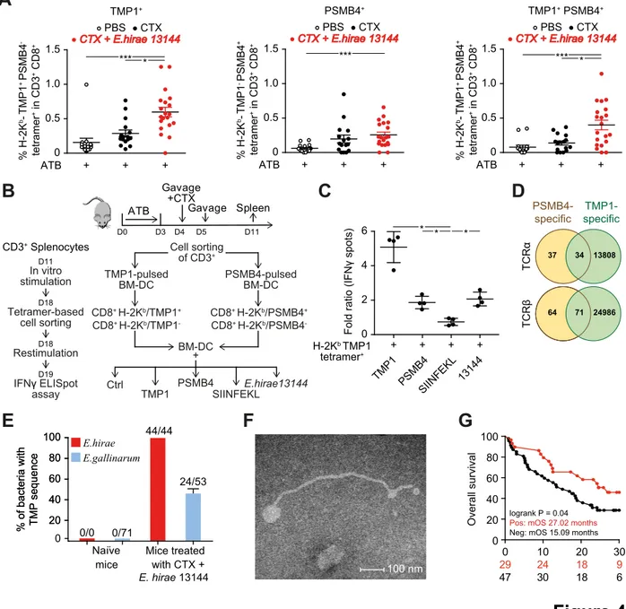
Reenforcing the notion of molecular mimicry between phage-encoded and cancer antigens, flow cytometric analyses using fluorescent-labelled tetramers H-2Kb/TSLARFANI (from TMP1) and H-2Kb/GSLARFRNI (from PSMB4) identified a subset of double-positive CTLs that infiltrate MCA205 tumors from CTX/E. hirae 13144-treated mice (Figure S6F) and that was as frequent as 245
CTLs recognizing the PSMB4 peptide only (Figure 4A). We purified the splenic CD8+ T cells using either the TMP1-H-2Kb or PSMB4-H-2Kb specific tetramers and stimulated them with irrelevant (OVA-derived-SIINFEKL) versus relevant (TMP-derived TSLARFANI or PSMB4-derived GSLARFRNI) peptides (Figure 4B). CD8+ T cells binding H-2Kb-TMP1 tetramers produced IFN not only in response to TMP1 (up to 5-fold increase in IFN secreting T cells) but 250
also in response to the PSMB4 epitope (2-fold increase, as much as with heat-killed E. hirae 13144 processed by DC) (Figure 4C, Figure S6G). Similarly, CD8+ T cells binding H-2Kb- PSMB4 tetramers functionally recognized TMP1, albeit less efficiently than the PSMB4 epitope (Figure S6G). We analyzed the T cell receptor (TCR) repertoire of these two tetramer-reactive CD8+ T cell subsets. In accordance with the functional data, half of the CD8+ T cells labelled 255
with PSMB4-H-2Kb tetramers shared clonotypes with the much wider TCR repertoire of T cells labelled with the TMP1-H-2Kb specific tetramers (Figure 4D, Table S4-S5) (but not with the negative fraction, Figure S6H). In sum, T cells recognizing the TMP1 epitope of immunogenic E.
hirae can crossreact with a peptide contained in the oncogenic driver PSMB4 and vice versa.
260
Temperate bacteriophages are bacterial viruses that can transfer virulence, antimicrobial resistance genes, and immunogenic sequences to new bacterial hosts (23). The TMP protein, which contains a variable number of tandem repeats with highly conserved tryptophan and phenylalanine residues at fixed positions is encoded by the genome of Siphoviridae phages (24,
25). To investigate the capacity of the E. hirae 13144 phage to lysogenize other bacterial species 265
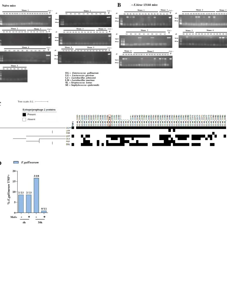
in vivo, we performed culturomic analyses of the ileal content from C57BL/6 mice subjected to
oral gavage with E. hirae 13144 and systemic CTX therapy, followed by PCR analyses seeking TMP sequences (Figure S8A-B). We tested 7 to 18 bacterial colonies from each animal and a total of 76 colonies. We only found lysogenic conversion of E. gallinarum by the E.
hirae-temperate phage in vivo, as confirmed by sequencing of the phage genome in the second
270
host (Figure 4E, Figure S8B-C). In contrast, none of the 90 colonies (mostly of E. gallinarum) isolated from naive mice harbored the TMP sequence (Figure S8A). Similarly, in vitro coculture
of TMP+ E. hirae 13144 together with TMP- E. gallinarum spp. at a 1:1 ratio uncovered a
significant (~15%) rate of lysogenic conversion (Figure S8D). Examination of a preparation admixing E. hirae 13144 and E. gallinarum at a 1:10 ratio by means of transmission electron 275
microscopy revealed numerous phages with the typical Siphoviridae morphology in the medium, whereas control cultures (bacteria separately) were free of such phages (Figure 4F). Altogether, these results indicate that the TMP1 peptide-encoding Siphoviridae phage from E. hirae 13144 is a virulent phage.
280
We next explored the possible pathophysiological relevance of these findings. We first screened a total of 3,027 adult and mother-infant metagenomes (26), validated by a second independent metagenomic-assembly based screening of 9,428 metagenomes (27) (28), to assess the breadth of coverage (BOC) of the E. hirae genome and its phages (Figure S9A). E. hirae was present with 100% confidence (i.e. BOC > 80%) in less than 150 fecal samples from disparate 285
geography, age and datasets. This phage (and its host) could be vertically transmitted from mothers to infants and then colonizes the neonate. There was an increased prevalence of the phage (57%) in fecal microbiomes from children (representing 16% of all metagenomes, Fisher’s test p-value <0.00001). Of note, the E. hirae 13144 phage was detectable in many samples lacking the presence of the E. hirae core genome, suggesting that other bacteria than E. hirae can 290
host this phage. All host genomes belonged to the Enterococcus genus (except two assigned to
Coprobacillus), in particular E. faecalis (80 genomes), E. faecium (23), and E. hirae (15),
suggesting that phage 13144 (and its homologues from E. hirae 708, and 13344) are genus-specific but not species-genus-specific.
295
Contrasting with metagenomics that has a low sensitivity to detect poor abundance species, culturomics followed by matrix-assisted laser desorption ionization time-of-flight mass spectrometry (MALDI-TOF) provides a technology for detecting rare E. hirae colonies in the stool of healthy individuals (29) or cancer patients (8). PCR analyses of each single cultivatable enterococcal colony (up to 5 per species and individual) from 76 cancer patients led to the 300
detection of the TMP sequence encompassing the TMP1 peptide in 34% of the patients, only in
E. faecalis and E. hirae (Figure S9B, Figure S10). Advanced renal and lung cancer patients
overall survival after therapy with immune checkpoint inhibitors targeting PD-1 (Figure 4G). Therefore, we screened sixteen TMP-derived nonapeptides predicted to bind the human MHC 305
class I HLA-A*0201 with high affinity for their ability to prime naive CD8+ T cells from six healthy volunteers in vitro. We found 6 out of 16 epitopes capable of triggering significant peptide-specific IFN release that were located in two distinct regions of the TMP protein (504-708 and 1397-1462, Figure S11A-B, Table S6). Using the NCBI BLASTP suite, we searched the human cancer peptidome (of the TCGA database) for a high degree of homology with these 6 310
HLA-A*0201 -restricted immunogenic nonapeptides. We found that only the TMP-derived peptide KLAKFASVV (aa 631-639) shared significant homology (7 out of 9 aa, with identical residues at the MHC anchoring positions 2 and 9) with a peptide contained in the protein glycerol-3-phosphate dehydrogenase 1-like (GPD1-L) (Figure S11C). GPD1-L reportedly counteracts the oncogenic HIF1-dependent adaptation to hypoxia, and its expression is 315
associated with favorable prognosis in head and neck squamous cell carcinomas (30–32). The TCGA transcriptomics database unveiled that high expression of GPD1-L is associated with improved overall survival in lung adenocarcinoma and kidney cancers (Figure S11D). Moreover, high expression of GPD1-L mRNA by tumors at diagnosis was associated with improved progression-free survival in three independent cohorts of non-small cell lung cancer (NSCLC) 320
patients (n=157, Table S7) treated with anti-PD1 Abs (Figure S11E-F). Expression of GPD-1L failed to correlate with that of PD-L1 in NSCLC (Figure S11G). Of note, mutations in or adjacent to the 631-639 amino acid sequence of GPD-1L gene could rarely be identified in several types of neoplasia (Figure S12).
325
We derived an HLA-A*0201-restricted, phage peptide (KLAKFASVV)-specific T cell line from peripheral blood mononuclear cells of a human volunteer. Clones from this line also recognized the HLA-A*0201 -restricted, GPD-1L epitope (KLQKFASTV) (Figure S13A-C). Moreover, we
detected CD8+ T cells binding HLA-A*0201/KLAKFASVV tetramers exhibiting hallmarks of
effector functions after in vitro stimulation of PBMC with the KLAKFASVV phage epitope in 3 330
out of 6 NSCLC patients (Fig. S13D-F). In the reverse attempt searching for molecular mimicry between well known and naturally processed non-mutated melanoma differentiation antigens recognized by human T cell clones (such as HLA-A*0201-binding MART-1 or MELOE epitopes) and gut commensal antigens, we found microbial analogs in the public microbiome data
bases (Figure S14, Table S8-Table S9, Figure S15, Table S8-S10). Some of these microbial 335
peptides are recognized by the corresponding TCR (Tables S9- S10) with similar affinities as the parental (tumoral) epitope.
Altogether the present results demonstrate that microbial genomes code for MHC class I-restricted antigens that induce a memory CD8+ T cell response, which then crossreacts with 340
cancer antigens. Several lines of evidence plead in favor of this interpretation, as exemplified for the TMP1 epitope found within a phage that infects enterococci. First, naturally occurring (‘mut3’ in E. hirae strain ATCC9790) or artificial mutations (‘mut2’ or ‘mut3’ in E.coli) introduced into the TMP1 epitope suppressed the tumor-prophylactic and therapeutic potential of bacteria expressing TMP1. Second, transfer of the TMP1-encoding gene into E. coli conferred 345
immunogenic capacity to this proteobacterium, which acquired the same antitumor properties as TMP1-expressing E. hirae. Third, when cancer cells were genetically modified to remove the TMP1-crossreactive peptide within the PSMB4 protein, they formed tumors that could no longer be controlled upon oral gavage with TMP1-expressing E. hirae. Fourth, cancer patients carrying the TMP phage sequence in fecal enterococci spp. or the GPD1-L tumoral antigen homologous to 350
TMP epitopes exhibited a better response to PD-1 blockade, suggesting that this type of microbe-cancer cross-reactivity might be clinically relevant.
Recent reports point to the pathological relevance of autoantigen-crossreactive, microbiota-derived peptides for autoimmune disorders such as myocarditis, lupus and rheumatoid arthritis 355
(34–36). Given the enormous richness of the commensal proteome (37), we expect the existence of other microbial antigens mimicking auto- and tumor antigens. In fact, we extended these findings to naturally processed melanoma-specific antigens that have microbial orthologs recognized by the same TCRs. Global phage numbers have been estimated to reach as high as 1031 particles with the potential of 1025 phage infections occurring every second (38, 39). Thus, 360
the perspective opens that, within the microbiota, bacteriophages may enrich the therapeutic armamentarium for modulating the intestinal flora and for stimulating systemic anticancer immune responses.
References and Notes:
365
1. P. Sharma, J. P. Allison, Immune checkpoint targeting in cancer therapy: toward combination strategies with curative potential. Cell. 161, 205–214 (2015).
2. L. Galluzzi, A. Buqué, O. Kepp, L. Zitvogel, G. Kroemer, Immunological Effects of Conventional Chemotherapy and Targeted Anticancer Agents. Cancer Cell. 28, 690–714 370
(2015).
3. L. Zitvogel, Y. Ma, D. Raoult, G. Kroemer, T. F. Gajewski, The microbiome in cancer immunotherapy: Diagnostic tools and therapeutic strategies. Science. 359, 1366–1370 (2018).
4. T. Tanoue, S. Morita, D. R. Plichta, A. N. Skelly, W. Suda, Y. Sugiura, S. Narushima, H. 375
Vlamakis, I. Motoo, K. Sugita, A. Shiota, K. Takeshita, K. Yasuma-Mitobe, D.
Riethmacher, T. Kaisho, J. M. Norman, D. Mucida, M. Suematsu, T. Yaguchi, V. Bucci, T. Inoue, Y. Kawakami, B. Olle, B. Roberts, M. Hattori, R. J. Xavier, K. Atarashi, K. Honda, A defined commensal consortium elicits CD8 T cells and anti-cancer immunity. Nature.
565, 600–605 (2019).
380
5. M. Vétizou, J. M. Pitt, R. Daillère, P. Lepage, N. Waldschmitt, C. Flament, S. Rusakiewicz, B. Routy, M. P. Roberti, C. P. M. Duong, V. Poirier-Colame, A. Roux, S. Becharef, S. Formenti, E. Golden, S. Cording, G. Eberl, A. Schlitzer, F. Ginhoux, S. Mani, T. Yamazaki, N. Jacquelot, D. P. Enot, M. Bérard, J. Nigou, P. Opolon, A. Eggermont, P.-L. Woerther, E. Chachaty, N. Chaput, C. Robert, C. Mateus, G. Kroemer, D. Raoult, I. G. Boneca, F.
385
Carbonnel, M. Chamaillard, L. Zitvogel, Anticancer immunotherapy by CTLA-4 blockade relies on the gut microbiota. Science. 350, 1079–1084 (2015).
6. R. Daillère, M. Vétizou, N. Waldschmitt, T. Yamazaki, C. Isnard, V. Poirier-Colame, C. P. M. Duong, C. Flament, P. Lepage, M. P. Roberti, B. Routy, N. Jacquelot, L. Apetoh, S. Becharef, S. Rusakiewicz, P. Langella, H. Sokol, G. Kroemer, D. Enot, A. Roux, A. 390
Eggermont, E. Tartour, L. Johannes, P.-L. Woerther, E. Chachaty, J.-C. Soria, E. Golden, S. Formenti, M. Plebanski, M. Madondo, P. Rosenstiel, D. Raoult, V. Cattoir, I. G. Boneca, M. Chamaillard, L. Zitvogel, Enterococcus hirae and Barnesiella intestinihominis Facilitate Cyclophosphamide-Induced Therapeutic Immunomodulatory Effects. Immunity. 45, 931– 943 (2016).
395
7. Y. Rong, Z. Dong, Z. Hong, Y. Jin, W. Zhang, B. Zhang, W. Mao, H. Kong, C. Wang, B. Yang, X. Gao, Z. Song, S. E. Green, H. K. Song, H. Wang, Y. Lu, Reactivity toward Bifidobacterium longum and Enterococcus hirae demonstrate robust CD8+ T cell response and better prognosis in HBV-related hepatocellular carcinoma. Exp. Cell Res. 358, 352–359 (2017).
400
8. B. Routy, E. Le Chatelier, L. Derosa, C. P. M. Duong, M. T. Alou, R. Daillère, A.
Fluckiger, M. Messaoudene, C. Rauber, M. P. Roberti, M. Fidelle, C. Flament, V. Poirier-Colame, P. Opolon, C. Klein, K. Iribarren, L. Mondragón, N. Jacquelot, B. Qu, G. Ferrere, C. Clémenson, L. Mezquita, J. R. Masip, C. Naltet, S. Brosseau, C. Kaderbhai, C. Richard,
H. Rizvi, F. Levenez, N. Galleron, B. Quinquis, N. Pons, B. Ryffel, V. Minard-Colin, P. 405
Gonin, J.-C. Soria, E. Deutsch, Y. Loriot, F. Ghiringhelli, G. Zalcman, F. Goldwasser, B. Escudier, M. D. Hellmann, A. Eggermont, D. Raoult, L. Albiges, G. Kroemer, L. Zitvogel, Gut microbiome influences efficacy of PD-1-based immunotherapy against epithelial tumors. Science. 359, 91–97 (2018).
9. N. R. Rose, Negative selection, epitope mimicry and autoimmunity. Curr. Opin. Immunol. 410
49, 51–55 (2017).
10. V. Rubio-Godoy, V. Dutoit, Y. Zhao, R. Simon, P. Guillaume, R. Houghten, P. Romero, J.-C. Cerottini, J.-C. Pinilla, D. Valmori, Positional scanning-synthetic peptide library-based analysis of self- and pathogen-derived peptide cross-reactivity with tumor-reactive Melan-A-specific CTL. J. Immunol. Baltim. Md 1950. 169, 5696–5707 (2002).
415
11. L. Vujanovic, M. Mandic, W. C. Olson, J. M. Kirkwood, W. J. Storkus, A mycoplasma peptide elicits heteroclitic CD4+ T cell responses against tumor antigen MAGE-A6. Clin.
Cancer Res. Off. J. Am. Assoc. Cancer Res. 13, 6796–6806 (2007).
12. M. E. Perez-Muñoz, P. Joglekar, Y.-J. Shen, Y.-J. Shen, K. Y. Chang, D. A. Peterson, Identification and Phylogeny of the First T Cell Epitope Identified from a Human Gut 420
Bacteroides Species. PloS One. 10, e0144382 (2015).
13. Y. Yang, M. B. Torchinsky, M. Gobert, H. Xiong, M. Xu, J. L. Linehan, F. Alonzo, C. Ng, A. Chen, X. Lin, A. Sczesnak, J.-J. Liao, V. J. Torres, M. K. Jenkins, J. J. Lafaille, D. R. Littman, Focused specificity of intestinal TH17 cells towards commensal bacterial antigens.
Nature. 510, 152–156 (2014).
425
14. J. N. Chai, Y. Peng, S. Rengarajan, B. D. Solomon, T. L. Ai, Z. Shen, J. S. A. Perry, K. A. Knoop, T. Tanoue, S. Narushima, K. Honda, C. O. Elson, R. D. Newberry, T. S.
Stappenbeck, A. L. Kau, D. A. Peterson, J. G. Fox, C.-S. Hsieh, Helicobacter species are potent drivers of colonic T cell responses in homeostasis and inflammation. Sci. Immunol. 2 (2017), doi:10.1126/sciimmunol.aal5068.
430
15. Q. Ji, A. Perchellet, J. M. Goverman, Viral infection triggers central nervous system autoimmunity via activation of CD8+ T cells expressing dual TCRs. Nat. Immunol. 11, 628–634 (2010).
16. V. P. Balachandran, M. Łuksza, J. N. Zhao, V. Makarov, J. A. Moral, R. Remark, B. Herbst, G. Askan, U. Bhanot, Y. Senbabaoglu, D. K. Wells, C. I. O. Cary, O. Grbovic-Huezo, M. 435
Attiyeh, B. Medina, J. Zhang, J. Loo, J. Saglimbeni, M. Abu-Akeel, R. Zappasodi, N. Riaz, M. Smoragiewicz, Z. L. Kelley, O. Basturk, Australian Pancreatic Cancer Genome
Initiative, Garvan Institute of Medical Research, Prince of Wales Hospital, Royal North Shore Hospital, University of Glasgow, St Vincent’s Hospital, QIMR Berghofer Medical Research Institute, University of Melbourne, Centre for Cancer Research, University of 440
Queensland, Institute for Molecular Bioscience, Bankstown Hospital, Liverpool Hospital, Royal Prince Alfred Hospital, Chris O’Brien Lifehouse, Westmead Hospital, Fremantle Hospital, St John of God Healthcare, Royal Adelaide Hospital, Flinders Medical Centre, Envoi Pathology, Princess Alexandria Hospital, Austin Hospital, Johns Hopkins Medical
Institutes, ARC-Net Centre for Applied Research on Cancer, M. Gönen, A. J. Levine, P. J. 445
Allen, D. T. Fearon, M. Merad, S. Gnjatic, C. A. Iacobuzio-Donahue, J. D. Wolchok, R. P. DeMatteo, T. A. Chan, B. D. Greenbaum, T. Merghoub, S. D. Leach, Identification of unique neoantigen qualities in long-term survivors of pancreatic cancer. Nature. 551, 512– 516 (2017).
17. C. P. Bradley, F. Teng, K. M. Felix, T. Sano, D. Naskar, K. E. Block, H. Huang, K. S. 450
Knox, D. R. Littman, H.-J. J. Wu, Segmented Filamentous Bacteria Provoke Lung
Autoimmunity by Inducing Gut-Lung Axis Th17 Cells Expressing Dual TCRs. Cell Host
Microbe. 22, 697-704.e4 (2017).
18. S. Viaud, F. Saccheri, G. Mignot, T. Yamazaki, R. Daillère, D. Hannani, D. P. Enot, C. Pfirschke, C. Engblom, M. J. Pittet, A. Schlitzer, F. Ginhoux, L. Apetoh, E. Chachaty, P.-L. 455
Woerther, G. Eberl, M. Bérard, C. Ecobichon, D. Clermont, C. Bizet, V.
Gaboriau-Routhiau, N. Cerf-Bensussan, P. Opolon, N. Yessaad, E. Vivier, B. Ryffel, C. O. Elson, J. Doré, G. Kroemer, P. Lepage, I. G. Boneca, F. Ghiringhelli, L. Zitvogel, The intestinal microbiota modulates the anticancer immune effects of cyclophosphamide. Science. 342, 971–976 (2013).
460
19. G. Y. Lee, P. M. Haverty, L. Li, N. M. Kljavin, R. Bourgon, J. Lee, H. Stern, Z. Modrusan, S. Seshagiri, Z. Zhang, D. Davis, D. Stokoe, J. Settleman, F. J. de Sauvage, R. M. Neve, Comparative oncogenomics identifies PSMB4 and SHMT2 as potential cancer driver genes.
Cancer Res. 74, 3114–3126 (2014).
20. Y.-C. Cheng, W.-C. Tsai, Y.-C. Sung, H.-H. Chang, Y. Chen, Interference with PSMB4 465
Expression Exerts an Anti-Tumor Effect by Decreasing the Invasion and Proliferation of Human Glioblastoma Cells. Cell. Physiol. Biochem. Int. J. Exp. Cell. Physiol. Biochem.
Pharmacol. 45, 819–831 (2018).
21. X. Zhang, D. Lin, Y. Lin, H. Chen, M. Zou, S. Zhong, X. Yi, S. Han, Proteasome beta-4 subunit contributes to the development of melanoma and is regulated by miR-148b. Tumour 470
Biol. J. Int. Soc. Oncodevelopmental Biol. Med. 39, 1010428317705767 (2017).
22. H. Wang, Z. He, L. Xia, W. Zhang, L. Xu, X. Yue, X. Ru, Y. Xu, PSMB4 overexpression enhances the cell growth and viability of breast cancer cells leading to a poor prognosis.
Oncol. Rep. 40, 2343–2352 (2018).
23. M. G. Weinbauer, Ecology of prokaryotic viruses. FEMS Microbiol. Rev. 28, 127–181 475
(2004).
24. M. Piuri, G. F. Hatfull, A peptidoglycan hydrolase motif within the mycobacteriophage TM4 tape measure protein promotes efficient infection of stationary phase cells. Mol.
Microbiol. 62, 1569–1585 (2006).
25. M. Belcaid, A. Bergeron, G. Poisson, The evolution of the tape measure protein: units, 480
26. E. Pasolli, L. Schiffer, P. Manghi, A. Renson, V. Obenchain, D. T. Truong, F. Beghini, F. Malik, M. Ramos, J. B. Dowd, C. Huttenhower, M. Morgan, N. Segata, L. Waldron, Accessible, curated metagenomic data through ExperimentHub. Nat. Methods. 14, 1023– 1024 (2017).
485
27. P. Ferretti, E. Pasolli, A. Tett, F. Asnicar, V. Gorfer, S. Fedi, F. Armanini, D. T. Truong, S. Manara, M. Zolfo, F. Beghini, R. Bertorelli, V. De Sanctis, I. Bariletti, R. Canto, R.
Clementi, M. Cologna, T. Crifò, G. Cusumano, S. Gottardi, C. Innamorati, C. Masè, D. Postai, D. Savoi, S. Duranti, G. A. Lugli, L. Mancabelli, F. Turroni, C. Ferrario, C. Milani, M. Mangifesta, R. Anzalone, A. Viappiani, M. Yassour, H. Vlamakis, R. Xavier, C. M. 490
Collado, O. Koren, S. Tateo, M. Soffiati, A. Pedrotti, M. Ventura, C. Huttenhower, P. Bork, N. Segata, Mother-to-Infant Microbial Transmission from Different Body Sites Shapes the Developing Infant Gut Microbiome. Cell Host Microbe. 24, 133-145.e5 (2018).
28. E. Pasolli, F. Asnicar, S. Manara, M. Zolfo, N. Karcher, F. Armanini, F. Beghini, P. Manghi, A. Tett, P. Ghensi, M. C. Collado, B. L. Rice, C. DuLong, X. C. Morgan, C. D. 495
Golden, C. Quince, C. Huttenhower, N. Segata, Extensive Unexplored Human Microbiome Diversity Revealed by Over 150,000 Genomes from Metagenomes Spanning Age,
Geography, and Lifestyle. Cell. 176, 649-662.e20 (2019).
29. B. Samb-Ba, C. Mazenot, A. Gassama-Sow, G. Dubourg, H. Richet, P. Hugon, J.-C. Lagier, D. Raoult, F. Fenollar, MALDI-TOF identification of the human Gut microbiome in people 500
with and without diarrhea in Senegal. PloS One. 9, e87419 (2014).
30. T. J. Kelly, A. L. Souza, C. B. Clish, P. Puigserver, A hypoxia-induced positive feedback loop promotes hypoxia-inducible factor 1alpha stability through miR-210 suppression of glycerol-3-phosphate dehydrogenase 1-like. Mol. Cell. Biol. 31, 2696–2706 (2011).
31. Z. Feng, J. N. Li, L. Wang, Y. F. Pu, Y. Wang, C. B. Guo, The prognostic value of glycerol-505
3-phosphate dehydrogenase 1-like expression in head and neck squamous cell carcinoma.
Histopathology. 64, 348–355 (2014).
32. S.-C. Liu, S.-M. Chuang, C.-J. Hsu, C.-H. Tsai, S.-W. Wang, C.-H. Tang, CTGF increases vascular endothelial growth factor-dependent angiogenesis in human synovial fibroblasts by increasing miR-210 expression. Cell Death Dis. 5, e1485 (2014).
510
33. S. Simon, Z. Wu, J. Cruard, V. Vignard, A. Fortun, A. Khammari, B. Dreno, F. Lang, S. J. Rulli, N. Labarriere, TCR Analyses of Two Vast and Shared Melanoma Antigen-Specific T Cell Repertoires: Common and Specific Features. Front. Immunol. 9, 1962 (2018).
34. C. Gil-Cruz, C. Perez-Shibayama, A. De Martin, F. Ronchi, K. van der Borght, R. Niederer, L. Onder, M. Lütge, M. Novkovic, V. Nindl, G. Ramos, M. Arnoldini, E. M. C. Slack, V. 515
Boivin-Jahns, R. Jahns, M. Wyss, C. Mooser, B. N. Lambrecht, M. T. Maeder, H. Rickli, L. Flatz, U. Eriksson, M. B. Geuking, K. D. McCoy, B. Ludewig, Microbiota-derived peptide mimics drive lethal inflammatory cardiomyopathy. Science. 366, 881–886 (2019).
35. T. M. Greiling, C. Dehner, X. Chen, K. Hughes, A. J. Iñiguez, M. Boccitto, D. Z. Ruiz, S. C. Renfroe, S. M. Vieira, W. E. Ruff, S. Sim, C. Kriegel, J. Glanternik, X. Chen, M. Girardi, 520
P. Degnan, K. H. Costenbader, A. L. Goodman, S. L. Wolin, M. A. Kriegel, Commensal orthologs of the human autoantigen Ro60 as triggers of autoimmunity in lupus. Sci. Transl.
Med. 10 (2018), doi:10.1126/scitranslmed.aan2306.
36. M. F. Konig, L. Abusleme, J. Reinholdt, R. J. Palmer, R. P. Teles, K. Sampson, A. Rosen, P. A. Nigrovic, J. Sokolove, J. T. Giles, N. M. Moutsopoulos, F. Andrade, Aggregatibacter 525
actinomycetemcomitans-induced hypercitrullination links periodontal infection to autoimmunity in rheumatoid arthritis. Sci. Transl. Med. 8, 369ra176 (2016).
37. J. Li, H. Jia, X. Cai, H. Zhong, Q. Feng, S. Sunagawa, M. Arumugam, J. R. Kultima, E. Prifti, T. Nielsen, A. S. Juncker, C. Manichanh, B. Chen, W. Zhang, F. Levenez, J. Wang, X. Xu, L. Xiao, S. Liang, D. Zhang, Z. Zhang, W. Chen, H. Zhao, J. Y. Al-Aama, S. Edris, 530
H. Yang, J. Wang, T. Hansen, H. B. Nielsen, S. Brunak, K. Kristiansen, F. Guarner, O. Pedersen, J. Doré, S. D. Ehrlich, MetaHIT Consortium, P. Bork, J. Wang, MetaHIT Consortium, An integrated catalog of reference genes in the human gut microbiome. Nat.
Biotechnol. 32, 834–841 (2014).
38. M. L. Pedulla, M. E. Ford, J. M. Houtz, T. Karthikeyan, C. Wadsworth, J. A. Lewis, D. 535
Jacobs-Sera, J. Falbo, J. Gross, N. R. Pannunzio, W. Brucker, V. Kumar, J. Kandasamy, L. Keenan, S. Bardarov, J. Kriakov, J. G. Lawrence, W. R. Jacobs, R. W. Hendrix, G. F. Hatfull, Origins of highly mosaic mycobacteriophage genomes. Cell. 113, 171–182 (2003). 39. K. E. Wommack, R. R. Colwell, Virioplankton: viruses in aquatic ecosystems. Microbiol.
Mol. Biol. Rev. MMBR. 64, 69–114 (2000).
540
Acknowledgments: We are thankful to the animal facility team of Gustave Roussy and all the
technicians from Centre GF Leclerc. We are very endebted to Dr Oliver Kepp, Gustave Roussy for figure design, and to Prof. Hans Georg Rammensee from the Department of Immunology, Institute for Cell Biology, University of Tübingen, Tübingen, Germany for his careful guidance 545
in peptide selection and reading of the paper. LZ and GK were supported by the Ligue contre le Cancer (équipe labelisée); Agence National de la Recherche (ANR) – Projets blancs; ANR under the frame of E-Rare-2, the ERA-Net for Research on Rare Diseases; Association pour la recherche sur le cancer (ARC); Cancéropôle Ile-de-France; Chancelerie des universités de Paris (Legs Poix), Fondation pour la Recherche Médicale (FRM); a donation by Elior; the European 550
Commission (Horizon 2020: Oncobiome); the European Research Council (ERC); Fondation
Carrefour; High-end Foreign Expert Program in China (GDW20171100085 and
GDW20181100051), Institut National du Cancer (INCa); Inserm (HTE); Institut Universitaire de France; LeDucq Foundation; the LabEx Immuno-Oncology; the RHU Torino Lumière; the Seerave Foundation; the SIRIC Stratified Oncology Cell DNA Repair and Tumor Immune 555
Elimination (SOCRATE); ONCOBIOME H2020 network, CARE network (directed by Prof. Mariette, Kremlin Bicêtre AP-HP), and the SIRIC Cancer Research and Personalized Medicine (CARPEM); RHU Torino Lumière (ANR-16-RHUS-0008). The results shown here are based upon data generated by the TCGA Research Network: http://cancergenome.nih.gov/. National Research, Development andInnovation Fund of Hungary Project no. FIEK_16-1-2016-0005. Z.S 560
was supported by the Research and Technology Innovation Fund NAP2-2017-1.2.1-NKP-0002, Breast Cancer Research Foundation (BCRF-17-156). Z.S and I.C were supported by the Novo Nordisk Foundation Interdisciplinary Synergy Programme Grant (NNF15OC0016584). PN was supported by the Italian Association for Cancer Research AIRC IG 19822. Mouse TCR sequencing was performed by the TRiPoD ERC-Advanced EU (322856) grants to Prof. David 565
Klatzmann.
Competing interests statement: RD, DR, LZ and GK are cofounders of everImmune, a biotech
company devoted to the use of commensal microbes for the treatment of cancers. RD is a full-time employee of everImmune. RD and LZ hold patents on immunogenic phage sequences. 570
Supplementary Materials:
Materials and Methods Figures S1-S15
Tables S1-S10 Reference (1-17) 575
% H-2K b-TMP1 + tetramer + CD8 +
Figure 1
D0 D3 D4 ATB Gavage +CTX Gavage D5 D11 D12 D18 D19 Tumor inoculation D25 Gavage +CTX Gavage Gavage +CTX Gavage Tumor size at sacrifice D0 D3 D4 ATB Gavage +CTX Gavage D5 D11 Tumor inoculation Spleen Ex vivo recall responses & flow cytometryCTX CTX + + + + + Tu m or s iz e (m IFN γ spots / 2x10 5 CD8 + T cells
E
D
20 40 6080 CTX CTX+E.hirae13144 CTX+E.hiraeIGR11 CTX+E.hirae ATCC9790 CTX +E.hirae13344 CTX +E.hirae708
+ + + + + - - + + 0 100 E.hirae 13344 E.hirae ATCC9790 0 0.5 1.0 1.5 2.0 No pe ptideTMP1TMP2TMP3 VSTNHYGLLNo pepti de TMP1TMP2TMP3 No pe ptideTMP1TMP2TMP3 No pe ptideTMP1TMP2TMP3 No pe ptideTMP1TMP2TMP3 No pe ptideTMP1TMP2TMP3 VSTNHYGLL VSTNHYGLL VSTNHYGLL VSTNHYGLL VSTNHYGLL
E.hirae13144 E.hirae IGR11 PBS Tetramer TMP1+ SIINFEKLTetramer+ ** * *** *** ** ** ** ns ns ns ns
Legends to Figures
Figure 1. Phage Tail Length Tape Measure Protein as the unique antigenic sequence in E. hirae 13144.
A, B. C57BL/6 mice bearing MCA205 sarcomas were conditioned with broad spectrum antibiotics (streptomycin, colistin, ampicillin, vancomycin) for 3 days before performing oral gavages with E. hirae strain 13144 and i.p. injections of cyclophosphamide (CTX), as indicated (A), and tumor size was recorded for each mouse at sacrifice on day 25 (B). C-E. Naïve C57BL/6 mice were conditioned with antibiotics, gavaged with distinct E. hirae strains and treated with CTX (C). Day 11 purified CD8+ T splenocytes were restimulated ex vivo in a recall assay with bone marrow-derived dendritic cells loaded with the indicated peptides (Table S2, group 7) to quantify IFNγ-secreting CD8+T cells (D). H-2Kb/TMP1 (TSLARFANI) or H-2Kb/SIINFEKL tetramer binding CD8+ splenocytes were detected by cytofluorometry at day 11 (E). Also refer to Figure S2. Each graph assembles results from 2-3 independent experiments containing groups of 5-6 mice. ANOVA statistical analyses (Kruskal-wallis test): *p<0.05, **p<0.01, ***p<0.001. Refer to the statistical report.
+ + + + + +
Figure 2
0 5 10 15 20 25 50 0 100 150 200Group 1 pulsed-DC TMP1 pulsed-DC
PBS DC E.hirae 13144 pulsed-DC
TMP1-mut3 pulsed-DC Group 1 pulsed-DC TMP1 pulsed-DC
PBS DC E.hirae 13144 pulsed-DC
E.coli EGFP
PBS E.hirae 13144
E.coli TMP1-mut3
E.coli TMP1 E.coli TMP1-mut2
TMP1-mut3 pulsed-DC
Days after tumor inoculation
CTX
B
C
D
E
DCDC Tumor size (mm 2) Tumor size (mm 2) 0 100 200 300 400 0 0.5 1.0 1.5 2.5 2.0 0 100 200 300 400 Tumor size (mm 2) + + + + + + E.coli EGFP PBS E.hirae 13144 E.coli TMP1-mut3E.coli TMP1 E.coli TMP1-mut2
CTX % H-2K b-TMP1 tetramer + CD8 * * * * * * ** * * ** * **
Figure 2. Prophylactic and therapeutic immunization using Phage Tail Length Tape Measure Protein (TMP) against sarcomas.
A. Sequence of the immunogenic epitope TMP1 (TSLARFANI) with the artificial and naturally occuring mutations in positions 2 and 3, respectively. B-C. Prophylactic vaccinations. TLR3 ligand-exposed dendritic cell (DC) were pulsed with peptides or heat-inactivated bacteria and then s.c. inoculated twice into mice. One month later, MCA205 sarcomas were implanted in the opposite flank, followed by monitoring of tumor size (means±SEM in B, individual results in C). D-E. Therapeutic settings. MCA205 tumor bearing mice were treated with cyclophosphamide (CTX) and gavaged with E. hirae 13144 or E. coli (like in Fig. 1A) that were genetically modified to express the indicated peptides or enhanced green fluorescent protein (EGFP) as a negative control. Tumor growth at sacrifice (D) and the frequency of H-2Kb/TMP1 tetramer binding splenic CD8+ T cells (E) were monitored. Results are shown for 12-18 animals, gathered from 2-3 independent experiments. ANOVA statistical analyses (Kruskal-wallis test): *p<0.05,
Figure 3
clone 3 clone 2
clone 1 clone 2
PSMB4-mut2 PSMB4-mut2 PSMB4-mut3 PSMB4-mut3 PSMB4-mut3
clone 1 WT clone 2 WT PSMB4-mut3 WT clone 1 MCA205 WT polyclonal
C
D
E
B
1 2 3 4 5 6 MCA205 TC1 MC38 Rel. expression of PSMB4 (fold change) MCA205 WT (clone 1) Tumor size (mm 2) 0 100 150 50 200 250 0 Tu m or s iz e (m m 2) 0 100 150 50 200 250 10 20 30Days after tumor inoculation
PBS CTX CTX + E.hirae 13144 CTX CTX + E.hirae 13144 CTX CTX + E.hirae 13144 0 10 20 30 Tumor size (mm 2) 0 100 150 50 200 250
MCA205 PSMB4-mut2 (clone 1)
Days after tumor inoculation
PBS CTX CTX + E.hirae 13144 Tumor size (mm 2) 0 100 150 50 200 250 0 10 20 30
Days after tumor inoculation
PBS CTX CTX + E.hirae 13144 * TC1 WT PBS CTX CTX + E.hirae 13144 TC1 PSMB4-mut3 PBS CTX CTX + E.hirae 13144 Tumor size (mm 2) 0 0 100 200 300 10 20 30
Days after tumor inoculation
Tumor size (mm 2) 0 100 200 400 300 Tumor size (mm 2) 0 100 200 400 300 0 10 20 30 40
Days after tumor inoculation
ns p= 0.0823 * * * * * * ** ns ns
Figure 3. Molecular mimicry between enterophage TMP and the oncogenic driver PSMB4 in two mouse cancers.
A. Sequence alignment of the enterophage TMP1 peptide and a PSMB4 epitope with its two experimental mutants. B. Relative expression of PSMB4 mRNA in MCA205 sarcoma, TC1 lung cancer and MC38 colon carcinomas as compared to their healthy tissue of origin (mean ratio+SEM, n=3). C-D. Therapeutic response of wild type versus knock-in mutants of MCA205 to cyclophosphamide (CTX) alone or in combination with immunogenic E. hirae strain 13144 (setting as in Fig. 1A). Results are shown as tumor growth kinetics (means±SEM) for selected MCA205 clones (C) or as individual results (one dot corresponds to one mouse) on day 25 (D). E. Therapeutic response of wild type versus mutated TC1 lung cancers to CTX alone or in combination with E. hirae 13144 (setting as in Fig. 1A, but without antibiotic preconditioning) reflected by tumor growth kinetics and individual tumor sizes at sacrifice. Results are shown as means +SEM. Mann Whitney test or ANOVA statistical analyses (Kruskal-wallis test): *p<0.05,
Figure 4
C
E
F
D
TCRβ TCRαG
PSMB4-specific specific TMP1-37 34 13808 64 71 24986 % H-2K b-TMP1 + PSMB4 - tetramer + in CD3 + CD8 + % H-2K b-TMP1 - PSMB4 + tetramer + in CD3 + CD8 + % H-2K b-TMP1 + PSMB4 + tetramer + in CD3 + CD8 + 0 0.5 1.5 1.0 0 0.5 1.5 1.0 0 0.5 1.5 1.0CTX + E.hirae 13144 CTX + E.hirae 13144 CTX + E.hirae 13144
+ + +
ATB ATB + + + ATB + + +
Naïve Mice treated
E. hirae 13144 44/44 24/53 0/71 0/0 E.hirae E.gallinarum 0 20 40 60 80 100
% of bacteria with TMP sequence 20
40 60 80 100
% of bacteria with TMP sequence
H-2Kb TMP1
tetramer+
TMP1 PSMB4SIINFEKL 13144
Fold ratio (IFN
γ spots) 0 2 6 4 + + + + D0 D3 D4 ATB Gavage +CTX CD3+ Splenocytes Gavage D5 D11 Spleen
B
Cell sorting of CD3+ Tetramer-based cell sorting D18 In vitro stimulation D11 D18 Restimulation IFNγ ELISpot assay D19 CD8+ H-2Kb/TMP1+ CD8+ H-2Kb/TMP1- CD8 + H-2Kb/PSMB4+ CD8+ H-2Kb/PSMB4 -TMP1-pulsed BM-DC PSMB4-pulsedBM-DC BM-DC Ctrl TMP1 PSMB4SIINFEKLE.hirae13144 + 0 10 20 30 29 47 2430 1818 96 logrank P = 0.04Pos: mOS 27.02 months
Neg: mOS 15.09 months
0 20 40 60 80 100 Overall survival * * * *** *** * *** * with CTX + mice 100 nm
Figure 4. TMP crossreacts with the PSMB4 cancer epitope and affects human anticancer immune responses.
A. Flow cytometry analysis of CD8+ tumor-infiltrating lymphocytes (from tumors treated as in Fig. 1A) after co-staining with two different tetramers (H-2Kb/TMP1 and H-2Kb/PSMB4, sequences in Fig. 3A). Each dot depicts one tumor. The graphs assemble the results of 3 independent experiments with 5 mice/group. B,C. Purified CD3+ T splenocytes from animals treated with CTX and E. hirae 13144 were restimulated ex vivo with bone marrow-derived dendritic cells (DC) loaded with TMP1 or PSMB4 peptide. One week after ex vivo restimulation, peptide- specific CD8+ T cells were purified after staining with the corresponding tetramer to measure IFNγ secretion in response to DC loaded with peptides (TMP1, PSMB4, SIINFEKL as negative control) or heat-inactivated E. hirae 13144. These results were performed in parallel on the tetramer-binding versus non-binding fraction and were normalized to the PBS controls (Ctrl). Each dot represents one culture. Mann Whitney test or ANOVA statistical analyses (Kruskal-wallis test): *p<0.05, ** p<0.01, ***p<0.001. D. Venn diagram of TCR and chains from tetramer positive CD8+ T cells specific for PSMB4 (yellow) or TMP1 (green). E. Lysogenic conversion of E. gallinarum by the E. hirae siphoviridae phage in vivo. Ileal content was obtained from naïve mice or from mice receiving E. hirae together with cyclophosphamide (CTX), followed by cultivation and isolation of bacterial colonies, MALDI-TOF identification and PCR-based detection of TMP. Results are from 5 mice/group. F. Transmission electron microscopy of the phage produced by E. hirae 13144. G. Kaplan Meier survival plots of 76 patients with non-small cell lung cancer or renal cell cancer subjected to PD-1-targeting immunotherapy, stratified according to the presence or absence of TMP in at least 5 E. faecalis or
E. hirae colonies/patient. Univariate Log-rank (Mantel-Cox) analysis. Refer to the statistical
Crossreactivity between MHC class I-restricted antigens from cancer cells and an
enterococcal bacteriophage.
Authors: Aurélie Fluckiger, Romain Daillère, Mohamed Sassi, Barbara Susanne Sixt, Peng Liu,
Friedemann Loos, Corentin Richard, Catherine Rabu, Maryam Tidjani Alou, Anne-Gaëlle
Goubet, Fabien Lemaitre, Gladys Ferrere, Lisa Derosa, Connie PM Duong, Meriem
Messaoudene, Andréanne Gagné, Luisa De Sordi, Laurent Debarbieux, Sylvain Simon, Clara-Maria Scarlata, Maha Ayyoub, Belinda Palermo, Francesco Facciolo, Romain Boidot, Richard Wheeler, Ivo Gomperts Boneca, Zsofia Sztupinszki, Krisztian Papp, Istvan Csabai, Edoardo Pasolli, Nicola Segata, Carlos Lopez-Otin, Zoltan Szallasi, Fabrice Andre, Valerio Iebba, Valentin Quiniou, David Klatzmann, Jacques Boukhalil, Saber Khelaifia, Didier Raoult,
Laurence Albiges, Bernard Escudier, Alexander Eggermont, Fathia Mami-Chouaib, Paola
Nistico, Nathalie Labarrière, François Ghiringhelli, Bertrand Routy, Vincent Cattoir, Guido Kroemer*, and Laurence Zitvogel*.
*Correspondence to: laurence.zitvogel@gustaveroussy.fr; kroemer@orange.fr
This PDF file includes:
Materials and Methods Figures S1 to S15 Tables S1 to S10 Statistical report
Methods:
Cell culture, reagents and tumor cell lines. MC38, TC1, MCA205 (WT or PSMB4-mutated)
tumor cell lines or clones were cultured at 37°C with 5% CO2 in RPMI 1640 medium containing
10% fetal calf serum (FCS), 2 mM L-glutamine, 100 UI/mL penicillin/streptomycin, 1 mM sodium pyruvate and MEM non-essential amino acids (henceforth referred to as complete RPMI 1640). All these reagents were purchased from Gibco-Invitrogen (Carlsbad, CA, USA).
Mice. All animal experiments were carried out in compliance with French and European laws
and guidelines and regulations. The local institutional board approved all mouse experiments (permission number: 2016-109-7450). All mouse experiments were performed at the animal facility in Gustave Roussy Cancer Campus where animals were housed in specific pathogen-free conditions. Female C57BL/6 were purchased from Harlan (Gannat, France). Mice were used at an age between 7 and 12 weeks of age.
Antibiotic treatments. Mice were treated during 3 days (biotinylated) an antibiotic (ATB)
solution containing ampicillin (1 mg/mL), streptomycin (5 mg/mL), colistin (1 mg/mL) and vancomycin (0.25 mg/mL) (Sigma-Aldrich) added to the sterile drinking water of mice. Antibiotic activity was confirmed by cultivating fecal pellets resuspended in brain heart infusion (BHI) broth + 15% glycerol at 0.1 g/mL on COS (BD Columbia agar with 5% sheep blood, BioMérieux) plates for 48h at 37°C in aerobic and anaerobic conditions. In the context of bacterial or fecal transplantation, mice received 3 days of ATB before undergoing bacterial or fecal transplantation the next day by oral gavage using animal feeding needles. ATB were not used for Figure 3E and Figure S6E.
Tumor challenge and treatment. Syngeneic C57BL/6 mice were inoculated subcutaneously
(s.c.) with 1× 10⁶ MC38 colon cancer cells, 0.8 × 10⁶ MCA205 sarcoma cells or 0.8 × 10⁶ TC1 lung cancer cells. When tumors reached 20 to 35 mm² in size, the mice were treated intraperitoneally (i.p.) with cyclophosphamide (CTX, 100mg/kg) (Endoxan Baxter, was provided by Institut de Cancérologie Gustave Roussy, Villejuif, France) or anti-PD-1 mAb (250μg/mouse; clone RMP1-14) or isotype control (clone 2A3) (BioXcell, NH, USA). Depending on the experimental setting, mice were injected with CTX once or 3 times at 1-week intervals. Mice
were injected 4 times at 3-day intervals with anti-PD-1 mAb. Tumor size was routinely monitored every 3 days by means of a caliper.
Gut colonization with dedicated commensal species. Enterococcus hirae 13144 were originally
isolated from spleens of SPF mice treated with CTX in our laboratory. E. hirae 708 was provided by INRA (P. Langella), while E. hirae 13344, ATCC9790 were provided by Prof. Cattoir, CHU de Caen, France. L. plantarum was provided by Prof. Ivo Gomperts Boneca from the Institut Pasteur strain repository, France. All E.hirae IGR strains were isolated from the stools of NSCLC patients in our laboratory, according to patient informed consent and local IRB approval (ancillary study "Oncobiotics"). All bacteria were grown in COS plates for 24 to 48 hours at 37°C in aerobic conditions. Colonization of ATB pre-treated mice was performed by oral gavage with 100 μl of suspension containing 1 × 10⁹
bacteria. For bacterial gavage, we used suspensions of 1010 CFU/mL, monitored using a fluorescence spectrophotometer (Eppendorf) at an optical density of 600 nm in PBS. Depending on the experimental setting, 2 or 6 bacterial gavages were performed for each mouse: the first, the same day as CTX injection, and then 24 hours after the injection of CTX. For anti-PD1 mAb, 5 bacterial oral gavages were performed for each mouse: the first, the same day and 24h before the first anti-PD1 injection, and the same day for the three other injections of anti-PD1 Abs. The efficacy of colonization was confirmed by culturing the feces 48 hours post-gavage. Fecal pellets were harvested and resuspended in BHI+15% glycerol at 0.1 g/mL. Serial dilutions of feces were plated onto COS plates and incubated for 48 hours at 37°C in aerobic and anaerobic conditions. After 48 hours, the identification of specific bacteria was accomplished using a Matrix-Assisted Laser Desorption/Ionisation Time of Flight (MALDI-TOF) mass spectrometer (Andromas, Beckman Coulter, France).
Culture and propagation of bone marrow-derived dendritic cells. Bone marrow-derived
dendritic cells (BM-DCs) were generated by flushing bone marrow precursors from the femurs and tibia of female C57Bl/6 WT mice aged between 8 and 12 weeks. Bones were collected in sterile PBS, washed in alcohol and Iscove’s medium (IMDM, Sigma-Aldrich) baths, extremities of bones were cut and flushed using a 26G needle. After red blood cell lysis, cells were cultured in IMDM supplemented with 10% of FCS + 2mM L-glutamine + 100 UI/mL penicillin/streptomycin + 50μM 2-mercaptoethanol (Sigma-Aldrich) (referred herein as complete
IMDM medium) at 0.5 × 10⁶/mL and treated with 40ng/mL of GM-CSF (supernatant of GM-CSF transfected-cells J558) and 10 ng/mL of recombinant interleukin-4 (IL-4) for BM-DCs (from Peprotech). Cells were split at day 3 and used in experiments on day 7 or 8.
Test of memory TC1 immune response and H-2Kb restricted-peptides on splenic CD8+ T cells. Interferon-γ (IFN-γ) ELISPOT assay were performed in 96-well PVDF bottomed sterile
plates (Millipore MSIP S4510) by means of a commercial kit (Cell sciences, Newburyport, US) according to the manufacturer's instructions. After PVDF membrane activation with ethanol 35%, plates were coated overnight with capture antibody to IFN-γ and washed before incubation of blocking buffer during 2 hours. BM-DC (1 × 10⁵/well) were exposed to heat-inactivated (2 hours at 65°C) bacterial strains (E. hirae 13144, E.hirae 708, E.hirae 13344 and L.plantarum at a multiplicity of infection [MOI] of 1:10) or pulsed with peptides (20μg/mL) and were added to CD8+ T cells (2 x 10⁵/well) for 20 hours at 37°C. Cells were then removed and plates were developed with a biotinylated antibody specific for IFN-γ during 1 hour and 30 minutes, followed by streptavidin-alkaline phosphatase during 1 hour. Finally, the substrate of streptavidin (BCIP/NBT buffer) was added for 5-20 min. Spots were counted by means of a CTL Immunospot Analyzer (Cellular Technology Limited, Cleveland, OH).
Vaccination of mice. BM-DCs were activated with poly I:C (10µg/mL, Invivogen) overnight
before infection with heat-inactivated (2 hours at 65°C) bacterial strains (MOI 10) or pulsed with peptides (20μg/mL, peptide 2.0). After 6 hours of incubation with bacteria or 1 hour of incubation with peptides, BM-DCs were washed 3 times with PBS before subcutaneous injection in the right flank of mice (1.5 x 10⁵ cells per mice). Mice were vaccinated twice at 10 days apart and challenged 4 weeks after the second vaccination with the minimal tumorigenic dose of MCA205 tumor cells in left flank.
Flow cytometry analyses. In experiments without tumor, spleens were harvested 7 days after the
injection of CTX. In tumor growth experiments, spleens, tumors and tumor draining lymph node were harvested at different time points, 7, 14 and 21 days after the first injection of CTX into mice bearing MCA205 tumors. Excised tumors were cut into small pieces and digested in RPMI medium containing LiberaseTM at 25 μg/mL and DNase1 at 150 UI/mL (Roche) for 30 minutes at
37°C and then crushed and filtered twice using 100 and 40μm cell strainers (Becton & Dickinson, BD). Lymph nodes and spleen were crushed in RPMI medium and subsequently filtered through a 70 μm cell strainer. Two million splenocytes, tumor cells or lymph node cells were pre-incubated with purified antimouse CD16/CD32 (clone 93; eBioscience) for 15 minutes at 4°C, before membrane staining. Dead cells were excluded using the Live/Dead Fixable Yellow dead cell stain kit (Life Technologies). Anti-mouse antibodies for CD3 (145-2C11), CD4 (GK1.5), CD8 (eBioH35-17.2), CXCR3 (CXCR3-173), CCR9 (CW-1.2), and TMP specific tetramer (BD, BioLegend, eBioscience and Cliniscience). Stained samples were acquired on Canto II 7 colors cytometer (BD) and analyses were performed with FlowJo software (Tree Star, Ashland, OR, USA).
Human T cell responses to HLA-A*0201 restricted-TMP epitopes. Cytapheresis cones were
collected from healthy volunteers (Etablissement français du sang, EFS) and peripheral blood mononuclear cells (PBMC) were separated using a Ficoll Hypaque (Sigma Aldrich) gradient. We selected only donors with the HLA-A02*01 haplotype determined by immunofluorescence and flow cytometry. PBMC were washed and resuspended in the separation medium (PBS, 1mM ethylenediaminetetraacetic acid, 2% human AB+ serum) for magnetic bead separation. CD14+ monocytic cells (human CD14 MicroBeads, Miltenyi) were enriched from 75 × 106 peripheral blood mononuclear cells (PBMC) and cultured at 0.5 x 10⁶/mL in IMDM supplemented with
10% human AB+ serum, 1% of 2 mmol/L glutamine (GIBCO Invitrogen), 1000 IU/mL GM-CSF
and 1000 IU/mL IL-4 (Miltenyi). Cells were split at day 3 and used in experiments on day 6 or 7. Such (DC-like) cells were seeded in 96-well plates at 1 × 10⁵ cells/well either alone or in the presence of peptides (20µg/mL) for 2 hours at 37°C, 5% CO2. The remaining autologous PBMC fractions were enriched for CD8+ T cells (CD8+ T Cell Isolation Kit, human, Miltenyi). The enriched CD8+ T cells were washed, counted and resuspended at 1 × 10⁵cells/well in RPMI-1640
supplemented with 10% human AB+ serum, 1% 2 mMol/L glutamine, 1%
penicillin/streptomycin (GIBCO Invitrogen) and 50 U/mL IL-2 (Proleukin). DC-peptide/ T cell co-cultures were incubated for one week at 37°C, 5% CO2 (medium was changed every 2 days).
Then, the pools of cells were seeded in 96-well ELIspot plates at 2 x 10⁵ cells/well and restimulated with or without peptides (20µg/mL) or anti-CD3/anti-CD28 coated beads (1μL/mL, Dynabeads T-Activator, Invitrogen) as a positive control for 20 hours at 37°C. IFN-γ ELISPOT