HAL Id: hal-02951491
https://hal-normandie-univ.archives-ouvertes.fr/hal-02951491
Submitted on 28 Sep 2020
HAL is a multi-disciplinary open access
archive for the deposit and dissemination of
sci-entific research documents, whether they are
pub-lished or not. The documents may come from
teaching and research institutions in France or
abroad, or from public or private research centers.
L’archive ouverte pluridisciplinaire HAL, est
destinée au dépôt et à la diffusion de documents
scientifiques de niveau recherche, publiés ou non,
émanant des établissements d’enseignement et de
recherche français ou étrangers, des laboratoires
publics ou privés.
The Shift from Local to Global Visual Processing in
6-Year-Old Children Is Associated with Grey Matter Loss
Nicolas Poirel, Grégory Simon, Mathieu Cassotti, Gaëlle Leroux, Guy
Perchey, Céline Lanoë, Amélie Lubin, Marie-Renée Turbelin, Sandrine Rossi,
Arlette Pineau, et al.
To cite this version:
Nicolas Poirel, Grégory Simon, Mathieu Cassotti, Gaëlle Leroux, Guy Perchey, et al.. The Shift
from Local to Global Visual Processing in 6- Year-Old Children Is Associated with Grey Matter
Loss. PLoS ONE, Public Library of Science, 2011, 6 (6), pp.e20879. �10.1371/journal.pone.0020879�.
�hal-02951491�
investigate changes in gray matter that underlie the shift from a bias for local to global visual information. Six-year-old children were assigned to groups according to their judgment on a global/local task. The first group included children who still presented with local visual processing biases, and the second group included children who showed global visual processing biases. VBM results indicated that compared to children with local visual processing biases, children with global visual processing biases had a loss of gray matter in the right occipital and parietal visuospatial areas.
Conclusions: These anatomical findings are in agreement with previous findings in children with neurodevelopmental disorders and represent the first structural identification of brain regions that allow healthy children to develop a global perception of the visual world.
Citation: Poirel N, Simon G, Cassotti M, Leroux G, Perchey G, et al. (2011) The Shift from Local to Global Visual Processing in 6-Year-Old Children Is Associated with Grey Matter Loss. PLoS ONE 6(6): e20879. doi:10.1371/journal.pone.0020879
Editor: Georges Chapouthier, Universite´ Pierre et Marie Curie, France Received March 31, 2011; Accepted May 11, 2011; Published June 8, 2011
Copyright: ß 2011 Poirel et al. This is an open-access article distributed under the terms of the Creative Commons Attribution License, which permits unrestricted use, distribution, and reproduction in any medium, provided the original author and source are credited.
Funding: The authors received no external funding sources for this study. Competing Interests: The authors have declared that no competing interests exist. * E-mail: nicolas.poirel@parisdescartes.fr
Introduction
Recent evidences from magnetic resonance imaging (MRI) show that human brain development is characterized by nonlinear dynamic loss of gray mater (GM) with age that varies according to brain region [1,2]. Even if the fine-tuning of GM is now well established, little is known about the relationship between brain structure variation and perception evolution in children [3]. Reduction in synaptic density, a phenomenon called ‘‘synaptic pruning,’’ is a fundamental neural plasticity mechanism that may underlie selective behavioral special-ization [4]. The present study investigated selective specialspecial-ization during a well-known developmental period in children in which the mode of visual perception changes.
The visual world consists of local elements (e.g. trees) that are arranged coherently into a global configuration (e.g. a forest). Converging paradigms using compound stimuli (large global forms composed of arrangements of small local forms; see [5] and Fig. 1) clearly indicate an evolution from local preference (also called local bias) in young children to an adult-like global preference (also called global bias) by 9 years of age, with a transition occurring around 6 years of age [6,7,8]. This transition may be due to a shift in visuospatial strategy, i.e. a shift from a strategy of local sampling of visual information processing to an exhaustive adult-like global exploration of the visual stimuli [8,9]. The brain regions allowing
this shift in visual preference have not been identified, although neuropsychological and neuroimaging studies have indicated that different brain regions process global and local information. Adult patients with right hemisphere injuries show impaired processing of global level information, whereas patients with left hemisphere injuries present with deficits in processing local elements [10,11]. These observations have been confirmed using functional imaging in healthy adults [12,13] and in 14-year-olds [14]. Interestingly, children with perinatal brain lesions to the left or right hemi-spheres present with visuospatial deficits that mirror those in adult patients (see [15] for a review). Longitudinal studies of 5- to 12-year-old children by Stiles et al. also showed that overall, children with right perinatal lesions can accurately perceive local but not global elements of visual information, whereas children with left perinatal lesions show the reverse pattern. The authors also noted that although all of the children showed improved performance in terms of visual perception as they got older, the deficit pattern persisted for both groups. These studies revealed important information about the relationships between brain lesions and visuospatial development. However, the shift in bias from local to global visual processing that occurs around age 6 in healthy children has never been investigated.
The current study used voxel-based morphometry (VBM) of anatomical MRI images of children’s brains to determine whether
the shift from a local to a global visual processing bias corre-sponded to changes in gray matter. It has been proposed that the right hemisphere supports global information processing; thus, we expected that compared to children with a local visual processing bias (hereafter termed the ‘‘local bias’’ group), those with a global visual processing bias (the ‘‘global bias’’ group) would show GM loss mostly in right brain regions. This GM loss would represent selective brain specialization for global visual processing. More specifically, we expected to find GM loss in the right primary visual cortex and in the right lingual gyrus, areas that are strongly implicated in global processing in adults [12,16]. Finally, the shift in visual processing bias might also induce GM loss in the right parietal regions [17]. Because the switch in visual preference concerns global visual processing, we did not expect differences between the two groups of children in the left hemisphere, which is involved in local visual processing, as noted above.
To test these hypotheses, we compared anatomical MRI images from 6-year-old children who presented with either a local or a global visual processing bias. In agreement with the principle of selective specialization, our hypothesis was that reduction in right hemisphere GM in children in the global bias group would be associated with the emergence of adult-like global visual perception.
Methods Participants
Twenty-five children from Caen (Calvados, France) participated in this study (mean age, 6 years61.6 months; 16 girls; 21 right handed children). The children had no history of neurological disease and no cerebral abnormalities as assessed by T1-weighted MRI. The local ethics committee (CPP Nord-Ouest III, France) approved the study. Written consent was obtained from the parents and the children themselves after detailed discussion and explanations.
MRI acquisition and analysis
Anatomical images were acquired for each child on the same 3 T MRI scanner (Achieva, Philips Medical System, the Nether-lands) using 3D T1-weighted spoiled gradient images (FOV: 256 mm; slice thickness: 1.33 mm; 128 slices; matrix size 1926192 voxels; 5 min 7 s duration). Brain images were acquired while the children passively watched a cartoon on an MRI-compatible
screen. The sedative effects of the audio/visual system on children in MRI scanners have been demonstrated: specifically, this system reduces motion, provides a positive experience, and decreases wait times [18].
The T1 images were spatially normalized and segmented with SPM5 software (Welcome Department of Cognitive Neurology, www.fil.ion.ucl.ac.uk/spm) using a specific template built using the T1 images of our sample of children (the anatomical images were acquired with the same MRI scanner). A factorial VBM analysis [19] was performed using SPM5 software on normalized, modulated, and smoothed GM images by contrasting the two groups of children on the basis of their local/global scores (see below). This included a total brain volume correction for each subject.
Local/global task
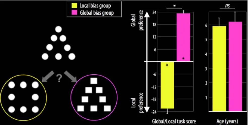
All children were presented with the global/local task at school after the laboratory MRI session [5,20]. A total of 24 compound stimulus triads were presented to measure global/local bias in visual perception. Specifically, children judged which of two figures was most similar to a reference figure (Fig. 1). The judgment could be made based on either the local or global aspect of the reference. Children were instructed to give their first, most immediate similarity judgment for each trial. A measure of global/local precedence was calculated afterwards for each participant by subtracting the number of local choices from the number of global choices. The value range was 224 to 24, with a more positive value indicating a greater bias toward global visual information.
Results
Behavioral results
The children were grouped according to their scores on the local/global task. Children with negative scores were included in the local bias group, and children with positive scores were included in the global bias group (Fig. 1). In this sample, seven children showed a local visual processing bias (6 girls; 7 right-handed; mean score on the global/local task, 222.660.8) and 18 children showed a global visual processing bias (10 girls; 14 right-handed; mean score on the global/local task, 23.360.3). The global/local task scores differed significantly between the local bias group and the global bias group (t(23) = 67, p,0.0001).
Figure 1. Representative example of a global/local triad stimulus (left), mean global/local task scores (middle), and mean age (right) for the local bias group (yellow) and the global bias group (pink). *p,0.05; ns = non-significant.
doi:10.1371/journal.pone.0020879.g001
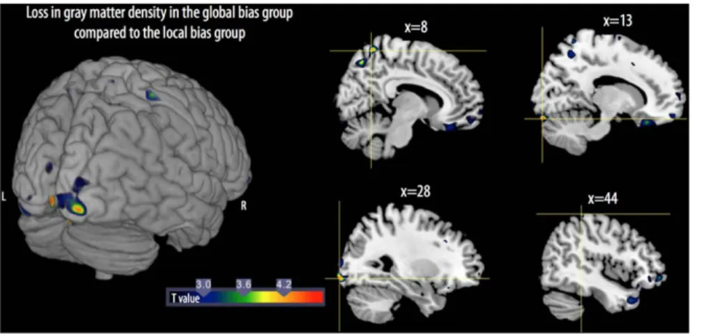
GM loss was also observed in the parietal cortex, including losses in the right precuneus and the postcentral gyrus. The reverse comparison, i.e., subtraction of the GM density results of the local bias group from those of the global bias group revealed no significant differences.
Discussion
This study is the first to directly examine changes in GM density during the developmental window in childhood when there is a shift from a local visual processing bias to an adult-like global visual processing bias. In agreement with our previous findings, children at the transition age of 6 years presented either a local visual processing bias or an adult-like global visual processing bias [8]. Using VBM, we showed GM loss along the right calcarine sulcus in the global bias group of children compared to the local bias group of children, suggesting a fine-tuning of the primary visual cortex for processing global visual information. GM differences were also found in the right lingual gyrus and in the right parietal region.
In adults, the right middle occipital cortex is more activated during global tasks than during local tasks [16]. This early visual area is predominant during processing of natural visual scenes with low spatial frequency and is known to convey global information during visual processing [21]. At the same time, the right lingual
guidance of attention toward the salient global form of a stimulus is disrupted. Our results are clearly in agreement with the aforementioned roles of the parietal cortex. Taken together, the data showing loss of GM in the right parietal and visual areas in some 6-year-olds may reflect anatomical maturation processes that allow children to shift from a mode of local to global processing of visual information. Our results also suggest that a neurodevelop-mental disorder of the dorsal stream, including in the right parietal and visual regions found in the present work, would create specific difficulties in processing global visual information. Recent neurodevelopmental data in individuals with Williams syndrome are in agreement with this idea [27]. Williams patients, usually defined as local spatial processors [28], present specifically reduced parietal and visual dorsal activation during global processing, whereas activation in the ventral occipito-temporal cortex is equivalent to controls. These results fit well with our findings that the emergence of a global visual preference in healthy children is accompanied by GM loss in the occipito-parietal dorsal pathway of the brain.
Finally, the present findings may provide a better understanding of some psychiatric disorders, such as schizophrenia. Indeed, global information processing is defective even in the early stages of perception in schizophrenia patients, resulting in a visual attraction toward the local properties of real-world scenes [29].
Table 1. Anatomic localization, localization extent, MNI coordinates, and Z scores for maximal gray matter volume differences between the local bias group and the global bias group of children.
Anatomic localization Number of voxels Hemisphere MNI coordinates Z score
X Y Z
Local biasminus Global bias
Calcarine 204 R 4 2100 25 3.94 Lingual 279 R 13 290 219 3.93 Inf/Mid Occipital 278 R 28 297 28 3.83 R 24 2102 22 3.16 Precuneus 224 R 8 256 65 3.73 60 R 8 267 52 3.56 Postcentral 56 R 44 239 66 3.51 Lingual 58 L 222 271 21 3.43
Global biasminus Local bias No significant difference L: left; R: right.
Difficulties in processing global information in schizophrenia patients are proposed to be due to impairment of the dorsal pathway [30]. According to the recent view that impairment in brain structure maturation is responsible, at least in part, for schizophrenia [31], the use of VBM with a focus on the brain regions identified in this study may be useful for exploring the neurodevelopmental origins of this pathology.
In conclusion, how we perceive the visual world as a coherent whole has been a central question in experimental psychology since the end of the 19thcentury [32]. Since the end of the 20th century, brain imaging techniques in adults and neuropsycholog-ical findings in children have given us a greater understanding of the neural basis of visual processing [12,15]. The present study is
the first to report specific brain regions that are involved in the perception of global visual information in healthy children.
Acknowledgments
The authors thank the children who participated in the study and their families.
Author Contributions
Conceived and designed the experiments: NP GL AP OH. Performed the experiments: GL GP CL AL MRT SR AP OH. Analyzed the data: NP GS MC GL. Contributed reagents/materials/analysis tools: NP GS MC OH. Wrote the paper: NP.
References
1. Casey B, Tottenham N, Liston C, Durston S (2005) Imaging the developing brain: what have we learned about cognitive development? Trends Cogn Sci 9: 104–110.
2. Shaw P, Kabani NJ, Lerch JP, Eckstrand K, Lenroot R, et al. (2008) Neurodevelopmental trajectories of the human cerebral cortex. The Journal of Neuroscience: The Official Journal of the Society for Neuroscience 28: 3586–3594.
3. O’Hare E, Sowell E (2008) Imaging Developmental Changes in Gray and White Matter in the Human Brain. Dans A. Nelson, M. Luciana, eds. Handbook of Developmental Cognitive Neuroscience (second edition, pp 23–38) The MIT Press.
4. Edelman GM (1993) Neural Darwinism: selection and reentrant signaling in higher brain function. Neuron 10: 115–125.
5. Kimchi R (1992) Primacy of wholistic processing and global/local paradigm: a critical review. Psychol Bull 112: 24–38.
6. Dukette D, Stiles J (2001) The effects of stimulus density on children’s analysis of hierarchical patterns. Developmental Science 4: 233–251.
7. Kimchi R, Hadad B, Behrmann M, Palmer S (2005) Microgenesis and ontogenesis of perceptual organization. Psychol Sci 16: 282–290.
8. Poirel N, Mellet E, Houde´ O, Pineau A (2008) First came the trees, then the forest: developmental changes during childhood in the processing of visual local-global patterns according to the meaningfulness of the stimuli. Developmental Psychology 44: 245–253.
9. Vurpillot E (1968) The development of scanning strategies and their relation to visual differentiation. Journal of Experimental Child Psychology 6: 632–650. 10. Delis D, Robertson L, Efron R (1986) Hemispheric specialization of memory for
visual hierarchical stimuli. Neuropsychologia 24: 205–214.
11. Robertson L, Lamb M (1991) Neuropsychological contributions to theories of part/whole organization. Cognit Psychol 23: 299–330.
12. Fink G, Halligan P, Marshall J, Frith C, Frackowiak R, et al. (1996) Where in the brain does visual attention select the forest and the trees? Nature 382: 626–628. 13. Martinez A, Moses P, Frank L, Buxton R, Wong E, et al. (1997) Hemispheric asymmetries in global and local processing: evidence from fMRI. Neuroreport 8: 1685–1689.
14. Moses P, Roe K, Buxton R, Wong E, Frank L, et al. (2002) Functional MRI of global and local processing in children. Neuroimage 16: 415–424.
15. Stiles J, Reilly J, Paul B, Moses P (2005) Cognitive development following early brain injury: evidence for neural adaptation. Trends Cogn Sci 9: 136–143. 16. Han S, Weaver J, Murray S, Kang X, Yund E, et al. (2002) Hemispheric
asymmetry in global/local processing: effects of stimulus position and spatial frequency. Neuroimage 17: 1290–1299.
17. Weissman DH, Woldorff MG (2005) Hemispheric asymmetries for different components of global/local attention occur in distinct temporo-parietal loci. Cerebral Cortex 15: 870–876.
18. Lemaire C, Moran GR, Swan H (2009) Impact of audio/visual systems on pediatric sedation in magnetic resonance imaging. Journal of Magnetic Resonance Imaging 30: 649–655.
19. Ashburner J, Friston KJ (2000) Voxel-based morphometry-the methods. Neuroimage 11: 805–821.
20. Kimchi R, Palmer S (1982) Form and texture in hierarchically constructed patterns. Journal of Experimental Psychology : Human Perception and Performance 8: 521–535.
21. Peyrin C, Baciu M, Segebarth C, Marendaz C (2004) Cerebral regions and hemispheric specialization for processing spatial frequencies during natural scene recognition. An event-related fMRI study. Neuroimage 23: 698–707. 22. Beaucousin V, Cassotti M, Simon G, Pineau A, Kotsova M, et al. (2011) ERP
evidence of a meaningfulness impact on visual global/local processing: When meaning captures attention. Neuropsychologia 49: 1258–1266.
23. Robertson L (1996) Attentional persistence for features of hierarchical patterns. journal of Experimantal Psychology: General 125: 227–249.
24. Himmelbach M, Erb M, Klockgether T, Moskau S, Karnath H (2009) fMRI of global visual perception in simultanagnosia. Neuropsychologia 47: 1173–1177. 25. Jung WH, Gu B, Kang D, Park J, Yoo SY, et al. (2009) BOLD response during
visual perception of biological motion in obsessive-compulsive disorder: an fMRI study using the dynamic point-light animation paradigm. European Archives of Psychiatry and Clinical Neuroscience 259: 46–54.
26. Mevorach C, Humphreys G, Shalev L (2006) Opposite biases in salience-based selection for the left and right posterior parietal cortex. Nature Neuroscience 9: 740–742.
27. Mobbs D, Eckert MA, Menon V, Mills D, Korenberg J, et al. (2007) Reduced parietal and visual cortical activation during global processing in Williams syndrome. Developmental Medicine and Child Neurology 49: 433–438.
Figure 2. 3D rendering (left) and sagittal views (right) show the loss of gray matter volume between the local bias group and global bias group of children. L: left; R: right. For illustrative purposes, the maps were thresholded at p = 0.01.
doi:10.1371/journal.pone.0020879.g002