HAL Id: tel-00988899
https://tel.archives-ouvertes.fr/tel-00988899
Submitted on 9 May 2014HAL is a multi-disciplinary open access archive for the deposit and dissemination of sci-entific research documents, whether they are pub-lished or not. The documents may come from teaching and research institutions in France or abroad, or from public or private research centers.
L’archive ouverte pluridisciplinaire HAL, est destinée au dépôt et à la diffusion de documents scientifiques de niveau recherche, publiés ou non, émanant des établissements d’enseignement et de recherche français ou étrangers, des laboratoires publics ou privés.
Environmental sensitivity of the C. elegans vulval
signalling network
Stéphanie Grimbert
To cite this version:
Stéphanie Grimbert. Environmental sensitivity of the C. elegans vulval signalling network. Agricul-tural sciences. Université Nice Sophia Antipolis, 2014. English. �NNT : 2014NICE4015�. �tel-00988899�
http://creativecommons.org/licenses/by-nc-sa/3.0/fr/ Université de Nice-Sophia Antipolis
THESE
pour l’obtention du grade de
DOCTEUR DE L’UNIVERSITE DE NICE-SOPHIA ANTIPOLIS Ecole Doctorale Sciences de la Vie et de la Santé – ED85
Mention : Interactions moléculaires et cellulaires
***
Présentée et soutenue publiquement par Stéphanie GRIMBERT
le 10 avril 2014
***
Sensibilité environnementale du réseau de
développement de la vulve de C. elegans
Environmental sensitivity of the
C. elegans vulval signalling network
Membres du jury:
Prof. DELAUNAY Franck Institut de Biologie Valrose, Nice (France) Président de jury Prof. STREIT Adrian Max-Planck-Institut, Tübingen (Allemagne) Rapporteur
Dr. EWBANK Jonathan CIML, Marseille (France) Rapporteur Dr. DELATTRE Marie LBMC-ENS Lyon, Lyon (France) Examinatrice Dr. BRAENDLE Christian Institut de Biologie Valrose, Nice (France) Directeur de thèse
1
Summary
How genetic and environmental factors interact during development is a key question in current biology, yet little is known about how molecular and cellular processes integrate environmental information. In my PhD research I aimed to address this problem using the network of C. elegans vulval signalling pathways as a model system. The principal objective of my project was to quantitatively examine how involved major signalling pathways, EGF-Ras-MAPK, Wnt and Delta-Notch, are modulated by specific environmental signals.
First, I analysed how a specific environmental factor (starvation) alters activities and interplay of signalling pathways underlying C. elegans vulval cell fate patterning. Using genetic approaches, I examined in detail how starvation signals are perceived and mediated to modulate vulval induction. I found that starvation consistently increased vulval induction through upregulation of the EGF-Ras-MAPK pathway activity independent of the Wnt pathway. This environmental effect is mediated by internal sensing of nutrient deprivation, likely acting through the TOR pathway, and affects vulval induction at the level or upstream of the EGF receptor. These findings highlight how developmental processes and involved evolutionarily conserved signalling pathways are modulated in response to environmental variation.
Second, I examined the environmental sensitivity of the Caenorhabditis vulval developmental system from an evolutionary perspective through comparative analysis of different C. elegans and C. briggsae isolates. I aimed to maximally disrupt this patterning process by exposure to extreme temperatures and to quantify which underlying developmental and cellular aspects are most environmentally sensitive and how such sensitivity evolves within and between species. I found that extreme temperature induced diverse developmental variants and defects, which were strongly genotype- and species-dependent. The occurrence of certain developmental defects induced by temperature extremes further revealed that vulval precursor cells and associated fates differ in temperature sensitivity, and this cell-specific sensitivity shows evolutionary variation. These results illustrate how sensitivity of different system parameters underlying Caenorhabditis vulval development are shaped by subtle, specific interactions between environmental perturbation and genetic background.
2
Résumé
Comprendre comment les facteurs génétiques et environnementaux interagissent au cours du développement est une question fondamentale en biologie. Cependant, peu de choses sont
connues à propos de l’intégration des informations environnementales par les processus
moléculaires et cellulaires. Au cours de ma thèse, je me suis intéressée à cette question en utilisant le réseau de développement de la vulve du nématode C. elegans comme système
modèle. L’objectif principal de mon projet était une étude quantitative de la modulation par l’environnement des voies de signalisation majeures impliquées dans ce processus telles que,
EGF-Ras-MAPK, Delta-Notch et Wnt.
J’ai tout d’abord analysé comment un facteur environnemental spécifique (la carence
nutritionnelle) modifie les activités et les interactions entre les voies de signalisation sous-jacentes au développement vulvaire chez C. elegans. L’utilisation d’approches génétiques m’a
permis d’examiner en détail comment les signaux environnementaux de carence sont perçus
et transmis afin de moduler l’induction vulvaire. J’ai ainsi mis en évidence que
l’augmentation de l’induction vulvaire par la carence passe par une augmentation de l’activité
de la voie EGF-Ras-MAPK et est indépendante de la voie Wnt. Cet effet de l’environnement
est assuré par la détection de la diminution de l’apport en nutriments, probablement par l’action de la voie TOR, et affecte l’induction vulvaire en parallèle ou en amont du récepteur à l’EGF. Ces résultats mettent en évidence comment les processus développementaux et les
voies de signalisation sous-jacentes évolutivement conservées répondent et intègrent la variation environnementale.
J’ai ensuite examiné la sensibilité environnementale du système de développement de
la vulve de Caenorhabditis dans une perspective évolutive et ce, grâce à l'analyse comparative de différents isolats naturels de C. elegans et C. briggsae. En perturbant au maximum le
réseau de développement vulvaire par l’exposition à des températures extrêmes, j’ai pu
quantifier quels aspects moléculaires et cellulaires de ce réseau étaient les plus sensibles à
l’environnement et analyser l’évolution de cette sensibilité au sein de différentes espèces et
souches de Caenorhabditis. J’ai pu observer que l’exposition à des températures extrêmes
induit des variants et des défauts de manière fortement dépendante de la souche et de l’espèce. L’occurrence de certains défauts développementaux induits par la température révèlent en
3 outre que certaines cellules précurseurs de la vulve et les voies de signalisation associées présentent une sensibilité environnementale différente. Ces résultats illustrent la manière dont la sensibilité des différents paramètres sous-jacents au développement de la vulve des
Caenorhabditis est façonnée par des interactions spécifiques entre les perturbations
4
“Not everything that can be counted counts, and not everything that counts can be counted”
5
Acknowledgements
First of all, I would like to thank my supervisor Christian Braendle. I really enjoyed working with him for these past five years. I started my project when the lab was just starting and he created such a great work environment. I am really happy for having been the first member of this super team! I want to thank him for his continuous support during my PhD studies, for his patience, his motivation and his enthusiasm. I really appreciated that his door was always open to discuss and ask questions. I also especially want to thank him for giving me the opportunity to present my work in many national and international meetings. Finally, I want to thank him for hosting so many great team dinners and especially the now traditional Christmas/Chinese fondue dinner. I deeply thank both Mr. Cricri and Dr. Braendle for these wonderful five years!
My PhD was funded by fellowships of the French Ministry of Research and the Fondation ARC pour la recherché sur le cancer.
I would like to thank the members of the thesis committee for their precious time. I thank my two “rapporteurs” Adrian Streit and Jonathan Ewbank, the ”examinatrice” Marie Delattre and the president of the jury, Franck Delaunay.
I am also thankful to all the people and institutions that provided me with help and materials. I especially thank Marie-Anne Félix, Michalis Barkoulas (thank you for being my
“centering” colleague) and Sébastien Schaub (your macro saved me a lot of time!)
I deeply want to thank all the past and present members of the Braendle team. I am really thankful to those who read and commented my papers/thesis/presentation during the
five past years. You should be relieved now, it’s done!
Nausicaa, thank you for being my co-PhD student during these four years, I couldn’t ask for a better partner in crime. We shared good and bad moments together and always supported each other. We also discovered our common passion (and gift!): making incredible cakes!
6 Clotilde, thank you for always being there to read and discuss my papers and presentations but also for the numerous conversations we had (serious or not). I always felt enriched after discussing with you.
Anne, the past years with you in the lab were so nice. I really enjoyed your presence. Your kindness and help were very valuable for me.
Paul, you always provided me with your help when I needed it. I also want to thank you for all your advice and encouragements. You are the wise man in this girls band!
Céline, thank you for always being there for all of us. It was really helpful to have someone to rely on.
Emilie, thank you for our discussions about babies/Canada/experiments...
I also want to thank all the other people of the 7th floor – it was such a nice environment to work in. I especially want to thank Micha for being my first colleague, you’ll always be a member of the Braendle team! Bruno, for all the songs we sang together! I’ll miss my weekly karaoke. Alex, for all your informatics tips!
I want to thank all my dear friends for allowing me to keep my sanity throughout this period:
Félicie, even with the distance you’ve always been there for me. “Come what may...”
Pierrick, for being my oldest friend. You are such a nice guy.
Sophie, thank you for your always opened door and our desperate lunches.= Coralie, thank you for your valuable support and your funny jokes.
Fabien and Nath, I really appreciated our discussions even if they were rare.
Jean-Baptiste, thank you for your kindness and your precious advice and Geordie, I really enjoy our new lunches and talks.
This is not an exhaustive list and I also think about Mallorie, Laurent, Zette, Damien, Nans...
I obviously want to thank my parents, for their enduring love and support and I am
also truly grateful to my little sis’ for her love, care and encouragement.
Last but not least, Lucas and Charlotte:
My little Charlotte I deeply thank you for being such a perfect daughter. Your smiles render every day wonderful. And, on a more serious note: thank you for letting us sleep!!
7 Lucas, having a baby while aiming to complete two PhDs – what a huge challenge!
But we did it! For all those years you’ve always been there for me. Thank you for
everything!!
8
Table of contents
Summary ... 1
Résumé ... 2
Acknowledgements ... 5
Table of contents ... 8
Table of figures ... 12
1. Introduction ... 15
1.1. Motivation: Understanding the environmental context-dependence of
organismal development ... 15
1.2. Environmental sensitivity of developmental systems ... 17
1.2.1. Developmental processes are inherently sensitive to the environment ... 17
1.2.2. Developmental plasticity versus robustness in response to environmental variation ... 19
1.3. The study organism Caenorhabditis elegans ... 22
1.3.1. General biology ... 22
1.3.2. Natural C. elegans habitat ... 25
1.3.3. Environmental sensitivity of C. elegans development ... 26
1.3.4. Perception and transduction of environmental cues in C. elegans ... 27
1.4. The study system: C. elegans vulval cell fate patterning ... 30
1.4.1. The C. elegans vulval signalling network ... 30
1.4.2. Robustness of C. elegans vulval cell fate patterning ... 36
1.4.3. Evolution of Caenorhabditis vulval cell fate patterning ... 36
9 1.5.1. Precision and robustness of the vulval cell fate patterning process in different
environments ... 38
1.5.2. Environmental sensitivity of the C. elegans vulval network ... 39
1.6. Objectives of PhD research project ... 42
1.7. Outline of PhD thesis ... 43
2. Nutrient deprivation modulates EGF-Ras-MAPK pathway activity
during C. elegans vulval induction ... 45
2.1. Introduction ... 45
2.2. Material and Methods ... 50
2.2.1. Strains and general procedures ... 50
2.2.2. Scoring of vulval phenotypes ... 50
2.2.3. Experimental environments ... 50
2.2.4. RNAi experiments ... 51
2.2.5. Quantification of pept-1 RNAi effects on Ras and Notch pathway activities ... 51
2.2.6. Statistical Analyses ... 52
2.3. Results ... 52
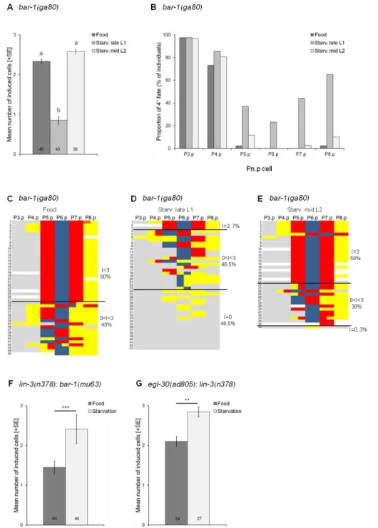
2.3.1. Starvation suppresses the Vulvaless phenotype of lin-3/egf(rf) mutations ... 52
2.3.2. Wnt pathway activity does not contribute to starvation suppression of lin-3/egf(rf) mutations ... 54
2.3.3. Starvation suppression of lin-3/egf(rf) acts independently of sensory signalling mediated by Insulin and TGF-β pathways ... 57
2.3.4. Disruption of the intestinal peptide transporter pept-1 mimics starvation suppression of lin-3/egf(rf) mutations ... 59
2.3.5. pept-1 RNAi increases EGF-Ras-MAPK and Delta-Notch pathway activities ... 61
2.3.6. Nutrient deprivation induced by pept-1 RNAi modulates vulval induction at the level of EGF/EGFR ... 64
10
2.4. Discussion ... 65
2.4.1. Antagonistic starvation signals modulate C. elegans vulval induction ... 66
2.4.2. Starvation promotes EGF-Ras-MAPK activity via the TOR pathway during C. elegans vulval induction ... 67
2.4.3. Effects and integration of PEPT-1/TOR into vulval signalling pathways ... 68
2.4.4. Environmental sensitivity versus robustness of C. elegans vulval cell fate patterning ... 69
2.4.5. Significance of interactions between environmental variation and molecular signalling pathways ... 70
3. Thermal perturbations reveal evolution of environmentally sensitive
parameters in Caenorhabditis vulval development ... 72
3.1. Introduction ... 72
3.2. Material and Methods ... 77
3.2.1. Strains ... 77
3.2.2. Temperature assays ... 77
3.2.3. Scoring of vulval cell fates and variant patterns ... 78
3.2.4. Measurement of temperature effects on lip-1::gfp activity ... 78
3.2.5. Statistical analysis and data presentation ... 79
3.3. Results ... 81
3.3.1. Extreme temperatures debuffer vulval development inducing diverse variants and defects ... 81
3.3.2. Genotype-dependence of temperature-induced vulval pattern variants ... 84
3.3.3. Environmental sensitivity of specific system features ... 85
3.3.4. Environmental versus mutational perturbation of vulval cell fate patterning ... 87
11 3.4.1. Environmental sensitivity of the vulval cell fate patterning process shows
evolutionary variation ... 89
3.4.2. Different features of the vulval patterning process vary in their environmental sensitivity in a genotype-dependent manner ... 90
3.4.3. Environmental and mutational perturbations reveal genotypic biases in the production of vulval developmental variants ... 90
3.4.4. Characterizing cryptic genetic variation to study developmental evolution of environmental sensitivity ... 92
4. Conclusions and perspectives ... 94
4.1. General conclusions ... 94
4.2. Nutrient deprivation modulates EGF-Ras-MAPK pathway activity during
C. elegans vulval induction (Chapter 2) ... 95
4.2.1. Summary ... 95
4.2.2. Perspectives and future experiments ... 96
4.2.3. Significance ... 99
4.3. Thermal perturbations reveal evolution of environmentally sensitive
parameters in Caenorhabditis vulval development (Chapter 3) ... 99
4.3.1. Summary ... 99
4.3.2. Perspectives and future experiments ... 101
4.3.3. Significance ... 101
12
Table of figures
Figure 1.1. Integration of the environment into the genotype-phenotype map ... 16
Figure 1.2. Drosophila body size in food versus starvation conditions ... 18
Figure 1.3. Visualizing the pattern of phenotypic responses using reaction norms ... 19
Figure 1.4. Developmental plasticity in Daphnia sp. In this picture two individuals of the same species ... 20
Figure 1.5. Robustness to different sources of variation ... 21
Figure 1.6. Distributed robustness versus redundancy ... 21
Figure 1.7. The C. elegans life cycle ... 23
Figure 1.8. Morphology of C. elegans hermaphrodites and males ... 25
Figure 1.9. Starvation responses during the C. elegans life cycle .. ... 27
Figure 1.10. Regulation of VPC competence in C. elegans . ... 31
Figure 1.11. Overview of the vulval signalling network in C. elegans ... 33
Figure 1.12. C. elegans vulva development ... 35
Figure 1.13. External and internal cues influencing C. elegans vulva development . ... 40
Figure 2.1. C. elegans vulval cell fate patterning ... 46
Figure 2.2. Starvation suppresses the Vulvaless phenotype caused by lin-3/egf(rf) mutations ... 53
Figure 2.3. Wnt pathway activity does not contribute to starvation suppression of lin-3/egf(rf) mutations ... 55
Figure 2.4. Starvation suppression of lin-3/egf(rf) acts independently of sensory signalling mediated by Insulin and TGF-β pathways ... 58
Figure 2.5. Disruption of the intestinal peptide transporter PEPT-1 mimics starvation suppression of lin-3/egf(rf) mutations ... 60
Figure 2.6. pept-1 RNAi increases EGF-Ras-MAPK and Delta-Notch pathway activities ... 62
Figure 3.1. Caenorhabditis vulval cell fate patterning ... 75
13 Figure 3.3. Effects of temperature and genotype on variant class frequencies ... 82 Figure 3.4. Effects of temperature and genotype on frequencies of specific vulval variants . 83 Figure 3.5. Effects of high temperature on C. elegans N2 vulval induction and Delta-Notch pathway activity . ... 86 Figure 3.6. Comparison of vulval variants induced by temperature versus mutation accumulation ... 87
Figure 4.1. Environmental sensitivity of C. elegans vulval cell fate patterning ... 94 Figure 4.2. New insights on external and internal cues influencing C. elegans vulva development ... 96 Figure 4.3. Model of the interplay of TOR and EGF-Ras-MAPK in C. elegans vulval development ... 97 Figure 4.4. Environmental sensitivity of vulval precursor cells depends on genotype and environment ... 100 Figure 4.5. Integrating environmental and mutational inputs in the Caenorhabditis vulval genotype-phenotype map ... 102
14
15
1. Introduction
1.1. Motivation: Understanding the environmental context-dependence of
organismal development
Organismal development relies on complex signalling networks involving a relatively small number of highly conserved molecular signalling pathways (e.g. Receptor Tyrosine Kinase, TGF-, Delta-Notch, Wnt, Nuclear Hormone Receptor, Hedgehog) (Gerhart, 1999; Pires-da Silva and Sommer, 2003). The same molecular cascades therefore participate in diverse developmental processes and their precise function is strongly dependent on genetic and cellular contexts, i.e. they are highly flexible. This flexibility and context-dependent activation of molecular pathways during development allows their diversified action in the same organism, e.g. in response to changing cellular environments, as well as across different species where the same molecular pathway may be utilized for divergent functions.
Organisms live, develop and evolve in highly variable, complex and fluctuating external environments. How organismal development responds to and integrates environmental variation is thus a fundamental question in biology. Given that any phenotype is the result of an interaction between genotype and environment, it is of particular importance to understand how developmental processes shape the translation of genotype into phenotype, and how variation in the external environment impacts this translation. While it is clear that environmental variation may strongly impact developmental processes and corresponding phenotypic outcomes, environmental variation has traditionally been ignored in genetic and developmental studies. Consequently, little is known about the detailed mechanisms underlying environmental modification of developmental processes.
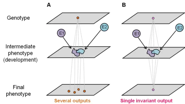
Fundamentally, we can distinguish two opposite developmental responses to environmental variation. First, development may vary in response to the environment, which generates changes in corresponding phenotypic outcomes, a phenomenon termed developmental or phenotypic plasticity (Figure 1.1A). Second, development may generate an invariant final phenotype in the presence of environmental variation irrespective of whether underlying development is sensitive or insensitive this variation. This phenomenon, i.e. developmental insensitivity to environmental variation, is frequently referred to as developmental robustness (Figure 1.1B). Importantly, such robustness does not exclude
16 environmental sensitivity of the underlying developmental mechanisms (Braendle and Félix, 2008).
Figure 1.1. Integration of the environment into the genotype-phenotype map. Two different genotypes and their response to two environments (E1 and E2) are represented in A and B. (A) The environment induces variation at the intermediate phenotypic level (e.g. gene expression, pathway activity) which results in variation of the final phenotypic output (plasticity). (B) The environment induces variation at the intermediate phenotypic level but not in the final phenotypic output. Adapted from Braendle et al. (2008).
Understanding the environmental sensitivity of developmental systems and underlying molecular and cellular processes is thus relevant to understand both how development maintains phenotypic stability despite environmental variation and how development generates phenotypic change in tune with prevailing environmental conditions.
In my PhD work, I focused on how environmental variation affects the functioning of a robust developmental system and its underlying genetic network. To address this question, I used the C. elegans vulval developmental system, a very well-characterized process involving highly conserved molecular signalling pathways (i.e. EGF-Ras-MAPK, Delta-Notch and Wnt). In this introduction, I will first present an overview of our current understanding of environmental sensitivity of developmental systems, followed by a brief introduction of the study organism, C. elegans. I will then summarize relevant aspects of the model
17 developmental system, C. elegans vulval cell fate patterning. Specifically, I will discuss previously obtained insights into the environmental sensitivity, robustness and evolution of this system.
1.2. Environmental sensitivity of developmental systems
1.2.1. Developmental processes are inherently sensitive to the environment
From the simplest unicellular to the most advanced multicellular form, all organisms live in complex environments that vary in diverse abiotic and biotic parameters, such as temperature, light, nutrients and pathogens. Such environmental variation may affect diverse phenotypic aspects (e.g. gene expression levels, protein synthesis, body size), which may reflect adaptive, neutral or maladaptive organismal responses. Environmental variation may, for example, profoundly affect global gene expression profiles as observed in the yeast Saccharomyces
cerevisiae (Causton et al., 2001; Gasch et al., 2000). A wide range of environmental
conditions, primarily stressors (e.g. temperature shocks, hydrogen peroxide, hyper- and hypo-osmotic shock, amino acid starvation or nitrogen source depletion), have been shown to modulate gene expression in S. cerevisiae. A large fraction of the genome responds in a stereotypical manner across tested environmental conditions. Screening of DNA microarrays led to the identification of almost 900 genes whose expression was commonly disrupted upon stress exposure irrespective of its precise nature, yet most conditions also showed specific regulation of specific gene subsets (Gasch et al., 2000). These and many other studies in yeast and other organisms show that gene expression, one of the most basal phenotypic characters, is strongly environmentally sensitive. However, for most of these observed changes it is unclear how they translate into later phenotypic consequences, e.g. how they impact reproductive features or survival.
Well-characterized metazoan developmental processes in response to changing environments are represented by growth and body size control in response to nutritional availability and status. Growth control, a fundamental process shaped by obvious interactions between genes and environment, has been particularly well elucidated in the fly, Drosophila
melanogaster (Nijhout, 2003). In Drosophila, growth occurs during larval stages and adults
emerge at their final size: individuals grown in starvation conditions are reduced by 50% in size compared to well-fed ones (Figure 1.2) (Hietakangas and Cohen, 2009). Consequently, the final size of an individual critically depends on the coordination of developmental timing and nutrient availability (Layalle et al., 2008).
18
Figure 1.2. Drosophila body size in food versus starvation conditions. A strong reduction of body size is observed under starvation conditions. The control fly (left) was grown in standard conditions (with plenty of food). The starved fly (right) was grown in a media containing only 10% of the standard nutritional value. Photograph from Hietakangas and Cohen (2009).
Systemic regulation of growth involves interplay between multiple tissues and signalling pathways (Hietakangas and Cohen, 2009). This regulation is mainly ensured by the Insulin-like signalling (Nijhout, 2003). The TOR (Target of Rapamycin) signalling pathway is also a major regulator of cell growth control – by regulating both cell size and proliferation (Zhang et al., 2000). In D. melanogaster, TOR activity is controlled by amino acids (Avruch et al., 2009; Colombani et al., 2003) and cellular energy levels (ATP/AMP ratio) – sensed by AMP-activated protein kinase (AMPK) (Hardie, 2007). The well-understood mechanisms underlying Drosophila growth control reveal how instructive environmental cues (e.g. nutrition) translate into developmental and metabolic changes and plastically modulate the final phenotype (e.g. body size) of an organism.
Environmental variability in yeast gene expression and nutritional control of
Drosophila growth represent just two examples out of a diverse spectrum of environmentally
sensitive processes. However, they clearly illustrate that environmental variation may have strong, wide-ranging phenotypic effects and that the environment may provide instructive information, critical in regulating key developmental decisions.
19
1.2.2. Developmental plasticity versus robustness in response to environmental variation
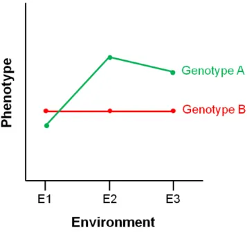
As already mentioned, developmental responses to the environment may generate plastic or invariant (robust) phenotypic outcomes (Figure 1.1). Differences in environmental sensitivity between genotypes for a given phenotype can be visualized using reaction norms (Figure 1.3), illustrating phenotypic changes across different environments.
Figure 1.3. Visualizing the pattern of phenotypic responses using reaction norms. Reaction norms are used to represent the phenotypic response of a given genotype across environments. Genotype A is plastic across environments. Genotype B is non-plastic (robust) across environments.
Developmental (or phenotypic) plasticity is the ability of a genotype to produce different phenotypes in response to environmental variation (Bradshaw, 1965; Pigliucci, 2001; Schlichting and Pigliucci, 1998; Stearns, 1992). Developmental plasticity is a universal organismal feature, but which is most commonly used to refer to adaptive, flexible changes of development in response to specific environmental conditions (West-Eberhard, 2003). A clear and famous example of adaptive developmental plasticity concerns the morphology of the
Daphnia water fleas (Woltereck, 1932) which grow a defensive “helmet” in response to
20
Figure 1.4. Developmental plasticity in Daphnia sp. In this picture two individuals of the same species. Daphnia water fleas display the ability to form a defensive “helmet” when exposed to predators. Left: exposed to predators. Right: absence of predators. Picture credits: Christian Laforsch.
Other striking examples of developmental plasticity include temperature-induced sex determination (Crews et al., 1994), plastic caste determination in social insects (Simpson et al., 2011) or alternative diapausing phenotypes (dauer) in C. elegans (Hu, 2007). Such developmental plasticity is common for many other traits and species, e.g. for body size in various organisms, as illustrated in the above example of Drosophila. Understanding the molecular and genetic mechanisms of such developmental plasticity is a current key focus both in developmental and evolutionary biology, and much progress has been made using the model systems A. thaliana (Komeda, 2004), D. melanogaster (Flatt et al., 2013) and C.
elegans (Braendle et al., 2008; Viney and Diaz, 2012).
Developmental plasticity, reflecting environmental sensitivity of development, contrasts with environmental insensitivity of development, often termed (environmental) robustness (de Visser et al., 2003; Waddington, 1942). Developmental robustness is an essential feature of organisms, which have to face diverse sources of variation, including genetic, environmental and stochastic variation (Felix and Wagner, 2008). Developmental robustness describes the ability of a biological system to generate invariant outputs when facing such variation (Figure 1.5).
21
Figure 1.5. Robustness to different sources of variation. In (a) the final phenotype is robust to internal stochastic variation (e.g. cellular concentration of a given molecule) represented in green and orange. In (b) the final phenotype is robust to genetic variation even if variation can occur at the intermediate level. In (c) the final phenotype is robust to environmental variation (E1 and E2, in purple and blue). Adapted from Félix and Wagner, 2008.
Fundamentally, robustness of biological processes to diverse sources of variation may result through distributed robustness or redundancy (Felix and Wagner, 2008; Wagner, 2005a) (Figure 1.6).
Figure 1.6. Distributed robustness versus redundancy. Both panel show hypothetical signalling cascades. The information from the upper circles (white) in transduced via numerous components
22
(different greys) to downstream effectors (black). In the case of distributed robustness (left) the information is distributed among several different paths – none of them performing the same function. In case of redundancy (right) the information processes through components that have exactly the same function (dark grey). Adapted from Félix and Wagner, 2008.
In a distributed system no sub-section plays the same role and they constitute alternative paths. They are tightly connected and strongly interact. Conversely, redundancy results from the equivalence of different parts of a developmental system. Whether either type of robustness results simply through emerging network properties or through adaptive evolution remains, however, difficult to evaluate (Felix and Wagner, 2008; Wagner, 2005a). Importantly, robustness of biological processes to a given perturbation may also render them robust to other sources of perturbations, i.e. robustness shows congruence (Masel and Siegal, 2009). Therefore, for example, robustness to environmental variation will make the system also robust to genetic or stochastic variation (de Visser et al., 2003; Meiklejohn and Hartl, 2002; Proulx and Phillips, 2005) .
An important consequence of developmental robustness is that it may generate robustness to mutations, leading to the accumulation of cryptic genetic variation, i.e. evolutionary variation in the absence of evolutionary change of the phenotype (Gibson and Dworkin, 2004). Thus, robustness of the phenotype can have seemingly paradoxical evolutionary consequences and lead to increased genetic evolvability of such system (Masel and Trotter, 2010).
1.3. The study organism Caenorhabditis elegans
1.3.1. General biologyThe free-living nematode Caenorhabditis elegans is a small (1 to 1.5mm), transparent and simple multicellular organism which lives in rotten matter, feeding on diverse microbes (Kiontke and Sudhaus, 2006; Kiontke et al., 2011). Since its introduction by Sydney Brenner (Brenner, 1974), C. elegans has become a well-characterized model organism for molecular, genetic and developmental studies. The invariant C. elegans cell lineage has been determined (Sulston and Horvitz, 1977; Sulston et al., 1983) and this nematodes was the first metazoan to have its genome completely sequenced (Consortium, 1998). Using C. elegans as a model
23 system has many advantages: easy culturing methods (individuals can be maintained on agar plates and fed with Escherichia coli), short life cycle (3.5 days at 20°C), and reproduction through self-fertilizing hermaphrodites, resulting in isogenic populations. Male production is facultative and results through spontaneous X-chromosome non-disjunction during meiosis.
Available resources (databases, literature, genetic maps and mutant libraries) and a wide range of established experimental techniques (RNAi, mutagenesis and transgenesis) as well as easy culturing (including cryopreservation of stocks) make C. elegans a model organism of choice.
Life cycle
Under laboratory conditions it takes about three and a half days to complete the C. elegans life cycle from egg to reproductive adult (Figure 1.7). The life cycle is composed of two phases: embryonic and post-embryonic development. The embryonic development starts in
utero and eggs are laid as early embryos. After hatching, both hermaphrodites and males
develop through four larval stages. At the end of each stage, larvae undergo a brief lethargus with arrest of pharyngeal pumping. This lethargus is associated with a moult during which a new stage-specific cuticle is synthesized. The last moult leads to the reproductively mature adult.
Figure 1.7. The C. elegans life cycle. The C. elegans life cycle is comprised of embryonic and post-embryonic development. Post-post-embryonic development is composed of four larval stages followed by
24
adulthood. During the L1 stage, individuals can arrest their development for several days under unfavourable conditions (i.e. starvation). When food becomes available again they re-enter the reproductive life cycle. At the end of the L1 stage, if the environment is harsh (high temperature, low food, crowding), they can undergo an additional developmental arrest: the dauer stage. Upon better conditions development will resume and individuals will form L4 larvae. Duration of each stage is indicated at 20°C. Image: Nausicaa Poullet.
Morphology and reproductive mode
All nematodes share the same unsegmented and cylindrical body plan. In C. elegans, both self-fertilizing hermaphrodite (XX) and male (XO) bodies are surrounded by a collagenous cuticle. This outer tube – composed of hypodermis and cuticle – surrounds a pseudocoelomic cavity containing both digestive and reproductive tracts. Body shape is maintained by hydrostatic pressure. In laboratory conditions, individuals are mainly fed on the cultures of the bacterium E. coli. The bacteria are ingested through the mouth and pass through a two-lobed pharynx – which acts as a pump and grinder.
The adult hermaphrodite is composed of 959 somatic cells, 302 of which are neurons and 95 of which are body wall muscles (White, 1988). C. elegans males are initially identical to the hermaphrodite larvae apart from a few male fate cells, but start to display typical budding shape of their posterior half during the L2 stage (Nguyen et al., 1999; Sulston and Horvitz, 1977; Sulston et al., 1980).The adult male is composed of 1031 somatic cells, 381 of which are neurons, mostly involved in mating behaviour (White, 1988). C. elegans males and hermaphrodites display sexual dimorphism in all tissues – except for the pharynx and the excretory system.
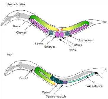
The hermaphroditic reproductive system is composed of two symmetrical U-shaped gonad arms containing the germline, connected by the central uterus and the vulva (Figure 1.8). The first germ cells produced are sperm. During the L4 stage, hermaphrodites produce around 160 sperm per gonadal arm and store them in the spermatheca. At the end of the L4 stage an irreversible switch occurs and hermaphrodites start to produce oocytes (Ellis and Kimble, 1995) – which will be fertilized by sperm when entering the spermatheca. Embryos start to develop in the uterus until gastrulation and are then laid through the vulva. In contrast, the male gonad is composed of one single arm. Males produce sperm throughout life. The male's copulatory apparatus is located in the tail. The fan extends from the tail and contains
25 nine pairs of sensory rays. The proctodeum ends in the cloaca and posteriorly links the intestine and the gonad. The spicules are two particular sensilla covered by cuticle and playing a major role during mating, i.e. locating the hermaphrodite vulva and hold it open during sperm transfer.
Figure 1.8. Morphology of C. elegans hermaphrodites and males. Schematic drawing of anatomical structures left lateral side. (A) Hermaphrodite. (B) Male. See text. Image: Nausicaa Poullet.
1.3.2. Natural C. elegans habitat
Despite the extensive knowledge accumulated on C. elegans development and genetics, little is known about its natural environment. However, C. elegans ecology has recently become a field of interest and led to the discovery of many new C. elegans wild isolates (Andersen et al., 2012) and Caenorhabditis species (Kiontke et al., 2011). Often described as a soil nematode, the cosmopolite C. elegans (Barriere and Felix, 2005) actually lives in decomposing vegetal matter, such as rotten plant, flowers or fruits. C. elegans wild populations have also been found in decomposing invertebrates. The C. elegans natural habitat is extremely variable in terms of food availability, temperature, oxygen and chemicals concentration. The C. elegans natural habitat thus represents a highly ephemeral environment undergoing strong fluctuations in nutritional conditions.
26 Compared to its wild habitat, the lab environment is very stable and standardized. Individuals are maintained on petri dishes filled with Nematode Growth Medium (NGM) (Brenner, 1974; Hope, 1999) and fed with a particular strain of E. coli (OP50). Plates are usually maintained at 20°C. This standard environment has been developed to optimize offspring production but as E. coli is a mammalian intestinal bacterium, thus unlikely to represent a highly relevant natural food source for C. elegans.
1.3.3. Environmental sensitivity of C. elegans development
Environmental variations can elicit both behavioural and developmental responses in C.
elegans, such as attraction/avoidance (Bargmann et al., 1993; Troemel et al., 1997),
morphological and locomotion changes in liquid culture (Szewczyk et al., 2006) or specific diapause-like states (i.e. L1 arrest, dauer and ARD) (Angelo and Van Gilst, 2009; Hu, 2007; Johnson et al., 1984). When encountering stressful conditions (i.e. starvation, crowding or high temperature), individuals can slow or arrest their development in several developmental stages (Ruaud and Bessereau, 2006). Food limitation (starvation) is one of the major environmental stressors that C. elegans individuals can encounter. If the embryos hatch in the absence of food, the L1 larvae arrest their development (Johnson et al., 1984). When food becomes available again, the arrested L1 are able to re-enter a normal life cycle (Slack and Ruvkun, 1997) (Figure 1.9). At the end of the L1 stage, if the environmental conditions are not favourable (low food availability, high temperature and crowding) the individuals enter a morphologically specific L2 stage, called L2d. L2d larvae retain the potential of forming L3 larva but if environmental conditions remain unfavourable, these L2d enter the L3 dauer larval stage (Albert and Riddle, 1988; Golden and Riddle, 1982, 1984). This dauer stage can last for months until dauer larvae experience favourable conditions. Upon food re-exposure, dauers start to develop, and after 10 hours, the L3/L4 moult occurs and individuals re-enter the normal life cycle at the L4 stage (Figure 1.9). During dauer, feeding is completely arrested
– defined as a non-aging state, and dauer larvae display a ‘‘waving behaviour’’ which may
serve to find or attract hosts that may carry them to a location providing adequate resources (Riddle, 1988; Riddle and Albert, 1997). Dauer formation represents one of the best-understood examples of developmental modulation by external cues – coupling both sensory and metabolic information. Dauer formation is regulated by a complex genetic network (Fielenbach and Antebi, 2008), revealed by the analysis of daf-c (dauer constitutive) and daf-d (dauer defective) mutants (Riddle and Albert, 1997). This network involves chemosensory
27 components of the cGMP pathway, such as G-protein, guanylyl cyclase and cGMP dependant cyclases (Birnby et al., 2000) but also metabolic components, such as Insulin (Li et al., 2003; Pierce et al., 2001). After the last moult, at the beginning of reproduction, individuals can also adopt a diapause-like state, called adult reproductive diapause (ARD) (Angelo and Van Gilst, 2009), in which the germline size is reduced to a minimal pool of stem cells. When conditions are favourable the germline regrows and reproduction starts again (Figure 1.9). During adulthood, hermaphrodites also have an additional strategy to face an unfavourable environment (e.g. starvation). They can retain their embryos, which will hatch inside the dead adult (contrary to what happens in ARD) (Chen and Caswell-Chen, 2003). This is called
“bagging” and may represent a strategy to protect progeny until they reach the resistant dauer
stage (Chen and Caswell-Chen, 2003; Chen and Caswell-Chen, 2004) (Figure 1.9).
Figure 1.9. Starvation responses during the C. elegans life cycle. Starvation during the L1 stage leads to a reversible L1 arrest. Starvation in late L1 and L2 causes adoption of the dauer stage. Starvation during the L4 stage leads to adult reproductive diapause whereas late L4 or adult starvation results in “bagging”, i.e. internal hatching of embryos. Adapted from Angelo and Van Gilst, 2009.
1.3.4. Perception and transduction of environmental cues in C. elegans Immunity and stress response
During its life C. elegans can experience highly stressful conditions (e.g. high temperature, hypo- and hyperoxia, starvation). Environmental perception (see below) and stress-triggered responses ensure the adjustment of cellular functions in critical environments and evolutionary conserved stress response mechanisms have been found in C. elegans (Koga et al., 2000; Lehtinen et al., 2006). DAF-21, for instance, is a member of the HPS90 family, expressed in all cells under stressful conditions (Inoue et al., 2003). C. elegans response to
28 hypoxia also involved evolutionary conserved mechanisms like the HIF (Hypoxia Inducible factor) complexes required for C. elegans physiological adaptation to hypoxic conditions (Jiang et al., 2001; Shen et al., 2005).
C. elegans can be infected by a large variety of pathogens (Darby, 2005). Many of
them colonize the C. elegans intestine, some adhere to the cuticle while others produce toxins and can kill C. elegans without any physical contact. C. elegans infection has been used as a powerful genetic system to study innate immunity (Engelmann and Pujol, 2010; Ewbank, 2006). General stress response mechanisms are involved in pathogen response but C. elegans immune response also relies on major conserved signalling pathways like ERK, p38 MAPK, Insulin or TGF- (Engelmann and Pujol, 2010).
Sensory perception
Chemosensation
The C. elegans chemosensory set of neurons is highly developed – 32 neurons can detect hundreds of chemicals (Bargmann, 2006), which are required to avoid noxious substances and to find food and mating partners. C. elegans males have numerous additional chemosensory neurons mainly involved in mating behaviour (Liu and Sternberg, 1995; Sulston et al., 1980).
C. elegans chemosensory neurons can be directly or indirectly exposed to the environment,
mainly through the amphid, the phasmid and the inner/outer labial organs (Ward et al., 1975). Only two specific neurons AQR and PQR– responsible with URX for oxygen sensing (Chang et al., 2006; Cheung et al., 2005; Gray et al., 2004) and social feeding (Gray et al., 2004), are directly exposed to body fluids. Chemosensory neurons usually belong to left-right pairs and each pair can be distinguished from another through morphological criteria (White et al., 1986). Chemicals are detected through hundreds of G protein-coupled receptors (GCPRs) (Robertson and Thomas, 2006) using cGMP as second messenger or relying on TRPV channels and allow C. elegans to integrate environment into both developmental and behavioural processes.
Thermosensation
In C. elegans a single pair of amphid neurons (AFD) is essential to for thermosensation and thermotaxis behaviour (Mori and Ohshima, 1995). The AFD neurons respond to warming and their ablation leads to cryophilic individuals (Kimura et al., 2004). Temperature sensing in C.
29
elegans acts in a small cellular circuit involving AIY, AIZ and RIA neurons (McKemy,
2007). AIY and AIZ act as antagonists in thermal responses: AIY-ablated animals are cryophilic and AIZ-ablated animals are thermophilic. In addition to other sensory defects RIA-ablated animals are partially thermosensory-deficient.
Mechanosensation
Mechanosensory neurons serve to detect collisions with particles (e.g. debris, other animals) as well as forces generated by its own movement. Mechanosensation is ensured by 30 putative mechanoreceptor neurons (MRNs) in the hermaphrodite and 52 extra MRNs exist in the male
– mainly involved in mating behaviour. In C. elegans, touch responses are involved in many
behaviours like locomotion (Chalfie et al., 1985; Wicks and Rankin, 1995), egg laying (Sawin, 1996), feeding (Chalfie et al., 1985; Keane and Avery, 2003), defecation (Thomas, 1990) and mating (Liu and Sternberg, 1995). Mechanical information is transduced by putative channels of the TRP (Transient Receptor Potential) superfamily in ciliated MRNs and of the DEG/ENaC (DEGenerin/Epithelial Na+ Channel) superfamily in non ciliated MRNs (Ernstrom and Chalfie, 2002; Goodman and Schwarz, 2003).
Metabolism
Being able to coordinate and adjust energy levels in tune with prevailing environmental conditions is critical for cellular and organismal survival. Metabolic sensors are key regulatory elements that allow individuals to perceive their environment and adapt to it (Lindsley and Rutter, 2004). On a cellular level, metabolic sensors detect and respond to levels of macronutrients (e.g. glucose, amino acids and fatty acids, AMP/ATP ratio). On an organismal level, coordination of energetic status from different tissues is controlled by hormonal signals (Lindsley and Rutter, 2004).
C. elegans presents highly conserved metabolic sensors. First, the IGF-1/Insulin
signalling pathway connects nutrient levels to growth, development and longevity mainly through the DAF-16/FoxO transcription factor (Murphy and Hu, 2013). The second key metabolic sensor is the LET-363/TOR (Target of Rapamycin) signalling pathway. It couples nutrient levels to cell size and proliferation. In C. elegans, inactivation of LET-363/TOR and its partner DAF-15/Raptor leads to developmental arrest and fat accumulation (Jia et al., 2004; Vellai et al., 2003). LET-363/TOR is directly regulated by nutrient levels but also by
30 the DAF-2-Insulin pathway to control dauer formation and longevity (Jia et al., 2004). The third major cellular sensor is the AMPK pathway. It responds to cellular AMP:ATP ratio as well as upstream kinase cascades (Kahn et al., 2005; Lindsley and Rutter, 2004). In C.
elegans AAK-2, the orthologue of AMPK, regulates lifespan in response to AMP:ATP ratio
and insulin-like signals. Nuclear hormone receptors (NHRs) also coordinate metabolic responses and function as regulators of metabolic gene expression (Van Gilst et al., 2005a; Van Gilst et al., 2005b).
1.4. The study system: C. elegans vulval cell fate patterning
C. elegans vulval development is an extensively studied and well-characterized
developmental process involving conserved signalling pathways (Félix, 2012a; Félix and Barkoulas, 2012; Sternberg, 2005). This process underlies the formation of an essential reproductive organ, required for egg laying and mating with males.
1.4.1. The C. elegans vulval signalling network
At hatching, the C. elegans L1 larva possesses six pairs of ventral blasts: the Pn cells. During L1 each pair rotates and the twelve Pn cell aligned along the antero-posterior axis (Sulston and Horvitz, 1977). Each Pn cell will then asymmetrically generate two daughters: Pn.a and Pn.p. The Pn.a cells will primarily develop into ventral cord neurons (Sulston and Horvitz, 1977) whereas the Pn.p cells adopt an hypodermal fate. During the L1 stage, P(3-8).p acquire competence to form vulval tissue: each of these cells is able to respond to the inductive signal and adopt a vulval fate (Kimble, 1981; Sternberg and Horvitz, 1986; Sulston and White, 1980). The specification of these vulval precursor cells (VPCs) is ensured by the expression of the lin-39/Hox5 gene (Maloof and Kenyon, 1998; Salser et al., 1993), which encodes a homeodomain protein required for specification of mid-body region cell fates (Figure 1.12A). SEM-4, a zinc finger protein, is necessary for full lin-39/Hox5 expression (Grant et al., 2000). LIN-39/Hox5 acts with two homeodomain proteins co-factors ceh-20/pbx1-3 and unc-62/meis which are transcribed in all the Pn.p cells, except P12.p (Yang et al., 2005). CEH-13/Hox1, encoded by the ceh-13/Hox1 gene and expressed in cell nuclei all along the ventral cord (Brunschwig et al., 1999), antagonises LIN-39/Hox5 and promotes Pn.p fusion (Tihanyi et al., 2010).
31 LIN-39/Hox5 activity is also required during the L2 stage to maintain VPCs competence (Chen and Han, 2001). During the L2 stage lin-39/Hox5 expression is under the control of the canonical Wnt pathway (Eisenmann et al., 1998). Combined with the Wnt pathway, the EGF/Ras/MAPK signalling is also involved in the maintenance of VPCs competence. For instance, gain of function mutations in the EGF-Ras-MAPK pathway diminishes fusion during the L2 stage (Chen and Han, 2001). Moreover and in a bar-1/
-Catenin mutant sensitized background, a let-23/egfr mutation aggravates the fusion phenotype
(Eisenmann et al., 1998).
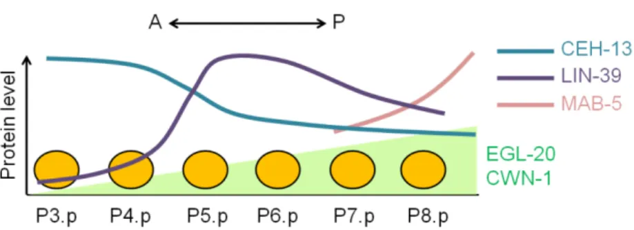
As mentioned before, ablation experiments revealed that only P(3-8).p have competence to form vulval tissue (Kimble, 1981; Sternberg and Horvitz, 1986; Sulston and White, 1980). Nevertheless, the competence level of each VPC is not equivalent (Clandinin et al., 1997). LIN-39 expression, regulating VPC competence (Maloof and Kenyon, 1998), is variable along the antero-posterior axis leading to a differential sensitivity of the VPCs to the inductive signal (Pénigault and Félix, 2011b). P7.p and P8.p are less sensitive to the LIN-3/EGF inductive signal due to the expression of mab-5/Hox7. mab-5/Hox7 encodes a homeodomain transcription factor required for specification of posterior cell fates. Moreover P3.p sensitivity can be explained by the graded action of two Wnt ligands egl-20 (Coudreuse et al., 2006; Whangbo et al., 2000) and cwn-1 (Hayashi et al., 2009) regulating lin-39/Hox5 expression in the VPCs (Figure 1.10).
Figure 1.10. Regulation of VPC competence in C. elegans. In C. elegans, vulval precursor cells are not equally sensitive to the inductive signal. This scheme represents the regulation of VPC competence along the antero-posterior axis. Disks represent the vulval precursor cells. The Wnt ligands egl-20 and cwn-1 are more expressed in the posterior part of larva and a long-range gradient diffuses until the midbody of the animal (Coudreuse et al., 2006). lin-39/Hox5 is expressed along the AP axis with low levels in P3.p and P4.p and a peak on P5.p. ceh-13/Hox1 and mab-5/Hox7
32
antagonize lin-39/Hox5 and are expressed respectively all along the AP axis and posteriorly. Adapted from Pénigault and Félix, 2011b.
During the second larval stage (L2) the anchor cell (AC) induces vulva cell fates by expressing the EGF-like ligand LIN-3 (Hill and Sternberg, 1992) (Figure 1.11 and Figure 1.12B). The AC is necessary and sufficient to induce vulva formation (Kimble, 1981). LIN-3/EGF disperses as a morphogen and the Pn.p adopt different cell fate according to their location. P6.p is closest to the AC and receives the highest level of LIN-3/EGF causing it to adopt a 1° cell fate. P6.p then expresses Delta ligands which activate the Delta-Notch pathway in its neighbours, P5.p and P7.p. This activation causes them to adopt a 2° fate (Greenwald et al., 1983) and prevents them from adopting a 1° fate by inhibiting the EGF-Ras-MAPK pathway (Sternberg, 1988; Yoo et al., 2004) through the mitogen-activated protein (MAP) kinase phosphatase LIP-1 (Berset et al., 2001) (Figure 1.11). This cross-talk between EGF-Ras-MAPK and Delta-Notch is a key to maintain a robust 2°-1°-2° spatial pattern. However, a lower dose of LIN-3/EGF may also be responsive of the adoption of the 2° fate in P5.p and P7.P (Katz et al., 1995). Morphogen induction and signalling crosstalk act together to ensure a precise 2°-1°-2° pattern (Kenyon, 1995). Moreover, it has been recently shown that a switch from the canonical LET-60/Ras-LIN-45/Raf pathway to a LET-60/Ras-RGL-1-RAL-1 signalling pathway can promote the 2° cell fate in P5.p and P7.p (Zand et al., 2011) (Figure 1.11). P3.p, P4.p and P8.p do not receive enough signal (LIN-3/EGF or Delta- LIN-12/Notch) and adopt a 3°, non vulval fate (Hill and Sternberg, 1993). The Wnt signalling pathway, involved in Pn.p competence, seems to have a partially redundant role with the EGF-Ras-MAPK pathway in Pn.p induction. Indeed, overactivation of the Wnt pathway can compensate vulval induction when the EGF-Ras-MAPK pathway is compromised (Gleason et al., 2002). The adult vulva is formed from the 22 descendants of P5.p, P6.p and P7.p according to an invariant and characterised lineage.
Negative regulation of vulval induction is also ensured by synMuv (Synthetic Multivulva) genes. SynMuv genes are transcriptional repressors that negatively regulate lin-3 expression in hyp7 (Cui et al., 2006; Sundaram, 2006; Thomas and Horvitz, 1999). There are three classes of synMuv genes: A, B and C. Two redundant sets of synMuv genes have been described: class A and class B. Mutation in only A or B genes leads to a normal vulval development whereas mutations in both A and B genes result in a Multivulva phenotype.
33
Figure 1.11. Overview of the vulval signalling network in C. elegans. Core components of the EGF-Ras-MAPK pathway are in the centre. The inductive signal LIN-3/EGF is sent by the anchor cell. This signal acts as a morphogen and is able to activate the EGF-Ras-MAPK pathway in the Pn.p cells.P6.p then adopts a primary (1°) cell fate. P6.p is the closest cell of the AC, thus the activation of the EGF-Ras-MAPK pathway is higher in this cell. In response, the Delta-Notch pathway is activated in P5.p and P7.p. This activation gives rise to the adoption of a secondary cell fate (2°) and to the repression of the EGF-Ras-MAPK pathway and upregulation of lip-1. This highly regulated process leads to an invariant pattern 2°-1°-2° vulval cell fate pattern.
As described before there are two different vulval fates: 1° and 2°. P6.p usually adopts a 1° cell fate and P5.p and P7.p a 2° cell fate. During the L3 stage, the vulval lineage consists of three division cycles (Figure 1.12C). The two first steps are the same for the 1° and 2° fates. It consists in two cycles of longitudinal divisions (L) – following the AP axis. The last division cycle is particular for each fate. The four primary cells divide transversally (T) – following the left/right axis and give rise to 8 progeny cells. The secondary lineage is more complex: two secondary progeny cells of both P5.p and P7.p divide longitudinally (LL), one divides transversally (T) and the last one does not divide (U). The secondary lineage then gives rise to 7 progeny cells. P5.p and P7.p lineages are symmetrical. The asymmetry of each secondary lineage is independent of the gonad (Katz et al., 1996) but the reverse polarity of
34 P7.p is gonad-dependent and under the control of the Wnt pathway (Inoue et al., 2004). The fibroblast growth factor (FGF) pathway acts in concert with LIN-17/Frizzled to influence the localization of SYS-1, a component of the Wnt/β-Catenin asymmetry pathway (Minor et al., 2013). The three remaining VPCs (P3.p, P4.p and P8.p) do not adopt a vulval fate. In 50% of the individuals of the lab reference strain N2, P3.p does not divide and fuses with the hypodermis during the L2 stage (F or 4° fate). In the other 50%, P3.p adopts a 3° cell fate like P4.p and P8.p. The 3° cell fate is a non-induced fate consisting in one division during the L3 stage followed by fusion with the syncitial hypodermis hyp7 (SS) (Sulston and Horvitz, 1977). During the L4 stage, the vulval cells start moving towards the anchor cell. The vulval invagination forms (Figure 1.12D), and cells fuse into seven toroids. The eversion occurs during the L4/adult moult.
35
Figure 1.12. C. elegans vulva development. The Caenorhabditis vulva develops from a subset of six ventral cells, P3.p to P8.p. (A) L1 stage: P3-8.p cells express the Hox gene lin-39 and acquire competence to form vulval tissue. (B) Late L2 stage: the anchor cell (AC) releases a LIN-3/EGF inductive signal. Only three of the six vulval precursor cells adopt a vulval fate. LIN-3/EGF acts as a morphogen and the receiving cells adopt different cell fates according to their location. P6.p receives the highest level of LIN-3/EGF causing it to adopt a 1° cell fate (blue). The expression of the Delta ligands in P6.p activates the Delta-Notch pathway in its neighbours, P5.p and P7.p. This activation causes them to adopt a 2° fate (red). The three remaining cells adopt a non-vulval 3° fate (yellow). However the fate of P3.p fate is variable and the undivided cell fuses with the hypodermis in approximately 50% of individuals (4° fate (brown)). The canonical Caenorhabditis vulval pattern is defined as 2°-1°-2°. (C) Late L3 stage: The invariant and fate-related cell division pattern leads to 22 vulval cells. Cell divisions T: transverse (left-right) division, L: longitudinal (antero-posterior) division, U: undivided, SS: fusion to the epidermal syncytium (hyp7) after single division (3° fate); F: fusion to the syncytium in the L2 stage with no prior division (4° fate). 3° and 4° fate are non vulval. (D) L4 stage: vulval morphogenesis takes place.
36
Robustness of C. elegans vulval cell fate patterning
Theoretical works show that many genetic and network features of developmental systems can contribute to their robustness, including: genetic epistasis and pleiotropy, redundancy, feedback loops and cross-talks (Meir et al., 2002; Siegal and Bergman, 2002; von Dassow et al., 2000; Wagner, 2005b). Due to its molecular and cellular organization, the C. elegans vulval developmental network displays high robustness (Figure 1.11 and Figure 1.12). This highly regulated process leads to an invariant pattern 2°-1°-2° of vulva cell fate. Two different and non-exclusive models have been proposed to ensure this precise cell fate pattern formation: morphogen-based versus sequential induction. The morphogen-based model relies on a series of experiments demonstrating that an isolated VPC (laser ablation) can adopt a secondary cell fate (Katz et al., 1995). Under the control of an heat-shock promoter, lin-3/egf can be expressed at different doses. A high dose of LIN-3/EGF produces a 1° cell fate, an intermediary dose produces a 2° cell fate whereas a low dose produces a 3° cell fate. The sequential model relies on genetic mosaics experiments. The let-23/egfr gene is not required autonomously in 2° cells (Koga and Ohshima, 1995; Simske and Kim, 1995). In the absence of LET-23/EGFR, the 2° cell fate is adopted by cells adjacent to a 1° cell. The secondary cell fate can then be induced by the primary cell fate. Moreover, positives feedback loops are found in both EGF-Ras-MAPK and Delta-Notch pathways (Berset et al., 2005; Stetak et al., 2006; Yoo and Greenwald, 2005). Multiple molecular cross-talks between these two pathways have been identified (Berset et al., 2001; Chen and Greenwald, 2004; Shaye and Greenwald, 2002; Yoo et al., 2004).
1.4.2. Evolution of Caenorhabditis vulval cell fate patterning Evolution within the Caenorhabditis genus
The canonical 3°-3°-2°-1°-2°-3° vulval pattern is invariant within the Caenorhabditis genus (Félix, 2007; Kiontke et al., 2007). Nevertheless, P3.p division frequency varies extensively among the Caenorhabditis species (Delattre and Félix, 2001). Comparative analysis of more than 20 Caenorhabditis species reveals a decrease of P3.p competence in C. briggsae, in which P3.p is never competent, and close relatives (Kiontke et al., 2011). The invariable pattern is then considered as 3°-2°-1°-2°-3°.
Another interesting difference between C. elegans and C. briggsae concerns the competence of P4.p and P8.p. In C. elegans, P4.p is more competent than P8.p (Katz et al.,
37 1995) whereas in C. briggsae P8.p is the more competent (Félix, 2007). This posterior reduction of competence in C. elegans could be explained by mab-5/Hox7 expression in P7.p and P8.p (Clandinin et al., 1997) (see 1.4.1). Anchor cell ablation in the Caenorhabditis species reveals that the AC is necessary and sufficient for vulval induction among these species, except for C. sp1 in which more gonadal cells are involved (Félix, 2007; Kiontke et al., 2007).
Uncovering cryptic genetic variation underlying Caenorhabditis vulval development Caenorhabditis vulval development robustly ensures the formation of an invariable
3°-2°-1°-2°-3° pattern governed by intercellular signalling. As described before (see 1.2.2), such robustness may allow accumulation of cryptic genetic variation buffered in standard conditions. Revealing such cryptic genetic variation underlying vulval development among different isolates or species shows that an evolutionarily invariant phenotype (cell fate pattern) may go in hand with extensive evolutionary divergence of underlying developmental mechanisms. Cryptic variation in the vulval signalling network among C. elegans wild isolates (genotypes) has been uncovered through introgression of vulval mutations, measurements of pathway activities or exposure to different environments (Braendle and Félix, 2008; Milloz et al., 2008).
By introgressing vulval mutations, e.g. let-23/egfr into different wild isolates, Milloz et al. (2008) found that the genetic background strongly modified the penetrance of such mutations, revealing an interaction between the introgressed mutation and the wild genetic background. Moreover, EGF-Ras-MAPK pathway activity in VPCs was measured in different genotypes, indicating that pathway varies up to two-fold across these wild isolates (Milloz et al., 2008). Duveau et al. (2010) mapped such cryptic genetic variation in let-23/egfr penetrance among isolates, identifying nath-10 locus as responsible for this variation. The cryptic effect on vulval signalling involving nath-10 seems to result from pleiotropy caused by nath-10 function in sperm production, and thus reproduction (Duveau and Felix, 2010). Cryptic genetic variation of vulval cell fate patterning was also revealed using accumulation of spontaneous random mutation: different C. elegans and C. briggsae isolates generated different frequencies and spectra of vulval patterning errors after 250 generations of mutation accumulation (Braendle et al., 2010). These experiments further suggested that mutational robustness of vulval cell fate patterning is subject to evolution.