HAL Id: halshs-02539238
https://halshs.archives-ouvertes.fr/halshs-02539238
Submitted on 15 Apr 2020HAL is a multi-disciplinary open access archive for the deposit and dissemination of sci-entific research documents, whether they are pub-lished or not. The documents may come from teaching and research institutions in France or abroad, or from public or private research centers.
L’archive ouverte pluridisciplinaire HAL, est destinée au dépôt et à la diffusion de documents scientifiques de niveau recherche, publiés ou non, émanant des établissements d’enseignement et de recherche français ou étrangers, des laboratoires publics ou privés.
Case of Sudanese Nubia
Francigny Vincent, Hege Hollund, Alex de Voogt, Eveline Altena, Camille
Fallet, Peter de Knijff
To cite this version:
Francigny Vincent, Hege Hollund, Alex de Voogt, Eveline Altena, Camille Fallet, et al.. Limits of Ancient DNA Extraction from Teeth: The Case of Sudanese Nubia. Nyame Akuma, Society of Africanist Archaeologists, 2013, p. 13-29. �halshs-02539238�
Introduction
Biological anthropology dominates fieldwork investigations on funerary archaeology throughout Nubia, but infrequently uses genetic resources (Buzon 2008; Krings et al. 1999; Lalueza Fox 1997; Manni et al. 2002). Using a range of archaeological skeletal samples from Sudan, we examine the relationship between a set of preservation parameters, and DNA preservation. The general aim is to optimize the methodology and chances of successful sampling in an arid milieu. The Nile Valley of Sudanese Nubia, often considered as an archaeological El Dorado because of its high potential for the preservation of ancient material and the marginal impact of modern development on its territory, remains surprisingly unknown as a field for ancient DNA studies. While a number of important historical questions could be addressed by archaeogenetics, few attempts have been made to collect samples in Nubia and use DNA analysis to explain the origin of the population settled in the valley, its evolution and exposure to genetic and cultural admixture. The situation is in contrast with the prominent role of a natural corridor that for thousands of years served as the main channel for exchanges (Török 2008) and migrations (Carlson and Van Gerven 1979; Dafa’Alla 1989) in East Africa. The rich historical background of the Nubian land, with its succession of chiefdoms and kingdoms, three phases of Egyptian conquests and the constant pressure of desert tribes on the valley settlers, logically generates questions about the Limits of Ancient DNA Extraction
from Teeth: The Case of Sudanese Nubia
Vincent Francigny
American Museum of Natural History African Ethnology / Division of
Anthropology
Central Park West at 79th Street 10024-5192 NY, New York USA E-mail: vincentsoudan@yahoo.fr Hege Hollund University of Stavanger Museum of Archaeology N-4036 Stavanger Norway E-mail: hege.hollund@uis.no Alex de Voogt
American Museum of Natural History African Ethnology / Division of
Anthropology
Central Park West at 79th Street 10024-5192 NY, New York USA
E-mail: adevoogt@amnh.org Eveline Altena
Forensic Laboratory for DNA Research Leiden University Medical Center PO Box 9600 / 2300 RC Leiden The Netherlands
E-mail: E.Altena@lumc.nl Peter de Knijff
Forensic Laboratory for DNA Research Leiden University Medical Center PO Box 9600 / 2300 RC Leiden The Netherlands E-mail: P.de_Knijff@lumc.nl SUDAN Camille Fallet Université de Neuchâtel Institut d’Archéologie
Espace Louis-Agassiz 1 - 2000 Neuchâtel Suisse
diversity of its population.
Thus far, mostly cranial materials were collected from Nubia and sent to the countries involved in survey excavations (England, France, Denmark, United States) (Buzon 2008; Stynder et
al. 2009), while locally no proper anthropological
storage facility or study was ever established. It is, therefore, not surprising that throughout the literature, only one study has been published with ancient DNA from Sudanese Nubia (Lalueza Fox 1997), based alas, on a collection brought to Europe almost 20 years before the examination took place, resulting in only 15 successful Polymerase Chain Reaction (PCR) amplifications among 29 samples analyzed. It demonstrated, through the presence of the sub-Saharan African mitochondrial marker Hpa I (np3,592) in the Meroitic population from the site of Amir Abdallah (Middle Nubia), that south-north gene flow occurred in this region during antiquity. This isolated result should be carefully considered since it can be particular to this settlement and local area. A few articles have, nevertheless, tried to contextualize this evidence of gene flow in the Nile Valley, highlighting the influence of the Nubian corridor on genetic admixture (Irish 2005) and the impact of colonization and military expeditions during antiquity (Keita 2005; Lucotte and Mercier 2003). Studies with modern DNA samples from the region also exist (Krings et al. 1999; Manni
et al. 2002), but with almost the same degree of
rareness, and without adequately addressing the environmental or historical questions that lie behind these movements of populations.
While it seems logical that the desert has been a significant limiting factor to gene flow between the African population living to the north and the south of the Sahara, the particular case of Egypt, where a more heterogeneous genetic composition of the population can be found (Terreros et al. 2005), confirms the possible relevance of the Nile Valley as a migratory path. But what is true for Nubia probably needs some adjustments when applied to the whole Egyptian territory, as its modern population gene pool shows a long history of admixture with
European and Middle-Eastern populations as well (Manni et al. 2002). While the importance of DNA analysis for the Nile Valley is immediately apparent, there has been only marginal success with DNA extraction. A discussion of the causes of DNA degradation in this area and the strategies for optimizing future extractions in Sudan is timely in the light of increasing technological advances in the field of DNA analysis and extraction.
DNA Degradation
Successful and reliable DNA extraction from ancient skeletal material is complicated by degradation and contamination. The DNA molecules of an organism are rapidly broken down post-mortem (Lindahl 1993). In the early 1990s, scientists found that fragmented remains of ancient DNA could survive within skeletal material (Willerslev and Cooper 2005). How and when this occurs is still incompletely understood although some inferences have been made using bone preservation parameters that assess the degree of diagenetic alteration. Diagenesis encompasses all the physical, biological and chemical alterations that skeletal material may undergo during burial, including uptake and exchange of ions, breakdown and leaching of collagen, alteration of the mineral phase, infilling by mineral deposits and microbial decay (Hedges 2002). The relationship between various bone preservation parameters, and the success rate of DNA extraction and sequencing, can elucidate the processes that determine DNA decay and survival. For example, the correlation between bone mineral crystallinity and successful DNA amplification, has led to the suggestion that DNA is preserved by adsorption onto the mineral surface, whereas the presence of intact collagen may also point to DNA survival (Campos et al. 2011; Götherström et al. 2002). Bioerosion, the tunnelling of skeletal material by fungi and bacteria, causes loss of both protein and DNA thus the absence of microorganisms promotes DNA preservation (Burger et al. 1999). In general, several studies show that if the bone is well preserved, both in terms of
the organic and the inorganic phase, the chances of obtaining DNA are higher. The burial environment determines the type and rate of diagenetic processes. For bone, water content, oxygen levels, local hydrology, pH and soil composition have been identified as important environmental parameters, determining the long-term integrity of the material (Jans 2005). For DNA itself, temperature is considered one of the most important factors for decay rates whereas salt content, water content and pH also play a part (Pruvost et al. 2007). Bone
and dentine share fundamental biochemical and structural features (Gilbert et al. 2005; Hillson 1986), thus the same preservation parameters can also be applied to dentine.
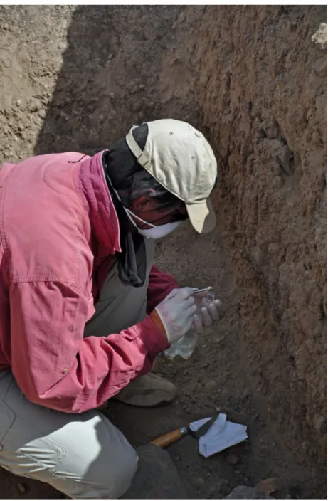
In order to elucidate bone/dentine and DNA preservation/degradation processes in ancient skeletal material from Sudan, we conducted a study on four samples from Nubia. Despite good macroscopic preservation, on-site sterile sampling (Figure 1) and fairly rapid analysis post-excavation,
they did not yield any or little DNA. A likely hypothesis for this decay is the high temperature at the site, where the average annual temperature is at present around 30° C. A review by Reed et al. (2003) shows that the success rate for DNA extraction correlates negatively with closeness to the equator, and that no DNA older than 5000 years has been successfully amplified at latitudes lower than 30 degrees. Bollongino and Vigne (2008) also observed that it is generally problematic to extract DNA in material from Near Eastern open arid sites due to exposure to high temperatures both after burial and during excavation and post-excavation storage and treatment. Smith et al. (2003) have defined a parameter thermal age that takes into account the local temperature at a site through time to assess the chances of obtaining any DNA. Thermal age is the time taken to produce a given degree of DNA degradation assuming that DNA degrades mainly by depurination with known temperature dependence in aqueous solutions. Higher thermal age means a lower chance of finding any preserved DNA. Conversely, Singh et al. (2011) reported recently on the extraction of 4000 years old cattle DNA from India, demonstrating that DNA can be found, despite high thermal ages and low latitudes.
Other environmental parameters also affect preservation. Low water content, for example, slows down hydrolytic and biological decay and in that sense promotes DNA and bone preservation. But Nubia, with a decline of rainfall and a drastic increase of temperature (Elagib and Elhag 2011), became an extremely arid location where complete desiccation may also have a negative effect causing accelerated strand breakage (Dose et al. 1991) and cross-linking (Bieger-Dose et al. 1992). In sum, the interplay of the different factors in a region such as Sudan remains unclear.
Archaeological Context
Teeth samples were taken from two sites, Sai Island and Kerma, located in distinct areas (Figure 2). They cover four different time periods and
cultural phases (Table 1). This prevents conclusions based on a single site and time period. To ensure the quality of the samples and allow other studies than DNA analyses, a set of two teeth per individual was taken directly from jaws at the moment of the discovery and with protective measures to avoid contamination. After the lack of successful PCR on four teeth, four others of the same skeletons were used to investigate the state of preservation of the samples:
• The first sample, S-2A, was taken from an individual, a female around 20 years old, recovered in the descendary in front of the entrance of the burial chamber of a grave (T 027) at Sai Island Meroitic necropolis 8-B-5.A (Francigny 2009, 2010). According to the material found on the site, it was dated to the classic or late Meroitic period, between the 1st and the 4th centuries AD.
The grave was entirely dug into alluvial silt, with a small layer of gravel and sand at the surface. The chamber was originally closed by a blocking system made of schist stones sealed with mud plaster, later opened by robbers who dragged out the body from the cavity to the descendary. Thus, the disarticulated human remains were found in a disturbed layer of wind-blown sand that also contained remnants of grave goods. • The second (S-4) and third samples (S-5)
come from the oldest part of the eastern cemetery of Kerma. This part contains graves of early C-Group and Ancient Kerma traditions that are C14 dated between 2600
and 2200 BC (Honegger 2011). The sample S-4 was taken from an intact grave (409) in the Ancient Kerma area, dated ca. 2400-2200 BC. The body was deposited in a pit at a depth of 173cm, which was filled with silt. A typical Kerma tumulus, consisting of indurate silt and rows of black and white stones, covered the grave. The individual, a man over 30 years old, lay on his right side in a flexed position. The head was facing north
and his hands were in front of his face. The general state of preservation was very good: some leather and vegetable matter (fragments of a braided mat and a plant pillow) were still preserved and the bones were in a good condition, despite some fragmentation due to the collapse of elements of the pit walls during the excavation.
• Sample S-5 was taken from a plundered grave (418) in the C-Group area dating to
ca. 2600–2400 BC. The pit was 71cm deep
and it was filled with sand and silt. The upper body (head, arms, some vertebrae and ribs) was disturbed and was found about 20cm from the surface, while the lower body was preserved at the bottom of the pit. The individual, a woman over 30 years old, was lying in the common position on the right side with the legs flexed. The preservation of the bones is good. We note that the skull has a greenish color on the right side (mandibular condyle, zygomatic process, mastoid, temporal and parietal bones), which could be due to the presence of metal (copper-alloy) in the grave. For more than a decade C14 analyses have been undertaken
on bones, teeth and leather coming from the graves of the Kerma necropolis. Despite the very good state of preservation of this material, the results were problematic. Some
bones did not contain any collagen and other samples (bones, tooth, leather) gave dates that were either too old or too recent. This points to the difficulty of extracting the little collagen that was preserved and the pollution by older carbonates and/or humic acid.
• The fourth sample, S-6, was taken from an intact grave on Sai island’s northern necropolis, dating to the beginning of the Christian era, between the 6th and the 9th
centuries AD. The individual, a male around 50 years old, was buried in a supine position, head to the west. He was tightly wrapped and placed in a narrow vertical shaft dug into alluvial silt, which was filled after the funerary ceremony took place.
Analyses of Nubian Teeth Samples
The Nubian samples stem from different periods and archaeological contexts, in an arid climate that has often produced advanced desiccation and natural mummification of soft tissues. Several preservation parameters were obtained on the teeth using UV autofluorescence, microscopic study and Fourier transform infrared spectrometry (FTIR).
Recently, the assessment of bone UV Sample
number Archaeological code Period Date discoveryDepth of Burial type S-2A Sp 27 / Individual 3 Meroitic 350 BC – 350 AD 2 m Burial chamber / multiple
grave S-4 Sp 409 Ancient Kerma 2500 – 2050 BC 1.7 m Vertical shaft S-5 Sp 418 C-group 2300 – 1600 BC 0.7 m Vertical shaft S-6 Sp 1 Christian 550 – 1500 AD 2 m chamber, Burial
vertical shaft Table 1: Description of the samples.
autofluorescence was put forward as a simple, high-throughput screening technique before archaeometric analyses (Hoke et al. 2011). It is not clear what the fluorescence represent, but a few studies have shown that it may be linked to the preservation of collagen (Prentice 1965), and integrity of the bone microstructure (Hoke et al. 2011). Three to 10mm thick transversal cross-sections of bone are polished and studied under UV light (365nm). The principle is simple: fresh or well-preserved materials will fluoresce bluish white, while degraded material display no or other colored fluorescence. A semi-quantitative index is made on the basis of the proportion of the cross-section surface that displays the blue fluorescence. For the current study, a fluorescence index (FI) completely analogous to the Oxford Histological Index (OHI) was applied (Table 2). The teeth were cut in half, longitudinally. Each sample was observed next to a negative and positive control of modern cattle bone, the negative control having been heated to 400° C and displaying a dark brown fluorescence. All samples, apart from S-4, showed bright blue fluorescence, similar to fresh bone, and thus received a FI score between 4 and 5. The sample S-4 had a light yellow fluorescence across the whole surface, and received a FI score of 0.
The OHI has shown some correlation with DNA contamination and amplification success
rate as it semi-quantifies the extent of bioerosion (Colson et al. 1997; Gilbert et al. 2005; Haynes et al. 2002). However, although the method is relatively cheap and simple, it is still quite time-consuming and destructive because the material has to be imbedded and ground to make a thin-section for high-magnification microscopic study. A simpler method is possible, where no preparation other than cutting a wedge from the bone is carried out, and an OHI value is given by studying it at low magnification (up to 60x). This approach can also be used on longitudinal sections of teeth, simply cut in half, using the same piece as is used for the fluorescence indexing. The OHI is used to semi-quantify the extent of bioerosion (Table 3), following Millard (2001). Additionally a general histological index (GHI) is given. It takes physical and chemical degradation into account, such as generalized destruction (bone mineral dissolution), staining and extensive cracking. No bioerosion or generalized destruction was observed in any of the samples, thus all were given an OHI and GHI of 5.
One of the Sudanese teeth showing light yellow fluorescence (S-4) was studied in more detail than the rest of the samples. It was prepared to assess the presence of inclusions and staining, and to obtain further preservation parameters such as the cracking index (Jans 2005). A thin-section was made, involving impregnation in epoxy resin and grinding Fluorescence
Index (FI) % Blue color Description 0 < 5 No or very little blue fluorescence
1 < 20 Small areas of blue, otherwise cross-section has other colors / no fluorescence 2 <50 Islands of blue in between larger areas of other colors / no fluorescence 3 > 50 Large areas of other colors / no fluorescence but more than 50% is blue 4 > 70 Small areas of other colors / no fluorescence, otherwise blue
5 > 95 Predominantly blue fluorescence Table 2: Fluorescence index.
Table 3: Oxford histological index.
OHI Approximate % of intact bone Description
0 < 5 No original features identifiable, except that Haversian canals may be identifiable. 1 <15 Small areas of well-preserved bone present, or the lamellate structure is preserved by the pattern of destructive foci. 2 <50 Some well-preserved bone present between destroyed areas. 3 >50 Larger areas of well-preserved bone present.
4 >85 Bone is fairly well preserved with minor amounts of destroyed areas. 5 >95 Very well preserved, similar to modern bone.
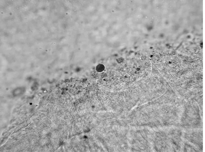
with a diamond grinding wheel to obtain 70μm thickness. It was studied in normal and polarized transmitted light at a magnification ranging from 40× to 400×. Diagenetic change is visible in bone at a microscopic level, using normal and polarized transmitted light, and consists of five categories: the presence and type of microscopic focal destructions (MFDs) assumed to be caused by micro-organisms (bioerosion), the presence of inclusive material and infiltrated material, the presence of micro-fissures, and the intensity of birefringence (Jans 2005). The thin-section displayed brown staining all the way through, while some cracking across the dentine and along the cementum and the pulp cavity surface were also evident in the form of larger
cracks (no micro-fissures) that could be related to sample preparation, and possibly to the fact that the sample was cut before being embedded in resin. A few possible fungal structures were observed within the pulp cavity (Figure 3). This might be a recent post-excavation phenomenon, and it seemed in any case that the fungus was not attacking the material. An unknown type of orange inclusions was also observed on the surface of the pulp cavity and on the root surface (Figure 4). These were only superficial and not likely to affect DNA preservation in this case.
A common and fairly simple method of assessing bone mineral integrity is by the crystallinity
index, or splitting factor (SF), obtained by Fourier-Transform Infrared spectrometry (FTIR). FTIR can also be used to obtain several other preservation parameters, such as the carbonate tophosphate peak ratio (C/P) that indicates the loss or uptake of carbonates, and the amide to phosphate peak ratio (Am/P) which reflects residual organic (Trueman
et al. 2008). Recently, a new FTIR-method has
been proposed as a tool in the analysis of fossil and archaeological bone, which is void of some of the methodological issues attached to the traditional method, and is faster and easier to use (Thompson
et al. 2009). This method, known as FTIR-ATR, is
an FTIR with an attenuated total reflection (ATR) unit. The sample is placed, without any sample preparation, directly onto an optically dense crystal. Dentine powders are drilled directly from a freshly cut cross-section using a hand-held drill with a tungsten-carbide drill-bit. The same piece that was
sampled for the histology and UV-work is also used here. All IR-parameters reported are made using FTIR-ATR (Shimadzu FTIR-8400S with an ATR from Specac, Golden Gate), averaging two replicate measurements. The SF, C/P and Am/P values are calculated based on the peak heights of the obtained spectra. Nubian samples were compared to fresh bone, and a highly altered bone sample (Figures 5 and 6). The results showed that the dentine of all samples was well preserved: some re-crystallization (increased SF, decreased C/P) and collagen loss (decreased Am/P) had occurred but this was not extensive.
DNA work was carried out in a dedicated ancient DNA laboratory, taking severe cautions against contamination with other DNA (including cleaning with bleach and UV-C light, wearing body-covering sterile suits, mouth caps and gloves). For
Figure 5: Plot of C/P versus SF values comparing the Nubian samples with a fresh bone sample and a highly altered bone sample.
DNA extraction, the teeth were cleaned with alcohol and chlorohexidine Digluconate containing tissues to remove dirt. Afterward they were irradiated four times with UV-C light for 45min to remove possible contaminating human DNA. They were ground to a fine powder with Mixer Mills from Retsch®. For each DNA extraction, 0.4gr of bone powder was used, to which 1ml of 0.5 M EDTA pH 8.0 with 5 % sarcosyl and 90µl Proteinase K was added and mixed overnight at 56°C. Negative controls were included in this process. After centrifugation the supernatant was removed and purified with the QIAquick PCR Purification kit® (Qiagen) using a centrifuge. The purified DNA was eluded in 40µl of sterile water. DNA concentration was measured with the Quantifiler Duo kit® (Applied Biosystems). This test also includes an indicator for inhibition, although not every possible inhibitor may be
detected. Whether or not a component acts as an inhibitor also depends on the type of reaction that is performed on a sample. Autosomal Short Tandem Repeats were typed with the PowerPlex® ESX 16 System (Promega), except for using half of all reagents. For each sample 5µl of DNA extract was used. PCR products were analysed on a 3100 Genetic Analyser® (Applied Biosystems). The data were analysed with GeneMarker® software version 1.75 (SoftGenetics LLC®). Per sample only one PCR was conducted, so results are not replicated and therefore not confirmed. DNA amplification was unsuccessful with the exception of the fourth sample S-6 dating to the Christian and, therefore, from the most recent time period in the total sample. But even in this case only three markers could be typed. This indicates severe loss and fragmentation of DNA in the four analysed samples. Typing autosomal STRs Figure 6: Plot of Am/P versus C/P values comparing the Nubian samples with a fresh bone sample and a highly altered bone sample.
with the PowerPlex® ESX 16 System (Promega) was just used as a quick scan method for quality in this study, because it is relatively fast and cost efficient. However, it is possible that with more sensitive techniques the DNA of these samples can be investigated to a level that allows the answering of the afore-mentioned research questions. It should also be kept in mind that the STR results are not confirmed, although they are in line with the results of the Quantifiler Duo kit® (Applied Biosystems).
Finally, thermal age was calculated using the recently developed DNA screening wizard available at http://beta.thermal-age.eu. Thermal age is the time taken to produce a given degree of DNA decay at a constant temperature of 10 °C and takes the thermal history of the site into account. The calculated thermal ages for the different samples ranged from about 25,000 to 164,000.
Concluding Remarks
Using a set of relatively simple methods
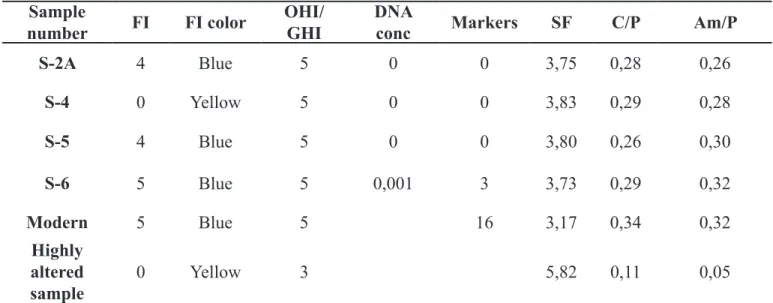
(summarized in Table 4), the diagenetic analysis of four teeth from two different sites in Sudanese Nubia, dating to four different periods, shows that most are well preserved according to several different preservation parameters. Most have a high organic content and low crystallinity with similar values to that of modern bone. The samples display no bioerosion. UV autofluorescence also indicates overall good preservation as all samples except one show bright bluish white fluorescence, similar to modern material. Both biological and chemical decay of the bone mineral and collagen has been inhibited. Despite this, DNA analyses were not or only partly successful.
Several factors are likely to affect the good preservation of this archaeological dental material. Aridity, burial depth and soil type, may all have promoted the preservation of both the organic and mineral phase due to low water and oxygen content, inhibiting both chemical and biological decay. The bone mineral is generally found to be well preserved at arid sites (Zazzo and Saliège 2011). In temperate and cold regions, this would mean a high Sample
number FI FI color OHI/GHI DNA conc Markers SF C/P Am/P
S-2A 4 Blue 5 0 0 3,75 0,28 0,26 S-4 0 Yellow 5 0 0 3,83 0,29 0,28 S-5 4 Blue 5 0 0 3,80 0,26 0,30 S-6 5 Blue 5 0,001 3 3,73 0,29 0,32 Modern 5 Blue 5 16 3,17 0,34 0,32 Highly altered sample 0 Yellow 3 5,82 0,11 0,05
[FI = Fluorescence Index; OHI/GHI = Oxford/General Histological Index; DNA conc = DNA
concentration (ng/µl); Markers = Number of typed DNA markers (max 16); SF = Splitting Factor; C/P = Carbonate/Phosphate ratio; Am/P = Amide/Phosphate ratio]
likelihood of obtaining well-preserved DNA. High FI has been found to correlate strongly with high DNA amplification success rate (Hoke et al. 2011). The fact that these Nubian teeth are well preserved according to various measures of dentine organic, inorganic and microstructural integrity suggests that temperature and desiccation have overruled other influencing factors, leading to rapid decay of DNA. This is also indicated by the fact that the one sample (S-6) that did yield some short fragments of DNA is the youngest sample, i.e., the one having the lowest thermal age. The high thermal age for all of the samples suggests that DNA survival is not likely although, evidently, some DNA was still present in the youngest sample (thermal age of 25 000). Furthermore, other bone samples from the site of Kerma have been used in a study exploring a new parameter for assessing collagen deterioration. This involves calculating a thermal age of the collagen and may also prove to be a useful DNA screening method. Out of more than 900 samples from 50 different sites, the Kerma-material was found to be the most altered, with extreme high thermal ages (van Doorn et al. 2012).
Though other aspects such as the correlation between successful DNA analysis and the nature of the soil, the depth of the grave and the level of disturbance of the burial might become relevant in the future (de Voogt and Francigny 2012), high temperature is left as the main factor influencing DNA decay in this case, causing rapid DNA depurination. However, alternative pathways of DNA decay are poorly understood. Some studies have shown that inter-strand cross-linking may be a significant damage type in certain environments (Willerslev and Cooper 2005). As mentioned above, Bieger-Dose et al. (1992) found that extreme dryness promotes DNA cross-linking. Although the graves from Sai Island and Kerma cemeteries have been out of reach from the Nile flood, it is unlikely, at least for the plundered ones, that the level of humidity inside remained constant for thousands of years. These conclusions are in line with the assumption made by Gilbert et al. (2005) on the low potential for DNA recovery of ancient material
collected in graves from the Nile Valley.
Despite the poor DNA results at Sai and Kerma, this diagenetic analysis, where microscopic and chemical techniques have been applied, identify important requirements for bioarchaeological sampling and future attempts at DNA extraction within the region of the eastern Sahara. While Sudanese Nubia has changed into a more arid place since antiquity, with a substantial decline of rainfall over the last century (Elagib and Elhag 2011; Hulme 1990), chances of successful ancient DNA extraction might potentially exist farther south in the Keraba area and the desert steppes of the Butana, also inhabited throughout antiquity. Studies based on larger data sets that would explore the relation between taphonomic contexts and DNA degradation processes would also increase the chances of future successful DNA extractions in Sudan.
Bibliography
Bieger-Dose, A., K. Dose, R. Meffert, M. Mehler and R. Risi
1992 Extreme dryness and DNA-protein cross- links. Advances in Space Research 12: 265-
270.
Bollongino, R., and J.-D. Vigne
2008 Temperature monitoring in archaeological animal bone samples in the Near East arid area, before, during and after excavation.
Journal of Archaeological Science 35: 873-
881.
1999 DNA preservation: A microsatellite- DNA study on ancient skeletal remains.
Electrophoresis 20: 1722-1728.
Buzon, M. R.
2008 A bioarchaeological perspective on Egyptian colonialism in Nubia during the New Kingdom. The Journal of
Egyptian Archaeology 94: 165-181.
Campos, P. F., O. E. Craig, G. Turner-Walker, E. Peacock, E. Willerslev and M. T. P. Gilbert
2011 DNA in ancient bone. Where is it located and how should we extract it? Annals of
Anatomy Anatomischer Anzeiger 194 (1):
7-16.
Carlson, D. S., and D. P. Van Gerven
1979 Diffusion, biological determinism, and biocultural adaptation in the Nubian Corridor. American Anthropologist 81: 561-580.
Colson, I. B., J. F. Bailey, M. Vercauteren and B. C. Sykes
1997 The preservation of ancient DNA and bone diagenesis. Ancient Biomolecules 1: 1-9.
Dafa’Alla, S. B.
1989 Distribution and migrations of the Nubian tribes during the Meroitic and X-Group Periods. Beiträge Zur Sudanforschung 4: 75-94.
de Voogt, A., and V. Francigny
2012 Opening a grave in antiquity. Formation and interpretation in the Kingdom of Meroe. Journal of African Archaeology 10(1): 59-70.
Dose, K., A. Bieger-Dose, O. Kerz and M. Gill 1991 DNA-strand breaks limit survival in extreme dryness. Origins of Life and
Evolution of Biospheres 21: 177-187.
Elagid, N. A., and M. N. Elhag
2011 Major climate indicators of ongoing drought in Sudan. Journal of Hydrology 409: 612-625.
Francigny, V.
2009 The Meroitic necropolises of Sai Island. First season at the cemetery 8-B-5.A. Sudan
& Nubia 13: 92-99.
2010 The Meroitic necropolises of Sai Island. Second season at the cemetery 8-B-5.A.
Sudan & Nubia 14: 56-61.
Gilbert, M. T. P., I. Barnes, M.J. Collins, C. Smith, J. Eklund, J. Goudsmit, H. Poinar and A. Cooper 2005 Long-term survival of ancient DNA in
Egypt: response to Zink and Nerlich (2003).
American Journal of Physiscal Anthropology 128: 110-114.
A. J. Hansen, C. Smith, K.E.H. Penkman, K. Prangenberg, C. M. Nielsen-Marsh, M. E. Jans, P. Arthur, N. Lynnerup, G. Turner-Walker, M. Biddle, B. Kjolbye-Biddle and M. J. Collins
2005 Biochemical and physical correlates of DNA contamination in archaeological human bones and teeth excavated at Matera, Italy. Journal of Archaeological
Science 32: 785-793.
Götherström, A., Collins, M. J., Angerbjörn, A. and K. Lidén
2002 Bone preservation and DNA amplification.
Archaeometry 44: 395-404.
Haynes, S., J. B. Searle, A. Bretmanand K. M. Dobney 2002 Bone preservation and ancient DNA:
the application of screening methods for predicting DNA survival. Journal of
Archaeological Science 29: 585-592.
Hedges, R. E. M.
2002 Bone diagenesis: an overview of processes.
Archaeometry 44: 319-328.
Hillson, S.
1986. Teeth. Cambridge: Cambridge University Press
Hoke, N., J. Burger, C. Weber, N. Benecke, G. Grupe and M. Harbeck
2011 Estimating the chance of success of
archaeometric analyses of bone:UV- induced bone fluorescence compared to histological screening.
Palaeogeography, Palaeoclimatology, Palaeoecology 310: 23-31.
Honegger, M.
2011 The beginning of the Kerma civilisation in the eastern cemetery. In M. Honegger and C. Bonnet editors, Archaeological
excavations at Kerma (Sudan), Documents de la mission archéologique suisse au Soudan. Neuchâtel: Université de
Neuchâtel, pp. 9-14.
Hulme, M.
1990 The changing rainfall ressources of Sudan.
Transactions of the Institute of british Geographers 15 (1): 21-34.
Irish, J. D.
2005 Population continuity vs. discontinuity revisited: dental affinities among late Paleolithic through Christian-Era Nubians.
American Journal of Physical Anthropology 125: 520-535.
Jans, M. M. E.
2005 Histologial Characterisation of
Diagenetic Alteration of Archaeological Bone. Amsterdam: VU University.
2005 History in the interpretation of the pattern of p49a,f TaqI RFLP Y-chromosome variation in Egypt: a consideration of multiple lines evidence. American
Journal of Human Biology 17: 559-567.
Krings, M., A. H. Salem, K. Bauer, H. Geisert, A.K. Malek, L. Chaix, C. Simon, D. Welsby, A. Di Rienzo, G. Utermann, A. Sajantila, S. Pääbo and M. Stoneking
1999 mtDNA analysis of Nile River Valley populations: A genetic corridor or a barrier to migration? The American Journal of
Human Genetics 64 (4): 1166-1176.
Lalueza Fox, C.
1997 mtDNA analysis in ancient Nubians supports the existence of gene flow between sub-Sahara and North Africa in the Nile Valley. Annals of Human Biology 24 (3): 217-227.
Lindahl, T.
1993 Instability and decay of the primary structure of DNA. Nature 362: 709.
Lucotte, G. and G. Mercier
2003 Brief communication: Y-chromosome haplotype in Egypt. American Journal of
Physical Anthropology 121: 63-66.
Manni, F., P. Leonardi, A. Barakat, H. Rouba, E. Heyer, M. Klintschar, K. Mcelreavey and L. Quintana-Murci
2002 Y-chromosome analysis in Egypt suggests a genetic regional continuity in northeastern Africa. Human Biology 74 (5): 645-658.
Millard, A. R.
2001 The deterioration of bone. In D. Brothwell and M. Pollard, editors, Handbook of
Archaeological Sciences. New York: Wiley,
pp. 633-643.
Prentice, A. I. D.
1965 Bone autofluorescence and mineral content. Nature 206: 1167.
Pruvost, M., R. Schwarz, V. Bessa Correia, C. Hamplot, S. Braugier, N. Morel, Y. Fernandez-Jalvo, T. Grange and E. Geigl
2007 Freshly excavated fossil bones are best for amplification of ancient DNA. Proceedings
of the National Academy of Science 104:
739-744.
Reed, F. A., E. J. Kontanis, K. A. R. Kennedy and C. F. Aquadro
2003 Ancient DNA prospects from Sri Lankan highland dry caves support an emerging global pattern. American Journal of
Physical Anthropology 121: 112-116.
Singh, N., P. Joglekar and K. Koziol
2011 First ancient bovine DNA evidence from India: difficult but not impossible. Journal
Smith, C. I., A. T. Chamberlain, M. S. Riley, C. Stringer and M. J. Collins
2003 The thermal history of human fossils and the likelihood of successful DNA amplification. Journal of Human Evolution 45: 203-217.
Stynder, D. D., J. Braga. and E. Crubézy 2009 Craniometric Evidence for Biological
Continuity between Meroitic and Post- Meroitic Populations Buried at the Necropolis of Missiminia, Middle Nubia.
South African Archaeological Bulletin 64:
122-129.
Terreros, M. C., L. Martinez and R. J. Herrera 2005 Polymorphic Alu insertions and genetic
diversity among African populations.
Human Biology 77 (5): 675-704.
Thompson, T. J. U., M. Gauthier and M. Islam 2009 The application of a new method of Fourier
Transform Infrared Spectroscopy to the analysis of burned bone. Journal of
Archaeological Science 36: 910-914.
Török, L.
2008 Between Two Worlds. The Frontier Region
between Ancient Nubia and Egypt 3700 BC – AD 500. Leiden: Brill.
Trueman, C. N., K. Privat and J. Field
2008 Why do crystallinity values fail to predict the extent of diagenetic alteration of bone mineral? Palaeogeography,
Palaeoclimatology, Palaeoecology 266:
160-167.
van Doorn, N. L., J. Wilson, H. Hollund, M. Soressi and M. J. Collins
2012 Site-specific deamidation of glutamine: a new marker of bone collagen deterioration.
Rapid Communications in Mass Spectrometry 26: 2319-2327.
Willerslev, E. and A. Cooper
2005 Ancient DNA. Proceedings of the Royal
Society - Biological Sciences 272: 3-16.
Zazzo, A. and J.F. Saliège
2011 Radiocarbon dating of biological apatites: A review. Palaeogeography,
Palaeoclimatology, Palaeoecology