HAL Id: inserm-00663677
https://www.hal.inserm.fr/inserm-00663677
Submitted on 27 Jan 2012
HAL is a multi-disciplinary open access
archive for the deposit and dissemination of
sci-entific research documents, whether they are
pub-lished or not. The documents may come from
teaching and research institutions in France or
abroad, or from public or private research centers.
L’archive ouverte pluridisciplinaire HAL, est
destinée au dépôt et à la diffusion de documents
scientifiques de niveau recherche, publiés ou non,
émanant des établissements d’enseignement et de
recherche français ou étrangers, des laboratoires
publics ou privés.
Free-breathing myocardial T2 measurements at 1.5T
Maelene Lohezic, Anne Menini, Jean-Marie Escanyé, Pierre-Yves Marie,
Damien Mandry, Pierre-André Vuissoz, Jacques Felblinger
To cite this version:
Maelene Lohezic, Anne Menini, Jean-Marie Escanyé, Pierre-Yves Marie, Damien Mandry, et al..
Free-breathing myocardial T2 measurements at 1.5T. 2011 SCMR/Euro CMR Joint Scientific Sessions, Feb
2011, Nice, France. pp.P11. �inserm-00663677�
P O S T E R P R E S E N T A T I O N
Open Access
Free-breathing myocardial T2
measurements at 1.5T
Maelene Lohezic
1*, Anne Menini
2, Jean-Marie Escanyé
3, Pierre-Yves Marie
4, Damien Mandry
4,
Pierre-André Vuissoz
3, Jacques Felblinger
2From 2011 SCMR/Euro CMR Joint Scientific Sessions
Nice, France. 3-6 February 2011
Introduction
Myocardial T2 mapping is a valuable tool for tissue characterization and oedema visualization. For instance, it is used to detect early rejection of heart transplant [1]. T2 values are usually estimated by performing several black blood Fast Spin Echo (FSE) sequences with differ-ent Echo Times (TE), what requires multiple breath holds. Successive apneas could lead to misregistration between images and to patient discomfort. A method allowing free breathing myocardial T2 measurements has been recently proposed and evaluated at 3T [2]. Results at 1.5T are presented here.
Purpose
This study aims at demonstrating the feasibility of free-breathing myocardial T2 mapping at 1.5T.
Methods
MRI experiments
Five healthy volunteers underwent cardiac examination at 1.5T (SIGNA HDxt, GE Healthcare, Milwaukee, WI). Two sets of ten images with different TE were acquired with a conventional cardiac-gated black blood FSE sequence at mid-cavity short axis view, one during breath hold and the other one during free breathing. The same parameters were used, except for echo train length (ETL) (Table 1).ETL was set at 24 to keep the breath hold duration short, whereas 16 echoes were used for free-breathing acquisitions. Raw data from the free breathing acquisitions were recorded. Signals from a respiratory belt were carried by a custom Maglife patient monitoring system (Schiller Medical, France) and recorded with a dedicated home-made hardware [3].
Post-processing
First, the ten breath-held images were registered manu-ally. Then, the T2 map was obtained using a mono-exponential model to fit the T2 signal versus the echo time decay curve, on a pixel-wise basis.
Free breathing reconstruction
Using physiological signals extracted from the respira-tory belt, the method presented in [2] was used to obtain an artefact-free proton density weighted image r0 and a T2 map from the free breathing raw data set. For the sake of comparison, six segments were drawn on the left ventricle myocardium. The mean value of each ROI was then used to get 6 myocardial T2 values.
Results
Like at 3T, there was no significant differences between the two sets of myocardial T2 values (paired Student T-test, p=0.17). The free breathing T2 maps were in good agreement with the breath-held ones and respiratory artefacts were widely reduced in r0 (Fig. 1).
Conclusions
The proposed free breathing method allows performing accurate T2 mapping at 1.5T with no additional acquisi-tion time.
1GE Healthcare / IADI Lab, Nancy, France
Full list of author information is available at the end of the article
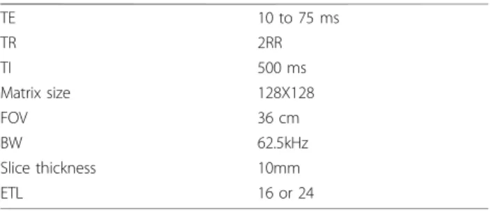
Table 1 Acqusition parameters
TE 10 to 75 ms TR 2RR TI 500 ms Matrix size 128X128 FOV 36 cm BW 62.5kHz Slice thickness 10mm ETL 16 or 24
Lohezic et al. Journal of Cardiovascular Magnetic Resonance 2011, 13(Suppl 1):P11 http://jcmr-online.com/content/13/S1/P11
© 2011 Lohezic et al; licensee BioMed Central Ltd. This is an open access article distributed under the terms of the Creative Commons Attribution License (http://creativecommons.org/licenses/by/2.0), which permits unrestricted use, distribution, and reproduction in any medium, provided the original work is properly cited.
Author details
1
GE Healthcare / IADI Lab, Nancy, France.2INSERM U947, Nancy, France.
3Nancy-Université, Nancy, France.4CHU Nancy, Nancy, France.
Published: 2 February 2011
References
1. Marie, et al: JACC 2001, 37:825-831. 2. Lohezic, et al: Proc. ISMRM 2010, 2958. 3. Odille, et al: IEEE TBME 2007, 54:630-640.
doi:10.1186/1532-429X-13-S1-P11
Cite this article as: Lohezic et al.: Free-breathing myocardial T2 measurements at 1.5T. Journal of Cardiovascular Magnetic Resonance 2011 13(Suppl 1):P11.
Submit your next manuscript to BioMed Central and take full advantage of:
• Convenient online submission
• Thorough peer review
• No space constraints or color figure charges
• Immediate publication on acceptance
• Inclusion in PubMed, CAS, Scopus and Google Scholar
• Research which is freely available for redistribution
Submit your manuscript at www.biomedcentral.com/submit
Figure 1 T2 maps obtained on one volunteer in breath hold (left) and in free breathing (right).
Lohezic et al. Journal of Cardiovascular Magnetic Resonance 2011, 13(Suppl 1):P11 http://jcmr-online.com/content/13/S1/P11