HAL Id: hal-01176910
https://hal-univ-perp.archives-ouvertes.fr/hal-01176910
Submitted on 26 Sep 2017
HAL is a multi-disciplinary open access
archive for the deposit and dissemination of
sci-entific research documents, whether they are
pub-lished or not. The documents may come from
teaching and research institutions in France or
abroad, or from public or private research centers.
L’archive ouverte pluridisciplinaire HAL, est
destinée au dépôt et à la diffusion de documents
scientifiques de niveau recherche, publiés ou non,
émanant des établissements d’enseignement et de
recherche français ou étrangers, des laboratoires
publics ou privés.
Smart designing of new hybrid materials based on
brushite-alginate and monetite-alginate microspheres:
Bio-inspired for sequential nucleation and growth
Walid Amer, Karima Abdelouahdi, Hugo Ronald Ramananarivo, Aziz Fihri,
Mounir El Achaby, Mohamed Zahouily, Abdellatif Barakat, Kamal Djessas,
James Clark, Abderrahim Solhy
To cite this version:
Walid Amer, Karima Abdelouahdi, Hugo Ronald Ramananarivo, Aziz Fihri, Mounir El Achaby, et
al.. Smart designing of new hybrid materials based on brushite-alginate and monetite-alginate
micro-spheres: Bio-inspired for sequential nucleation and growth. Materials Science and Engineering: C,
Elsevier, 2014, 35, pp.341-346. �10.1016/j.msec.2013.11.012�. �hal-01176910�
Smart designing of new hybrid materials based on brushite-alginate
and monetite-alginate microspheres: Bio-inspired for sequential
nucleation and growth
Walid Amer
a, Karima Abdelouahdi
b, Hugo Ronald Ramananarivo
a, Aziz Fihri
a, Mounir El Achaby
a,
Mohamed Zahouily
c, Abdellatif Barakat
d, Kamal Djessas
e, James Clark
f, Abderrahim Solhy
a,⁎
a
MAScIR Foundation, INANOTECH, Rabat Design, Rue Mohamed El Jazouli, Madinat El Irfane 10100 Rabat, Morocco
bCentre National pour la Recherche Scientifique et Technique (CNRST), Division UATRS, Angle Allal Fassi/FAR, B.P. 8027, Hay Riad, 10000 Rabat, Morocco c
Laboratoire de Matériaux, Catalyse et Valorisation des Ressources Naturelles, URAC 24, Faculté des Sciences et Techniques, Université Hassan II, Mohammedia B.P. 146, 20650, Morocco
dSUPAGRO-INRA-CIRAD-UMR IATE 1208, Ingenierie des Agropolymères et Technologies Emergentes, 2, Place Pierre Viala-Bât 31, 34060 Montpellier cedex 1, France e
CNRS-PROMES Tecnosud, F-66100 Perpignan, France
f
Green Chemistry, Centre of Excellence, University of York, York YO10 5DD, UK
a b s t r a c t
a r t i c l e i n f o
Article history:
Received 15 November 2012
Received in revised form 30 September 2013 Accepted 5 November 2013
Available online 19 November 2013 Keywords: Hybrid materials Biopolymers Alginate Phosphates Brushite Monetite
In this report new hybrid materials based on brushite-alginate and monetite-alginate were prepared by self-assembling alginate chains and phosphate source ions via a gelation process with calcium ions. The alginate served as nanoreactor for nucleation and growth of brushite or/and monetite due to its gelling and swelling prop-erties. The alginate gel framework, the crystalline phase and morphology of formed hybrid biomaterials were shown to be strongly dependent upon the concentration of the phosphate precursors. These materials were char-acterized by thermogravimetric analysis (TGA), Fourier transform infrared spectroscopy (FTIR), X-ray diffraction (XRD), scanning electron microscopy (SEM) and energy dispersive x-ray analysis (EDX).
© 2013 Elsevier B.V. All rights reserved.
1. Introduction
Hybrid materials offer the opportunity to combine the desirable properties of an organic matrix with those of inorganic solids[1–3]. This material category has a diverse spectrum of applications ranging from pharmaceutical to wastewater treatment[4–6]. On the other hand, the calcium phosphate materials contain a multitude of crystal-line phases and have attracted considerable interest both in the scientif-ic community and industry due to their physscientif-ico-chemscientif-ical properties and their value in manyfields of technology[7–17]. However, the com-bination of calcium phosphates and biopolymer can confer favorable mechanical properties, including strength due to the inorganic phase, toughness and plasticity due to the biopolymer phase. Biopolymers differ from each other in chemical composition, molecular weight, poly-dispersity, crystallinity, hydrophobicity, solubility, chain length, and thermal transitions[18]. Different biopolymer-calcium orthophosphate composites have been successfully obtained with alginate[19], chitosan
[20,21], cellulose[22]and starch[23,24]. Alginate is a natural polysac-charide extracted from the marine brown algae and it is composed of
two uronic acid monomers derived from mannose: the acid β-L-guluronic (G) and the acidα-D-mannuronic (M) are linked by glycoside bondsβ-(1–4) and α-(1–4) (see Fig. S1 in Supporting information)
[25,26]. Alginate gelation occurs when multivalent ions take their place between the G blocks of guluronic monomers, forming junctions of electrostatic nature, and the bond strength varies depending on the type of cation[27,28]. Most of the published works on hybrid composite alginate–calcium phosphate are based on preparation, nucleation and growth of apatite particles in a matrix of alginate biopolymer. In this context, Rajkumar et al. synthesized a nano-hydroxyapatite alginate nanocomposite with different weight percentages of sodium alginate; its biological and mechanical properties were studied[29]. Zhang et al. prepared a series of new nanocomposite beads of sodium alginate/ hydroxyapatite, in order tofind a new way to slow drug release, but unfortunately they did not show the XRD spectra which can confirm if that it was really the hydroxyapatite or another phase[30].
Brushite (dicalcium phosphate dihydrate (DCPD)) and monetite (dicalcium phosphate (DCP)) cements have raised considerable interest in the last decade, because they are metastable under physiological con-ditions and can be resorbed more quickly than hydroxyapatite cements
[31]. Several authors have suggested that it's a precursor of bone miner-alization, including biological apatites[32,33]. The brushite has been
⁎ Corresponding author. Fax: +212 53 027 9827. E-mail address:a.solhy@mascir.com(A. Solhy).
0928-4931/$– see front matter © 2013 Elsevier B.V. All rights reserved.
http://dx.doi.org/10.1016/j.msec.2013.11.012
Contents lists available atScienceDirect
Materials Science and Engineering C
widely used in the reconstruction materials, dental cements, as well as in formulation chemistry[34,35]. These phosphates are usually synthe-sized in an aqueous medium by double decomposition of a calcium salt and a phosphate salt or by neutralization of phosphoric acid in lime
[36,37]. Other routes have been also explored to synthesis these calcium phosphates such as sol–gel[38], solid state[39], chemical precipitation
[40], hydrothermal[41], hard templating[42],flame-spray route[43], dual irradiation of the microwave and ultrasound[44], and physiologi-cal conditions[45]. More recently, Gomez-Morales et al. have controlled the precipitation of calcium phosphate such as brushite by using vapor diffusion method in microdroplets and amino acids like organic additives[46].
This current study demonstrates for thefirst time that the synthesis of hybrid material phosphate–biopolymer can be achieved using gelation of alginate mixed with a phosphate source via complexation of calcium ions. The synthetic protocol reported here takes advantage of the controlled gelation of the mixture of brushite or monetite struc-ture at room temperastruc-ture.
2. Experimental section
2.1. Chemical reagents
(NH4)2HPO4and Ca(NO3)2·4H2O were purchased from Aldrich and used as a precursor of the HPO42−ligand and for gelation, respectively. Other phosphate sources are also tested namely: sodium phosphate di-basic (Na2HPO4) and ammonium phosphate monobasic (NH4H2PO4). Sodium alginate was purchased from Aldrich and used as supplied. Deionized water was used in all experiments.
2.2. Synthesis of hybrid materials
Hydrogel material was achieved via complexation of alginate/ phosphate by calcium ion (Fig. S2). Aqueous solutions of phosphorus precursor were prepared by dissolving different amounts of (NH4)2HPO4: 0.1, 0.3, 0.5, and 1 M into 100 mL of distilled water, and then sodium alginate was added to diammonium phosphate solution with a concentration of 1%. The mixture was stirred for 1 h at room tem-perature. This gel was added dropwise using a syringe with a 0.8 mm diameter needle at room temperature to the stirred Ca(NO3)2·4H2O solution (0.25 M). The formed beads immediately are abandoned overnight to ensure effective diffusion of calcium ions, and thereafter the homogeneity of the system and the growth of hybrid materials.
After that, the beads werefiltered with a 100 mesh screen, and washed three times with distilled water to remove the Ca2+excess and impuri-ties on the beads surface. In the end, the beads were dried at room tem-perature for 24 h before various characterizations. For different (NH4)2HPO4concentration (0.1, 0.3, 0.5 and 1 M), the dried hybrid materials will be henceforth identified as Phos–Alg_1 (phosphate– alginate), Phos–Alg_2, Phos–Alg_3, and Phos–Alg_4 respectively. 2.3. Thermal and structural characterization techniques
TGA were conducted under air in a TA Instrument Q500 apparatus with a 10 °C/min ramp between 25 and 1000 °C. X-ray diffraction pat-terns of the samples were obtained at room temperature on a Bruker AXS D-8 diffractometer using Cu-Kα radiation in Bragg–Brentano geometry (θ–2θ). Fourier transform infrared spectra of samples in KBr pellets were measured on a Bruker Vector 22 spectrometer. Scanning electron microscopy pictures were recorded on a FEI Quanta 200 micro-scope after carbon metallization.
3. Results and discussion
In order to conduct this study, we investigated several parameters, especially the concentration of phosphate precursor and the drying temperature, that influence the nucleation and growth of the inorganic matrix and obviously the hybrid material by afterwards. Thus, the con-centration of the precursor of the phosphate plays an important role in the appearance of the prepared hybrid xerogels.Fig. 1shows pictures taken by a digital camera and SEM at low magnification of the as synthe-sized hybrid xerogel microspheres (Phos–Alg_1 and Phos–Alg_2), micro-lentils (Phos–Alg_3) and granulated powder (Phos–Alg_4). This change in the external shape can be explained by the increase of the concentration of HPO42−ligand which occupies the space in the alginate chains and subsequently distorts the spherical shape of the droplet.
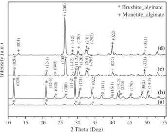
The XRD patterns have been used to investigate the phases in our hybrid xerogels at different phosphate concentrations.Fig. 2shows dia-grams of the samples such as Phos–Alg_1, Phos–Alg_2, Phos–Alg_3 and Phos–Alg_4 corresponding to (NH4)2HPO4 concentrations: 0.1 M, 0.3 M, 0.5 M and 1 M respectively. The Phos–Alg_1 is poorly crystalline maybe because the amount of phosphates is poor, but has a pure brushite phase or dicalcium phosphate dihydrate with monoclinic crystal structure (JCPDS 4-013-3344). Brushite crystallized in the monoclinic system (space group Ia) with the crystallographic pa-rameters a = 6.239 Å, b = 15.180 Å, c = 5.812 Å,α = γ = 90° and
β = 116°42′[47]. The intensity of the diffraction peaks indicates that the sample Phos–Alg_2 is well crystallized. This is made up of a brushite phase with trace amount of monetite or dicalcium phosphate with triclinic crystal (JCPDS 4-009-4184). The Phos–Alg_3 represents a co-existence of both phases which is confirmed by the high intensity of dif-fraction peaks of the monetite {020} facet and brushite {200} facet. Finally, the Phos–Alg_4 sample is composed predominantly of the monetite phase. Monetite crystallized in the triclinic system (space group P− 1) with the crystallographic parameters: a = 6.91 Å, b = 6.998 Å, c = 6.627 Å,α = 96.34°, β = 91.67° and γ = 76.18°. The concentration of (NH4)2HPO4plays a crucial role in the growth of inorganic phases. The selectivity for DCPD or DCP was clearly influenced when the amount of phosphate in the alginate matrix was increased. The oversaturation of alginate matrix by phosphate leads to a high per-centage of the monetite phase in Phos–Alg_4. High concentrations of phosphate source favor monetite formation (Fig. S3). This can be ex-plained by increased steric hindrance exerted on the alginatefibers by ligand HPO42−, which will reduce necessary space for keeping the struc-tural water. We have also studied the influence of the drying tempera-ture on samples: Phos–Alg_2 and Phos–Alg_3. The analysis of these bio-composites dried at 100 °C overnight by XRD shows that we have a total selectivity to synthesis monetite (Fig. 3). This can be explained by the conversion of brushite phase into monetite, which is simply due to the dehydration of brushite (removal of water molecules from the structure). Thus we can get simply the monetite phase by increasing the drying temperature. It's also noteworthy that the use of other sources of HPO42−ligand gives similar results. The semi-qualitative analysis by energy dispersive spectroscopy (EDAX) was carried out, when taking SEM photographs (Fig. S4). The study allowed us to dem-onstrate the presence of the chemical elements that constitute calcium phosphates: i) phosphorus, ii) oxygen, and ii) calcium. The data from semi-qualitative analysis give us an idea as to the composition of xerogels prepared in terms of molar ratio Ca/P. The results of the Ca/P ratio are in the range of: 1.12, 0.94, 0.97 and 1.24 for Phos–Alg_1, Phos–Alg_2, Phos–Alg_3 and Phos–Alg_4 respectively. These values are consistent with the molar ratio of brushite and monétite Ca/P = 1 reported in the literature.
The crystal unit cell contains four motifs of CaHPO4·2H2O and the structure is shown inFig. 4. The latter shows that the crystals are com-posed of brushite corrugated sheets of composition CaHPO4, arranged in parallel to each other, normal to the axis b and linked together by a dou-ble layer of water molecules. In the same way,Fig. 5gives a projection of monetite viewed along the b-axis. This projection shows the spatial
10 15 20 25 30 35 40 45 50 55
*
*
*
*
*
(200) + (11-2) (020) (-1-12) (001) (200) +*
(d) (c) (b) (a) * Brushite_alginate + Monetite_alginate 2 Theta (Deg) Intensity (a.u.) + + (120) + +(201) (022) + (-1-12) + (120) + +(201) (11-2)*
*
(12-1) (022) + (200) +*
(040) (020)*
*
(12-1) (040)*
*
(121)*
(022)*
(141)*
*
(16-1) (15-2)*
*
(240)*
(170)*
(062) (14-3)*
(-202) + (-202) + +(-321) (-321) +Fig. 2. XRD patterns of hybrid materials: Phos–Alg_1 (a), Phos–Alg_2 (b), Phos–Alg_3 (c) and Phos–Alg_4 (d). 10 15 20 25 30 35 40 45 50 55 + Intensity (a.u.) 2 Theta (Deg) (-1-12) (001) + Monetite_alginate + (-1-21) + (201) + (022) + (030) + (-301) + (131) + (301) + (-1-31) + (-321) + (112) + (200) + (3-21) + (-202) + (-3-13) + (321) +
Fig. 3. XRD pattern of dried hybrid materials (Phos–Alg_2 and Phos–Alg_3) overnight at 100 °C.
distribution and the unit cell of monetite illustrating the sequence of PO4units and the location of calcium ions within the framework.
Fig. 6shows the FT-IR spectra of the hybrid composite beads. The ab-sorptions observed at 1421 cm−1in all samples are due to the symmet-ric and asymmetsymmet-ric stretching modes of the COO−bands from the carboxylate groups[48,49]. The spectra of Phos–Alg_1, Phos–Alg_2, and Phos–Alg_3 exhibit similar bands. Brushite is characterized by the O\H stretching modes of the crystallization water and also for alginate matrix, with two peak doublets, respectively, 3550 and 3466 cm−1[48]. The H\O\H bending gives rise to absorption at 1651 cm−1[50]. The main IR bands characterizing the PO4group can be detected at 1128, 1059 and 998, and 1012 cm−1, due to PO stretching modes of the PO4 fragment[50]. Weaker sharp bands at 1210 and 871, and 875 cm−1 are due, respectively, to the P\O\H stretching mode and the P\OH bending mode[51]. Bands at frequencies 521, 590, and 650 cm−1 are assigned mainly to PO deformation modes of the tetrahedral PO4 group[52]. The FTIR also identifies the functional groups of
C-monetite sample[53], water vibrations at 3550 and 3466 cm−1are not seen in the spectrum of samples“d” corresponding to Phos–Alg_4.
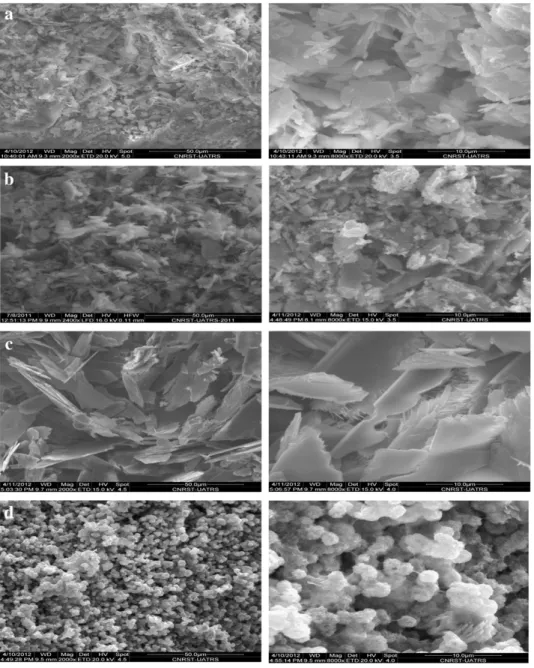
Morphologies of the hybrid bio-composites are shown inFig. 7. These show that the morphology of the crystals prepared by this route is very much dependent on the precursor concentration.Fig. 7a shows the SEM image of Phos–Alg_1, where the inorganic matrix is the brushite crystal prepared with 0.1 M of the (NH4)2HPO4solution. The morphology of the crystal is small plate-like and needle-like structures with a heterogeneous size.Fig. 7b presents the SEM image of Phos– Alg_2, where the inorganic matrix is a mixture of the brushite and monetite crystals prepared with 0.3 M. This image consists of heteroge-neous shapes like thefirst sample. In contrast, the platelet arrangement shown inFig. 7c, which was prepared with 0.5 M concentration of HPO42−ligand, gives rise to this single morphology with homogeneous dimensions, even though we have two phases. Generally, for thefirst three samples (a), (b) and (c), the morphology consists of merged plate-lets forming irregular chunky materials with various dimensions (a), (b) and (c). The fast rate of nucleation and crystal growth as a result of the high precursor concentration led to better defined plate-like structures corresponding to the predominant brushite crystals[54]. Finer spherical-like monetite particles are formed when we have in-creased the source concentration of HPO42−ligand (1 M). The morphol-ogy of monetite crystals is similar to snowballflowers with a size at around 2μm (Fig. 7d).
The thermal behavior of these dried beads was investigated by ther-mogravimetric analysis. The curves (Fig. 8a, b, c and d) showed different profiles characterized by different ranges of thermal degradation. These thermograms (a, b, c, d) showed continuous weight loss in the range of 25–330 °C and 330–750 °C. The first weight loss at ca. 200 °C can be assigned to the loss of absorbed water that can lead to conversion of DCPD to DCP[54]and also the conversion of DCP to calcium pyrophos-phate (Ca2P2O7(CCP))[53,55]. These observations were confirmed by the analysis of inorganic materials recovered after calcination at 750 °C by XRD and FTIR (Figs. 9 and 10). The weight loss shown by the curve (a) at 750 °C is around 4% and can be explained by thermal degradation of amorphous carbon. The total weight loss was found to be around 55%, 37%, 27% and 19% as shown inFig. 8(a, b, c and d) re-spectively, these results prove that increasing the amount of phosphate in an alginate gel matrix leads to decrease of the mass loss directly caus-ing the formation ofβ-TCP and CCP.
From a mechanistic point of view, the fact that the mixture of the phosphate source with alginate gives rise to a homogeneous gel, sug-gests an interaction between the biopolymer and the HPO42−. The stabil-ity and homogenestabil-ity of this gel can be explained by the possible interactions between the HPO42−ligand and the carboxylate group of the alginate. This physical bonding translates to mixtures that are rich in electronic pairs showing higher reactivity and mineralization poten-tial to be transformed into hybrid hydrogel materials. When the mixture of alginate and phosphate source meets the solution of Ca2+, divalent ions, interchain cross-linking occurs as a result of calcium coordination with pliers formed by two phosphated GG blocks as the“egg-box” model[56–58]. In addition, the nucleation and the growth of brushite and monetite can be due to interactions between inorganic minerals and alginate chains in the biopolymer. These observations are in agree-ment with those reported by M. Rajkumar[29].
4. Conclusions
In summary, a new type of hybrid materials was obtained via a straightforward synthetic route with alginate biopolymer as organic matrix and phosphate source ions with calcium ions. All the synthesis has been carried out at room temperature; thus greatly expanding the applicability of the method for the preparation of materials loaded with thermally unstable substances. Four hybrid beads were prepared with different inorganicfiller contents. Structure and physicochemical characterization of these hybrid phosphates were evaluated by FT-IR,
Fig. 5. Projection view of monetite.
4000 3500 3000 2500 2000 1500 1000 500 1651 1598 1421 1332 550 871 512 1012 3550 3466 1128 998 1598 1421 1337 1210 875 1059 820 650 590 521 wave number (cm-1)
Transmittance (a.u.) (a) (b) (c) (d)
Fig. 6. FT-IR spectra of hybrid beads: Phos–Alg_1 (a), Phos–Alg_2 (b), Phos–Alg_3 (c) and Phos–Alg_4 (d).
Fig. 7. SEM micrographs of hybrid xerogels: Phos–Alg_1 (a), Phos–Alg_2 (b), Phos–Al_3 (c) and Phos–Alg_4 (d). 0 100 200 300 400 500 600 700 800 900 1000 40 50 60 70 80 90 100 (a) (b) (c) (d) Temperature (°C) Weight %
Fig. 8. Thermogravimetric curves for hybrid xerogels: Phos–Alg_1 (a), Phos–Alg_2 (b), Phos–Alg_3 (c) and Phos–Alg_4 (d).
20 25 30 35 40 45 50 55 60 65 70 Intensity (a.u.) * (024) (024) (024) (024)
*
(c) (b) (a) ° °*
*
° ° ° ° ° ° ° ° ° ° (008)*
°° ° ° -(d)-*
-(002)*
*
-*
(211) -+*
-(300)-*
*
+*
-*
--*
-**
--*
-+*
-*
(311) +-*
-+*
-Ca 10(PO4)6(OH)2 -+ CaO Ca 3PO4-*
-(002)*
-* -*
--*
-*
*
-*
-*
--*
-**
--*
-*
-*
-*
Ca 2O7 P2 °*
° (203) ° ° °*
° -° ° - ° °°-*
-° ° ° ° ° ° ° ° ° ° ° ° ° (008)*
° ° ° °*
* *
*
*
*
*
*
* * ** *
*
**
*
° (203) ° ° °*
° °°°*
° ° ° ° °*
*
° ° ° ° ° ° ° ° ° °*
° ° ° °* *
*
*
* *
*
° (203) ° ° °*
° °°° ° ° ° ° ° ° ° ° ° ° ° °*
° ° ° °*
*
° (203) ° ° ° ° °°° ° ° ° ° (008) ° °° ° ° ° ° ° ° °*
*
° (203) ° ° ° °°°° ° ° ° ° ° ° ° ° ° °*
° (203) ° ° ° °°°° ° ° ° ° ° ° ° ° °*
° (203) ° °°°° ° °*
*
° ° ° ° ° ° ° ° ° (008)*
° ° °*
* *
*
*
*
*
* * ** *
*
**
*
° ° ° °*
° °°°*
° ° ° ° ° ° ° ° ° ° ° ° ° ° ° ° °*
° ° °*
° °°°° ° ° ° ° ° ° ° ° ° ° °*
° ° °*
° °°° ° 2 Theta (Deg) °°XRD, SEM and TGA. Experimental results for all hybrid materials indicat-ed that the phosphate source concentration is a prindicat-edominant factor for the sequential nucleation and growth and also the crystalline selectivity. The structural characterization results suggested that the particles formed into the biopolymeric matrix are brushite and/or monetite. Moreover, the observation of particle morphology by SEM proved that the shape changes according to the concentration of phosphate. The thermal behavior of these materials depends on the amount of water physisorbed, or structural, and the phosphate concentration. Character-ization of the recovered phosphates after heat treatment reveals the for-mation of a mixture of several types of calcium phosphate. Studies of the applications of these materials are underway in our laboratory.
Acknowledgments
Thefinancial assistance of the Office Chérifien des Phosphates in the Moroccan Kingdom (OCP Group): R&D Department towards this research is hereby acknowledged. The authors are also grateful to Mr. Mohamed Lasry for his help to achieve this work within the framework of the IMEDER and AMISOLE. Warm thanks are due to Dr. Mohamed Oudanane and for his admirable team from the clinic of the United Nations in Rabat for the professional qualifications and the kindness that they have shown during my hospitalization and for my successful surgery.
Appendix A. Supplementary data
Supplementary data to this article can be found online athttp://dx. doi.org/10.1016/j.msec.2013.11.012.
References
[1] A.P. Wight, M.E. Davis, Chem. Rev. 102 (2002) 3589–3614.
[2] Z. Ahmad, J.E. Mark, Mater. Sci. Eng. C 6 (1998) 183–196.
[3] S.V. Dorozhkin, M. Epple, Angew. Chem. Int. Ed. 41 (2002) 3130–3146.
[4] M.F. Ashby, Y.J.M. Bréchet, Acta Mater. 51 (2003) 5801–5821.
[5] L. Nicole, L. Rozes, C. Sanchez, Adv. Mater. 22 (2012) 3208–3214.
[6] K.E. Lee, N. Morad, T.T. Teng, B.T. Poh, Chem. Eng. J. 203 (2012) 370–386.
[7] G.S. Sailaja, S. Velayudhan, M.C. Sunny, K. Sreenivasan, H.K. Varma, P. Ramesh, J. Mater. Sci. 38 (2003) 3653–3662.
[8] A. Solhy, W. Amer, M. Karkouri, R. Tahir, A. El Bouari, A. Fihri, M. Bousmina, M. Zahouily, J. Mol. Catal. A Chem. 336 (2011) 8–15.
[9] P. Becker, Phosphates and phosphoric acid: raw materials technology and econom-ics of the wet process, Fertilizer Science and Technology Series, 2nd ed., Marcel Dek-ker, New York, 1989, p. 6.
[10] L.L. Hench, J. Am. Ceram. Soc. 81 (1998) 1705–1728.
[11] F.C.M. Driessens, M.G. Boltong, O. Bermu dez, J.A. Planell, M.P. Ginebra, E. Fernandez, J. Mater. Sci. Mater. Med. 5 (1994) 164–170.
[12] K.A. Hing, S.M. Best, K.E. Tanner, W. Bonfield, P.A. Revell, J. Biomed. Mater. 68 (2004) 187–200.
[13]D.C. Fredericks, J.A. Bobst, E.B. Petersen, J.V. Nepola, J.E. Dennis, A.I. Caplan, A.V. Burgess, R.J. Overby, O.H. Schulz, Orthopedics 27 (2004) S167–S173.
[14] M. Komath, Bull. Mater. Sci. 26 (2003) 415–422.
[15] A.K. Cherian, A.C. Rana, S.K. Jain, Drug Dev. Ind. Pharm. 26 (2000) 459–463.
[16] S. Patil, S.S. Pancholi, S. Agrawal, G.P. Agrawal, Drug Deliv. 11 (2004) 193–199.
[17] I. Roy, S. Mitra, A. Maitra, S. Mozumdar, Int. J. Pharm. 250 (2003) 25–33.
[18] S.M. Rea, W. Bonfield, J. Am. Ceram. Soc. (2004) 40–43.
[19] O. Smidsrod, K. Draget, Carbohydr. Eur. 14 (1996) 6–13.
[20] K.D. Patel, A. El-Fiqi, H.-Y. Lee, R.K. Singh, D.-A. Kim, H.-H. Lee, H.-W. Kim, J. Mater. Chem. 22 (2012) 24945–24956.
[21] Q. Hu, B.Q. Li, M. Wang, J.C. Shen, Biomaterials 25 (2004) 779–785.
[22] J. Liuyun, L. Yubao, X. Chengrong, J. Biomed. Sci. 16 (65) (2009) 1–10.
[23] A.P.T. Marques, R.L. Reis, Mater. Sci. Eng. C 25 (2005) 215–229.
[24] I.B. Leonora, A. Itob, K. Onumab, N. Kanzakib, R.L. Reisa, Biomaterial 24 (2003) 579–585.
[25] E.C.C. Stanford, Chem. News 47 (1883) 254–267.
[26] S.N. Pawar, K.J. Edgar, Biomaterials 33 (2012) 3279–3305.
[27] A. Haug, O. Smidsrod, Acta Chem. Scand. 24 (1970) 843–854.
[28] E. Torres, Y.N. Mata, M.L. Blazquez, J.A. Munoz, F. Gonzalez, A. Ballester, Langmuir 21 (2005) 7951–7958.
[29] M. Rajkumar, N. Meenakshisundaram, V. Rajendran, Mater. Charact. 62 (2011) 469–479.
[30] J. Zhang, Q. Wang, A. Wang, Acta Biomater. 6 (2009) 319–714.
[31] G. Vereecke, J. Lemaître, J. Cryst. Growth 104 (1990) 820–832.
[32] A.H. Roufosse, W.J. Landis, W.K. Sabine, M.J. Glimcher, J. Ultrastruct. 68 (1979) 235–255.
[33] M.J. Glimcher, L.C. Bonar, M.D. Grynpas, W.J. Landis, A.H. Roufosse, J. Crystal. Growth 53 (1981) 100–119.
[34] A.A. Mirtchi, J. Lemaître, N. Terao, Biomaterials 107 (1989) 475–480.
[35] S.V. Dorozhkin, Materials 2 (2009) 399–498.
[36] A.T. Jensen, J. Rathlev, Inorg. Synth. 4 (1953) 19–22.
[37] P.D.S. St Pierre, J. Am. Chem. Soc. 77 (1955) 2197–2198.
[38]Y. Tokuoka, Y. Ito, K. Kitahara, Y. Niikura, A. Ochiai, N. Kawashima, Chem. Lett. 35 (2006) 1220–1221.
[39] G. Thomas, H. Dehbi, Mater. Chem. Phys. 15 (1986) 1–13.
[40] R.I. Martin, P.W. Brown, J. Cryst. Growth 183 (1998) 417–426.
[41] Y. Fujishiro, H. Yabuki, K. Kawamura, T. Sato, A. Okuwaki, J. Chem. Technol. Biotechnol. 57 (1993) 349–353.
[42] H. Lim, A. Kassim, N. Huang, R. Hashim, S. Radiman, P. Khiewe, W. Chiu, Ceram. Int. 35 (2009) 2891–2897.
[43] D. Mohn, N. Doebelin, S. Tadier, R.E. Bernabei, N.A. Luechinger, W.J. Stark, M. Bohner, J. Mater. Chem. 21 (2011) 13963–13972.
[44] Z. Zou, X. Liu, L. Chen, K. Lin, J. Chang, J. Mater. Chem. 22 (2012) 22637–22641.
[45] D. Lee, P.N. Kumta, Mater. Sci. Eng. C 30 (2010) 934–943.
[46] J. Gomez-Morales, J.M.D. Lopez, M. Iafisco, A. Hernandez-Hernandez, M. Prat, Cryst. Growth Des. 11 (2011) 4802–4809.
[47] D.W. Jones, J.A.S. Smith, J. Chem. Soc. (1962) 1414–1420.
[48] C.C. Ribeiro, C.C. Barrias, M.A. Barbosa, Biomaterials 25 (2004) 4363–4373.
[49] S. Teng, J. Shi, B. Peng, L. Chen, Compos. Sci. Technol. 66 (2006) 1532–1538.
[50] R. Stulajterova, L. Medvecky, Colloids Surf. A Physicochem. Eng. 316 (2008) 104–109.
[51]A. Shkilnyy, J. Brandt, A. Mantion, O. Paris, H. Schlaad, A. Taubert, Calcium Chem. Mater. 21 (2009) 1572–1578.
[52] S. Mandel, A.C. Tas, Mater. Sci. Eng. C 30 (2010) 245–254.
[53] Q. Ruan, Y. Zhu, Y. Zeng, H. Qian, J. Xiao, F. Xu, L. Zhang, D. Zhao, J. Phys. Chem. B 113 (2009) 1100–1106.
[54] M. Cusack, A. Freer, Chem. Rev. 108 (2008) 4433–4454.
[55] L.R. Frost, J. Sara, Thermochim. Acta 521 (2011) 14–17.
[56] H. Monma, T. Kamiya, J. Mater. Sci. 22 (1987) 4247–4250.
[57] I. Braccini, S. Perez, Biomacromolecules 2 (2001) 1089–1096.
[58] P. Sikorski, F. Mo, G. Skjak-Bræk, B.T. Stokke, Biomacromolecules 8 (2007) 2098–2103. 4000 3500 3000 2500 2000 1500 1000 500 1035 1035 3580 1643 1643 972 1160 3665 3580 1421 728 (d) 750°C (c) 750°C (d) 750°C (a) 750°C Wave number (cm-1) Transmittance (a.u.) 1223 981 1090 875 605 566 940 1090 566 728 498

![Fig. 6 shows the FT-IR spectra of the hybrid composite beads. The ab- ab-sorptions observed at 1421 cm −1 in all samples are due to the symmet-ric and asymmetsymmet-ric stretching modes of the COO − bands from the carboxylate groups [48,49]](https://thumb-eu.123doks.com/thumbv2/123doknet/13962608.453002/5.892.56.422.76.459/spectra-composite-sorptions-observed-samples-asymmetsymmet-stretching-carboxylate.webp)