HAL Id: tel-01466738
https://tel.archives-ouvertes.fr/tel-01466738
Submitted on 13 Feb 2017HAL is a multi-disciplinary open access
archive for the deposit and dissemination of sci-entific research documents, whether they are pub-lished or not. The documents may come from teaching and research institutions in France or abroad, or from public or private research centers.
L’archive ouverte pluridisciplinaire HAL, est destinée au dépôt et à la diffusion de documents scientifiques de niveau recherche, publiés ou non, émanant des établissements d’enseignement et de recherche français ou étrangers, des laboratoires publics ou privés.
separation
Agathe Puszka
To cite this version:
Agathe Puszka. Diffuse optical tomography : a time-resolved approach for reflectance measurements at short source-detector separation. Optics [physics.optics]. Université de Grenoble, 2013. English. �NNT : 2013GRENY051�. �tel-01466738�
THÈSE
Pour obtenir le grade de
DOCTEUR
DE
L’UNIVERSITÉ
DE
GRENOBLE
Spécialité : Physique
Arrêté ministériel : 7 août 2006
Présentée par
Agathe PUSZKA
Thèse dirigée par Jacques DEROUARD codirigée par Anne KOENIG
préparée au sein du Laboratoire Images et Systèmes
d’Acquisition du CEA-LETI de Grenoble
dans l'École Doctorale de Physique
Tomographie optique diffuse : une
approche résolue en temps pour les
mesures en réflectance à courtes
distances entre sources et détecteurs
Thèse soutenue publiquement le 5 décembre 2013, devant le jury composé de :
M. Hamid DEHGHANI
Professeur à l’université de Birmingham (Rapporteur)
M. Patrick POULET
Maître de conférences à l’université de Strasbourg (Rapporteur)
M. Antonio PIFFERI
Maître de conférences au Politecnico di Milano (Membre)
M. Adam LIEBERT
Maître de conférences à « Institute of Biocybernetics and Biomedical Engineering » (Varsovie) (Membre)
M. Eric LACOT
Professeur à l’université de Grenoble (Président)
Mme Anne KOENIG
Ingénieur-chercheur au CEA de Grenoble, (Membre)
Université de Grenoble
Ecole Doctorale de Physique
Thèse
Pour obtenir le grade de : Docteur de l’Université de Grenoble
Spécialité : Physique
Agathe PUSZKA
Diffuse optical tomography:
a time-resolved approach for reflectance measurements
at short source-detector separation
(Tomographie optique diffuse: une approche résolue en temps pour les
mesures en réflectance à courtes distances entre sources et détecteurs)
Directeur de thèse : Dr. Jacques DEROUARD Encadrant CEA : Dr. Anne KOENIG
Laboratoire d’Images et Systèmes d’Acquisition - CEA-LETI - Grenoble Laboratoire Interdisciplinaire de Physique - Université de Grenoble
« Dans la vie, rien n’est à craindre, tout est à comprendre. » Marie Curie
Remerciements
Ce travail de thèse a été réalisé au Laboratoire d’Images et Systèmes d’Acquisition (LISA) du DTBS au CEA-LETI, sous la direction de Jacques Derouard, de l’Université de Grenoble.
Ces trois années de thèse m’ont permis de confirmer mon envie de poursuivre ma carrière professionnelle dans la recherche et le développement de nouvelles techniques pour des applications biomédicales. A cet égard, je dois beaucoup aux personnes avec qui j’ai travaillé au LISA ou au cours de collaborations et je souhaite les remercier ici.
Je tiens avant tout à remercier mon encadrante au CEA-LETI : Anne Koenig, mon directeur de thèse : Jacques Derouard ainsi que mon chef de laboratoire : Jean-Marc Dinten, de m’avoir confié ce sujet de thèse et de m’avoir conseillée tout au long de ce travail. A travers nos discussions, vous m’avez transmis votre passion pour la recherche ainsi que votre esprit critique et votre rigueur d’analyse.
Je remercie également mon jury de soutenance, dont mes rapporteurs : Hamid Dehghani et Patrick Poulet, mes examinateurs : Antonio Pifferi, Adam Liebert et le président du jury : Eric Lacot. J’ai apprécié le soin que vous avez apporté à la relecture, l’analyse et la critique de mon travail. Je remercie en particulier ceux qui sont venus de loin pour assister à cette soutenance.
Ce travail de thèse a été fait en synergie avec le projet Premabrain du LISA. Je dois beaucoup à l’énergie et à la patience de ses acteurs : Anne Planat, Lionel Hervé, Michel Berger et Mathieu Debourdeau. Avoir échangé avec vous sur les questions et problèmes auxquels j’ai été confrontée m’a permis d’avancer considérablement dans mon travail. Ces échanges ont été cruciaux pour moi et je vous remercie sincèrement pour le temps passé à travailler ensemble. Et pour tous les bons moments qui vont avec !
Au cours de ma thèse, j’ai aussi eu la chance d’avoir un second laboratoire d’accueil : le Département de Physique du Politecnico di Milano, qui m’a accueillie à plusieurs reprises pour presque 4 mois en tout. Je remercie grandement Antonio Pifferi qui a accepté cette collaboration et m’a aidée dans de nombreuses démarches. Le travail expérimental à Milan, au côté de Laura Di Sieno, Alberto Dalla Mora et Davide Contini a été extrêmement enrichissant. Je remercie également les chercheurs du Département d’Electronique, Alberto Tosi et Gianluca Boso pour nous avoir permis d’utiliser leurs fast-gated SPADs.
D’autres personnes ont été précieuses pour l’aboutissement de ce travail. Je remercie Eric Gros d’Aillon du LDET au CEA-LETI pour ses multiples conseils sur les SPADs, ainsi que la société Auréa technologie pour nous avoir aidés à tester leurs détecteurs.
Mes séjours au Politecnico de Milano n’auraient pas été possibles sans le soutien financier des bourses de LASERLAB Europe et Explora’Doc de la Région Rhône-Alpes.
Pour finir, un élément vital pour moi afin d’arriver au bout de ces trois ans de dur labeur: la joie de vivre ! Cette force m’a été apportée au quotidien par mes collègues du LISA, croisés au cours de ces trois ans : les thésards : Anne-Sophie, Frédéric, Gaëlle, Blandine, Srikanth, les stagiaires : Anthony, Alice, Daniel, Amir, les CDD : François, Yann, Ophélie, la nouvelle recrue : Charlotte, ainsi que les “légendes” du LISA : Cédric, Mathieu, Thomas, Vincent… Je remercie sincèrement tout le LISA pour ces trois bonnes années passées ensemble.
Pour finir, c’est à mes proches que je dédie ce travail : mes parents, mes deux sœurs et mon compagnon Baptiste.
Abstract
Diffuse optical tomography (DOT) is an emerging medical imaging technique using near-infrared light to probe biological tissues. This technique can retrieve three-dimensional maps of absorption and scattering coefficients inside organs from non-invasive measurements. With a multispectral approach, the spatial distribution of endogenous chromophores (hemoglobin, water) can even be obtained.
For some clinical applications, it is desirable to carry out the measurements for DOT with a compact probe including all sources and detectors. However, the depth sensitivity is a real challenge in this configuration. We proposed to tackle this challenge by using time-resolved measurements.
A time-resolved approach is developed to perform DOT with reflectance measurements at short source-detector separation. This approach involves methodological aspects including the processing of time-resolved signals by DOT algorithms based on the Mellin-Laplace transform. Then, this approach consists in optimizing the detection chain on two aspects for enhancing the detection and localization of absorption contrast in depth in diffusive media. First, the impact of the temporal response of the detector is studied with commercially available single-photon detectors (classical and hybrid photomultipliers). Second, the enhancements in probed depth permitted with fast-gated single-photon avalanche diodes are explored in a joint work with the Politecnico di Milano. To finish, a study is carried out to illustrate the performance of the proposed approach with respect to spatial resolution in depth for different configurations of sources and detectors in the optical probe.
Probes with a width limited to a few centimeters open the gate to multiple clinical interests. They could access intern organs like the prostate or facilitate the measurements on extern organs like the breast or the brain.
Table of contents
Introduction ... 1
1. Diffuse optical imaging and tomography ... 7
1.1. Optics in biological tissues ... 8
1.1.1. Optical properties of biological tissues ... 8
1.1.2. Physical models for light propagation in diffusive media ... 12
1.2. NIR imaging techniques in biological tissues ... 16
1.2.1. Review of existing NIR imaging techniques... 16
1.2.2. Diffuse optical tomography (DOT) ... 17
1.3. Summary ... 22
2. Reconstruction algorithm for time-resolved DOT in reflectance at short source-detector separation ... 25
2.1. Reconstruction algorithms for time-resolved DOT ... 25
2.1.1. Direct problem ... 25
2.1.2. Inverse problem ... 27
2.1.3. Datatypes ... 29
2.1.4. Choice of datatypes: a statistical approach ... 41
2.2. The time-resolved DOT algorithm developed at CEA-LETI ... 52
2.2.1. Including a non-ideal IRF ... 52
2.2.2. Applying the Mellin-Laplace transform ... 54
2.2.3. Solving the inverse problem ... 55
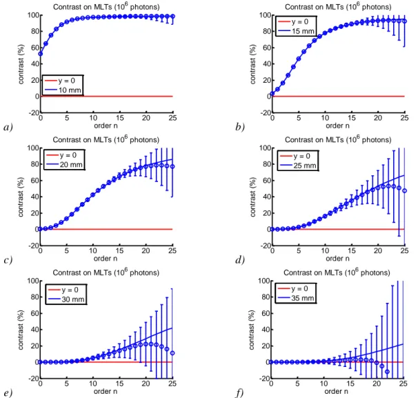
2.3. Optimal use of our algorithm for DOT in reflectance at short source-detector separation: a simulation study ... 57
2.3.1. Objectives ... 57
2.3.2. Method ... 57
2.3.3. Results ... 60
2.3.4. Conclusions ... 67
2.4. Summary ... 67
3. Time-resolved acquisition chain with free-running single-photon detectors 71 3.1. State-of-the-art of time-resolved instruments for DOT ... 71
3.2. Choice of a time-resolved acquisition chain ... 72
3.2.1. Existing time-resolved acquisition chains ... 72
3.2.2. Choice of an acquisition chain ... 76
3.3. Setup of a TCSPC acquisition chain ... 77
3.3.1. Global view of the setup ... 77
3.3.2. Laser ... 77
3.3.3. Optical attenuators ... 78
3.3.4. Synchronization signal ... 78
3.3.5. Optical fibers ... 79
3.3.6. TCSPC board ... 80
3.3.7. Single-photon detectors for TCSPC ... 80
3.3.8. IRF measurements ... 86
3.4.2. Method ... 89
3.4.3. Results ... 94
3.4.4. Conclusions ... 101
3.4.5. Discussion ... 101
3.5. Choice of a single-photon detector: experimental validation ... 102
3.5.1. Objective of the study ... 102
3.5.2. Measurement protocol ... 102 3.5.3. Data analysis ... 107 3.5.4. Results ... 108 3.5.5. Conclusions ... 117 3.5.6. Discussion ... 117 3.6. Summary ... 117
4. Enhanced dynamic range for time-resolved DOT with fast-gated SPADs . 121 4.1. Principle of TCSPC with a time-gated detector ... 121
4.2. Requirements on the time-gating operation ... 122
4.2.1. Rise time of the gate ... 122
4.2.2. Flatness of the gate ... 124
4.2.3. Limited impact of early photons ... 125
4.3. Considered solutions for time-gating the detector ... 125
4.4. Choice of single-photon avalanche diodes (SPADs) for fast-gating ... 127
4.4.1. The SPAD detector: general characteristics ... 127
4.4.2. Electronic module for fast-gating ... 127
4.4.3. IRF of the fast-gated SPAD ... 129
4.5. DOT with fast-gated SPADs: a proof of principle study ... 130
4.5.1. Objective of the study ... 130
4.5.2. Experimental setup ... 131 4.5.3. Measurement procedure ... 134 4.5.4. Signal pre-processing ... 137 4.5.5. Reconstruction method... 140 4.5.6. Results ... 141 4.5.7. Conclusions ... 146 4.5.8. Discussion ... 146
4.6. Limits and perspectives of DOT with fast-gated SPADs ... 148
4.6.1. Limits ... 148
4.6.2. Perspectives ... 149
4.7. Summary ... 149
5. Spatial resolution in depth of time-resolved DOT with optical probes ... 153
5.1. Simulation study on the spatial resolution of optical probes ... 153
5.1.1. Objectives ... 153
5.1.2. Method ... 153
5.1.3. Case studies ... 155
5.1.4. General conclusions ... 173
5.2. Experimental study ... 174
5.2.1. Setup and measurement protocol ... 174
5.2.2. Data processing and DOT reconstruction ... 176
5.2.3. Results ... 176
5.3. Summary ... 193
6. Conclusion and perspectives ... 197
6.1. Conclusions ... 197
6.2. Limitations ... 198
7. Appendix ... 202
7.1. Data sheet SPC-130/134 ... 202
7.2. Data sheet PMC-100-20 ... 203
7.3. Data sheet HPM-100-50 ... 205
7.4. Application note on DOT with a hybrid PMT ... 207
7.5. Liquid phantoms ... 210
7.6. Solid phantoms ... 211
7.7. Estimation of the number of collected photons at the detector in an infinite homogeneous medium ... 212
Publications ... 216
Acronyms
2D: two-dimensional 3D: three-dimensional
ADC: analog to digital converter APD: avalanche photodiode CCD: charge-coupled device
CEA: commissariat à l’énergie atomique CFD: constant fraction discriminator DE: diffusion equation
DA : diffusion approximation DOT: diffuse optical tomography DTOF: distribution of times-of-flight FWHM: full width at half maximum IR: infrared
IRF: instrument response function
LETI: laboratoire d’électronique et de technologie de l’information LISA: Laboratoire Images et Systèmes d’Acquisition
MCP: micro-channel plate MLT: Mellin-Laplace transform NIR: near-infrared
PMT: photomultiplier tube SNR: signal to noise ratio
SPAD: single-photon avalanche diode SYNC: synchronization
TAC: time-to-amplitude converter
TCSPC: time-correlated single-photon counting TPSF: time point-spread function
TR-DOT: time-resolved diffuse optical tomography VOA: variable optical attenuator
Introduction
Since the first X-ray image of a hand in 1895 by Wilhelm Röntgen, medical imaging techniques have been intensively developed and have enabled a considerable progress in medicine in various fields of applications like diagnostic, assisted surgery and medical research. X-ray radiography, X-ray computed tomography, magnetic resonance imaging, ultrasound imaging and nuclear medicine are currently the most spread modalities at the clinics. Nevertheless, given the variety of human biological tissues and relevant information for understanding all pathologies, a unique imaging technique answering all questions remains a utopia. Each modality can provide specific information and the images from different techniques are often exploited for medical diagnostics. Nowadays, new medical imaging techniques are still sought to address unmet clinical needs.
Optical imaging of biological tissues has been emerging since the 1980’s. The field of biomedical optics is now very broad and embodies various imaging techniques enabling to visualize biological tissues at different scales, from the cell to the organ, and to collect diverse information dealing with tissue composition or structure. The common approach to most of these techniques is to shine light onto a biological sample, tissue or organ, and to analyze the output light in order to extract information on this sample. Why using light? Mainly because the major constituents of biological tissues like water, lipids and hemoglobin in its oxygenated and deoxygenated forms possess different absorption spectra in the band of the electromagnetic spectrum ranging from the visible to the near-infrared. Additionally, in this band called the “therapeutic window”, the absorption coefficients of all these endogenous chromophores are lower than in the neighboring spectral band, which allows looking deeper in the tissues. With this respect, optical imaging can bring new information, not available with other techniques up to now. This modality also encompasses other advantages like being non-ionizing contrary to X-ray and nuclear techniques and allowing compact imaging devices compatible with bed-side monitoring. These points become attractive for clinical applications requiring frequent imaging follow-ups or measurements on weakened patients. Nevertheless, challenges arise when imaging tissues with light, the main ones being the penetration depth and the loss of spatial resolution due to the diffusion of light by the cells and other biological structures.
The emblematic applications of optical imaging at the scale of the organ, called diffuse optical imaging, are breast and brain imaging (Durduran et al 2010). Whereas X-ray mammography remains the gold standard for cancer detection, research has been carried out in the past ten years in order to determine which complementary information optics could bring. Clinical studies have revealed different optical properties between normal tissue and malignant breast tumors (Cerussi et al 2006) and suggested the possibility to use optical spectroscopic measurements to assess breast cancer risk (Taroni et al 2010). Currently, different research teams investigate the possibility to follow, with diffuse optical imaging, the response to a breast cancer therapy called “neoadjuvant therapy” (Zhou et al 2007), (Cerussi et al 2010), (Busch et al 2012). Optical imaging is also relevant for brain activation measurements because it enables to quantify the amount of hemoglobin in its oxygenated and deoxygenated forms, signatures of oxygen consumption. Many clinical studies investigate this possibility (Montcel et al 2006), (Wabnitz et al 2010), (Kirilina et al 2012). In this context, optics competes with magnetic resonance imaging and promises more compact and portable devices. Other reported applications for diffuse optical imaging
(Jiang et al 2011). Even if the optical modality is not yet used in routine at the clinics, all these recent clinical results encourage further improvements of this technique.
Among different techniques for diffuse optical imaging, diffuse optical tomography (DOT) provides three-dimensional maps of the distribution of optical coefficients of absorption and diffusion inside an organ. These maps are obtained thanks to non-invasive measurements by placing sources of light and detectors around the organ and analyzing the collected light. These maps are calculated by tomographic reconstruction algorithms: based on a direct model of light propagation in diffuse media, they propose maps of optical properties inside the organ predicting at best the measurements. Most DOT instruments consist in spherical or cylindrical configurations of sources and detectors all around the organ to image, for the brain and the breast. Nevertheless, for some organs, the anatomy does not allow this geometry, like for the prostate. In other cases, for practical reasons, a compact instrument, easy to install on the patient can be preferred to one featuring a large number of optical fibers to attach all over the imaged organ. An alternative is an “optical probe”: it consists in a combination of optical fibers for illumination and detection distributed on a few cm² and enabling reflectance measurements at short source-detector separation. Such a probe can be positioned at the surface of an organ and image locally its optical properties.
The scope of this PhD work is to propose an approach for DOT in the configuration of reflectance measurements at short source-detector separation, and to study the performances of such an optical probe.
Conceiving an optical probe for DOT providing useful clinical information is not straightforward. Indeed, such devices allowing only reflectance measurements at short source-detector separation are generally limited by their depth sensitivity. Spatial resolution and accurate quantification of absorption and diffusion coefficients are other important challenges. Currently, the most spread approach for DOT, called continuous wave, consists in using continuous light sources and detectors. However, in this case the measurements are very sensitive to shallow layers of the organ. A few probes for continuous wave DOT have been developed, either for prostate imaging or for breast imaging. In the first case, the results have shown limited depth sensitivity (Xu et al 2008), in the second case, long source-detector distances have been included in the probe design to increase depth sensitivity (Gonzalez et al 2012).
To tackle the challenge of depth sensitivity, the laboratory LISA of CEA-LETI has preferred a time-resolved approach. The principle is very intuitive: when a short pulse of light (a few picoseconds) is sent in a diffusive medium, most photons which stayed in the shallow layers are detected fast at the surface because they have undergone few diffusion events. On the contrary, the photons which have reached deep layers and come back to the surface again have stayed longer in the medium and are detected at the surface later (a few nanoseconds). Therefore, these so-called “late photons” carry the depth information. The bottleneck is that they are very few of them, as most photons reaching deeper layers are absorbed. Combining a pulsed light source and a time-resolved detection chain enables to select the photons depending on their time-of-flight and to be specifically sensitive to shallow and deep layers in the biological tissue.
Time-resolved DOT was pioneered at the University College of London in the 1990’s, by Dr. Arridge and Dr. Hebden for algorithmic and instrumental aspects. They have issued the first software package for time-resolved DOT reconstructions (TOAST) and realized the
first corresponding instrument (MONSTIR). Since then, different research teams have published studies on the improved depth sensitivity with time-resolved DOT (Selb et al 2007), (Liebert et al 2012), (Ducros et al 2011). In the field of spectroscopy, the null source-detector approach was developed at Politecnico di Milano, and showed improved contrast in depth with time-resolved measurements at short interfiber-distances (Torricelli et al 2005), (Bianco et al 2002). All this work has motivated our choice of a time-resolved approach to tackle the challenge of DOT in reflectance at short source-detector separation. However, it will be further justified with our own method in this manuscript.
This PhD work aims at studying the potential of optical probes for DOT, in the light of new developments of the algorithms and instruments. On the first point, the optimal choice of the pieces of information extracted from the time-resolved measurements has not yet been specifically optimized for DOT in reflectance at short interfiber distances. About the instrument, new time-resolved detectors are now available, like hybrid photomultipliers and single-photon avalanche diodes, and their contribution was not quantified yet for DOT in reflectance. Our goal is to develop a global time-resolved approach encompassing the optimal choices both at the algorithm level and for the instrument in order to determine the best performances currently achievable by DOT with optical probes.
This work is restricted to DOT images in absorption acquired at a single wavelength. The developed method should be applied to multispectral measurements in the future.
This manuscript starts with a description of the physics of diffuse optics and an introduction to diffuse optical tomography in order to position this PhD work and uncover its challenges (Chapter 1).
A simulation framework is then put in place to quantify the limits in detection of absorption contrast in depth under different noise conditions (Chapter 2). Different “datatypes”, pieces of information extracted from the time-resolved measurements, are compared and we study the importance of their choice in order to optimize the robustness of detection in depth. In this chapter, we also introduce the time-resolved DOT (TR-DOT) reconstruction algorithm developed at our laboratory and propose a method to optimize its use for time-resolved measurements acquired in reflectance at short source-detector separation. The simulation work of Chapter 2 confirms the need to increase the dynamic range of time-resolved measurements so as to better measure the late-photons and increase the imaged depth range of an optical probe. This motivates the search of corresponding experimental implementations.
A first experimental setup is proposed in Chapter 3, involving free-running single-photon detectors and a time-correlated single-single-photon counting (TCSPC) electronics. We discuss the choices made to optimize this setup and more specifically investigate the impact of the temporal response of the detector on the performance of TR-DOT. This study is based on two existing single-photon detectors offering significantly different temporal responses: a classical and a hybrid photomultipliers. This two-step study, with a simulation part and an experimental demonstration, shines a light on the importance of a fast temporal response of the detector in order to optimize the performance of TR-DOT in reflectance at short source-detector separation.
A second experimental setup is then proposed in order to acquire faster time-resolved measurements with a large dynamic range (Chapter 4). This approach is based on the time-gating of the single-photon detectors associated to TCSPC electronics. A technical
developed at Politecnico di Milano is tested for DOT. We detail the methods and protocols put in place for this proof-of-concept study and conclude on the interest of the fast-gated approach for TR-DOT probes.
After optimizing the processing of time-resolved measurements for DOT (Chapter 2) and their experimental acquisition (Chapter 3 and 4), we finish this work by better studying the imaging potential of a TR-DOT optical probe (Chapter 5). We focus on the performances in terms of spatial resolution in depth and study the different parameters influencing it. This study is delimited by simulations carried out on specific case studies and is then illustrated by experimental results obtained with the optimized setup of Chapter 4. To conclude, we illustrate this work by showing the spatial resolution of different real TR-DOT probe designs.
Chapter 1: Diffuse optical imaging and
tomography
In this first chapter, we introduce the basic notions of diffuse optical imaging and tomography required to understand the research work presented in the following chapters of this manuscript.
We first present the basic physical interactions between near-infrared light and biological tissues and mention the different existing models describing light propagation in scattering media.
In a second section, we present the medical imaging technique studied in our research work: diffuse optical tomography. After mentioning recent clinical results in this field, we discuss the need of optical probes to perform diffuse optical tomography for certain clinical applications. We conclude this chapter by introducing the challenges associated to an optical probe for diffuse optical tomography and our specific approach to tackle them.
1. Diffuse optical imaging and tomography
For the first time in 1895, looking inside the human body without cutting it open becomes possible. Experimenting with X-rays, Wilhelm Röntgen discovers that they are strongly absorbed by bones and much less by flesh. The first image formed with X-rays, transmitted through the hand of Röntgen’s wife, assesses this very clearly (Figure 1-1).
Since more than one century now, the field of medical imaging has considerably grown and plays a crucial role for diagnostic and therapy. The X-ray radiography was considerably improved to provide structural images with an excellent spatial resolution. 3D images of the human body can now be obtained by X-ray computed tomography.
Other techniques have arisen, involving other physical interactions between energy and matter. Ultrasound imaging works on the same principle as radars: sound waves are reflected at the interface between different tissues which allows seeing structural contrast. It has the advantage of requiring a relatively simple instrumentation, enabling bed-side monitoring.
Magnetic resonance imaging is based on the interaction between the magnetic field and matter, and more precisely with protons present in water molecules. This technique is very powerful to image structurally soft tissues and can be used to detect tumors inside an organ. It also encompasses another modality, the BOLD signal (blood-oxygen-level dependent), which can follow variations of the paramagnetic deoxygenated haemoglobin and give indirect information on the oxygenation of tissues. This functional technique is widely used for brain studies.
Another functional imaging technique is nuclear medicine. The patients receive an injection of tracers specifically bonding to some tissues and localized thanks to associated radioactive agents emitting gamma rays.
Figure 1-1 First medical image: X-ray of a hand by Wilhelm Röntgen in 1895. We can distinguish in dark the dense matter absorbing X-rays: bones and a metal ring. The flesh around is hardly visible.
Optical imaging is an emerging technique exploiting another part of the electromagnetic spectrum: the near-infrared range (NIR) (Figure 1-2). These electromagnetic waves are less
differently with biological tissues: they are absorbed by tissues and above all strongly scattered. This is why the scientific field studying optics in tissues is also called « diffuse optics ». In the following section, we describe the main interactions of NIR light with tissues to uncover the additional information about tissues that NIR images can provide.
Figure 1-2 The near-infrared range (NIR) in the electromagnetic spectrum
1.1. Optics in biological tissues
1.1.1. Optical properties of biological tissues
In the NIR range, biological tissues behave as diffuse media. The main interactions between matter and light are absorption and elastic scattering. Other phenomena can occur like emission of light called fluorescence, inelastic scattering like Raman scattering, and non-linear interactions. However, these phenomena are less preponderant and neglected in the context of diffuse optical tomography so they will not be further discussed here. We will now introduce the three main quantities used to describe optics in diffusive media: refractive index, absorption coefficient and scattering coefficient.
1.1.1.1 Refractive index
The refractive index of a medium is defined as the ratio between the speed of light in vacuum and its speed in the medium. In water, =1.33, whereas it can reach 1.5 in pure fat. Biological tissues contain structures of different natures, with different values of refractive index. Therefore an average value is used to describe it. Depending on their composition, the refractive index of biological tissues can range from = 1.35 to 1.45 (Bolin 1989). We will use the value of = 1.4 in our work.
1.1.1.2 Absorption
The absorption of light by matter is described by quantum physics. The energy of a photon is absorbed by an atom which can excite one of its energy level thanks to the energy of the photon. This absorption event can be followed by an emission of light (fluorescence) for certain molecules or the energy can simply be dissipated by thermal effect.
Figure 1-3 Absorption of light for a collimated beam in a non-scattering medium
Infrared Micro-wave
Near-infrared (NIR) Visible Ultraviolet X-ray 100 nm 400 nm 700 nm 1000 nm 50 µm (wavelength) 10 pm γ-ray
µ
aI
0I
x
At the macroscopic level, the absorption in a non-scattering medium is described by the Beer-Lambert law. If we consider a tissue of thickness x illuminated by a collimated beam of intensity I0 at wavelength λ (Figure 1-3), the output intensity I is express as follows:
(1-1)
is the absorption coefficient which depends on the wavelength λ and is expressed in cm-1. The inverse of the absorption coefficient is the average path of the photon in the medium before being absorbed.
The constituents of the tissues absorbing light are called chromophores. Each chromophore is characterized by its molar extinction coefficient in mol-1
.L.cm-1 which links its absorption coefficient to its concentration C in mol.L-1 in the medium:
(1-2)
In a medium containing a mix of n chromophores homogenously distributed, the absorption coefficient can be calculated as follows:
(1-3)
The main chromophores of human tissues are:
- Water: it is the major constituent of biological tissues, which can reach up to 80% in certain organs like the brain. Water absorbs strongly wavelengths in the infrared, above 900 nm.
- Hemoglobin: this protein located inside red blood cells ensures the transport of oxygen in the body. It is present under two main forms: the oxyhemoglobin (HbO2)
saturated with oxygen molecules and the deoxyhemoglobin (Hb) desaturated with oxygen molecules. These two forms having different absorption spectra in the NIR range, it is possible to deduce their relative concentrations from a measurement at multiple wavelengths. This is the working principle of commercially available pulse oximeters. A measure of oxygenation of blood is oxygen saturation SO2 (Hillman
2002):
(1-4)
- Lipids: similarly to water, these molecules strongly absorb light above 900 nm. - Melanin: this biological pigment is strongly absorbing molecule in the NIR but only
present in the epidermis (few µm thick). Whereas it is crucial to take it into account when probing small volumes like for skin spectroscopy, it becomes less preponderant for probing larger ones, like in tomography.
Figure 1-4 displays the NIR absorption spectra of the main chromophores of tissues. The wavelength range 700-900 nm, between the absorption peak of hemoglobin and the absorption peaks of water and lipid, is commonly called the “therapeutic window”.
For illustrative purpose, we show the image of a human hand seen in reflectance at λ=850 nm (Figure 1-5). The veins are very dark, due to the strong absorption of hemoglobin. This image illustrates that information provided by NIR in biological tissues is different from X-ray (compare with Figure 1-1).
Figure 1-4 Absorption spectra of the main constituents of biological tissues in the NIR. The range between 700 and 900 nm is commonly called the “therapeutic window”. Data extracted from
http://omlc.ogi.edu/spectra.
Figure 1-5 A human hand seen in reflectance with a NIR wavelength of λ=850 nm (false color). Image extracted from (Becker 2012).
1.1.1.3 Scattering
Scattering of light in biological tissues originates from the mismatch of refractive index between the different biological entities like cells, their nuclei, their membranes etc. There are different types of scattering regimes, depending on the relative size of the scattering object and the wavelength of the electromagnetic wave interacting with this object. The Rayleigh theory describes the isotropic scattering of light by particles significantly smaller than the wavelength. The Mie scattering encompasses the previous regime and the regime in which the sizes of particles are comparable to the wavelength. The latter regime generally produces anisotropic forward scattering. Finally, for objects larger than the wavelength, the laws of diffraction can be used.
Figure 1-6 Scattering of light for a collimated beam in a non-absorbing medium
6000 700 800 900 0.05 0.1 0.15 0.2 0.25 wavelength (nm) a b so rp ti o n co e ff ici e n t µ a (cm -1 ) HbO 2 (50 µM) Hb (50 µM) fat(mammalian) water (pure)
µ
sI
0I
x
A scattering coefficient is determined similarly to the absorption coefficient. If we consider a homogenous non-absorbing medium with a thickness x (Figure 1-6), the scattering coefficient links the measured output intensity I for a given incident collimated beam of wavelength λ and intensity I0:
(1-5)
is then the mean path of the photon before undergoing a scattering event.
The scattering coefficient perfectly describes isotropic scattering. However, most biological tissues have a preferential scattering direction and tend to scatter light forward. To describe this, the phase function is introduced: it gives the probability for a photon of an incident direction to be scattered in the direction . This problem is generally simplified by assuming that the phase function only depends on the angle between and (Figure 1-7).
Figure 1-7 Scattering of light by a particle (in grey) with an angle
The anisotropy of scattering is then defined as the mean value of weighted by the phase function :
(1-6)
In an isotropic medium, , whereas for pure forward scattering. Typically, the value of g ranges from 0.7 to 0.99 in biological tissues (Tuchin 2007). We will use a value of in our work.
The directional effects of scattering can be included in the reduced scattering coefficient which can be interpreted as an equivalent isotropic scattering coefficient:
(1-7)
Propagation of light in absorbing and scattering media using these physical properties will be detailed in a following section (1.1.2). In particular, the model used in the rest of our work, the diffusion equation, involves the coefficients and .
1.1.1.4 Values of optical properties in biological tissues
Depending on the concentration of chromophores present in a biological tissue and its own
θ
s
the human body. This can be seen on Table 1-1, gathering some values of and reported in literature for the most studied organs of the human body with diffuse optical imaging.
Table 1-1 Optical properties of bulk biological tissues commonly imaged with diffuse optics.
Tissue µa (cm-1) µ’s (cm-1) λ (nm) Reference
Breast 0.041 +/- 0.025 8.5 +/- 2.1 786 (Durduran et al 2002)
Brain (cortex) > 0.2 10 674 (Bevilacqua et al 1999)
Prostate 0.4 +/- 0.1 7.1 +/- 1.6 786 (Svensson et al 2007)
1.1.2. Physical models for light propagation in diffusive media
In this section, we detail the models describing light transport in diffusive media, and therefore in biological tissues. We first present the most rigorous model of the radiative transport and secondly introduce the diffusion approximation, a simplified model most often used for diffuse optical imaging as it enables faster computation.
1.1.2.1 Radiative transfert equation (RTE) Definition
The radiative transfert equation (RTE), also called the Boltzmann equation, mathematically describes phenomena of diffusion. It has first been used in astrophysics (Chandrasekhar 1960) and for the diffusion of neutrons (Case and Zweifel 1967) before being applied to diffuse optics (Ishimaru 1997).
The RTE is a differential equation describing the radiance and taking into account the conservation of energy inside an elementary volume of a diffusive medium (Splinter 2007).
Before stating the RTE, let us first explain the radiance . It is defined by the light power incident on a cross-sectional area flowing within a solid angle at a given time t. It is expressed in W.sr-1.m-2. In these notations, is the position at which radiance is evaluated, the direction vector and t the time (Figure 1-8).
Figure 1-8 Formalism in which we describe the radiative transfert equation (RTE)
s’
s
r
dω
dω’
Let us now state the RTE, before detailing the meaning of each terms involved in this equation:
(1-8)
With :
- speed of light in the medium defined as , being the speed of light in vacuum and the refractive index of the medium
- and are the absorption and scattering coefficients at the point
- is the phase function, giving the probability for a photon of an incident direction to be scattered in the direction .
Without going into further details about RTE, we will just mention here the meaning of the five main terms of this energy balance:
- Term 1: temporal variations of radiance,
- Term 2: radiance lost through the boundaries of the elementary volume, - Term 3: radiance lost due to absorption and diffusion into another direction,
- Term 4: recovery of radiance into the original direction as a result of scattering from direction into ,
- Term 5: angular and spatial distribution of the light source at time t, expressed in W.m-3.sr-1.
Resolution
Analytical solutions of the RTE exist, but only from simple medium geometries (Patterson et al 1991). Monte-Carlo simulations can numerically compute solutions of the RTE in complex media, by using a statistical approach (Wilson and Adam 1983). Monte-Carlo simulations are a gold standard extensively used in the field of biomedical optics (Wang et al 1995), (Sassaroli and Martelli 2012). Different versions of Monte-Carlo codes can be downloaded from the website of the Oregon Medical Laser Center at
http://omlc.ogi.edu/software/mc/. The bottleneck of this approach is still its high
computation time, required to have a good statistics. However, strategies are currently proposed to speed it up by using parallelization on graphic processing units (GPU) (Alerstam et al 2010).
Still, RTE and Monte-Carlo simulations are hardly used for diffuse optical tomography. A different model is preferred for being much faster: it is the diffusion equation, detailed in
1𝜕 , , 𝜕 = ∙ ∇ , , 𝑎 + , , + ( , ) 4𝜋 , , 𝜔 + , , 1 2 3 4 5
1.1.2.2 Diffusion equation (DE) Definition
The diffusion equation (DE) is obtained after making successive approximations to the RTE. The calculation steps to deduct the DE from the RTE can be found in several references like (Arridge 1999). We will not detail them here but only give the formulation of the DE and stress the various approximations made to deduct the DE from the RTE.
Like RTE, the diffusion equation is also a differential equation. However, it does not describe the radiance but the photon density in W.m-2
defined as the integral of the radiance over the solid angle 4 𝜋:
𝜔 (1-9)
The different crucial approximations which are made to deduct the DE from the RTE are the following:
- The phase function only depends on the diffusion angle θ.
- The temporal variations of the flux or current density (defined below) are negligible. This hypothesis is true only if .
𝜔 (1-10)
- The light source is isotropic (which means that the non-isotropic terms are not taken into account) and expressed in W.m-3.
Under all these approximations, the diffusion equation describes the photon density as a function of space and time t as follows:
𝜕
𝜕 ∙ 𝑎 (1-11)
With:
- the photon density, is the Green’s function of this differential equation. - is the spatial and temporal distribution of the light source.
- is the spatial distribution of the absorption coefficient in the medium.
- is the spatial distribution of the diffusion coefficient in the medium, which depends both on and on :
(1-12)
When looking at the diffusion equation, one can make various observations. First, by removing the angular dependence , only four variables are now necessary to compute it (coordinates x, y, z and t). Second, neither the phase function nor the anisotropy factor g is present. The DE assimilates the medium to an isotropic medium of scattering coefficient .
Limitations
The DE is a useful model for computational reasons. Nevertheless, it is important to keep in mind its limitations, derived from the various approximations made to deduct it from the RTE. First, the hypothesis is true in many biological tissues but not in all of them. It was demonstrated that when is 100 larger than and the medium is large, RTE and DE give similar results (Hielscher et al 1998). Second, the propagation distances have to be large with respect to to consider isotropic scattering. This is not necessarily true for small volumes like fingers. For the same reason, continuous wave measurements at short source-detector distances are not well described by the DE, because most detected photons have only undergone few diffusion events. For time-resolved measurements in this configuration, the early times are poorly estimated by the DE for the same reason but the error decreases for late times.
Resolution
Analytical solutions of the DE exist for specific cases, in simple media and can be used to some extent in biological tissues. We can cite the solution for a spatially punctual and temporally brief light source in a homogenous infinite medium (Jacques and Pogue 2008):
𝜋
(1-13)
By adding boundary conditions to this solution, other solutions can be expressed in semi-infinite infinite homogenous medium (Laidevant et al 2006) or slabs (Patterson et al 1989), (Contini et al 1997) or multi-layer media (Liemert and Kienle 2010).
Otherwise if the shape is complex and the medium heterogeneous, with layers and inclusions, the DE can solved with numerical methods like finite-elements methods (Jacques and Pogue 2008), finite difference or finite volumes. We use the latter method in our work.
Boundary conditions
When considering a non-infinite medium, boundary conditions have to be added to the model. Different types have been proposed in literature, the three main ones being partial current boundary condition (PCBC), zero boundary condition (ZBC), and extrapolated boundary conditions (EBC), all described in (Haskell et al 1994). (Hielscher et al 1995) have compared these three conditions and concluded that PCBC and EBC fit best the solutions obtained with Monte-Carlo in reflectance. We describe here the principle of the EBC, used in the rest of this work.
For the EBC, the photon density is extrapolated to be null at a distance from the surface of the medium. This distance is expressed as follows (Moulton 1990):
(1-14)
With
(1-15)
1.2. NIR imaging techniques in biological tissues
1.2.1. Review of existing NIR imaging techniques
The field of biomedical optics is very broad, and encompasses many aspects including imaging but also microscopy, spectroscopy, etc. We briefly introduce here the main NIR imaging techniques.
Surprisingly, the first reported medical study using NIR images was carried out in the 1930’s. Max Cutler observed breast tissues with light in transillumation and noticed different contrasts depending on the content of fat, fibrous tissue, epithelial elements, and blood (Cutler 1931). He also pointed the possibility to observe breast tumors (Figure 1-9). Nevertheless his reports already mention some of the main difficulties inherent to this technique: obtaining a homogeneous illumination, interpreting images, etc.
Figure 1-9 “Opacity on transillumination of a solid tumor of the breast” (Cutler 1931). The veins and a vascularized tumor are darker than surrounding tissues.
Since then, technological developments in light sources (lasers, diodes), detectors (charged coupled devices, avalanche photodiodes) and in computers have strongly pushed forward NIR imaging techniques, allowing better measurements and physical modeling. Since the 1980’s, various imaging techniques have emerged, we mention here those imaging at the scale of an organ (> cm).
Diffuse optical tomography (DOT) provides three-dimensional maps of the distribution of optical coefficients of absorption and diffusion inside an organ. This contrast is called endogenous as produced by chromophores and structures naturally present in tissues. These 3D maps are obtained thanks to non-invasive measurements by placing sources of light and detectors around the organ and analyzing the collected light. They are calculated by tomographic reconstruction algorithms involving a model of light propagation in tissues. The contrast can be enhanced by adding exogenous fluorescent agents emitting light at a different wavelength from the excitation. This modality is called fDOT or fluorescence molecular tomography (FMT).
Diffuse correlation spectroscopy (DCS) enables a point measurement of blood flow (and of the contributions of oxy and deoxyhemoglobin). It is used to follow oxygenation of the brain (Durduran et al 2004) and the muscle. Tomographic images of blood flow can be obtained with diffuse correlation tomography (DCT) (Durduran 2004).
To finish, photoacoustic imaging of biological tissues has appeared in the 2000’s and is now quickly developing (Xu and Wang 2006). It combines optical excitation of tissues and detection of ultrasonic waves emitted after the absorption of photons by chromophores. The advantage is a better spatial resolution allowed by the ultrasound detection, not degraded by diffusion like for light detection.
In this work, we focus on endogenous DOT, providing intrinsic information on the tissue and potentially the chromophore composition thanks to multispectral measurements. In the next paragraph, we introduce the working principle of DOT, highlight some clinical results and introduce our approach in this context.
1.2.2. Diffuse optical tomography (DOT)
1.2.2.1 How to make DOT images?Tomography refers to forming images of sections inside an object. In the context of medical imaging this is done with non-invasive measurements obtained with waves penetrating inside the tissue, generally by irradiating the tissue from one side and measuring on the other side.
For DOT, we proceed as follows. Light sources and detectors are positioned all around the object to image. The measurements of collected light for each excited source provide different “views” on the object. Each source-detector pair probes a certain volume inside the organ and the associated measurement is affected by the optical properties within this volume. Overlapping measurements provide complementary information from overlapping regions in the observed tissue. This set of measurements from all source-detector pairs carries the information on the distribution of the optical properties in terms of absorption and diffusion within the observed tissue.
Concretely, the mathematical link between these measurements and the 3D images of and in the object is done by a tomographic reconstruction algorithm. The latter numerically calculates the maps predicting the measurements at best.
The prediction of measurements for a given distribution of optical properties requires the knowledge of the so-called direct model, describing light propagation in tissues (section 1.1.2). Proposing maps of and explaining at best the measurements requires solving an inverse problem.
The basis of DOT algorithms was developed in the 1990’s and many approaches have been proposed. We mention here the most common method described as the “perturbation approach” (Arridge and Hebden 1997), which will be further detailed in Chapter 2. It mathematically links with a linear system Y=WX:
- Y: the differences between the measurements and the predicted measurements for a given map of optical properties,
- X: the variations in optical properties (absorption or diffusion), - W: the sensitivity matrix (jacobian).
We have to stress that the inverse problem is difficult to solve for DOT. It is an ill-posed problem, which means that the measurements only are not enough to determine all the parameters. Different algorithms can be used to solve the inverse problem: gradient
descent, conjugate gradient method, singular value decomposition, algebraic reconstruction technique, etc.
1.2.2.2 Possible techniques and instruments
In the previous section, we have described DOT in a generic way. There are actually three possible implementations of this technique: continuous wave (CW), time-resolved (TR) and intensity modulated (Figure 1-10).
a) b) c)
Figure 1-10 Three approaches for DOT: a) continuous wave (CW), b) time-resolved (TR), c) intensity modulated. Adapted from (Delpy and Cope 1997)
Continuous wave (CW) uses a continuous light source and detector and registers differences in intensity. Time-resolved uses a pulse of light (typically faster than 100 ps) and records the broadening of this pulse when passing through a diffusive medium with time-resolved detectors (gated cameras, photomultipliers and time-correlated single-photon counting electronics). The third approach consists in modulating light and recording the amplitude M of the modulated intensity and its phase φ, at the output of the medium.
Formally, the CW approach is described as the integral over time of the time resolved approach. The link between this latter approach and the intensity modulated one is given by the Fourier transform.
The information content is potentially richer with time-resolved and frequency-domain approaches, compared with CW. In theory, the measurements at all frequencies offer the same information content of the full of-flight distribution of photons. However, time-resolved instruments are very sensitive to low photon counts. We will detail this aspect later.
The description of the direct and inverse problems can be found for these three approaches in literature. DOT software packages are now available online for the 3 methods. For frequency domain and CW, the software Nirfast was developed at Dartmouth College. The University College of London has created the software TOAST for time-resolved DOT. In Chapter 2, we will detail the formalism of the direct (2.1.1) and inverse problem (2.1.2) for the time-resolved approach.
1.2.2.3 Clinical applications
Since the 1990’s, various clinical studies with DOT have been reported. A recent review paper on diffuse optical imaging and tomography summarizes them (Durduran et al 2010). We highlight here some results obtained for the two most common applications for DOT, breast and brain imaging, and two other applications showing promising clinical results.
Breast
Numerous clinical studies of the breast with DOT have been reported (Choe 2005), (Durduran et al 2010). DOT is relatively easy in the breast thanks to its simple internal
in ten sity continuous intensity I0 I in te n sity time-resolved intensity I0(t) I(t) in ten sity modulated intensity I0 Φ0 M0 I Φ M
structure (compared to the brain) and its low absorption. Nevertheless, in the context of breast cancer detection, X-ray mammography remains the gold standard. The trend is now to investigate further which information can be brought by NIR images and not by methods like X-ray and MRI. Multispectral DOT, allowing chromophore decomposition is gaining interest. Practically, different studies investigate the possibility to follow breast cancer treatment with CW or frequency-domain DOT monitoring (Zhou et al 2007), (Cerussi et al 2010), (Busch et al 2012). In this context, DOT has the advantage of being non-ionizing and allowing bed-side measurement.
Brain
DOT was naturally applied to brain imaging for its potential to distinguish oxy and deoxy hemoglobin and provide information on oxygenation, often correlated to brain activity. However, these measurements are very challenging: the head is a highly heterogeneous multi-layered medium with non-diffusive layers. Moreover, there are 1 to 2 cm between the surface of the head and the cortex. So measuring functional activity requires good depth sensitivity. In this context, both continuous and time-resolved approaches have been developed for DOT in the brain.
The brain of premature infants has also been imaged with DOT in order to detect potential lesions. In this context, DOT has the advantage of offering a bed-side solution, compared to MRI. We can cite the clinical studies with TR-DOT carried out at the University College of London (Austin et al 2006). Figure 1-11 shows 2D images of a premature infant with a hemorrhage. Its hemorrhage is distinguished by a larger blood volume (yellow in Figure 1-11a) and a local low oxygen saturation (purple in Figure 1-11 b)). The helmet of optical probes designed to perform such measurements is shown in Figure 1-12.
Figure 1-11 Images of DOT from the head of a premature infant having an haemorrhage produces with the TR-DOT system of the University College of London. “A coronal section from infant 11 showing (a) regional blood volume, (b) regional oxygen saturation, and (c) corresponding cranial ultrasound scan. There is an increase in regional haemoglobin concentration and decrease in regional oxygen saturation in the area corresponding to the intraventricular haemorrhage and haemorrhagic parenchymal infarct. The lesion is outlined in the ultrasound scan.” Extracted from (Austin et al 2006).
Figure 1-12 Helmet of optical fibers developed for TR-DOT measurements on the head of
premature infants at University College of London. Extracted from (Hebden et al 2002).
Prostate
Prostate cancer detection still lacks an adapted imaging modality. Not all tumors can be detected with ultrasound and numerous blind biopsies are often performed. Some research team are investigating the possibility to detect tumors in the prostate with CW DOT probes (Xu et al 2008). However, the strong absorption of the prostate is challenging (Svensson et al 2007). Recently, a CW DOT study has shown changes in total volume of hemoglobin associated with development of a rapidly growing tumor in the canine prostate (Jiang et al 2011).
Peripheral artery disease
DOT has recently been applied to study the perfusion of the foot in patients with peripheral artery disease (Khalil et al 2012). In this case, DOT measurements were done dynamically: before and after the occlusion of the foot. The obtained DOT images enabled to visualize differences in hemoglobin concentration between the two states (Figure 1-13). This work concluded on different spatial patterns between healthy subjects and patients with peripheral artery disease.
Figure 1-13 Left: anatomy of the cross-section of a foot. Right: changes in haemoglobin volumes before and after occlusion of the foot. Extracted from (Khalil et al 2012).
1.2.2.4 A probe for DOT? Clinical motivations
Current clinical systems for DOT involve the use of multiple optical fibers for illumination and detection, positioned all around the organ, as it is often done for imaging the breast and the brain. Whereas this configuration has the advantage of imaging the entire organ, it has some drawbacks. First, for practical aspects, positioning all these optical fibers in contact
with the organ and knowing their precise position are tedious tasks. It is even more complicated for weakened patients having undergone surgery or being injured (e.g. brain measurements in intensive care). It is also the case for measurements on the head of premature infants. Secondly, incorrect estimation of the positions of sources and detectors causes errors on the reconstructed images.
Of course, this configuration of sources and detectors is unavoidable when the full image of the organ is required. However, when a small volume of interest is enough, a different approach based on a compact probe can be preferred. A probe of a few centimeters wide can be imagined, gathering a bundle of illumination and collection fibers. For breast measurements, some probes are actually being developed to account for these earlier mentioned problems (Gonzalez et al 2012)
Such an approach also makes sense for internal organs like the prostate where in any case a large probe is impossible.
Technical challenges
Such optical probe implies measurements in reflectance, which means that the source and detectors are placed on the same side of the object. It also restricts the measurements to short source-detector separations.
The main challenge of this source-detector configuration is the depth sensitivity. Other important challenges are the absolute quantification of optical properties and the spatial resolution of images. In particular, the proper quantification of the absorption coefficient is crucial for a multispectral approach aiming at chromophore decomposition.
Choice of a time-resolved approach
In reflectance, time-resolved measurements allow a depth selection in the medium by performing a time-selection of detected photons. This can be easily understood. When a short pulse of light (a few picoseconds) is sent in a diffusive medium, most photons which stayed in the shallow layers are detected fast at the surface because they have undergone few diffusion events. On the contrary, the photons which have reached deep layers and come back to the surface again have stayed longer in the medium and are detected at the surface later (a few nanoseconds). Therefore, these so-called “late photons” carry the depth information. The bottleneck is that they are very few of them, as most photons reaching deeper layers are absorbed. Combining a pulsed light source and a time-resolved detection chain enables to select the photons depending on their time-of-flight and to be specifically sensitive to shallow and deep layers in the biological tissue.
The CW approach consists in integrating all photons: it is therefore most sensitive to the surface as the majority of measured photons are early ones.
With this respect, we have selected the time-resolved approach to tackle the challenge of depth sensitivity. Moreover, recent work in the field of spectroscopy has demonstrated that depth sensitivity is dependent on time-of-flight of photons but not on source-detector separation (Bianco et al 2002). Additionally, other work has shown that theoretically, contrast to noise in the presence of an absorbing inclusion in depth is higher at short source-detector separation (Torricelli et al 2005). In DOT, studies have also demonstrated a better depth detection and localization with TR-DOT than with CW (Selb et al 2007). All this work has motivated our choice of time-resolved measurements.
The work presented in the following chapters consists in proposing a complete TR-DOT approach optimizing the detection of absorption contrast in depth in diffusive media. It encompasses methodological aspects linked to the processing of measurements by TR-DOT algorithms and instrumental aspects in order to acquire measurements with the largest dynamic range possible. Having developed this method, an applicative study finally investigates the spatial resolution allowed by different optical probes.
We want to mention different restrictions to our work. First, we focus on DOT reconstructions of the absorption coefficients. Second, we consider simple objects to image, including a homogenous background with punctual absorbing inclusions. Third, the optical properties of this background are fixed at = 0.1 cm-1 and = 10 cm-1, average values between coefficients measured for breast and brain in the NIR range (Table 1-1). Finally, for experimental studies, measurements are carried out on optical phantoms, mimicking optical properties of absorption and diffusion in biological tissues.
1.3. Summary
In this first chapter, we have introduced the main physical interactions between near-infrared light and biological tissues: absorption and scattering. Different models can mathematically describe light propagation in scattering media. Whereas the radiative transfer equation is the most accurate, it can be simplified by the diffusion approximation to obtain a less computationally intense model. The latter model, the diffusion equation, is broadly used for diffuse optical imaging but is accurate only under certain conditions.
We have presented the technique further studied in this research work: diffuse optical tomography. This imaging modality provides 3D maps of absorption and scattering coefficients inside an organ from non-invasive measurements. The multispectral approach can also retrieve 3D maps of chromophores. We presented recent clinical results showing the interest of the information brought by near-infrared light.
We finished this chapter by discussing the need for optical probes with limited sizes to acquire local images of diffuse optical tomography in some organs. The biggest challenge of this technique is its depth sensitivity. Other challenges are accurate quantification of optical properties and spatial resolution.
The next four chapters will present our time-resolved approach to optimize the performances of optical probes for diffuse optical tomography.
Chapter 2: Reconstruction algorithm for
time-resolved DOT in reflectance at short
source-detector separation
In the previous chapter, we explained briefly how tomographic images could be obtained in diffusive media: it involves solving the direct and inverse problem. We have introduced this concept generally, without specifying which kind of measurement was done. Then, we have introduced the idea that time-resolved measurements provided a way to probe a diffusive medium at different depths and to increase the contrast in depth compared to a continuous wave approach.
This chapter now investigates how to reconstruct diffuse optical tomography (DOT) images with time-resolved measurements and how to optimize this reconstruction in the configuration of DOT in reflectance at short source-detector separation.
In a first section, we focus on the specificity of time-resolved DOT algorithms. We present the problem of the choice of “datatypes”, time-filters extracting information of the time-resolved measurements. In particular, we first study which datatype (Mellin, Laplace or Mellin-Laplace transforms) is more relevant in order to robustly detect the deepest absorption contrast. For this purpose, we have put in place a methodological framework based on simulations of measurements and addition of statistical noise representative of experiments. This proposed method enables to draw conclusions independently of any reconstruction algorithm.
In a second section, we introduce the main features of a versatile algorithm developed at the laboratory LISA of CEA-LETI for processing time-resolved data with the Mellin-Laplace transform. This algorithm was used for reconstructing all DOT images presented in this manuscript.
In a third and last section, we study how to best process time-resolved measurements depending on the available dynamic range or signal to noise ratio. We conclude on the performance in terms of detection and localization in depth of a single absorbing inclusion, in a given medium depending on the dynamic range of the measurement. This method developed on simulated measurements will be used in the next three chapters to process real measurements.