HAL Id: hal-01478632
https://hal-amu.archives-ouvertes.fr/hal-01478632
Submitted on 31 May 2021HAL is a multi-disciplinary open access archive for the deposit and dissemination of sci-entific research documents, whether they are pub-lished or not. The documents may come from teaching and research institutions in France or abroad, or from public or private research centers.
L’archive ouverte pluridisciplinaire HAL, est destinée au dépôt et à la diffusion de documents scientifiques de niveau recherche, publiés ou non, émanant des établissements d’enseignement et de recherche français ou étrangers, des laboratoires publics ou privés.
Non-contiguous finished genome sequence and
description of Gorillibacterium massiliense gen. nov, sp.
nov., a new member of the family Paenibacillaceae
Mamadou Bhoye Keita, Roshan Padhmanabhan, Aurélia Caputo, Catherine
Robert, Eric Delaporte, Didier Raoult, Pierre-Edouard Fournier, Fadi Bittar
To cite this version:
Mamadou Bhoye Keita, Roshan Padhmanabhan, Aurélia Caputo, Catherine Robert, Eric Delaporte, et al.. Non-contiguous finished genome sequence and description of Gorillibacterium massiliense gen. nov, sp. nov., a new member of the family Paenibacillaceae. Standards in Genomic Sciences, BioMed-Central, 2014, 9 (3), pp.807 - 820. �10.4056/sigs.5199182�. �hal-01478632�
Standards in Genomic Sciences (2014) 9: 807-820 DOI:10.4056/sigs.5199182
Non-contiguous finished genome sequence and description
of
Gorillibacterium massiliense
gen. nov, sp. nov., a new
member of the family
Paenibacillaceae
Mamadou Bhoye Keita1, Roshan Padhmanabhan1, Aurélia Caputo1, Catherine Robert1,
Eric Delaporte2, Didier Raoult1,3, Pierre-Edouard Fournier1, and Fadi Bittar1* 1URMITE, Aix-Marseille Université, Faculté de médecine, Marseille, France 2IRD, University Montpellier 1, Montpellier, France
3King Fahad Medical Research Center, King Abdul Aziz University, Jeddah, Saudi Arabia
*Correspondence: Fadi Bittar (fadi.bittar@univ-amu.fr)
Keywords: Gorillibacterium massiliense, genome, culturomics, taxono-genomics
Strain G5T gen. nov., sp. nov. is the type strain of Gorillibacterium massiliense, a newly
proposed genus within the family Paenibacillaceae. This strain, whose genome is de-scribed here, was isolated in France from a stool sample of a wild Gorilla gorilla subsp.
gorilla from Cameroon. G. massiliense is a facultatively anaerobic, Gram negative rod. Here we describe the features of this bacterium, together with the complete genome se-quence and annotation. The 5,546,433 bp long genome (1 chromosome but no plasmid) contains 5,145 protein-coding and 76 RNA genes, including 69 tRNA genes.
Introduction
Strain G5T (= CSUR P290 = DSM 27179) is the
type strain of Gorillibacterium massiliense gen. nov., sp. nov. This bacterium which is proposed to belong to the family Paenibacillaceae, is a Gram-negative, flagellated, facultative anaerobic, indole-negative bacillus that was isolated from a fecal sample of a wild western lowland gorilla from Cameroon, through a culturomics study of the bacterial diversity of the feces of wild goril-las. This technique was used successfully to ex-plore the human gut microbiota allowing the iso-lation of many new species and genera [1-3]. The newly proposed strategy of applying high throughput genome sequencing, MALDI-TOF spectral analysis of cellular proteins, coupled with more traditional methods of phenotypic characterization has been demonstrated as a useful approach for the description of new bac-terial taxa [4-15]. A principle advantage is that this method circumvents the vagaries of meth-ods that rely mainly on DNA-DNA hybridization to delineate species. Here, we applied this polyphasic approach to describe G. massiliense gen. nov., sp. nov. strain G5T.
The family Paenibacilliaceae [16] belongs to the phylum Firmicutes and includes the 9 following genera [17]: Paenibacillus [18,19], Ammoniphilus [20], Aneurinibacillus [21], Brevibacillus [21], Thermobacillus [22], Fontibacillus [23], Cohnella [24], Saccharibacillus [25] and Oxalophagus [26].
Members belonging to this family were isolated mainly from soil, roots, blood, feces and other sources [16]. To the best of our knowledge, this is the first report of the isolation of a novel ge-nus from the fecal flora of a gorilla.
Here we present a summary classification and a set of features for G. massiliense gen. nov., sp. nov. strain G5T (= CSUR P290 = DSM 27179)
to-gether with the description of the complete ge-nomic sequencing and its annotation. These characteristics support the circumscription of a novel genus, Gorillibacterium gen. nov. within
the family Paenibacillaceae, with
Gorillibacterium massiliense gen. nov., sp. nov. as the type species.
Classification and features
In July 2011, a fecal sample was collected from a wild Gorilla gorilla subsp. gorilla near Minton, a village in the south-central part of the DJA FAU-NAL Park (Cameroon). The collection of the stool sample was approved by the Ministry of Scientific Research and Innovation of Cameroon. No experiments were conducted on this gorilla. The fecal specimen was preserved at -80°C after collection and sent to Marseille. Strain G5T
(Ta-ble 1) was isolated in August 2012 by aerobic cultivation at 37°C on sterilized soil medium (12 g of soil (Latitude: N 43° 17' 20.151''; Longitude: E 5° 24' 15.3822'') /agar (14g/l). This strain ex-hibited a 93.72% 16S rRNA nucleotide sequence
similarity with Paenibacillus turicensis, the phylogenetically closest validly published Paenibacillus species (Figure 1). This value was lower than the 95.0% 16S rRNA gene sequence
threshold recommended by Stackebrandt and Ebers to delineate a new genus without carrying out DNA-DNA hybridization [37].
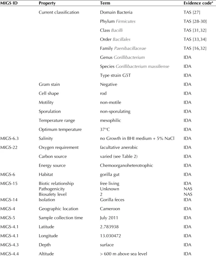
Table 1. Classification and general features of Gorillibacterium massiliense strain G5T
MIGS ID Property Term Evidence codea
Current classification Domain Bacteria TAS [27]
Phylum Firmicutes TAS [28-30]
Class Bacilli TAS [31,32]
Order Bacillales TAS [33,34]
Family Paenibacillaceae TAS [16,32]
Genus Gorillibacterium IDA
Species Gorillibacterium massiliense IDA
Type strain G5T IDA
Gram stain Negative IDA
Cell shape rod IDA
Motility non-motile IDA
Sporulation non-sporulating IDA
Temperature range mesophilic IDA
Optimum temperature 37°C IDA
MIGS-6.3 Salinity no Growth in BHI medium + 5% NaCl IDA
MIGS-22 Oxygen requirement facultative anerobic IDA
Carbon source varied (see Table 2) IDA
Energy source Chemoorganoheterotrophic IDA
MIGS-6 Habitat gorilla gut IDA
MIGS-15 Biotic relationship free living IDA
MIGS-14 Pathogenicity Biosafety level Isolation Unknown 2 Gorilla feces NAS NAS IDA
MIGS-4 Geographic location Cameroon IDA
MIGS-5 Sample collection time July 2011 IDA
MIGS-4.1 Latitude 2.783938 IDA
MIGS-4.1 Longitude 13.030472 IDA
MIGS-4.3 Depth surface IDA
MIGS-4.4 Altitude > 600 m above sea level IDA
aEvidence codes - IDA: Inferred from Direct Assay; TAS: Traceable Author Statement (i.e., a direct report exists in
the literature); NAS: Non-traceable Author Statement (i.e., not directly observed for the living, isolated sample, but based on a generally accepted property for the species, or anecdotal evidence). These evidence codes are from the Gene Ontology project [35]. If the evidence is IDA, then the property was directly observed for a live isolate by one of the authors or an expert mentioned in the acknowledgements.
Figure 1. Phylogenetic tree highlighting the position of Gorillibacterium massiliense strain G5T relative to other type
strains within the Paenibacillaceae family. GenBank accession numbers are indicated in parentheses. Sequences were aligned using CLUSTAL X (V2), and phylogenetic inferences obtained using the maximum-likelihood method within the MEGA 5 software [36]. Numbers at the nodes are percentages of bootstrap values obtained by repeating the analysis 1,000 times to generate a majority consensus tree. Brevibacillus brevis was used as out-group. The scale bar represents a 2% nucleotide sequence divergence.
Different growth temperatures (25, 30, 37, 45°C) were tested. No growth occurred at 45°C, growth occurred between 25°and 37°C, and op-timal growth was observed at 37°C. Colonies were bright grey with a diameter of 1.0 mm on 5% blood-enriched Columbia agar. Growth of the strain was tested under anaerobic and microaerophilic conditions using GENbag anaer and GENbag microaer systems, respectively (BioMérieux), and under aerobic conditions, with or without 5% CO2. Growth was observed
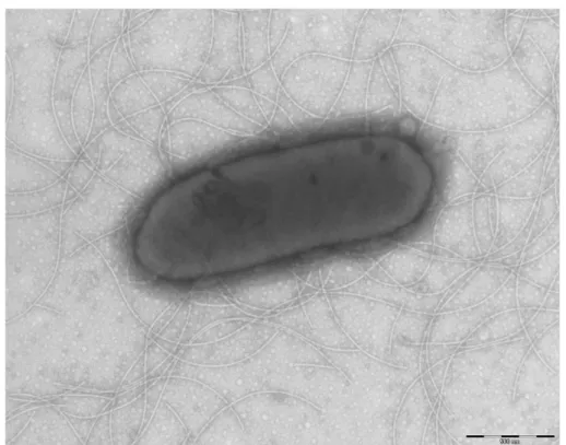
under anaerobic and microaerophilic conditions, but optimal growth was obtained aerobically. Moreover, the Gram staining showed Gram-negative rod (Figure 2). A motility test produced a negative result. Cells grown on agar did not sporulate and the rods exhibited peritrichous flagella and had a mean length of 1.75 µm and a mean diameter of 0.67 µm as determined by negative staining transmission electron micros-copy (Figure 3).
Strain G5T exhibited catalase activity but not
ox-idase activity. Using the API 50CH system (BioMerieux), a positive reaction was obtained for D-xylose, D-glucose, D-fructose, D-mannose, N-acetylglucosamine, aesculin, salicin, cellobi-ose, maltcellobi-ose, lactcellobi-ose, melibicellobi-ose, saccharose, trehalose, inulin, melezitose, D-raffinose, glycogen, gentiobiose, D-turanose, Me-thyl-α-D-glucopyranoside and hydrolysis of starch. A weak positive reaction was observed for L-arabinose. A negative reaction was ob-served for glycerol, ribose, D-galactose, L-rhamnose, L-sorbose, dulcitol, inositol, mann-itol, sorbmann-itol, methyl-αmannopyranoside, D-arabinose, amygdalin, arbitin, potassium gluconate, potassium 2-cetogluconate, potassi-um 5-cetogluconate, adonitol and D-tagatose. Using the API ZYM system, positive reactions were obtained only for naphthol-AS-BI-phosphohydrol-ase, α-galactosidase, β-galactosidase, β-gluco-sidase, arginine
arylamidase and arginine dihydrolase. The pro-duction of α-glucosidase, β-glucuronidase, ester-ase lipester-ase, leucine arylamidester-ase, cystine arylamidase, valine arylamidase, glycine arylamidase, phenylalanine arylamidase, lipase, alkaline phosphatase, acid phosphatase, N-acetyl-β-glucosaminidase and a-chymotrypsin were negative. Urease reaction and reduction of nitrates to nitrogen were also positive. Indole production was negative. G. massiliense was
sus-ceptible to ticarcillin, amoxicillin, tobramycin, imipenem, vancomycin and rifampin but re-sistant to ceftazidime (Caz 30), colistin (CT50) and metronidazole.
When compared with representative species from the family Paenibacillaceae [38-42], G. massiliense gen. nov., sp. nov. strain G5T
exhibit-ed the phenotypic differences detailexhibit-ed in Table 2.
Figure 2. Gram staining of G. massliensis strain G5T.
Figure 3. Transmission electron microscopy of G. massiliense strain G5T using a
Morgani 268D (Philips) at an operating voltage of 60kV. The scale bar represents 500 nm.
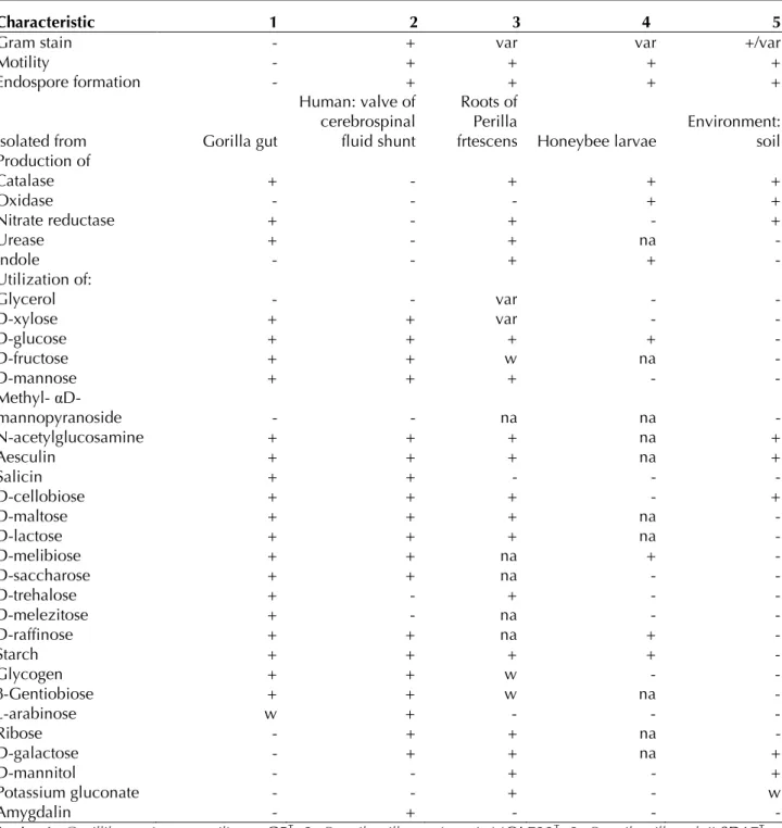
Table 2. Differential phenotypic characteristics between Gorillibacterium massiliense gen. nov., sp. nov., strain G5T
and phylogenetically close Paenibacillaceae species.
Characteristic 1 2 3 4 5
Gram stain - + var var +/var
Motility - + + + +
Endospore formation - + + + +
Isolated from Gorilla gut
Human: valve of cerebrospinal fluid shunt
Roots of Perilla
frtescens Honeybee larvae
Environment: soil Production of Catalase + - + + + Oxidase - - - + + Nitrate reductase + - + - + Urease + - + na - Indole - - + + - Utilization of: Glycerol - - var - - D-xylose + + var - - D-glucose + + + + - D-fructose + + w na - D-mannose + + + - - Methyl- αD-mannopyranoside - - na na - N-acetylglucosamine + + + na + Aesculin + + + na + Salicin + + - - - D-cellobiose + + + - + D-maltose + + + na - D-lactose + + + na - D-melibiose + + na + - D-saccharose + + na - - D-trehalose + - + - - D-melezitose + - na - - D-raffinose + + na + - Starch + + + + - Glycogen + + w - - β-Gentiobiose + + w na - L-arabinose w + - - - Ribose - + + na - D-galactose - + + na + D-mannitol - - + - + Potassium gluconate - - + - w Amygdalin - + - - -
Strain: 1, Gorillibacterium massiliense G5T; 2, Paenibacillus turicensis MOL722T; 3, Paenibacillus elgii SD17T ; 4,
Paenibacillus alvei BCRC 11220T; 5, Brevibacillus brevis NBRC 15304T.
-: negative result, +: positive result, var: variable, na: data not available, w: weak positive result
Matrix-assisted laser-desorption/ionization time-of-flight (MALDI-TOF) MS protein analysis was carried out as previously described [15] us-ing a Microflex spectrometer (Bruker Daltonics, Leipzig, Germany). Twelve distinct deposits were done for strain G5T from 12 isolated
colo-nies. The 12 G5T spectra were imported into the
MALDI BioTyper software (version 2.0, Bruker) and analyzed by standard pattern matching (with default parameter settings) against 6,252 bacterial spectra used as reference data, in the BioTyper database. A score enabled the
pre-sumptive identification of the isolated based on the following heuristicpecies: a score > 2 with a validated species enabled the identification at the species level, a score > 1.7 but < 2 enabled the identification at the genus level; and a score < 1.7 did not enable any identification. For strain G5T, a significant score was not obtained,
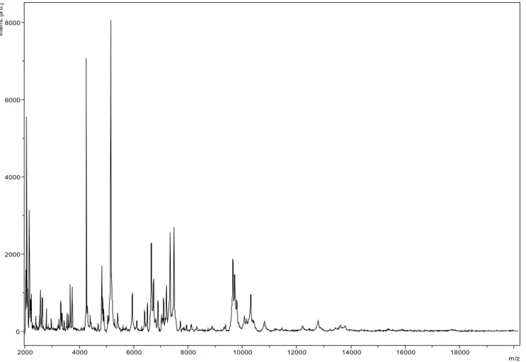
sug-gesting it was not a member of any known spe-cies or genus. We incremented our database with the spectrum from strain G5T (Figure 4).
Spectrum differences with other of Paenibacillaceae family are shown in Figure 5.
0 2000 4000 6000 8000 In te ns . [a .u .] 2000 4000 6000 8000 10000 12000 14000 16000 18000 m/z
Figure 4. Reference mass spectrum from G. massiliense strain G5T. Spectra from 16 individual colonies were
com-pared and a reference spectrum was generated.
Figure 5. Gel view comparing Gorillibacterium massilinensis gen. nov., sp. nov strain G5T spectra with other
mem-bers of the Paenibacillaceae family. The Gel View displays the raw spectra of all loaded spectrum files arranged in a pseudo-gel like look. The x-axis records the m/z value. The left y-axis displays the running spectrum number originating from subsequent spectra loading. The peak intensity is expressed by a Gray scale scheme code. The color bar and the right y-axis indicate the relation between the color a peak is displayed with and the peak intensi-ty in arbitrary units. Displayed species are indicated on the left.
Genome sequencing information
Genome project history
The organism was selected for sequencing on the basis of its phylogenetic position and 16S rRNA similarity to other members of the family Paenibacillaceae, and is part of a “culturomics” study of the gorilla flora aiming at isolating all bacterial species within gorilla feces. It was the 81st genome of the Paenibacillaceae family and
the first genome of Gorillibacterium massiliense gen. nov., sp. nov. A summary of the project in-formation is shown in Table 3. The Genbank ac-cession number is CBQR000000000 and consists of 176 large contigs. Table 3 shows the project information and its association with MIGS ver-sion 2.0 compliance [43].
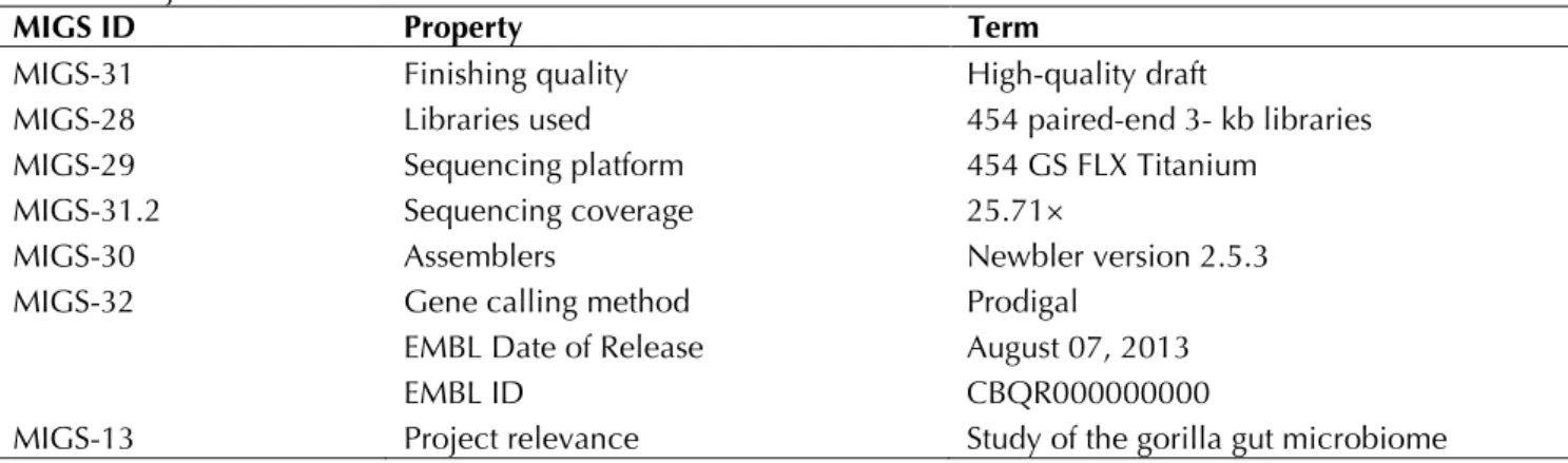
Table 3. Project information
MIGS ID Property Term
MIGS-31 Finishing quality High-quality draft
MIGS-28 Libraries used 454 paired-end 3- kb libraries
MIGS-29 Sequencing platform 454 GS FLX Titanium
MIGS-31.2 Sequencing coverage 25.71×
MIGS-30 Assemblers Newbler version 2.5.3
MIGS-32 Gene calling method Prodigal
EMBL Date of Release August 07, 2013
EMBL ID CBQR000000000
MIGS-13 Project relevance Study of the gorilla gut microbiome
Growth conditions and DNA isolation
Gorillibacterium massiliense gen. nov., sp. nov., strain G5T (= CSUR P290 = DSM 27179) wasgrown aerobically on 5% sheep blood-enriched Columbia agar at 37°C. Four petri dishes were spread and resuspended in 3×500µl of TE buffer and stored at 80°C. Then, 500 µl of this suspen-sion were thawed, centrifuged 3 minutes at 10,000 rpm and resuspended in 3×100 µL of G2 buffer (EZ1 DNA Tissue kit, Qiagen). A first me-chanical lysis was performed by glass powder on the Fastprep-24 device (Sample Preparation sys-tem, MP Biomedicals, USA) using 2×20 seconds cycles. DNA was then treated with 2.5µg/µL ly-sozyme (30 minutes at 37°C) and extracted us-ing the BioRobot EZ1 Advanced XL (Qiagen). The DNA was then concentrated and purified using the Qiamp kit (Qiagen). The yield and the con-centration were measured by the Quant-it Picogreen kit (Invitrogen) on the Genios Tecan fluorometer at 50ng/µl.
Genome sequencing and assembly
The paired-end library was prepared with 5 µg of bacterial DNA using DNA fragmentation on a Covaris S-Series (S2) instrument (Woburn, Mas-sachusetts, USA) with an enrichment size at 4.5kb. DNA fragmentation was visualized with an Agilent 2100 BioAnalyzer on a DNA labchip 7500. The library was constructed according to the 454 GS FLX Titanium paired-end protocol (Roche). Circularization and nebulization were performed and generated a pattern with an
op-timum at 510 bp. After PCR amplification through 17 cycles followed by double size selec-tion, the single stranded paired-end library was quantified using a BioAnalyzer 2100 on a RNA pico 6000 labchip at 68 pg/µL. The library con-centration equivalence was calculated as 2.45E+08 molecules/µL. The library was stored at -20°C until further use.
The paired-end library was clonally amplified with 0.25 cpb and 0.5 cpb in 2 emPCR reactions with the GS Titanium SV emPCR Kit (Lib-L) v2 (Roche). The yield of the emPCR was respective-ly of 5 and 6% as expected of the yield ranging from 5 to 20% recommended by the Roche pro-cedure.
Approximately 790,000 beads were loaded twice (i.e. two runs were performed using the same paired-end library) on a ¼ region of the GS Titanium PicoTiterPlate PTP Kit 70×75 and se-quenced with the GS FLX Titanium Sequencing Kit XLR70 (Roche). The two runs were per-formed overnight and then analyzed on the clus-ter through the gsRunBrowser and Newbler as-sembler (Roche). A total of 387,157 passed filter wells were obtained and generated 142.7 Mb of sequences with a length average of 369 bp. The passed filter sequences were assembled using Newbler with 90% identity and 40-bp as over-lap. The final assembly identified 12 scaffolds with 176 large contigs (>1.5kb), generating a genome size of 5.5 Mb which corresponds to a genome coverage of 25.71×.
Genome annotation
Open Reading Frames (ORFs) were predicted us-ing Prodigal [44] with default parameters but the predicted ORFs were excluded if they spanned a sequencing gap region. The predicted bacterial protein sequences were searched against the GenBank database [45] and the Clters of Orthologous Groups (COG) databases us-ing BLASTP. The tRNAScanSE tool [46] was used to find tRNA genes, whereas ribosomal RNAs were found by using RNAmmer [47] and BLASTn against the GenBank database. ORFans were identified if their BLASTP E-value was lower than 1e-03 for alignment length greater than 80 amino acids. If alignment lengths were smaller than 80 amino acids, we used an E-value of 1e-05.
To estimate the mean level of nucleotide se-quence similarity at the genome level between G. massiliense and another 2 members of the family Paenibacillaceae and Brevibacillus brevis, we use the Average Genomic Identity of Orthologous gene Sequences (AGIOS), a custom application we developed. Briefly, the AGIOS software
com-bines the Proteinortho software [48] for detect-ing orthologous proteins between genomes compared two by two, then retrieves the corre-sponding genes and determines the mean per-centage of nucleotide sequence identity among orthologous ORFs using the Needleman-Wunsch global alignment algorithm.
Genome properties
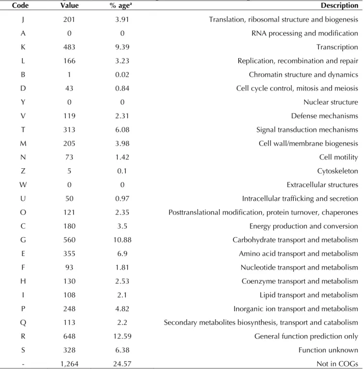
The genome is 5,546,433 bp long with a 50.39% G+C content (Figure 6 and Table 4). It is com-posed of 189 Contigs (176 large contigs, 12 scaf-folds). Of the 5,221 predicted genes, 5,145 were protein-coding genes, and 76 were RNAs (1 gene is 16S rRNA, 1 gene is 23S rRNA, 5 genes are 5S rRNA, and 69 are tRNA genes). A total of 3,865 genes (75.12%) were assigned a putative func-tion (by cogs or by NR blast). In addifunc-tion, 272 genes were identified as ORFans (5.29%). The remaining genes were annotated as hypothetical proteins (680 genes => 13.22%). The distribu-tion of genes into COGs funcdistribu-tional categories is presented in Table 5. The properties and the sta-tistics of the genome are summarized in Table 4 and 5.
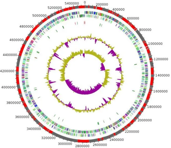
Figure 6. Graphical circular map of the chromosome. From outside to the center: Genes on the forward strand colored by COG categories (only genes assigned to COG), genes on the reverse strand colored by COG categories (only gene assigned to COG), RNA genes (tRNAs green, rRNAs red), G+C content and GC skew. Purple and olive indicating negative and positive values, respectively.
Table 4. Nucleotide content and gene count levels of the chromosome
Attribute Value % of totala
Genome size (bp) 5,546,433 100
DNA G+C content (bp) 2,794,611 50.39
DNA coding region (bp) 4,888,209 88.13
Total genes 5,221 100
RNA genes 76 1.46
Protein-coding genes 5,145 98.54
Genes with function prediction 3,865 75.12
Genes assigned to COGs 3,881 75.43
Genes with peptide signals 709 13.78
Genes with transmembrane helices 1,267 24.63
a The total is based on either the size of the genome in base pairs or the total number of protein-coding genes in
the annotated genome
Table 5. Number of genes associated with the 25 general COG functional categories
Code Value % agea Description
J 201 3.91 Translation, ribosomal structure and biogenesis
A 0 0 RNA processing and modification
K 483 9.39 Transcription
L 166 3.23 Replication, recombination and repair
B 1 0.02 Chromatin structure and dynamics
D 43 0.84 Cell cycle control, mitosis and meiosis
Y 0 0 Nuclear structure
V 119 2.31 Defense mechanisms
T 313 6.08 Signal transduction mechanisms
M 205 3.98 Cell wall/membrane biogenesis
N 73 1.42 Cell motility
Z 5 0.1 Cytoskeleton
W 0 0 Extracellular structures
U 50 0.97 Intracellular trafficking and secretion
O 121 2.35 Posttranslational modification, protein turnover, chaperones
C 180 3.5 Energy production and conversion
G 560 10.88 Carbohydrate transport and metabolism
E 355 6.9 Amino acid transport and metabolism
F 93 1.81 Nucleotide transport and metabolism
H 130 2.53 Coenzyme transport and metabolism
I 108 2.1 Lipid transport and metabolism
P 248 4.82 Inorganic ion transport and metabolism
Q 113 2.2 Secondary metabolites biosynthesis, transport and catabolism
R 648 12.59 General function prediction only
S 328 6.38 Function unknown
- 1,264 24.57 Not in COGs
Genomic comparison of G. massili
ense and other members of the
fami-ly
Paenibacillaceae
The genome of G. massiliense strain G5T was
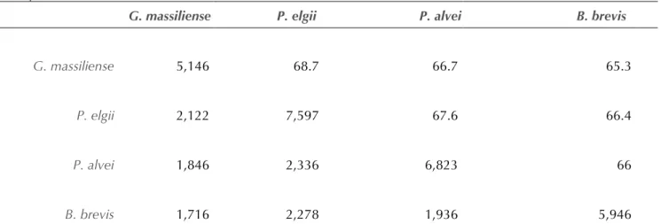
compared to those of P. elgii strain B69, P. alvei strain DSM 29 and B. brevis strain NBRC 100599 (Table 6A and Table 6B). The draft genome of G. massiliense is smaller in size than those of P. elgii, P. alvei and B. brevis (5.54 vs 7.96, 6.83 and 6.3 Mb respectively). G. massiliense has a lower G+C content than P. elgii (50.39% vs 52.6%) but higher than those of P. alvei and B. brevis (50.39% vs 45.9% and 47.3% respectively). The protein content of G. massiliense is lower than those of P. elgii, P. alvei and B. brevis (5,146 vs 7,597, 6,823 and 5,946 respectively) (Table 6 and Table 6B). In addition, G. massiliense shares 2,122, 1,846 and 1,716 orthologous genes with P. elgii, P. alvei and B. brevis, respectively (Table
6). The nucleotide sequence identity of ortholo-gous genes ranges from 66 to 67.6% among pre-viously published genomes, and from 65.3 to 68.7% between G. massiliense and other studied genomes (Table 6A and Table 6B). Table 6 summarizes the number of orthologous genes and the average percentage of nucleotide se-quence identity between the different genomes studied.
Conclusion
On the basis of phenotypic, phylogenetic and ge-nomic analyses, we formally propose the crea-tion of Gorillibacterium massiliense gen. nov., sp. nov., that contains the strain G5T. This bacterium
has been found in stool sample of wild gorilla collected in Cameroon.
Table 6A. Genomic comparison of G. massiliense gen. nov., sp. nov., strain G5T with four other members of the
family Paenibacillaceae†
Species Strain Genome accession num-ber Genome size (Mb) G+C content
Gorillibacterium massiliense G5T CBQR000000000 5.54 50.39
Paenibacillus elgii B69 AFHW00000000 7.96 52.6
Paenibacillus alvei DSM 29 AMBZ00000000 6.83 45.9
Brevibacillus brevis NBRC 100599 AP008955 6.3 47.3
†Species and strain names, GenBank genome accession numbers, sizes and G+C contents
Table 6B. Genomic comparison of G. massiliense gen. nov., sp. nov., strain G5T with four other members of the
family Paenibacillaceae†
G. massiliense P. elgii P. alvei B. brevis
G. massiliense 5,146 68.7 66.7 65.3
P. elgii 2,122 7,597 67.6 66.4
P. alvei 1,846 2,336 6,823 66
B. brevis 1,716 2,278 1,936 5,946
†Numbers of orthologous protein shared between genomes (lower left triangle), average percentage similarity of
nucleotides corresponding to orthologous proteins shared between genomes (upper right triangle). Bold numbers indicate numbers of proteins per genome.
Description of
Gorillibacterium
gen. nov.
Gorillibacterium (go.ri.li.bac.te.ri’um. gor.il.i NL gen fem, the genus name of the great ape; bac.ter’i.um N.L. neut. n., bacterium a rod; gorillibacterium a rod-shaped bacterium isolat-ed from a gorilla).Gram-negative rod. Facultatively anaerobic. Me-sophilic. Non-motile. Oxidase negative, catalase positive. Positive for urease, nitrate reduction, α- and β-galactosidase, arginine dihydrolase, argi-nine arylamidase, and β-glucosidase. Habitat: gorilla gut. Type species: Gorillibacterium massiliense.
Description of
Gorillibacterium massiliense
gen. nov., sp. nov.
Gorillibacterium massiliense (ma.si.li.en′se. L. gen. neut. n. massiliense, of Massilia, the ancient Roman name for Marseille, France, where the type strain was isolated).
G. massiliense is Gram-negative rod. Facultatively anaerobic. Mesophilic. Optimal growth is achieved at 37°C. Non-sporulating and non-motile bacterium. Colonies are bright gray and 0.5-1 mm in diameter on blood-enriched Colum-bia agar. Cells are rod-shaped and have a mean diameter of 0.67 µm and a mean length of 1.75 µm.
Catalase positive, oxidase negative. Using the API 20NE system, positive reactions are ob-served for nitrate reduction and urease reaction, but indole production was negative. Using the API 50CH system (BioMerieux), a positive reac-tion was obtained for the fermentareac-tion of D-xylose, D-glucose, D-fructose, D-mannose, N-acetylglucosamine, aesculin, salicin, D-cellobi-ose, D-maltD-cellobi-ose, D-lactD-cellobi-ose, D-melibiD-cellobi-ose, D-saccha-rose, D-trehalose, inulin, D-melezitose,
D-raffin-ose, glycogen, gentiobiD-raffin-ose, D-turanD-raffin-ose, Methyl- αD-glucopyranoside and starch. Negative reac-tions are observed for glycerol, ribose, D-galactose, L-rhamnose, L-sorbose, dulcitol, inosi-tol, D-manniinosi-tol, D-sorbiinosi-tol, methyl-αD-manno-pyranoside, D-arabinose, amygdalin, arbitin, po-tassium gluconate, popo-tassium 2-cetogluconate, potassium 5-cetogluconate, adonitol and D-taga-tose. Using the API ZYM system, positive reac-tions were observed for the production of naphthol-AS-BI-phosphohydrolase, α-galactosidase, β- α-galactosidase, β- glucosidase, Arginine arylamidase and Arginine dihydrolase. The production of α- glucosidase, β- glucuronidase, esterase lipase, leucine arylamidase and cystine arylamidase, valine arylamidase, glycine arylamidase, phenylalanine arylamidase, lipase, alkaline phosphatase, acid phosphatase, N-acetyl-β-glucosaminidase and a-chymotrypsin are negative. Susceptible to ticarcillin, amoxicillin, tobramycin, imipenem, vancomycin and rifampin but resistant to ceftazidime, colistin and metronidazole.
The G+C content of the genome is 50.39%. The 16S rRNA and genome sequences are deposited in GenBank under accession numbers KC193239 and CBQR000000000, respectively. The type strain G5T (= CSUR P290 = DSM 27179) was
iso-lated from the fecal flora of a Gorilla gorilla goril-la from Cameroon.
Acknowledgements
Fadi Bittar was supported by a Chair of Excel-lence IRD provided by the Institut de Recherche pour le Développement/Méditerranée-Infection foundation. Mamadou Bhoye Keita was funded by the Méditerranée-Infection Foundation. The authors thank the Xegen company for automat-ing the genome annotation process.
References
1. Lagier JC, Armougom F, Million M, Hugon P, Pagnier I, Robert C, Bittar F, Fournous G, Gimenez G, Maraninchi M, et al. Microbial culturomics: paradigm shift in the human gut microbiome study. Clin Microbiol Infect 2012; 18:1185-1193. PubMed
2. Dubourg G, Lagier JC, Armougom F, Robert C, Hamad I, Brouqui P, Raoult D. The gut
microbiota of a patient with resistant tuberculo-sis is more comprehensively studied by
culturomics than by metagenomics. Eur J Clin
Microbiol Infect Dis 2013; 32:637-645. Pub-Med http://dx.doi.org/10.1007/s10096-012-1787-3
3. Pfleiderer A, Lagier JC, Armougom F, Robert C, Vialettes B, Raoult D. Culturomics identified 11 new bacterial species from a single anorexia nervosa stool sample. Eur J Clin Microbiol Infect
Dis 2013; 32:1471-1481. PubMed
http://dx.doi.org/10.1007/s10096-013-1900-2
4. Mishra AK, Lagier JC, Robert C, Raoult D, Four-nier PE. Non-contiguous finished genome se-quence and description of Clostridium
senegalense sp. nov. Stand Genomic Sci 2012; 6:386-395. PubMed
5. Lagier JC, Armougom F, Mishra AK, Ngyuen TT, Raoult D, Fournier PE. Non-contiguous finished genome sequence and description of Alistipes timonensis sp. nov. Stand Genomic Sci 2012;
6:315-324. PubMed
http://dx.doi.org/10.4056/sigs.2685971
6. Lagier JC, El Karkouri K, Nguyen TT, Armougom F, Raoult D, Fournier PE. Non-contiguous fin-ished genome sequence and description of
Anaerococcus senegalensis sp. nov. Stand
Ge-nomic Sci 2012; 6:116-125. PubMed http://dx.doi.org/10.4056/sigs.2415480
7. Roux V, El Karkouri K, Lagier JC, Robert C, Raoult D. Non-contiguous finished genome se-quence and description of Kurthia massiliensis
sp. nov. Stand Genomic Sci 2012; 7:221-232.
PubMedhttp://dx.doi.org/10.4056/sigs.3206554
8. Mishra AK, Lagier JC, Robert C, Raoult D, Four-nier PE. Non-contiguous finished genome se-quence and description of Peptoniphilus timonensis sp. nov. Stand Genomic Sci 2012; 7:1-11. PubMed
http://dx.doi.org/10.4056/sigs.2956294
9. Hugon P, Ramasamy D, Lagier JC, Rivet R, Couderc C, Raoult D, Fournier PE. Non contig-uous-finished genome sequence and descrip-tion of Alistipes obesi sp. nov. Stand Genomic
Sci 2013; 7:427-439. PubMed
http://dx.doi.org/10.4056/sigs.3336746
10. Ramasamy D, Lagier JC, Nguyen TT, Raoult D, Fournier PE. Non contiguous-finished genome sequence and description of Dielma fastidiosa
gen. nov., sp. nov., a new member of the Fami-ly Erysipelotrichaceae. Stand Genomic Sci
2013; 8:336-351. PubMed
http://dx.doi.org/10.4056/sigs.3567059
11. Mishra AK, Lagier JC, Robert C, Raoult D, Four-nier PE. Genome sequence and description of
Timonella senegalensis gen. nov., sp. nov., a new member of the suborder Micrococcinae.
Stand Genomic Sci 2013; 8:318-335. PubMed http://dx.doi.org/10.4056/sigs.3476977
12. Mishra AK, Hugon P, Lagier JC, Nguyen TT, Couderc C, Raoult D, Fournier PE. Non contig-uous-finished genome sequence and descrip-tion of Enorma massiliensis gen. nov., sp. nov., a new member of the Family Coriobacteriaceae. Stand Genomic Sci 2013; 8:290-305. PubMed http://dx.doi.org/10.4056/sigs.3426906
13. Ramasamy D, Lagier JC, Gorlas A, Raoult D, Fournier PE. Non contiguous-finished genome sequence and description of Bacillus
massiliosenegalensis sp. nov. Stand Genomic
Sci 2013; 8:264-278. PubMed
http://dx.doi.org/10.4056/sigs.3496989
14. Hugon P, Mishra AK, Lagier JC, Nguyen TT, Couderc C, Raoult D, Fournier PE. Non-contiguous finished genome sequence and de-scription of Brevibacillus massiliensis sp. nov.
Stand Genomic Sci 2013; 8:1-14. PubMed http://dx.doi.org/10.4056/sigs.3466975
15. Keita MB, Diene S, Robert C, Raoult D, Four-nier PE, Bittar F. Non-contiguous finished ge-nome sequence and description of Bacillus massiliogorillae sp. nov. Stand Genomic Sci 2013; 9:93-105. PubMed
http://dx.doi.org/10.4056/sigs.4388124
16. De Vos P, Ludwig W, Schleifer KH, Whitman WB. Family IV. Paenibacillaceae fam. nov. In Bergey's Manual of Systematic Bacteriology, 2nd Edition, vol 3 (The Firmicutes), Springer; New York, 2009, p. 269.
17. Abstract for the family Paenibacillaceae. NamesforLife, LLC. Retrieved October 21, 2013.
http://doi.namesforlife.com/10.1601/tx.5108 18. Ash C, Priest FG, Collins MD. Molecular
identi-fication of rRNA group 3 bacilli (Ash, Farrow, Wallbanks and Collins) using a PCR probe test. Proposal for the creation of a new genus
Paenibacillus. Antonie van Leeuwenhoek 1993;
64:253-260. PubMed
http://dx.doi.org/10.1007/BF00873085
19. The type species of the genus Paenibacillus Ash et al. 1994 is Paenibacillus polymyxa. Opinion 77. Judicial Commission of the International Committee on Systematics of Prokaryotes. Int J
Syst Evol Microbiol 2005; 55:513.
20. Zaitsev GM, Tsitko IV, Rainey FA, Trotsenko YA, Uotila JS, Stackebrandt E, Salkinoja-Salonen MS. New aerobic ammonium-dependent obligately oxalotrophic bacteria: description of
Ammoniphilus oxalaticus gen. nov., sp. nov. and Ammoniphilus oxalivorans gen. nov., sp. nov. Int J Syst Bacteriol 1998; 48:151-163.
PubMed http://dx.doi.org/10.1099/00207713-48-1-151
21. Shida O, Takagi H, Kadowaki K, Komagata K. Proposal for two new genera, Brevibacillus gen. nov. and Aneurinibacillus gen. nov. Int J Syst
Bacteriol 1996; 46:939-946. PubMed
http://dx.doi.org/10.1099/00207713-46-4-939
22. Touzel JP, O'Donohue M, Debeire P, Samain E, Breton C. Thermobacillus xylanilyticus gen. nov., sp. nov., a new aerobic thermophilic xylan-degrading bacterium isolated from farm soil. Int J Syst Evol Microbiol 2000; 50:315-320.
PubMed http://dx.doi.org/10.1099/00207713-50-1-315
23. Saha P, Krishnamurthi S, Bhattacharya A, Shar-ma R, Chakrabarti T. Fontibacillus aquaticus
gen. nov., sp. nov., isolated from a warm spring. Int J Syst Evol Microbiol 2010; 60:422-428. PubMed
24. Kämpfer P, Rosselló-Mora R, Falsen E, Busse HJ, Tindall BJ. Cohnella thermotolerans gen. nov., sp. nov., and classification of 'Paenibacillus hongkongensis' as Cohnella hongkongensis sp. nov. Int J Syst Evol Microbiol 2006; 56:781-786.
PubMedhttp://dx.doi.org/10.1099/ijs.0.63985-0
25. Rivas R, García-Fraile P, Zurdo-Piñeiro JL, Mateos PF, Martínez-Molina E, Bedmar EJ, Sánchez-Raya J, Velázquez E. Saccharibacillus sacchari gen. nov., sp. nov., isolated from sugar cane. Int J Syst Evol Microbiol 2008; 58:1850-1854. PubMed
http://dx.doi.org/10.1099/ijs.0.65499-0
26. Collins MD, Lawson PA, Willems A, Cordoba JJ, Fernandez-Garayzabal J, Garcia P, Cai J, Hippe H, Farrow JA. The phylogeny of the genus Clos-tridium: proposal of five new genera and eleven new species combinations. Int J Syst Bacteriol 1994; 44:812-826. PubMed
http://dx.doi.org/10.1099/00207713-44-4-812
27. Woese CR, Kandler O, Wheelis ML. Towards a natural system of organisms: proposal for the domains Archae, Bacteria, and Eukarya. Proc
Natl Acad Sci USA 1990; 87:4576-4579. Pub-Medhttp://dx.doi.org/10.1073/pnas.87.12.4576
28. Gibbons NE, Murray RGE. Proposals concern-ing the higher taxa of Bacteria. Int J Syst
Bacteriol 1978; 28:1-6.
http://dx.doi.org/10.1099/00207713-28-1-1
29. Garrity GM, Holt JG. The Road Map to the Manual. In: Garrity GM, Boone DR, Castenholz RW (eds), Bergey's Manual of Systematic Bacte-riology, Second Edition, Volume 1, Springer, New York, 2001, p. 119-169.
30. Murray RGE. The Higher Taxa, or, a Place for Everything...? In: Holt JG (ed), Bergey's Manual of Systematic Bacteriology, First Edition, Vol-ume 1, The Williams and Wilkins Co., Balti-more, 1984, p. 31-34.
31. Ludwig W, Schleifer KH, Whitman WB. Class I.
Bacilli class nov. In: De Vos P, Garrity G, Jones D, Krieg NR, Ludwig W, Rainey FA, Schleifer KH, Whitman WB (eds), Bergey's Manual of Sys-tematic Bacteriology, Second Edition, Volume 3, Springer-Verlag, New York, 2009, p. 19-20. 32. List of new names and new combinations
pre-viously effectively, but not validly, published. List no. 132. Int J Syst Evol Microbiol 2010; 60:469-472.
http://dx.doi.org/10.1099/ijs.0.022855-0
33. Skerman VBD, McGowan V, Sneath PHA. Ap-proved Lists of Bacterial Names. Int J Syst
Bacteriol 1980; 30:225-420.
http://dx.doi.org/10.1099/00207713-30-1-225
34. Prévot AR. In: Hauderoy P, Ehringer G, Guillot G, Magrou. J., Prévot AR, Rosset D, Urbain A
(eds), Dictionnaire des Bactéries Pathogènes, Second Edition, Masson et Cie, Paris, 1953, p. 1-692
35. Ashburner M, Ball CA, Blake JA, Botstein D, Butler H, Cherry JM, Davis AP, Dolinski K, Dwight SS, Eppig JT, et al. Gene ontology: tool for the unification of biology. The Gene Ontol-ogy Consortium. Nat Genet 2000; 25:25-29.
PubMedhttp://dx.doi.org/10.1038/75556 36. Tamura K, Peterson D, Peterson N, Stecher G,
Nei M, Kumar S. MEGA5: Molecular Evolution-ary Genetics Analysis using Maximum Likeli-hood, Evolutionary Distance, and Maximum Parsimony Methods. Mol Biol Evol 2011; 28:2731-2739. PubMed
http://dx.doi.org/10.1093/molbev/msr121
37. Stackebrandt E, Ebers J. Taxonomic parameters revisited: tarnished gold standards. Microbiol
Today 2006; 33:152-155.
38. Bosshard PP, Zbinden R, Altwegg M.
Paenibacillus turicensis sp. nov., a novel bacte-rium harbouring heterogeneities between 16S rRNA genes. Int J Syst Evol Microbiol 2002; 52:2241-2249. PubMed
http://dx.doi.org/10.1099/ijs.0.02105-0
39. Kim DS, Bae CY, Jeon JJ, Chun SJ, Oh HW, Hong SG, Baek KS, Moon EY, Bae KS.
Paenibacillus elgii sp. nov., with broad antimi-crobial activity. Int J Syst Evol Microbiol 2004; 54:2031-2035. PubMed
http://dx.doi.org/10.1099/ijs.0.02414-0
40. Ueda J, Yamamoto S, Kurosawa N. Paenibacillus thermoaerophilus sp. nov., a moderately
thermophilic bacterium isolated from compost.
Int J Syst Evol Microbiol 2013; 63:3330-3335.
PubMed http://dx.doi.org/10.1099/ijs.0.048090-0
41. Lee FL, Kuo HP, Tai CJ, Yokota A, Lo CC.
Paenibacillus taiwanensis sp. nov., isolated from soil in Taiwan. Int J Syst Evol Microbiol 2007; 57:1351-1354. PubMed
http://dx.doi.org/10.1099/ijs.0.64764-0
42. Takebe F, Hirota K, Nodasaka Y, Yumoto I.
Brevibacillus nitrificans sp. nov., a nitrifying bacterium isolated from a microbiological agent for enhancing microbial digestion in sewage treatment tanks. Int J Syst Evol Microbiol 2012; 62:2121-2126. PubMed
http://dx.doi.org/10.1099/ijs.0.032342-0
43. Field D, Garrity G, Gray T, Morrison N, Selengut J, Sterk P, Tatusova T, Thomson N, Al-len MJ, Angiuoli SV, et al. The minimum infor-mation about a genome sequence (MIGS) speci-fication. Nat Biotechnol 2008; 26:541-547.
PubMedhttp://dx.doi.org/10.1038/nbt1360
45. GenBank database.
http://www.ncbi.nlm.nih.gov/genbank 46. Lowe TM, Eddy SR. t-RNAscan-SE: a program
for improved detection of transfer RNA gene in genomic sequence. Nucleic Acids Res 1997; 25:955-964. PubMed
http://dx.doi.org/10.1093/nar/25.5.0955
47. Lagesen K, Hallin P, Rodland EA, Staerfeldt HH, Rognes T, Ussery DW. RNAmmer: consistent
and rapid annotation of ribosomal RNA genes.
Nucleic Acids Res 2007; 35:3100-3108. Pub-Medhttp://dx.doi.org/10.1093/nar/gkm160
48. Lechner M, Findeib S, Steiner L, Marz M, Stadler PF, Prohaska SJ. Proteinortho: Detection of (Co-)orthologs in large-scale analysis. BMC
Bioinformatics 2011; 12:124. PubMed http://dx.doi.org/10.1186/1471-2105-12-124
![Figure 1. Phylogenetic tree highlighting the position of T relative to other type strains within the were aligned using CLUSTAL X (V2), and phylogenetic inferences obtained using the maximum-likelihood method within the MEGA 5 software [36]](https://thumb-eu.123doks.com/thumbv2/123doknet/13783746.439859/4.892.109.731.86.621/phylogenetic-highlighting-position-relative-clustal-phylogenetic-inferences-likelihood.webp)