Advances in the Endoscopic Management of Esophageal Neoplasia using Ultrahigh Speed Endoscopic Optical Coherence Tomography
by
Hsiang-Chieh Lee B.S. Civil Engineering
National Taiwan University, Taiwan, 2004
S.M. Electro-Optical Engineering National Taiwan University, Taiwan, 2006
MASSACHUSETTS INSTITUTE OF TECHNOLOGY
MA
R 1
73Z17
LIBRARIES
ARCHIVES
SUBMITTED TO THE DEPARTMENT OF ELECTRICAL ENGINEERING AND COMPUTERSCIENCE IN PARTIAL FULFILLMENT OF THE REQUIREMENTS FOR THE DEGREE OF
DOCTOR OF PHILOSOPHY IN ELECTRICAL ENGINEERING AND COMPUTER SCIENCE
AT THE
MASSACHUSETTS INSTITUTE OF TECHNOLOGY
FEBRUARY 2017
C 2017 Massachusetts Institute of Technology. All rights reserved.
The author hereby grants MIT permission to reproduce and to distribute publicly paper and electronic copies of this thesis document in whole or in part in any medium now known or hereafter created.
Signature of Author:
Signature redacted
Department of Electrical Engineering and Computer Science January 31, 2017
Certified
Signature redacted
K-/.
Certified by:
72
(
James G. Fujimoto Professor of Electrical Engineering and Computer Science Thesis Supervisor
Signature redacted
7
Associate Professor of!Hiroshi Mashimo Medicine, Harvard Medical School Thesis Supervisor
Accepted by:
Signature redacted
(
Leslie A. KolodziejskiLeslie A. Kolodziejski 1rofessor of Electrical Engineering and Computer Science Chair, Committee for Graduate StudentsAdvances in the Endoscopic Management of Esophageal Neoplasia using Ultrahigh Speed Endoscopic Optical Coherence Tomography
by
Hsiang-Chieh Lee
Submitted to the Department of Electrical Engineering and Computer Science on January 31, 2017 in Partial Fulfillment of the Requirements for the Degree of Doctor of Philosophy in Electrical Engineering and Computer Science at the Massachusetts Institute of Technology
ABSTRACT
Esophageal cancer is one of the most lethal malignancies, with a five-year survival rate of only 16.7%. Barrett's esophagus (BE) is a premalignant condition with increased risk of developing into esophageal adenocarcinoma, the most common type of esophageal cancer in the West. In current BE surveillance protocol, random biopsies are used to diagnose and detect dysplasia in BE, which is time-consuming and suffers from sampling error. Endoscopic optical coherence tomography (OCT) is a unique imaging technique that can provide micrometer scale, two- and three-dimensional imaging of esophageal tissue without requiring exogenous contrast agents. Although various studies have investigated the feasibility of endoscopic OCT in the human GI tract, the diagnostic accuracy of endoscopic OCT in the detection of dysplasia in BE was still suboptimal.
Most previous endoscopic OCT studies were limited to investigating the changes in tissue architecture but not microvasculature, due to hardware limitations of the OCT system and optical scanning instability of the imaging catheters. Alteration of microvasculature has been demonstrated as a critical marker of the neoplastic progression of dysplasia in BE. Although OCT has been demonstrated to provide vascular contrast with OCT angiography (OCTA) in ophthalmology, the translation of OCTA to endoscopic applications has been challenging. The aims of this thesis are: 1) Development of distally actuated imaging devices including a micromotor balloon catheter allowing wide field OCTA imaging and a hollow shaft micromotor catheter enabling unobstructed endoscopic OCT imaging in the gastrointestinal tract. 2) A clinical feasibility study investigating the association of OCTA microvascular features with the detection of dysplasia in patients with BE. 3) Laboratory studies using OCT to monitor radiofrequency ablation (RFA) dynamics with concurrent OCT imaging in ex vivo swine specimens. The scope of this thesis includes the design and development of novel imaging devices, laboratory imaging studies with both ex vivo and in vivo animal models, and a collaborative clinical imaging study with patients. The ultimate goal of this thesis work is to facilitate the implementation of endoscopic OCT and OCTA techniques in the endoscopic management of BE.
Thesis Supervisor: James G. Fujimoto
Acknowledgements
Through my Ph.D. journey in the department of the Electrical Engineering and Computer Science at MIT, it has been a great privilege to have the opportunity working with many brilliant scientists, physicians, collaborates, and graduate students. Without their support and active involvement, this thesis work cannot be completed. Many of them continued dedicating their careers in the field of optical coherence tomography (OCT) and played important roles in the advances of OCT in various biomedical applications. I want to especially thank my advisor Professor James Fujimoto for providing me the opportunity of working in this world-class laboratory, as well as his guidance and support over these years. The research environment he offered is more than a graduate student can expect. I also thank my thesis co-advisor Dr. Hiroshi Mashimo for his long-term mentoring and support on the endoscopic OCT projects conducted at the VABHS and the friendship over these years. I thank my thesis committee member, Professor Elfar Adalsteinsson for his input and advice to polish my thesis. In addition, I want to thank Dr. Chao Zhou and Dr. Aaron Aguirre for their mentorship, allowing me to continue the development of optical coherence microscopy (OCM) technique. I also want to thank Dr. Tsung-Han Tsai for his guidance and friendship since the first day I joined Professor Fujimoto's lab, patiently teaching me the secrets of Fourier-domain mode-locked laser as well as the hard-core OCT catheter engineering.
It was a great pleasure to work on the endoscopic OCT project (the "GI team") since the beginning of my Ph.D. journey at MIT. I had a great time working with various former or current GI team members including Dr. Desmond Adler, Dr. Chao Zhao, Dr. Tsung-Han Tsai, Dr. Zhao Wang, Osman Ahsen, and Kaicheng Liang, as well as the support from Dr. Yuankai Tao, Dr. Michael Giacomelli, and Dr. Ning Zhang. I am sure I will never forget the "VA Mashimo Monday" and the RLE van we often reserved for the commute between the MIT and VA. In particular, I want to thank Osman Ahsen and Kaicheng Liang, being such incredible and extremely helpful colleagues and friends. It was a truly an honor for me to closely working with you over the past four years. I would also like to express my appreciation to Dr. Hiroshi Mashimo, Dr. Qin Huang, Dr. Laren Becker, Dr. Tejas Kirtane, Dr. Walter Chan, Marisa Figueiredo, Dr. Frances Achee, Rachel Bereiweriso, Rayhme Collins, Caroline Costa, Greg Daniels, the nursing staff and the technicians in the endoscopic unit at VABHS, in particular, Annalee Murphy and Joanne Hill for their putting up with the space being occupied by the OCT instruments, the fragile OCT imaging catheters, and sometimes the last minute add-on of the OCT schedule.
I thank Dr. Aaron Aguirre, Dr. Chao Zhou, and Dr. Jonathan Liu at MIT, as well as Dr. David Cohen, Dr. Dejun Shen, Dr. Yuri Sheikine, and Dr. James Connolly in the Department of Pathology at Beth Israel Deaconess Medical Center for their support on the development and application of OCM for the path lab imaging. In addition, I would like to thank both former and current members of the Biomedical Optics Imaging and Biophotonics Group at RLE and MIT including Dr. Shu-Wei Huang, Dr. Bernhard Baumann, Dr. Ireneusz Grulkowski, Dr. Yueli Chen, Dr. Umit Demirbas, Dr. Alphan Sennaroglu, Dr. Yu Gu, Dr. Al-Hafeez Dhalla, Dr. Duo Li, Jing Wang, Dr. WooJhon Choi, Dr. Martin Kraus, Chen Lu, ByungKun Lee, Eric Moult, Tadayuki Yoshitake, Lucas Cahill, Kathrin Mohler, Lennart Husvogt, Stefan Ploner, Julia Schottenhamml.
Although I did not have a chance to work with you on the projects in ophthalmology and microscopy, the exchanges of opinions on various topics during the daily interaction and the assistance of the lab-related works make my lab life more interesting and fulfilling. And of course, I thank Ms. Dorothy Fleischer for all the administrative assistance and the heartwarming encouragement whenever I felt down or frustrated over these years. I thank Professor Erich Ippen and Professor Franz Kaertner for being part of my delightful MIT life, in particular, the Oktoberfest and the Christmas parties, makes me feel welcome while studying aboard alone.
I also want to thank the strong industrial support making my thesis work possible. I thank Dr.
Benjamin Potsaid and Mr. Alex Cable from Thorlabs, Inc., as well as Dr. Vijarsekhar Jayaraman from Praevium Research, Inc. for their continuing support of VCSEL light source and other critical electronic components to facilitate the development of the ultrahigh speed endoscopic
OCT engine. I thank Mr. Eric Swanson, Dr. Long Chen, and Dr. Chris Doerr from Acacia
Communication, Inc. for their support of the development of the silicon photonic integrated
SS-OCT system. I must thank the National Institute of Health and the Air Force Office of Scientific
Research for the funding assistance to support my thesis work.
Lastly, I want to save the most appreciative thanks for my friends and family. I want to thank my parents Feng-Liang Lee and Mei-Hua Lai for their sacrifice and unwavering love over these years supporting me to chase my dream studying aboard. My wife, Li-Hsin Chen, who stopped her career temporarily and came to the US to take care of me during the most challenging period of my Ph.D. journey. I am fortunate and blessed to have the support from my sister, Ying-Ling Lee, and brother Zhung-Fu Lee taking care of the family while I am not at home. Thanks to all my wonderful friends, in particular, Chien-Jen Lai, Cheng-Wei Cheng, I-Hua Chen, Tsung-Han Tsai, Chun-Ting Su, and Jonathan Liu.
TABLE OF CONTENTS
CHAPTER 1: INTRODUCTION: ENDOSCOPIC MANAGEMENT OF BARRETT'S
ESO PA H G U S... 9
1.1 Background - Barrett's Esophagus and Esophageal Carcinoma ... 10
1.2 Advanced Endoscopic Imaging Methods for Dysplasia Detection in BE ... 11
1.2.1 C hrom oendoscopy ... 11
1.2.2 Narrow Band Imaging (Virtual Chromoendoscopy) ... 12
1.2.3 Confocal Laser Endomicroscopy ... 13
1.3 Endoscopic Treatment Methods for the Eradication of Dysplasia in BE... 14
1.3.1 Endoscopic Mucosal Resection ... 15
1.3.2 Radiofrequency A blation ... 15
1.3.3 C ryospray A blation ... 16
1.4 Endoscopic OCT in the Endoscopic Management of BE ... 18
1.5 Scope of the Thesis... 20
CHAPTER 2: ENDOSCOPIC OPTICAL COHERENCE TOMOGRAPHY IMAGING USING A 360-DEGREE UNOBSTRUCTED MICROMOTOR IMAGING CATHETER 23 2.1 M otivation... 24
2.2 Development of the 360-degree Unobstructed Imaging Catheter ... 26
2.2.1 Ultrahigh speed endoscopic OCT system... 26
2.2.2 360-degree micromotor imaging catheter ... 27
2.2.4 Endoscopic OCT angiography (OCTA) and data visualization ... 30
2.3 R esults ... 31
2.3.1 N U RD characterization... 31
2.3.2 Human skin and buccal mucosa imaging... 31
2.3.3 Human lower GI tract imaging - dentate line... 32
2.4 D iscussion... 33
2.5 F igures... 36
2.6 T ables ... 41
CHAPTER 3: CIRCUMFERENTIAL OPTICAL COHERENCE TOMOGRAPHY ANGIOGRAPHY USING A MICROMOTOR BALLOON CATHETER... 43
3.1 M otivation... 44
3.2 Development of the Micromotor Balloon Catheter ... 46
3.2.1 Swept source OCT imaging system... 46
3.2.2 Micromotor balloon imaging catheter ... 47
3.2.3 Nonuniform rotation distortion (NURD) correction... 48
3.2.4 Endoscopic OCT angiography and data visualization... 50
3.2.5 Animal imaging procedures... 50
3.3 Im aging R esults... 51
3.4 D iscussion... 53
CHAPTER 4: ENDOSCOPIC OPTICAL COHERENCE TOMOGRAPHY
ANGIOGRAPHY MICROVASCULAR FEATURES ASSOCIATED WITH DYSPLASIA
IN BE...65
4.1 Motivation... 66
4.2 Endoscopic OCTA and Study Design ... 67
4.2.1 Patient enrollment ... 67
4.2.2 Endoscopic OCT and OCT Angiography (OCTA) imaging procedure ... 68
4.2.3 Endoscopic OCT imaging system... 70
4.2.4 Endoscopic OCTA and data visualization ... 70
4.2.5 OCTA reading criteria and protocol ... 71
4.2.6 Development of the OCTA criteria - initial learning phase ... 72
4.2.7 Training/validation session ... 73
4.2.8 Workflow of the OCTA reading protocol... 73
4.2.9 Statistical analysis... 74
4.3 Performance of the OCTA Criteria ... 74
4.3.1 Baseline characteristics... 74
4.3.2 Diagnostic performance of the OCTA criteria... 74
4.4 Discussion... 76
4.5 Figures... 80
4.6 Tables ... 83
4.7 Appendix - OCTA Reading Protocol (Training Materials) ... 88
CHAPTER 5: COMPREHENSIVE ASSESSMENT OF RADIOFREQUENCY ABLATION DYNAMICS WITH OPTICAL COHERENCE TOMOGRAPHY...114
5.1 Motivation... 115
5.2 Experimental Setup and Study Design ... 117
5.2.1 High speed swept-source OCT system ... 117
5.2.2 Radiofrequency ablation (RFA) setup ... 118
5.2.3 Specimen handling, imaging, and RFA protocol... 119
5.2.4 Histology processing and coagulated tissue analysis... 120
5.3 Results ... 121
5.3.1 RFA coagulum thickness analysis ... 121
5.3.2 Volumetric OCT imaging of the RFA coagulum at different energy settings.... 121
5.3.3 Concurrent OCT imaging of the RFA process ... 123
5.4 Discussion... 125
5.5 Figures... 130
5.6 Tables ... 137
CHAPTER 6: CONCLUSION AND FUTURE WORK ... 139
6.1 Summary of Thesis Work ... 140
6.2 Future Work... 142
6.2.1 Future improvement of the endoscopic OCT system. ... 142
6.2.2 Computer-aided diagnosis in the endoscopic OCTA imaging ... 143
6.2.3 Clinical studies of GI diseases ... 144
6.3 Figures... 145
CHAPTER 1
1.1 Background - Barrett's Esophagus and Esophageal Carcinoma
Barrett's esophagus (BE) is a common esophageal disease affecting 1% to 2% of the general population in the West [1] and represents a strong precursor in the neoplastic progression of esophageal cancer. In particular, esophageal adenocarcinoma (EAC), the most common type of esophageal cancer in the West, has a poor 5-year survival rate of 17% [2]. The incidence of EAC has increased 3-6 fold over the past 3-4 decades [3-5]. BE is highly associated with several risk factors including male gender, white race, age, and the presence of chronic gastroesophageal reflux disease (GERD) [6]. Histopathologically, BE is characterized by the replacement of stratified esophageal squamous epithelium with metaplastic columnar epithelium containing goblet cells, termed specialized intestinal metaplasia (IM). Endoscopically, BE is visualized under white light illumination as salmon-colored mucosa above the gastric folds. The neoplastic progression of BE to EAC involves a multistep process from non-dysplastic BE (NDBE) to low-grade dysplasia (LGD), high-low-grade dysplasia (HGD), and EAC [7]. For patients diagnosed with NDBE but not dysplasia, the annual risk of EAC is about 0.1-0.3% [8, 9]. In contrast, The incidence of EAC is significantly increased in patients with any grade of dysplasia [8], and HGD is associated with 10-60% increased risk of developing into EAC within 3-5 years [7, 10, 11]. The detection of dysplasia relying on conventional white light endoscopy (WLE) is challenging due to the patchy nature of dysplasia in the background of NDBE [12] and also the minute differences in surface and vascular pattern between lesions with dysplasia and neighboring NDBE tissue. Only 13% of dysplastic lesions are present in the nodular or raised form, and discernible on WLE [13].
Therefore, for patients with prior diagnosis of NDBE, surveillance EGD with standard Seattle biopsy protocol is recommended, which involves the collection of biopsy specimens at 4 quadrants of the esophagus with an interval of every 2 cm till the top of the BE mucosa is reached. Prior to the biopsy procedure, the BE length is documented carefully following the Prague C&M criteria [14] where C and M correspond to the distance from the incisor to the circumferential and maximum extent of the BE mucosa (salmon-color appearance) respectively to facilitate performing Seattle biopsy protocol. Although this rigorous biopsy showed a significant increase in the number of detected HGD cases [15], it is challenging to adhere to the exact protocol in community centers. Even among endoscopists, the adherence becomes worse
for cases with long segment BE [16, 17], suggesting the need for advanced imaging tools to enhance the contrast of dysplastic regions in the background of NDBE, and enable a more cost-effective and sensitive targeted biopsy approach.
1.2 Advanced Endoscopic Imaging Methods for Dysplasia Detection in BE
To date, various advanced endoscopic imaging modalities have been developed and investigated to facilitate the detection of dysplasia in BE. In the later sections, the background and current opinion on selected advanced imaging modalities including chromoendoscopy, narrow band
imaging, and confocal laser endomicroscopy will be briefly reviewed and discussed.
1.2.1 Chromoendoscopy
Chromoendoscopy involves the topical spray of dyes to enhance the surface mucosal or vascular pattern, and is often used in combination with magnification endoscopy to better differentiate dysplasia from neighboring NDBE tissue because of improved resolution. Various dyes have been used in chromoendoscopy including methylene blue (MB), indigo carmine, cresyl violet, and acetic acid (AA) in the examination of BE. Since the clinical utility of MB- or AA-based chromoendoscopy has been investigated most comprehensively, the following discussion will focus on chromoendoscopy utilizing these two dyes. In practice, chromoendoscopy involves two parts: the use of a spray catheter to distribute dyes evenly over the esophageal luminal surface, and the subsequent examination of the surface mucosa with white light magnification endoscopy.
In MB based chromoendoscopy, which typically uses 10-20 mL of 0.5% of MB for every 5 cm BE mucosa [18], a regular, homogeneous dark blue colored mucosa is observed in BE. In contrast, dysplasia is characterized as irregular, heterogeneous, and either very dark or light blue colored mucosa [19]. Despite its initial success in differentiating IM and dysplasia in BE [20], subsequent studies showed varying accuracy in the detection of dysplasia [21-24]. A recent meta-analysis study including 450 patients concluded that targeted biopsy by MB-based chromoendoscopy showed no additional benefit compared to conventional Seattle protocol [25]. In addition, MB is a vital stain that can induce oxidative damage to DNA in response to white light illumination [26]. Therefore, MB based chromoendoscopy is not regularly used in clinical practice today.
On the other hand, acetic acid based chromoendoscopy (AAC) uses acetic acid to enhance the visualization of surface mucosa patterns because of reversible acetylation of nuclear proteins within the BE mucosa [27]. Typically, a 1.5% to 3% acetic acid solution is sprayed over the BE mucosa. AAC was first demonstrated in combination with magnification endoscopy investigating the feasibility of detecting SIM in BE based on surface mucosa patterns [28]. For gastric epithelium, the surface mucosal pattern is characterized by circular pits. Surface mucosa patterns showing the absence of circular pits, and the presence of villous or ridged (cerebriform) patterns are suggestive of SIM [28]. Subsequent studies demonstrated high accuracy in the detection of dysplasia using targeted biopsies guided by AAC without magnification [29, 30]. In addition, a high correlation of the surface mucosal patterns with the histology was observed [29]. Although studies have shown the improved yield of targeted biopsy guided by AAC, randomized studies still need to be performed. In addition, acetic acid alters the electrical conductivity of the surface of BE mucosa, which might affect the efficacy of certain endoscopic treatments, such as radiofrequency ablation (RFA) [31]. Lastly, compared to virtual or optical chromoendoscopy, the spray procedure in AAC is still relatively time-consuming.
1.2.2 Narrow Band Imaging (Virtual Chromoendoscopy)
Virtual or optical chromoendoscopy is an imaging technique extended from existing WLE platforms that use optical filters and post processing to enhance the visualization of surface mucosal and vascular patterns. Compared to Fujinon FICE or Pentax i-scan techniques, narrow band imaging (NBI, Olympus Inc.) is the most widely utilized optical chromoendoscopy technique in clinical practice today, and thus this section will solely focus on NBI. NBI, first described by Gono et al. in 2004 [32], uses optical filters to limit illumination from white light to two narrow spectral bands with specific central wavelengths of 415 nm and 540 nm, that are strongly absorbed by hemoglobin and penetrate only the surface of the esophageal mucosa. The feasibility of NBI on dysplasia detection in BE was first investigated in combination with magnification endoscopy [33, 34]. For NDBE or IM, it is characterized by a regular villous or ridge surface mucosal pattern with a regular vascular pattern and absence of abnormal vessels. On the contrary, the dysplastic region is characterized by an irregular/distorted surface mucosal pattern with an irregular vascular pattern and increased vascularity. A recent meta-analysis of 8
studies including 446 patients showed a high detection accuracy of dysplasia in BE with a 96% sensitivity and 94% specificity using NBI with magnification endoscopy. However, the detection accuracy decreased to 95% sensitivity and 65% specificity on NDBE pathology. Furthermore, when comparing the accuracy for the detection of patients with dysplasia, NBI without using magnification endoscopy (targeted biopsies) showed comparable performance (sensitivity/specificity: 52.7%/100%) to standard high-definition (HD) WLE (targeted + random biopsies (Seattle protocol), sensitivity/specificity: 63.6%/100%) [35]. The sensitivity of NBI is significantly decreased compared to using magnification endoscopy.
1.2.3 Confocal Laser Endomicroscopy
Confocal laser endomicroscopy (CLE) is an emerging endoscopic imaging technique allowing real time and high resolution imaging in the human GI tract during endoscopy [36]. Unlike previous 'red flag,' wide field imaging techniques such as chromoendoscopy or NBI, the imaging field of CLE is more limited. In the literature reported to date, there exists two different CLE platforms: (1) a special endoscope integrating CLE into the distal end of the endoscope (eCLE, Pentax Medical, NJ) or (2) a small size probe comprising a 30,000-core fiber bundle that can be introduced into the 2.8 mm instrument channel of a standard endoscope (pCLE, CellVizio, Mauna Kea Technologies, France) [37]. In CLE, in vivo fluorescence imaging was performed by scanning the tissue surface with a blue laser following either topical or intravenous (IV) application of contrast agents. In current practice, IV application of 3-5 mL of 10% fluorescein is the preferred method due to its superior safety over other agents such as cresyl violet or acriflavine [38]. The specifications of CLE imaging performance vary between the two platforms. Although eCLE has larger imaging coverage (-475 x 475 pm2) over pCLE (240 x 240 pm2), the
frame rate of eCLE is limited to <2 frames per second (fps), which is slower than 12 fps in pCLE.
The initial study by Kiesslich et al. in 2006 demonstrated the feasibility of using CLE to predict the pathology of BE and dysplasia with 98.1% and 92.9% sensitivity as well as 94.1% and 98.4% specificity, respectively [36] albeit only approximately 38% of the CLE images acquired exhibiting good quality. Later studies developed a consensus image analysis criteria in differentiating HGD/EAC vs. NDBE [39]. A recent large scale multicenter randomized controlled study of 192 patients demonstrated the added clinical utility of CLE for improving the
sensitivity in detecting dysplasia from 40% to 96% by using a combination of endoscopic examination of HD-WLE with eCLE [40]. In addition, the diagnostic yield of targeted biopsy has been increased by 3x fold. Despite the promising results in the detection accuracy of dysplasia in BE, there exist several limitations in CLE. First, most of the high accuracy studies were reported with the eCLE modality, using the relatively high image quality of the eCLE platform. However, eCLE is no longer available commercially. In addition, the limited imaging field of CLE, in particular, pCLE makes it challenging to survey wide areas of the esophagus, and vulnerable to sampling error when correlating the histopathological diagnoses with the CLE images [41].
Recently, studies have demonstrated high resolution imaging of BE using a low cost, confocal laser endomicroscopy platform with a 1 mm diameter fiber bundle imaging probe, termed high resolution microendoscopy (HRME) [42]. The configuration of HRME is very similar to pCLE, without requiring the optical beam scanning unit. In addition, a high speed 2D camera was used to detect fluorescence images rather than a single detector. For the fluorescence imaging, a topical application of 0.01% proflavine is used, followed by light illumination over the tissue surface using a 455 nm blue light emitting diode. Based on these hardware implementations, HRME systems can be built at a low cost of <$3500 [43]. Using HRME, a recent study has reported a detection accuracy of HGD and early EAC with 90% sensitivity and
82% specificity. Although HRME showed high detection accuracy in initial results, the contrast
agents used (proflavine) are still under investigation, and carry a mutagenic effect on DNA. In
addition, the imaging field is still relatively limited (-720 x 720 Rm2).
1.3 Endoscopic Treatment Methods for the Eradication of Dysplasia in BE
The ultimate goal of the treatment of dysplasia in BE is the resection or ablation of dysplastic BE mucosa followed by acid suppression medication to promote the re-epithelization (generation of the neosquamous mucosa) over treated BE mucosa. In the endoscopic resection approaches, the histopathological information of the resected specimen is available, which can be used to accurately stage the resected specimen if the tumor is involved, as well as assessing the lateral and deep resection margins. On the contrary, the histological information over regions treated with ablative methods is not available. In the following sections, the most commonly used endoscopic resection and ablative treatments methods including endoscopic mucosal resection
(EMR), radiofrequency ablation (RFA) and cryoablation, will be briefly reviewed and discussed below.
1.3.1 Endoscopic Mucosal Resection
Endoscopic mucosal resection (EMR) is a treatment method that not only removes the neoplastic tissue (potential curative value) but also provides histopathological information of the resected specimens. The practice of EMR was first described by Inoue et al. in 1992 [44]. The EMR procedures can be performed in three different approaches: injection assisted, cap assisted, or ligation assisted [45]. Among these three approaches, due to its ease of use, cost and time [46], the ligation assisted EMR procedure has been widely performed. In the ligation assisted EMR procedure (Duette multiband mucosectomy kit (Cook Medical, IN)), the suspected lesion is suctioned into a cylindrical end cap, and the rubber band over the end cap is released to create a pseudopolyp, which can be resected via hot snare and retrieved for histological assessment.
Several prospective studies showed the complete remission rate of dysplastic and early EAC lesions with EMR, and a high 5-year survival rate of 84-98% [47-49]. Although attempts to resect entire lengths of BE mucosa have been demonstrated via stepwise EMR procedures, a high rate of esophageal stenosis has been observed [50]. Therefore, along with the advances in ablative treatment methods as described in the later section, a treatment regimen combining the use of EMR to remove nodular regions and subsequent ablative treatments has been adopted in the current practice of complete eradication of dysplasia in BE [51]. Apart from the therapeutic value of EMR, previous studies also reported a greater interobserver agreement in the pathologic diagnosis by EMR than that of pinch biopsies (LGD, K=0.33 vs 0.22, P < 0.001; HGD, K =0.43 vs 0.35, p < 0.02) [52]. A 25% change of diagnosis was observed among patients with HGD and early EAC lesions by using EMR as a diagnostic tool [53]. In addition, compared to endoscopic ultrasound (EUS), EMR allows accurate T staging and precise assessment of the lymphovascular involvement by providing a larger resected specimen over the pinch biopsy [54].
1.3.2 Radiofrequency Ablation
Radiofrequency ablation (RFA) uses an electrode array to deliver RF energy superficially to the dysplastic esophageal tissue [31, 55, 56]. It has been demonstrated as a safe and effective
treatment method toward the eradication of dysplasia in BE compared to other endoscopic treatment methods. In RFA, the low-frequency electromagnetic field results in rapid oscillation of charged ions within the tissue and creates molecular friction leading to the thermal injury of the treated tissue [57]. The commercially available RFA devices (Barrx series, Medtronic (formerly BARRX Medical, CA), MN) can be generally divided into two categories based on the treatment area: (1) circumferential ablation catheters which consist of an inflatable balloon with a variety of balloon sizes to accommodate the varying diameter of the esophagus among individuals (18-31 mm in diameters), and (2) focal ablation catheters which can be mounted to the distal end of the endoscope or introduced through the instrument channel of the endoscope for targeting specific lesions inside the esophagus under endoscopic guidance.
To date, various studies have demonstrated the efficacy and durability of RFA in the complete eradication of dysplasia (CE-D) in BE. Among patients who received RFA treatments for either NDBE (IM) or any grade of dysplasia, 78% and 91% of the patients achieved complete eradication of IM (CE-IM) and CE-D, respectively, based on a recent large scale, meta-analysis study [58]. In addition, CE-IM was achieved in 92% of patients at the five-year follow-up [56]. Furthermore, a low stricture rate (<6%) was reported among the patients treated with RFA compared to previous treatment methods [31, 59]. Therefore, RFA has become the standard treatment for patients with BE and dysplasia. However, RFA also has several limitations. Repeated RFA sessions were required to achieve CE-IM or CE-D. On average, 3.4 and 3.5 sessions were required for patients with IM alone [60] or any grade of dysplasia to achieve CE-IM [31], respectively. In addition, abrasion of coagulum between multiple RFA applications at each treated site is critical to ensure sufficient treatment efficacy. Lastly, the assessment of the effectiveness of each RFA application mainly relied on visual inspection, which might be challenging due to bleeding from the ablated sites. Recently, a high recurrent rate of IM has been reported in 13% [31], and 25.9% [61] of the patients who received RFA treatment for dysplasia at 1 year after CE-IM was achieved, suggesting that current RFA practice might not be optimal.
1.3.3 Cryospray Ablation
Cryospray ablation utilizes the application of cryogen over the dysplastic BE mucosa, with repeated cycles of fast freezing and slow thawing to generate tissue injury. The mechanism of
tissue injury due to cryogen application is relatively complex compared to RFA and associated with several factors, such as the number of spray cycles, the temperature at the tissue surface, and the durations of the freezing/thawing in each cycle [62]. Although cryospray ablation can also be performed using compressed carbon dioxide, the cryospray ablation (CSA) system using liquid nitrogen (CSA Medical Inc., MA) has been the most investigated. In CSA, as a non-contact ablation method, liquid nitrogen is sprayed over the tissue surface using a 7 French catheter introduced through the 2.8 mm instrument channel of a standard endoscope, while a decompression tube is placed in advance to ventilate the nitrogen gas. Ablation with 15-20 secs of liquid nitrogen (-176 degrees Celsius) and followed by a complete thawing is recommended by the manufacturer. In addition, for each targeted lesion, 3-4 cycles of freezing-thawing is recommended.
CSA was first demonstrated by Johnston et al. to treat BE [63]. In this study, 9 of 11 patients with BE and dysplasia achieved complete eradication of intestinal metaplasia (CE-IM) after treatment with CSA. High CE-D rates of 88% and 87% were reported from two separate studies by Greenwald et al. and Shaheen et al., respectively [64, 65]. However, the CE-IM rates reported in these two studies were limited to -50%, suggesting the potential of recurrent BE. A subsequent study of 36 patients who achieved CE-IM after CSA for HGD prior to being enrolled in the study reported a recurrent rate of 30% patients at a median 6.5 months follow-up period [66].
Recently, with better hardware designs, which include lower flow, better venting and more even spray (leading to less distention for the patients), the complexity of the CSA procedure (freezing, thawing, and the need for decompression tube for ventilation) has been reduced (truFreeze, CSA Medical Inc., MA). Nevertheless, in general, the CSA procedure time is usually longer than RFA. In addition, the dosing in CSA procedures is relatively imprecise compared to RFA. Lastly, the CSA system footprint is still relatively bulky, and limited by the liquid nitrogen tank inside the system. Recently, a novel focal cryoablation catheter (The Coldplay CryoBalloon, C2 Therapeutics, CA) became commercially available. It combines an inflated balloon with a spray catheter and can be controlled via a compact battery-powered handle [67]. The cryoballoon catheter can be introduced through the 3.7 mm instrument channel of a therapeutic endoscope.
The balloon is inflated after reaching the targeted lesion and centered with respect to the size of the esophagus lumen, followed by a continuous and stable release of nitrous oxide (-85 degrees Celsius). The nitrous oxide flow stops once reaching the preset ablation duration. Initial studies demonstrated the feasibility of squamous regeneration over previously treated regions. Future
large scale studies are required to validate its clinical utility in eradicating dysplasia in BE.
1.4 Endoscopic OCT in the Endoscopic Management of BE
Optical coherence tomography (OCT) is an imaging technique that can perform two and three-dimensional (3D) imaging of tissue architecture with near microscopic resolution [68]. Since its first demonstration in the ex vivo imaging of human retina and coronary artery tissue in 1991
[68], OCT technology has been successfully applied to a wide variety of applications, such as
ophthalmology [69], cardiology [70, 71], gastroenterology [72-74], urology [75], dermatology
[76, 77], and gynecology [78-80]. In 1997, Tearney et al. first demonstrated in vivo endoscopic OCT imaging of the rabbit esophagus and trachea using a 1 mm diameter fiber-optic catheter [81]. The architectural features of the rabbit esophagus and trachea identified in the OCT images
were well correlated with corresponding histology. Endoscopic OCT was later applied to investigate the architectural features of the gastrointestinal (GI) tract such as the esophagus and stomach [73, 74, 82-88], small and large intestines [74, 83, 89, 90], and bile ducts [91, 92].
BE architectural features are clearly differentiated from the normal esophagus due to the metaplastic process during BE progression. Endoscopic OCT has been previously demonstrated to differentiate SIM or NDBE from normal squamous and gastric mucosa [85, 93]. Recently, two separate studies investigated the accuracy of detecting dysplasia in BE [94, 95]. Isenberg et al. reported a 68% sensitivity and an 82% specificity for the detection of dysplasia from 33 patients with BE [94]. Evans et al. reported an 83% sensitivity and 75% specificity for detecting HGD and intramucosal carcinoma (IMC) with a scoring system using blinded OCT images from 55 patients [95]. Recently, Leggett et al. reported a decision tree based diagnostic algorithm comprising three OCT structural features: (1) effacement of the mucosal layer, (2) OCT signal contrast between the surface and subsurface regions, and (3) the number of atypical glands [96].
86% sensitivity and 88% specificity were reported for detecting dysplasia on 50 ex vivo EMR
OCT datasets. In addition to performing manual analysis on the endoscopic OCT images, Qi et al.
reported an 82% sensitivity and 74%specificity for identifying dysplasia in 13 patients based on a computer-aided analysis algorithm [97].
Although studies have demonstrated the feasibility using of endoscopic OCT to differentiate dysplasia from NDBE, the slow imaging speed limited endoscopic OCT imaging coverage of the esophagus. With the recent development of Fourier-domain (FD) detection techniques, both the imaging speed and quality of the OCT datasets have significantly improved
[98, 99]. Volumetric endoscopic OCT imaging of the esophagus was first demonstrated in
animal models in vivo using high speed FD-OCT systems [86, 87]. Subsequent studies have investigated endoscopic OCT in various gastroenterology applications either using a small size probe to provide high resolution imaging over the targeted area [100-103] or a balloon imaging catheter to perform endoscopic OCT imaging with large field coverage [104, 105]. Because of the rapid development of high speed OCT technology, endoscopic OCT has been used to image patients who received prior treatments for dysplasia. Adler et al. showed identification of subsquamous specialized intestinal metaplasia (SSIM) in patients treated with RFA in previous
EGD visits [100]. In conventional biopsy procedures, both the sampling depth and area are
limited. Therefore, SSIM is hard to detect without endoscopic OCT. Two other studies have shown improved detection of SSIM using OCT images from patients in vivo [103] and also from the analysis of the esophagectomy specimens [106] compared to standard biopsy.
As a result of limited catheter scanning speeds and OCT system imaging speeds, the majority of previous endoscopic OCT studies were limited to investigating changes in tissue architecture during BE progression identified in cross-sectional OCT images [95, 96, 107]. Several architectural features in BE progression have been identified in previous studies, for example, the difference in the OCT signal intensity between surface and subsurface mucosal layers (surface maturation), the presence of distorted glandular architecture (number and shape), and the effacement of the mucosal layer [95, 96].
Angiogenesis has been identified as a precursor in tumor progression and spreading [108]. The formation of new vessels from a pre-existing vascular network has been demonstrated as an
early event in the neoplastic progression from NDBE to dysplasia [109, 110]. OCT angiography
(OCTA) has been used to visualize 3D microvasculature using the Doppler effect to isolate blood
flow from static tissue [111, 112]. OCTA was later performed using motion contrast, by calculating the amplitude, phase, or complex amplitude variation of the OCT signals between neighboring B-scan frames [113]. Amplitude-based OCTA relaxes phase stability requirements of the OCT system and has good sensitivity to slow blood flows in the capillaries [114-118]. However, performing OCTA endoscopically has been challenging. Early studies suggested the feasibility of identifying 2D blood flow in the human GI tract and 3D blood flow in the living swine but did not visualize microvasculature [119, 120]. Recently, using an ultrahigh speed endoscopic OCT system and a micromotor imaging catheter, our group performed the first demonstration of OCT angiographic imaging to detect microvasculature in a patient with NDBE [121]. Due to technical advances, these methods can provide detailed information on the structural/functional changes in the subsurface glandular/microvascular features, which might facilitate the early detection of dysplasia in BE using OCT/OCTA techniques.
1.5 Scope of the Thesis
The aims of this thesis work are described as follows. 1) Development of distally actuated imaging devices including a micromotor balloon catheter allowing wide field OCTA imaging and a hollow shaft micromotor catheter enabling unobstructed endoscopic OCT imaging in the gastrointestinal tract. 2) A clinical feasibility study investigating the association of OCTA microvascular features with the detection of dysplasia in patients with BE. 3) Laboratory studies using OCT to monitor radiofrequency ablation (RFA) dynamics with concurrent OCT imaging in
ex vivo swine specimens.
This thesis is organized according to these aims. Chapter 2 describes the design and specification of the novel 360-degree unobstructed imaging micromotor imaging catheter. Chapter 2 also presents the preliminary results using this novel micromotor imaging catheter in combination with an ultrahigh speed OCT system to perform OCT and OCTA imaging of human tissue. Chapter 3 describes the design and specification of the micromotor imaging catheter as well as the imaging results of circumferential wide field OCTA imaging of the swine esophagus.
associated with dysplasia in BE. Chapter 4 also reports the preliminary results of the detection accuracy of using OCTA images on multiple blinded readers testing the developed OCTA
criteria. Chapter 5 details the feasibility study of using OCT to identify and measure the changes
of tissue architecture with respect to RFA applications with different RF energy density settings. Chapter 6 concludes the thesis work.
CHAPTER 2
2.0
Endoscopic Optical Coherence Tomography Imaging using a
2.1 Motivation
To date, a variety of endoscopic OCT imaging catheters has been developed to address the special need for different luminal organs such as the size and imaging coverages. In general, these catheters can be divided into two categories based on the beam scanning methods: forward viewing or side viewing catheters. In particular, because of the potential to provide wide field imaging over the luminal surface, side viewing catheters has gained relatively strong interests over the forward viewing imaging catheters.
Among various scanning methods observed in the side viewing catheters, a spiral (helical) scanning is the most common method to achieve a 2D optical beam scanning over the tissue surface with side viewing catheters. Conventionally, the helical scanning pattern is achieved by rotating the distal optics assembly using the torque transmitted from the proximal rotary motor through the torque cable while proximally translating the entire catheter longitudinally. Although it can enable very small size catheters and exhibits a low manufacturing cost, the proximal rotary actuation is vulnerable to the relatively poor frame-to-frame repeatability, resulting in nonuniform rotation distortion (NURD) observed in the OCT images. The NURD becomes exacerbated if the catheter needs to pass through multiple bends in order to reach the imaged site because of the complex organ geometry. In addition, the rotary frame rate is limited by the fiber-optic rotary joint (FORJ) used in the proximal rotary actuation scheme, which makes endoscopic
OCT imaging more vulnerable to motion artifacts, such as respiration or cardiac motion, even
though high speed imaging is used.
Alternatively, distal scanning method either using a miniature piezoelectric transducer (PZT)
[122-125] or microelectromechanical system (MEMS) [126-130] scanner allows precise beam
scanning over the tissue surface and has demonstrated endoscopic OCT imaging with resolution near cellular level. However, the size of most MEMS imaging catheters is still relatively bulky, in particular, those with electromagnetic actuation albeit a lower driving voltage (<10 volts) [128] compared to electric static ones (-tens of volts) [129]. On the other hand, the imaging field of the PZT based imaging catheters is relatively limited although the small catheter size. Also, the assembling process of PZT catheter is relatively complicated. Different from the approaches aforementioned, a distal micromotor imaging catheter enables precise circumferential beam
scanning without requiring an FORJ [131, 132]. In combination with a proximal pullback actuation, micromotor imaging catheters can provide a wider imaging coverage and less vulnerable to the flexing of the catheters. Various studies have investigated the feasibility of endoscopic OCT imaging using micromotor imaging catheters in different luminal organs. More importantly, as a result of low NURD, our group has successfully demonstrated endoscopic OCT angiography (OCTA) in the human GI tract using an ultrahigh speed OCT system and micromotor catheters [121, 133]. Endoscopic OCTA enables volumetric imaging of the subsurface microvasculature of esophageal mucosa at a frame rate of 400 frames per second (fps) and an imaging speed of 600,000 axial scans per second.
However, one crucial drawback of the micromotor imaging catheter is the motor wiring shadowing, which obscures a portion of the imaging coverage along the circumferential/rotary direction [134-136]. In addition, due to the scanning mechanism of the micromotor, a metal housing was used to facilitate the alignment and mounting of the micromotor as well as the focusing optical components in the micromotor imaging catheters [133, 135]. In our previous study [137], a customized brass housing with a single large window was used to enable -70% imaging coverage in the rotary/circumferential direction, while still providing sufficient mechanical strength over the strut part. As a result of the imaging field blockage, prior to the imaging acquisition, the operator needs to align the imaging window facing toward the regions of the interest (ROts), which prolongs the imaging session and also affects the yield of the OCT images. One potential approach would be to replace the metal housing with transplant material, such as plastic tubing or a hollow glass rod. However, this will introduce aberration into the focused light beam as well as increasing back-reflection from multiple interfaces, which can affect the quality of the OCT images.
Recently, several groups have demonstrated unobstructed full circumference imaging with PZT based squiggle motors, which allows light propagating through the central hollow space of the motor and being focused afterward with either a GRIN lens or ball-lensed fiber [138, 139]. However, the imaging speed was very slow (2 fps). In addition, the vibration resulted from the PZT benders or cubes might affect the fiber positioning inside the motor during the rotation. In this study, we developed a micromotor imaging catheter allowing 360-degree unobstructed
circumferential/rotary imaging coverage using a novel DC brushless hollow shaft motor. In combination with an ultrahigh speed OCT system, volumetric OCT images of a human finger/buccal mucosa as well as a healthy human rectal-anal junction (dentate line) were demonstrated at a 200 fps frame rate and a 600 kHz axial scan rate. In addition, an NURD correction algorithm similar to the version recently demonstrated by our group [137, 140] was implemented to suppress NURD from the brushless hollow shaft micromotor, enabling endoscopic OCT angiography (OCTA) [121]. Volumetric imaging of the subsurface microvasculature naturally coregistered to the structural volumetric OCT dataset was demonstrated. This study showed unobstructed coregistered endoscopic OCT and OCTA imaging using a novel 360-degree micromotor imaging catheter.
The prototype ultrahigh speed OCT engine used in this study was developed in a team effort led by a previous graduate student, Tsung-Han Tsai with the assistance from Drs. Benjamin Potsaid and Yuankai Tao, and Osman Ahsen in Professor Fujimoto's group. Osman Ahsen designed and built the 2 generation of the patient interface for clinical OCT imaging as well as the optimization of the acquisition software enabling streamline large size OCT data storage and on site enface visualization of the OCT dataset immediately post acquisition. Both features were critical to facilitate the workflow of the OCT imaging session in the endoscopy suite. Kaicheng Liang and Osman Ahsen participated in the discussion of the design of the 360-dgree micromotor catheter and the development of the NURD correction algorithms used in this study. The OCT data of the human lower GI tract presented in this study was taken in collaboration with Dr. Hiroshi Mashimo, MD. PhD., who performed the endoscopy session. All the data processing and analysis were performed by the author of the thesis work.
2.2 Development of the 360-degree Unobstructed Imaging Catheter
2.2.1 Ultrahigh speed endoscopic OCT system
In this study, an ultrahigh speed endoscopic OCT system as described in detail previously [121,
133] was used (Figure 2.1). Briefly, a 1310nm MEMS-tunable vertical-cavity surface-emitting
laser (VCSEL) light source was driven a 300 kHz sinusoidal signal to provide an effective 600kHz Ascan rate (bidirectional sweep) with a -120 nm wavelength sweep range and an output
optical power of -50 mW. Five percent of the light source output was connected to a Mach-Zehnder interferometer (MZI) and detected using a dual balanced optical clock generator (Thorlabs Inc., NJ). A clock signal with a maximum clock frequency of -1.1GHz, as determined by the optical path length difference in the MZI, and the optical bandwidth and sweep rate of the wavelength swept light source was used to external clock a high speed, l2bit, A/D acquisition card (ATS 9373, AlazarTech, Canada). The implementation of optical clock eliminates conventional wavelength calibration steps in the post-processing and hence decreases the computation cost of OCT imaging.
The remaining 95% of light output was connected to a dual circulator based Michelson interferometer comprising a 95/5 fiber-optic coupler (AC Photonics, CA). Light returning from the reference arm and sample arm were interfered at the 50/50 fiber-optic coupler (AC Photonics, CA) and connected to a dual balanced photodetector (PDB480C-AC, Thorlabs, Inc., NJ). The detected OCT signal was acquired using the A/D acquisition card aforementioned. Overall, this system allows ultrahigh speed OCT imaging with an axial resolution of -8 pm (in tissue), an imaging range of-2.4 mm (in tissue), and a detection sensitivity of -101dB. In the sample arm, a customized patient interface unit (PIU) was used to connect the imaging catheter and translating the imaging catheter longitudinally via a high precision translational motorized stage inside the PIU (Parker, CA).
2.2.2 360-degree micromotor imaging catheter
Figure 2.2 shows the schematic diagram of the 360-degree unobstructed micromotor imaging catheter. As shown in Fig. 2.2(a), a DC brushless, hollow shaft micromotor (DBLO24-05, Namiki Precision, CA) was used, enabling 360-degree unobstructed OCT imaging at a frame rate
of >100 fps. The hollow shaft micromotor has an outer diameter (OD) of 2.4 mm and a rigid length of 5 mm (6.35 mm, if including a 1.35 mm distal extrusion of the central shaft). The inner diameter (ID) and length of the central shaft are 0.55 mm and 6.35 mm, respectively. Details on the specification of the electrical properties of the hollow shaft motor and a conventional 2 mm micromotor (SBLO2-06, Namiki Precision, CA) used in our group's previous studies [133] served as the benchmark were summarized in Table 2.1.
Due the small size of individual components, a customized brass housing was used to
facilitate the centering, alignment, and mounting of the torque coil (2.2 mm OD, Asahi Intecc,
CA), hollow shaft micromotor, and a 1.4 mm OD optical focuser (10.5 mm working distance,
Go!Foton Corp., NJ). In addition, a small customized micro prism (40-90-50 degrees, 1 mm length, Shanghai Optics, China) was used to deflect light transmitting from the optical focuser and propagating through the central shaft of the micromotor subsequently toward the tissue surface. To facilitate the assembly process, a prism holder encapsulating the prism was attached the distal end of the motor shaft. The angle of the prism was specifically designed to avoid specular reflection from the fluorinated ethylene propylene (FEP) plastic sheath (AWG 9, 3 mm
ID, 3.4 mm OD, Zeus Industrial Products, SC) outside the imaging catheter. The focused spot
size was -28 pm (FWHM) at a distance of -900 pm beneath the tissue surface. As shown in Figure 2.2, this design further simplifies the assembly process and most importantly enables an unobstructed imaging along the circumferential/rotary direction.
Here, the specification of the optical focuser was specifically designed to optimize the focused spot size and the throughput of light propagating through the central shaft at a given working distance, with respect to the rigid length of the imaging catheter. Figure 2.3 shows the
simulated optical beam size (1/e2 diameter) as a function of the distance from the exit lens
surface and the designated focused spot size based on Gaussian beam optics. Due to the limitation on the central shaft ID (550 gm), a small focused spot size yields a large beam diameter at the proximal end of the motor shaft, resulting in a decreased light throughput after the micromotor. Therefore, a focused spot size of~28 pim (FWHM), yielding to a beam diameter of <240 pm (FWHM) at the proximal end of the motor shaft was used. In addition, the beam tilt of light transmitting from the optical focuser was set close to zero to facilitate the alignment and a constant light throughput when the motor rotates. Although the beam diameter can be further decreased to facilitate the alignment of light propagating through the shaft, the increased focused spot size might prohibit the capability of visualizing fine architectures within the tissue specimen. The overall measured light throughput (transmission ratio) of the imaging catheter was >80%.
In this study, the motor rotation speed was set as 12,000 revolutions per minute (rpm, equivalent to a frame rate of 200 fps) to avoid applying a high driving voltage on the micromotor
itself and hence damage/degrade the performance/lifetime of the micromotor (Table 2.1). The designated motor speed yielded a sampling interval of -3.6 pm per Ascan, which is -4 times Nyquist sampling along the circumferential/rotary direction. The pullback speed of the imaging catheter was 1 mm per second (mm/s), resulting in a 5 pm sampling interval between sequential
OCT frames (-3 times Nyquist), which is crucial for the generation of OCTA imaging. Each
volumetric OCT dataset imaged a surface area of 10 x 18 mm2 in 18 seconds, comprising 3000 x
3600 Ascans (rotary x pullback). Fig. 2.2(b) shows the photograph of the distal end of the
360-degree micromotor imaging catheter.
2.2.3 Non-uniform rotation distortion correction
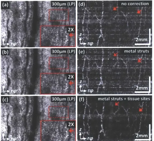
Due to the small size of the motor, a feedback control was not possible for the hollow shaft motor used in the 360-degree micromotor imaging catheter. Therefore, it is expected the presence of residual NURD in the OCT images and requires a correction algorithm to remove or suppress the NURD in the OCT images. Figure 2.4 shows the cross-sectional OCT images of (a) human finger and inner cheek as well as (b) rectum where no imaging field obstruction due to either motor electric wirings or metal housing struts was observed. Our group previously demonstrated the use of metal strut edges in the cross-sectional OCT images as fiducial markers to measure the instantaneous rotational speed of the micromotor to develop an NURD correction algorithm [137]. These locations were used to cubic spline resample the OCT data, such that the pixels in the transverse direction were spatially equally spaced. However, as shown in Fig. 2.4, no metal strut edges can be identified in the OCT images and thus an alternative method to measure the instantons motor speed needs to be developed.
Recently, our group successfully demonstrated the use of multiple regions of interest (ROts) within individual OCT frames to measure the instantaneous motor speed as an extension to the original method using the metal strut edges [140]. In this method, individual cross-sectional OCT images were divided into multiple ROIs with an identical number of Ascans. Then, the relative of shift of the middle Ascan in individual ROIs between adjacent frames was computed by calculating the cross-correlation similar to those demonstrated previously [141] but on a sub-pixel level [142]. Rotational speeds for all transverse sub-pixels were then estimated by applying a cubic spline interpolation to the rotation speed of the ROIs (the motion trace). Finally, a cubic
spline resampling was applied to the OCT data to produce an equal spacing between the transverse pixels. The same subpixel, cross-correlation based correction algorithm was used in this study to suppress the NURD in the OCT images. As shown in Figs. 2.4(a, b), multiple ROIs (red dashed box) were selected within the OCT frames and used to estimate the instantaneous rotation speed following the same algorithm described above [140]. Here, in particular, the motion trace of individual ROIs was manually examined afterward to remove those exhibiting slowly varying motion components such as the varying catheter-tissue contact due to the physiological motion prior to cubic spline interpolation step.
2.2.4 Endoscopic OCT angiography (OCTA) and data visualization
Before calculating the intensity decorrelation (D) between sequential OCT frames to identify the motion contrast from the moving erythrocytes within the microvascular network, an NURD correction algorithm was performed to remove or suppress the NURD exhibited in the volumetric OCT datasets (section 2.2.3). Then, the intensity decorrelation was calculated pixel-by-pixel between sequential NURD-corrected cross-sectional OCT frames in linear OCT signal intensity scale, following the formula listed below where A, is the OCT signal amplitude.
DYx2)1 [' : +"' .* (1):)
After the decorrelation calculation, a moving average of three consecutive decorrelation images was taken to suppress the background decorrelation noise. Finally, a threshold mask was applied to the averaged cross-sectional OCTA images to remove regions with low OCT signal where the
OCTA data is invalid [118, 121, 137, 140].
Prior to generating the depth-resolved en face OCT and OCTA images, the surface of the plastic sheath in the NURD corrected OCT images were identified using a graph cut, automatic segmentation algorithm [143]. Individual cross-sectional OCT and OCTA images in the volumetric OCT and OCTA dataset were shifted radially with respect to the plastic sheath identified to generate surface flattened volumetric OCT and OCTA datasets, respectively afterward. The depth resolved enface OCT and OCTA images were computed by using a mean projection over a 50 and 100 gm window at various depth level beneath sheath surface,
respectively. For example, an enface OCT image at 200 pLm below sheath surface was the mean projection of the OCT signal intensities from 176 to 225 pm below sheath surface. The enface OCT images were displayed using square root compression gray scale and cross-sectional OCT images were displayed using logarithmic gray compression scale. En face OCTA images were displayed in a linear gray scale. Here, the black color corresponds to a low signal level while white color as a high signal (inverted gray scale).
2.3 Results
2.3.1 NURD characterization
Figure 2.5 shows the NURD characterization of the 360-degree micromotor imaging catheter based on the measurement of 100 continuously acquired cross-sectional OCT images of human fingers. Noted that these images were acquired without pulling back the imaging catheter. Figure
2.5(a) shows the characteristic layered architectures of the human finger including epidermis and
dermis. As described in section 2.2.3, multiple ROIs with an identical number of Ascans within individual frames were used to measure the instantaneous motor speed (red dashed box, Fig.
2.5(a)). Figure 2.5(b) shows the angular deviation of the shift of the middle Ascan of one ROI in
Fig. 2.5(a). Compared to the same measurement using a micromotor catheter wth a conventional 2 mm OD, DC brushless micromotor (SBL02-06, Namiki Precision, CA) as shown in Fig. 2.5(c), the NURD was more severe in the 360-degree micromotor imaging catheter. Given a 3.4 mm catheter OD (including the plastic sheath), the standard deviation of the angular deviation was 113.4 pm (66.7 milliradians (mrad)), which is worse than 11.6 pm (6.8 mrad) of the conventional micromotor. The NURD performance of the 360-degree micromotor catheter makes it difficult to completely remove or suppress the NURD in the OCT images and enabling endoscopic OCTA with low decorrelation noise.
2.3.2 Human skin and buccal mucosa imaging
Figure 2.6 shows the coregistered enface OCT and OCTA images of the human finger and inner cheek (oral mucosa). The en face OCT and OCTA images were reconstructed from the surface flattened, NURD-corrected volumetric OCT and OCTA dataset at a depth of 260 pim beneath the tissue surface, which is within the epidermis and epithelium layer (EP) of the skin (finger) and