HAL Id: hal-01377207
https://hal.sorbonne-universite.fr/hal-01377207
Submitted on 6 Oct 2016
HAL is a multi-disciplinary open access archive for the deposit and dissemination of sci-entific research documents, whether they are pub-lished or not. The documents may come from
L’archive ouverte pluridisciplinaire HAL, est destinée au dépôt et à la diffusion de documents scientifiques de niveau recherche, publiés ou non, émanant des établissements d’enseignement et de
Low level exercise echocardiography helps diagnose early
stage heart failure with preserved ejection fraction: a
study of echocardiography versus catheterization
Nadjib Hammoudi, Florent Laveau, Gérard Helft, Nathalie Cozic, Olivier
Barthelemy, Alexandre Ceccaldi, Thibaut Petroni, Emmanuel Berman, Michel
Michel, Pierre-Louis Michel, et al.
To cite this version:
Nadjib Hammoudi, Florent Laveau, Gérard Helft, Nathalie Cozic, Olivier Barthelemy, et al.. Low level exercise echocardiography helps diagnose early stage heart failure with preserved ejection fraction: a study of echocardiography versus catheterization. Clinical Research in Cardiology, Springer Verlag, 2016, pp.1-10. �10.1007/s00392-016-1039-0�. �hal-01377207�
Low level exercise echocardiography helps diagnose early stage heart failure with
preserved ejection fraction: a study of echocardiography versus catheterization
Nadjib Hammoudi, MDa;
Florent Laveau, MDa; Gérard Helft, MD, PhDa; Nathalie Cozicb; Olivier Barthelemy, MDa; Alexandre Ceccaldia, MD; Thibaut Petroni, MDa; Emmanuel Bermana; Michel Komajda, MDa; Pierre-Louis Michel, MDa; Alain Mallet MD, PhDb; Claude Le Feuvre, MDa; Richard Isnard, MDa
a Université Paris 6, Institut de Cardiologie (AP-HP), Centre Hospitalier Universitaire Pitié-Salpêtrière, Institute
of Cardiometabolism and Nutrition (ICAN), INSERM UMRS 1166, Paris, France, ACTION Study Group.
b Université Paris 6, Département de Biostatistiques, Centre Hospitalier Universitaire Pitié-Salpêtrière, Paris,
France.
Address for correspondence
Nadjib Hammoudi, Institut de Cardiologie, Groupe Hospitalier Pitié-Salpêtrière, AP-HP, 47-83, boulevard de l'Hôpital, 75651 Paris Cedex 13. E-mail: nadjib.hammoudi@psl.aphp.fr. Telephone number: +33142165535. Fax number: +33142163020.
Acknowledgements
We thank Anissa Bouzamondo, Anne-Claire Coyne and Justine Pollet (French Society of Cardiology) for logistical and data-managing support as well as Sylvie Lang for careful review of the manuscript.
Funding
This work was supported by the French Federation of Cardiology and the French Society of Cardiology.
ABSTRACT
Background: Increased left ventricular end diastolic pressure (LVEDP) with exercise is an
early sign of heart failure with preserved left ventricular ejection fraction (LVEF). The
abnormal exercise increase in LVEDP is nonlinear, with most change occurring at low-level
exercise. Data on noninvasive approach of this condition are scarce. Our objective was assessing E/e’ to estimate low level exercise LVEDP using a direct invasive measurement as
the reference method.
Methods and Results: Sixty patients with LVEF>50% prospectivelyunderwent exercise cardiac catheterization and echocardiography. E/e’ was measured at rest and during low-level
exercise. Abnormal LVEDP was defined as>16mmHg. Patients with a history of coronary
artery disease and/or abnormal LV morphology were classified as having apparent cardiac
disease (CD).
Thirty-four (57%) patients had elevated LVEDP only during exercise. Most of the change in
LVEDP occurred since the first exercise level (25Watts). There was a correlation between LVEDP and septal E/e’ at rest and during exercise. Lateral E/e’ and E/average e’ ratio had
worse correlations with LVEDP.In the whole population, exercise septal E/e’ at 25 Watts had
the best accuracy for abnormal exercise LVEDP, area under curve (AUC)=0.79. However,
while low-level exercise septal E/e’ had a high accuracy in CD patients (n=26, AUC=0.96), E/e’ was not linked to LVEDP in patients without CD (n=34).
Conclusion: Low-level exercise septal E/e’ is valuable for predicting abnormal exercise
LVEDP in patients with preserved LVEF and apparent CD. However, this new diagnosis
approach appears not reliable in patients with normal LV morphology and without coronary
INTRODUCTION
Heart failure (HF) with preserved ejection fraction (pEF) is increasing in prevalence and is
becoming the predominant form of HF.[1–3] While cardiac dysfunction is readily apparent
when the left ventricular ejection fraction (LVEF) is reduced,[4] the diagnosis of HFpEF is
often not straightforward,[5] particularly in the early stages of the disease.[6] Identifying
HFpEF at an early stage, when the structural changes in the heart and vasculature are still
reversible, may be important for improving the prognosis of the disease.[7] Invasive
hemodynamics have demonstrated relevance for the diagnosis of early stage of HFpEF, which
is characterized by exercise-induced left ventricular filling pressure (LVFP) elevation despite
normal resting pressures.[6],[8] Interestingly, the abnormal exercise increase in LVFP is
nonlinear, with most change occurring at low level exercise.[6]
Data on noninvasive approach of exercise LVFP more suitable for routine clinical use are
scarce. The value of Doppler echocardiography using the ratio of early diastolic transmitral
velocity to tissue velocity (E/e’) for the noninvasive evaluation of LVFP has been reported.[9,
10] Nevertheless, there are limited and conflicting data on the validation of exercise E/e’ with
invasively determined LVFP in patients with preserved LVEF.[11, 12] Moreover, whereas
latent heart failure could be unmasked during exercise in patients with apparently normal LV
structure and function at rest,[6, 13] E/e’ was reported as less valuable in patients without
evidence of cardiac disease (CD) at steady state.[14–16] Thus the clinical integration of
exercise E/e’ in daily clinical practice is debated.[17–19]
The aim of this study was to assess the E/e’ ratio for the estimation of low level exercise
LVFP using a direct invasive measurement as the reference method in patients at high risk of
METHODS
Ethics statement
This study was carried out in accordance with the Helsinki Declaration and was approved by
the local institutional committee on human research. Written informed consent was obtained
from each patient before participation.
Patient population
We prospectively enrolled 60 patients in sinus rhythm with LVEF >50% and undergoing
clinically indicated left heart catheterization using a transradial approach, which allowed for
the invasive measurement of left ventricular end-diastolic pressure (LVEDP). The risk factors
of coronary artery disease, especially age, hypertension and diabetes, are also recognized risk
factors of HFpEF. Thus, the patients referred for coronary angiography are also at high risk of
HFpEF.[2]
Patients in whom the radial artery puncture was not feasible and those who were unable to
pedal were excluded. The other exclusion criteria were more than mild mitral regurgitation,
severe aortic stenosis, mitral stenosis, valvular prosthesis, severe mitral annular calcification,
hypertrophic cardiomyopathy, heart transplantation, LV thrombus, severe renal failure
(<30ml/min/1.73m²), atrial fibrillation, an acute coronary syndrome in the previous 3 months
and/or the diagnosis during coronary angiography of a stenosis requiring revascularization.
The baseline evaluation comprised the collection of medical history, a physical examination,
electrocardiogram and blood sampling for laboratory measurements including N-terminal
pro-brain natriuretic peptide (NT-pro BNP).
Patients with apparent CD were defined as having either clinical history of coronary artery
>50% on coronary angiography or echocardiographic evidence of LV structural abnormality
(hypertrophy or remodeling) as defined by the echocardiography guidelines.[15, 16, 20]
Invasive hemodynamic assessment
After coronary angiography, a fluid-filled pigtail catheter was positioned in the mid-LV
cavity. Transducers were balanced before the study with the zero level at the mid-axillary
line. LV pressure tracings were recorded at baseline and during exercise. Measurements were
made off-line by two experienced physicians who were blinded to echocardiography and
clinical data. LVEDP were averaged over five cardiac cycles, and ectopic or post-ectopic
beats weredisregarded. LVEDP >16 mmHg was used as a threshold to define abnormal
LVFP at rest and during exercise.[21, 22]
Echocardiographic studies
Transthoracic echocardiography was performed in all patients using the Vivid 9 system. The
images were transferred to a workstation equipped with the Echopac PC software (GE
Vingmed; Horten, Norway). All the examinations were analyzed off-line by two senior
cardiologists who were blinded to the clinical data and invasive measurements. All
projections were obtained according to the guidelines.[20] All measurements were averaged
over 3 to 6 cardiac cycles. Ectopic and post-ectopic beats were disregarded.
In M-mode, the following measurements were made at end diastole: LV internal diameter and
inter-ventricular septal and posterior wall thicknesses. LV mass was derived and indexed to
body surface area. Relative wall thickness was also calculated.[20]
From the 2-dimensional mode, the LA maximal volume was measured by the biplane
Simpson method and was indexed to body surface area (LAVi). LV volumes and LVEF were
measured using the biplane Simpson method.[20] From the continuous wave Doppler, peak
From the pulsed wave Doppler mode positioned at the tip of the mitral valve, early (E) and
late (A) peak diastolic velocities of the mitral inflow and E-wave deceleration time were
measured. The septal, lateral and average early diastolic velocities were recorded (e’), and the ratio of mitral flow E to e’ for each of these annular velocities was calculated.[16, 23] In
addition to steady state measures, the E and e’ wave velocities were recorded, and the
different E/e’ ratios were calculated during exercise. In cases of complete merging of E and A
for the mitral inflow, peak E was measured at the peak of the single wave form. With
complete merging of e′ and a’, the resulting velocity was taken as e′.[24]
Exercise protocol
A similar two leg pedaling exercise was performed within 24hours in the catheterization
laboratory using ERG 911 BP/XRAY and in the echocardiography laboratory using ERG 911
L/LS (Schiller, Switzerland). Patients in the supine position pedaled at a constant speed
beginning at a workload of 25 Watts for 3 minutes, followed by a workload of 50 Watts for an
additional 3 minutes. Invasive and non-invasive data were stored at baseline and during the
last minute of both levels of exercise. Relatively low levels of exercise were chosen because
obviously all the patients can reach this threshold,[25] andinvasive LV pressure tracings, as
well as echocardiographic image quality, are well maintained at this workload.[19, 26]
Moreover, most of the changes in LVFP occur during this low level of exercise.[6] Patient
medications were not modified between invasive and non-invasive studies.
Statistical analysis
All quantitative data are expressed as medians and interquartile ranges, and qualitative data
are expressed as numbers and percentages. The patients were classified according to LVEDP:
group 1 was normal LVEDP at rest and during exercise; group 2 was normal LVEDP at rest
during exercise. The data were compared using the Fisher and Kruskal–Wallis exact tests. The
Wilcoxon signed rank test was used for paired data analysis.
Correlations were evaluated using the Pearson test. To detect a correlation >0.4 between
LVEDP and echocardiography parameters with 90% power and a 2-sided alpha level of 0.05,
a sample size of 60 patients was calculated. Receiver-operator characteristics (ROC) curves
were plotted to assess E/e’ to predict exercise LVEDP >16 mmHg. The analyses were
performed in the overall population and in subgroups of patients with or without apparent CD.
Intraobserver and interobserver variabilities for the measurement of E/e’ ratio at rest and
during exercise were assessed in a subset of 10 patients. The coefficient of variation and
intraclass correlation coefficient were determined and presented in supplemental table 1.
MedCalc Statistical Software version 12.7.7 (MedCalc Software, Belgium) was used for the
calculations. A p<0.05 indicated statistical significance.
RESULTS
Population characteristics
The baseline characteristics of the patients are presented in Table 1. The patients' median age
was 63.5 years. Thirty-eight (63%) patients had hypertension and 16 (27%) had diabetes.
Thirteen (22%) patients had coronary stenosis >50%; however, none of these patients had a
stenosis that required a percutaneous intervention. Among these patients, we observed no
functional ischemia based on fractional flow reserve (n=6), stenosis of a small artery with a
diameter <2 mm (n=6) and a right coronary artery occlusion without significant stenosis on
other arteries (n=1). No patient had a history of congestive HF.
Invasive hemodynamic measurements
The invasive hemodynamic data are presented in Table 2. LVEDP at rest and in at least 1
after the first level of exercise. In 2 other patients, the LV pressure curve could not be
properly analyzed at one of the two exercise stages.
LVEDP was normal in 46 (77%) patients at rest. In 12 (20%) of these patients, LVEDP
remained normal during exercise (group 1), and in 34 (57%) patients, it became elevated
(group 2). The remaining 14 (23%) patients had elevated LVEDP at rest and during exercise
(group 3). Most of the change in LVEDP occurred within the first 3 minutes of exercise at a
very low-level workload (Figure 1), 82% of the patients classified in group 2 had abnormal
LVEDP since the first exercise level.
NT-pro BNP level was significantly higher in group 3 compared to group 1 and to group 2
respectively; however, NT-pro BNP levels in group 1 and group 2 were similar (Table 1).
Twenty-four (40%) patients experienced a dyspnea during low level exercise, and all of these
patients had an abnormal LVEDP at rest and/or during exercise (Table 2).
Echocardiography
The median time between echocardiography and catheterization was 4h36 min [3h05min-8 h].
The ultrasound measurements are reported in Table 3.
E/e’ ratio
E and A waves were fused in 4 patients at 25 Watts and in 8 patients at 50 Watts.
In whole population, there was a correlation between LVEDP and septal E/e’ at rest (r=0.28)
and during exercise (r=0.34). Lateral E/e’ was associated with LVEDP at 25 Watts of exercise
(r=0.27). However, lateral E/e’ was not linked to LVEDP at rest or at 50 Watts of exercise
(supplemental figure). Compared with septal E/e’, the E/average e’ ratio had a worse
correlation with LVEDP (Table 4).
Although, septal E/e’ ratio at rest was similar between group 1 and group 2; exercise septal
The ROC curve analysis for prediction of abnormal LVEDP from E/e’ ratios showed that Septal E/e’ at 25 Watts with an area under the curve (AUC) of 0.79 had the best diagnostic
value (Figure 2). The optimal septal E/e’ at 25 Watt threshold ≥8 had a sensitivity of 71% and
a specificity of 83% for predicting abnormal LVEDP.
Performance of E/e’ in patients with and without apparent CD
Because E/e’ ratio has been reported as a non-optimal index of LVFP in patients without
apparent heart disease,[14–16] the population was divided into patients with (n=26) and
without (n=34) apparent CD. The both groups had similar demographic and hemodynamic
characteristics (supplemental table 2). In particular, the prevalence of abnormal LVEDP only
during exercise (p=0.61) and the median time between echocardiography and catheterization
(p=0.96) were similar in both groups.
Compared with the whole population, Doppler-derived parameters were better linked to
LVEDP in patients with apparent CD. The best observed correlation was between septal E/e’
and LVEDP at 25 Watts of exercise (r=0.59, p=0.002). In contrast, no significant correlation was observed between E/e’ and LVEDP in patients without CD (supplemental table 3).
In patients with CD, the ROC curves analysis for prediction of abnormal LVEDP showed that
septal E/e’ at 25 Watts had the best diagnostic value (AUC=0.96). The optimal septal E/e’ at 25 Watts ≥8 had a sensitivity of 90% and a specificity of 100%. In patients without CD, the
DISCUSSION
The results of the study can be summarized as follows. First, we confirmed that exercise
induced abnormal LVEDP in a number of patients with normal LVEDP at rest and that most
of the changes in LVEDP occurred at a very low-level workload. Second, we observed a correlation between LVEDP and septal E/e’ at rest and during exercise; however, lateral E/e’
and E/average e’ ratio had a worse correlation with LVEDP. Finally, we found that septal E/e’
at 25 Watts of exercise was valuable for predicting abnormal exercise LVEDP. However, in
subgroup analysis, although exercise septal E/e’ has a very good diagnostic performance in
patients with apparent CD, this parameter had no significant diagnostic value in the subgroup
of patients with apparently normal LV morphology and no history of coronary artery disease.
Invasive hemodynamics
While cardiac dysfunction is readily apparent when the LVEF is reduced, the diagnosis of HF
is not straightforward when LVEF is preserved.[6] Recent data indicate that the early stage of
HFpEF, which is characterized by exercise-induced LVFP elevation despite normal resting
pressures, represents a pathological entity linked to symptoms and long-term survival.[8] The
invasive assessment of LVFP at peak exercise is feasible;[6, 8] however, this time-consuming
approach is not optimal for routine clinical use and limits its dissemination. As we reported in
our study and as observed previously,[6] the abnormal increase in LVFP with exercise is
nonlinear, with most change occurring during the first level of exercise. Thus, the LVFP
achieved at the first level of exercise could help diagnose an early stage of HFpEF in clinical
practice.[7]
E/e’ ratio
recently challenged [27] and validation data on exercise E/e’ in patients with preserved LVEF
are limited. In fact, of the 37 patients included in the validation study of exercise E/e’
compared to direct invasive measures of LV diastolic pressure, six (16%) had LVEF
<45%.[10] Moreover, using pulmonary capillary wedge pressure (PCWP) as reference
method, two smaller studies including 12 and 22 patients respectively, reported conflicting
results.[11, 12]
For the non-invasive estimation of LVFP at rest, current guidelines encourage the use the ratio
between the preload-dependent transmitral E velocity and e’ velocity, which is considered
relatively load independent.[16, 28] However, preload affects e’ in ventricles with normal
relaxation,[29] and E/e’ has lower accuracy in patients without apparent CD because e’ itself
is altered by volume shifts.[14, 15] Consistently in the guidelines, it is stated that E/e’ ratio
should not be used to assess LVFP in subjects with likely normal LV relaxation as defined by
no history of heart disease along with normal cardiac structure and function.[16, 28] This
physiological fact may partially explain the conflicting results in previously reported studies investigated E/e’ for exercise LVFP estimation.[10–12] In the former study showing the
reliability of E/e’ to identify patients with elevated exercise LVFP, the prevalence of coronary
artery stenosis was high (75%).[10] In another study reporting conflicting results, patients
with coronary artery disease were excluded.[12] Finally, another study excluding patients
with coronary stenosis showed that E/e’ was not valuable to estimate PCWP variation in
patients with preserved LVEF subjected to preload manipulation.[30]
Using a direct sampling of LV cavity during left heart catheterization as reference method, our study is the largest prospective study aimed to investigate exercise E/e’ in patients with
preserved LVEF. The relatively large and carefully phenotyped population included allowed a
subgroup analysis of patients with and without apparent CD. We found that exercise E/e’ had a mild diagnostic value of abnormal LVEDP in the whole population. However, although E/e’
had no diagnostic value in patients without CD, this index had a good value to predict
abnormal LVEDP in patients with apparent CD. Our data further indicate that E/e’ should not
be used for the assessment of LVFP in patients with preserved LVEF and without apparent
CD.[16] To choose the appropriate ultrasound approach, our findings suggest the collection of
patient clinical history and the assessment of LV morphology as the first step of the
noninvasive estimation of exercise LVFP.As reported previously, we observed that latent HF
could be unmasked during exercise in high risk patients with apparently normal LV structure
and function at rest.[6, 13] The best non-invasive approach of exercise LVFP in patients
without apparent LV structural abnormality and/or history of coronary artery disease remains
to be investigated.
We found that septal E/e’ had better accuracy compared with lateral E/e’ for the diagnosis of
exercise LVEDP >16 mmHg. In previously reported studies on the same field, lateral E/e’
was not investigated.[10–12] The septal side of the mitral annulus, in contrast to the lateral
side, moves in a direction more parallel to the ultrasound beam and is less affected by the
translational movement of the heart.
Clinical implications
The risk factors of HFpEF, especially age, hypertension and diabetes, are also recognized risk
factors of coronary stenosis.[2] Moreover, the symptoms of coronary diseases are not specific
and might also be suggestive of HFpEF.[2]In our study, 34 (57%) of the patients with normal
or subnormal coronary arteries and normal LVEDP at rest were diagnosed as having abnormal
exercise LVEDP. These data indicate that the stress protocol used in our study, which
combined radial artery puncture and low level stress exercise, could be clinically relevant in
lesion explains their symptoms and LVEDP is normal at rest. However, the efficiency of this
diagnostic strategy should be validated in larger populations.
Concerning non-invasive diagnosis of abnormal exercise LVFP, our data indicate that
exercise echocardiography at very low workload helps diagnose early stage HFpEF. This
approach is suitable for routine clinical use. Compared to E/e’ evaluation at peak exercise, low level workload E/e’ could be combined to the resting ultrasound evaluation in selected
population at high risk. This approach could be also easily integrated in current stress
echocardiography protocols designed to avoid LV ischemia. Moreover, the uncommon rate of
complete E and A merging at low level exercise facilitates Doppler analysis.[19, 26] Exercise
septal E/e’ appears to be superior to lateral and E/ average e’ ratio. Finally, E/e’ appears to be
reliable only in patients with apparent CD and should not be used for the assessment of
exercise LVFP in patients with normal LV morphology and no history of coronary artery
disease.
Study limitations
We included a relatively small population of patients. However, the sample size was larger
compared with prior reports that investigated the ultrasound for similar purpose.[10–12] The
invasive and ultrasound acquisitions were not performed simultaneously. Performing
simultaneous assessments may have enhanced the correlations between the two methods;
however, it would be difficult to obtain good quality images in the catheterization laboratory,
and this attempt would introduce another type of bias by excluding patients due to inadequate
CONCLUSION
The early stage of HFpEF is characterized by exercise-induced LVFP elevation despite
normal resting measurements. The abnormal increase in LVFP with exercise is nonlinear,
with most change occurring during the first level of exercise. Low level exercise septal E/e’ is
valuable for predicting abnormal exercise LVEDP in patients with preserved LVEF and
apparent CD. However, the non-invasive approach of LVEDP in patients with normal LV
morphology and no history of coronary disease needs further investigations. The reliable
noninvasive identification of early stages of HFpEF is the first step for clinical trials that
DISCLOSURES
Dr. Laveau was supported by a grant from the French Federation of Cardiology. Dr. Komajda
has performed consulting/advisory activities for Servier, Bristol-Myers Squibb, AstraZeneca,
Menarini, Novartis, MSD, and Sanofi-Aventis. The other authors have no conflict of interest
REFERENCES
1. Steinberg BA, Zhao X, Heidenreich PA, et al (2012) Trends in patients hospitalized with heart failure and preserved left ventricular ejection fraction: prevalence, therapies, and outcomes. Circulation 126:65–75. doi: 10.1161/CIRCULATIONAHA.111.080770 2. Borlaug BA (2014) The pathophysiology of heart failure with preserved ejection
fraction. Nat Rev Cardiol 11:507–515. doi: 10.1038/nrcardio.2014.83
3. Meinertz T, Diegeler A, Stiller B, et al (2015) German Heart Report 2013. Clin Res Cardiol Off J Ger Card Soc 104:112–123. doi: 10.1007/s00392-014-0799-7
4. von Scheidt W, Zugck C, Pauschinger M, et al (2014) Characteristics, management modalities and outcome in chronic systolic heart failure patients treated in tertiary care centers: results from the EVIdence based TreAtment in Heart Failure (EVITA-HF) registry. Clin Res Cardiol Off J Ger Card Soc 103:1006–1014. doi: 10.1007/s00392-014-0743-x
5. Kasner M, Sinning D, Burkhoff D, Tschöpe C (2015) Diastolic pressure-volume quotient (DPVQ) as a novel echocardiographic index for estimation of LV stiffness in HFpEF. Clin Res Cardiol Off J Ger Card Soc 104:955–963. doi: 10.1007/s00392-015-0863-y
6. Borlaug BA, Nishimura RA, Sorajja P, et al (2010) Exercise Hemodynamics Enhance Diagnosis of Early Heart Failure With Preserved Ejection Fraction. Circ Heart Fail 3:588–595. doi: 10.1161/CIRCHEARTFAILURE.109.930701
7. Borlaug BA (2014) Exercise haemodynamics and outcome in patients with dyspnoea. Eur Heart J 35:3085–3087. doi: 10.1093/eurheartj/ehu350
8. Dorfs S, Zeh W, Hochholzer W, et al (2014) Pulmonary capillary wedge pressure during exercise and long-term mortality in patients with suspected heart failure with preserved ejection fraction. Eur Heart J 35:3103–3112. doi: 10.1093/eurheartj/ehu315
9. Ha J-W, Oh JK, Pellikka PA, et al (2005) Diastolic stress echocardiography: A novel noninvasive diagnostic test for diastolic dysfunction using supine bicycle exercise Doppler echocardiography. J Am Soc Echocardiogr 18:63–68. doi:
10.1016/j.echo.2004.08.033
10. Burgess MI, Jenkins C, Sharman JE, Marwick TH (2006) Diastolic Stress
Echocardiography: Hemodynamic Validation and Clinical Significance of Estimation of Ventricular Filling Pressure With Exercise. J Am Coll Cardiol 47:1891–1900. doi: 10.1016/j.jacc.2006.02.042
11. Talreja DR, Nishimura RA, Oh JK (2007) Estimation of Left Ventricular Filling Pressure with Exercise by Doppler Echocardiography in Patients with Normal Systolic Function: A Simultaneous Echocardiographic–Cardiac Catheterization Study. J Am Soc Echocardiogr 20:477–479. doi: 10.1016/j.echo.2006.10.005
12. Maeder MT, Thompson BR, Brunner-La Rocca H-P, Kaye DM (2010) Hemodynamic basis of exercise limitation in patients with heart failure and normal ejection fraction. J Am Coll Cardiol 56:855–863. doi: 10.1016/j.jacc.2010.04.040
13. Tan YT, Wenzelburger F, Lee E, et al (2010) Abnormal left ventricular function occurs on exercise in well-treated hypertensive subjects with normal resting echocardiography. Heart Br Card Soc 96:948–955. doi: 10.1136/hrt.2009.185181
14. Firstenberg MS, Levine BD, Garcia MJ, et al (2000) Relationship of echocardiographic indices to pulmonary capillary wedge pressures in healthy volunteers. J Am Coll Cardiol 36:1664–1669.
15. Dokainish H, Zoghbi WA, Lakkis NM, et al (2004) Optimal noninvasive assessment of left ventricular filling pressures: a comparison of tissue Doppler echocardiography and B-type natriuretic peptide in patients with pulmonary artery catheters. Circulation 109:2432–2439. doi: 10.1161/01.CIR.0000127882.58426.7A
16. Nagueh SF, Appleton CP, Gillebert TC, et al (2009) Recommendations for the
evaluation of left ventricular diastolic function by echocardiography. Eur J Echocardiogr J Work Group Echocardiogr Eur Soc Cardiol 10:165–193. doi:
10.1093/ejechocard/jep007
17. Oh JK, Kane GC (2014) Diastolic stress echocardiography: the time has come for its integration into clinical practice. J Am Soc Echocardiogr Off Publ Am Soc Echocardiogr 27:1060–1063. doi: 10.1016/j.echo.2014.08.013
18. Hammoudi N, Achkar M, Isnard R (2015) Exercise E/e’ for the assessment of left ventricular filling pressures: with caution in clinical practice? J Am Soc Echocardiogr Off Publ Am Soc Echocardiogr 28:376–377. doi: 10.1016/j.echo.2014.11.001
19. Erdei T, Smiseth OA, Marino P, Fraser AG (2014) A systematic review of diastolic stress tests in heart failure with preserved ejection fraction, with proposals from the EU-FP7 MEDIA study group. Eur J Heart Fail 16:1345–1361. doi: 10.1002/ejhf.184
20. Lang RM, Badano LP, Mor-Avi V, et al (2015) Recommendations for cardiac chamber quantification by echocardiography in adults: an update from the American Society of Echocardiography and the European Association of Cardiovascular Imaging. Eur Heart J Cardiovasc Imaging 16:233–270. doi: 10.1093/ehjci/jev014
21. Penicka M, Bartunek J, Trakalova H, et al (2010) Heart Failure With Preserved Ejection Fraction in Outpatients With Unexplained Dyspnea. J Am Coll Cardiol 55:1701–1710. doi: 10.1016/j.jacc.2009.11.076
22. Borlaug BA, Paulus WJ (2011) Heart failure with preserved ejection fraction: pathophysiology, diagnosis, and treatment. Eur Heart J 32:670–679. doi: 10.1093/eurheartj/ehq426
23. Buss SJ, Krautz B, Schnackenburg B, et al (2014) Classification of diastolic function with phase-contrast cardiac magnetic resonance imaging: validation with
echocardiography and age-related reference values. Clin Res Cardiol Off J Ger Card Soc 103:441–450. doi: 10.1007/s00392-014-0669-3
24. Nagueh SF, Mikati I, Kopelen HA, et al (1998) Doppler estimation of left ventricular filling pressure in sinus tachycardia. A new application of tissue doppler imaging. Circulation 98:1644–1650.
25. Voss F, Schueler M, Lauterbach M, et al (2016) Safety of symptom-limited exercise testing in a big cohort of a modern ICD population. Clin Res Cardiol Off J Ger Card Soc 105:53–58. doi: 10.1007/s00392-015-0885-5
26. Hammoudi N, Achkar M, Laveau F, et al (2014) Left atrial volume predicts abnormal exercise left ventricular filling pressure. Eur J Heart Fail 16:1089–1095. doi:
10.1002/ejhf.131
27. Santos M, Rivero J, McCullough SD, et al (2015) E/e’ Ratio in Patients With
Unexplained Dyspnea: Lack of Accuracy in Estimating Left Ventricular Filling Pressure. Circ Heart Fail 8:749–756. doi: 10.1161/CIRCHEARTFAILURE.115.002161
28. Nagueh SF, Smiseth OA, Appleton CP, et al (2016) Recommendations for the Evaluation of Left Ventricular Diastolic Function by Echocardiography: An Update from the American Society of Echocardiography and the European Association of Cardiovascular Imaging. J Am Soc Echocardiogr Off Publ Am Soc Echocardiogr 29:277–314. doi: 10.1016/j.echo.2016.01.011
29. Firstenberg MS, Greenberg NL, Main ML, et al (2001) Determinants of diastolic myocardial tissue Doppler velocities: influences of relaxation and preload. J Appl Physiol Bethesda Md 1985 90:299–307.
30. Bhella PS, Pacini EL, Prasad A, et al (2011) Echocardiographic indices do not reliably track changes in left-sided filling pressure in healthy subjects or patients with heart failure with preserved ejection fraction. Circ Cardiovasc Imaging 4:482–489. doi: 10.1161/CIRCIMAGING.110.960575
FIGURE LEGENDS
Figure 1. LVEDP at rest and during exercise in the 3 groups of patients. Most of the changes
in LVEDP occurred during the first level of exercise.
Group 1, normal LVEDP; Group 2, abnormal LVEDP only during exercise; Group 3,
abnormal LVEDP at rest and during exercise; LVEDP, left ventricular end diastolic pressure. Within group analyses: *p<0.05 versus at rest; †p<0.0001 versus at rest; ‡p<0.0001 versus at
25Watts.
Figure 2. Receiver-operator characteristic curves for the prediction of exercise LVEDP >16
mmHg in the whole study population using septal (A) and lateral (B) E/e’ ratios at rest and
during exercise.
AUC, area under the curve; LVEDP, left ventricular end diastolic pressure.
Figure 3. Typical low level exercise echocardiography findings in two patients with exercise
induced abnormal LVEDP. While exercise septal E/e’ increases in the patient with apparent
CD, this parameter was not modified in the patient without CD.
CD, patient with apparent cardiac disease (ie, coronary artery disease or echocardiographic evidence of left ventricular structural abnormality); E/e’, the ratio of early diastolic transmitral
TABLES
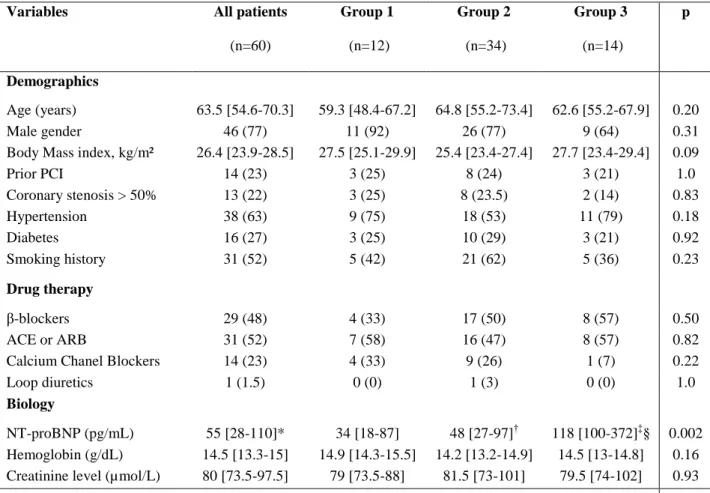
Table 1: Clinical characteristics of the study population.
Data are expressed as median and interquartile range or number (%). *n=57, † n=33, ‡ n=12
§ p < 0.05 compared to group 1 and to group 2.
ACE, Angiotensin converting enzyme inhibitor; ARB, Angiotensin receptor blocker; Group 1, normal LVEDP; Group 2, abnormal LVEDP only during exercise; Group 3, abnormal LVEDP at rest; LVEDP, left ventricular end diastolic pressure; NT-proBNP, N-terminal pro brain natriuretic peptide; PCI, percutaneous coronary intervention.
Variables All patients
(n=60) Group 1 (n=12) Group 2 (n=34) Group 3 (n=14) p Demographics Age (years) 63.5 [54.6-70.3] 59.3 [48.4-67.2] 64.8 [55.2-73.4] 62.6 [55.2-67.9] 0.20 Male gender 46 (77) 11 (92) 26 (77) 9 (64) 0.31 Body Mass index, kg/m²
(((kg/m²)(kg/m2) 26.4 [23.9-28.5] 27.5 [25.1-29.9] 25.4 [23.4-27.4] 27.7 [23.4-29.4] 0.09 Prior PCI 14 (23) 3 (25) 8 (24) 3 (21) 1.0 Coronary stenosis > 50% 13 (22) 3 (25) 8 (23.5) 2 (14) 0.83 Hypertension 38 (63) 9 (75) 18 (53) 11 (79) 0.18 Diabetes 16 (27) 3 (25) 10 (29) 3 (21) 0.92 Smoking history 31 (52) 5 (42) 21 (62) 5 (36) 0.23 Drug therapy β-blockers 29 (48) 4 (33) 17 (50) 8 (57) 0.50 ACE or ARB 31 (52) 7 (58) 16 (47) 8 (57) 0.82 Calcium Chanel Blockers 14 (23) 4 (33) 9 (26) 1 (7) 0.22 Loop diuretics 1 (1.5) 0 (0) 1 (3) 0 (0) 1.0
Biology
NT-proBNP (pg/mL) 55 [28-110]* 34 [18-87] 48 [27-97]† 118 [100-372]‡§ 0.002 Hemoglobin (g/dL) 14.5 [13.3-15] 14.9 [14.3-15.5] 14.2 [13.2-14.9] 14.5 [13-14.8] 0.16 Creatinine level (µmol/L) 80 [73.5-97.5] 79 [73.5-88] 81.5 [73-101] 79.5 [74-102] 0.93
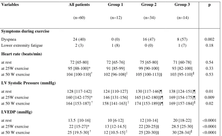
Table 2: Invasive hemodynamic data.
Variables All patients
(n=60) Group 1 (n=12) Group 2 (n=34) Group 3 (n=14) p
Symptoms during exercise
Dyspnea 24 (40) 0 (0) 16 (47) 8 (57) 0.002 Lower extremity fatigue 2 (3) 1 (8) 0 (0) 1 (7) 0.18
Heart rate (beats/min)
at rest 72 [65-80] 72 [65-76] 75 [65-80] 71 [60-78] 0.54 at 25W exercise 95 [88-100]* 91 [85-99] 99 [90-100] 93 [82-100] 0.33 0ln,nb,,;n ;,n;,n;,n 0.53 at 50 W exercise 104 [100-110]† 102 [96-108]‡ 105 [100-113]§ 103 [95-110]║ 0.53 LV Systolic Pressure (mmHg) at rest 128 [117-142] 124 [110-127] 130 [117-146]¶ 138 [124-151]¶ 0.01 at 25W exercise 160 [142-175]* 146 [131-156] 165 [142-180]§¶ 169 [154-175]¶ 0.009 at 50 W exercise 164 [153-187] † 158 [141-163] ‡ 174 [153-189]§¶ 169 [157-184]║ * 0.02 LVEDP (mmHg) at rest 13.5 [10-16] 10 [6-12] 12 [10-14] 20 [18-22] <0.0001 at 25W exercise 22 [15-27]* 13 [12-14.5] 22 [20-25]§ 28.5 [25-30] <0.0001 at 50 W exercise 25 [19.5-30] † 12 [10.5-15] ‡ 25 [20-30]§ 30 [28-34]║ <0.0001
Data are expressed as median and interquartile range.
* n=59, † n=57, ‡ n=11, § n=33, ║ n=13, ¶p<0.05 compared to group 1
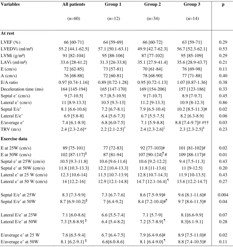
Table 3: Echocardiographic data.
Variables All patients
(n=60) Group 1 (n=12) Group 2 (n=34) Group 3 (n=14) p At rest LVEF (%) 66 [60-71] 64 [59-69] 66 [60-72] 63 [59-71] 0.29 LVEDVi (ml/m²) 55.2 [44.1-62.5] 57.1 [50.1-63.1] 49.9 [42.7-62.3] 56.7 [52.3-62.1] 0.53 LVMi (g/m²) 91 [82-104] 95 [88-106] 87 [77-102] 95 [85-109] 0.29 LAVi (ml/m²) 33.6 [28-41.2] 31.3 [26-33.8] 35.1 [27.9-41.4] 35.6 [28.9-43.7] 0.21 E (cm/s) 72 [62-85] 73 [57-81] 70 [61-84] 76 [69-98] 0.11 A (cm/s) 76 [68-88] 72 [60-81] 78 [68-90] 77 [71-88] 0.40 E/A ratio 0.97 [0.74-1.16] 0.89 [0.72-1.28] 0.95 [0.72-1.13] 1.07 [0.87-1.36] 0.38 Deceleration time (ms) 164 [145-194] 165 [147-170] 169 [154-206] 157 [123-186] 0.33 Septal e’ (cm/s) 9 [7-10.5] 9.7 [8.5-10.9] 9 [7-10.7] 8.9 [7-9.7] 0.45 Lateral e’ (cm/s) 11 [8.9-13.3] 10.5 [9.3-13] 11.2 [9-13.3] 10.9 [8-12.3] 0.86 Septal E/e’ 8.1 [6.6-10.6] 7.2 [6.7-8.1] 7.9 [6.5-10.4] 10.2 [8.5-11.3]# 0.02 Lateral E/e’ 6.9 [5.8-8] 6.4 [5.6-7.3] 6.7 [5.5-7.5] 8.2 [6.3-8.9] 0.06 E/average e’ 7.4 [6.1-8.9] 6.8 [6.0-7.5] 7.1 [5.9-8.8] 8.8 [7.4-9.7]# #†† 0.03 TRV (m/s) 2.4 [2.3-2.6]* 2.2 [2.1-2.5]† 2.4 [2.3-2.6]‡ 2.3 [2.3-2.5]§ 0.23 Exercise data E at 25W (cm/s) 89 [75-101] 77 [72-83] 92 [77-103]# 101 [81-102]# 0.02 E at 50W (cm/s) 102 [87-117]║ 87 [81-94] 107 [90-124]¶ # 109 [88-117]# 0.01 Septal e’ at 25W (cm/s) 10.5 [9.3-11.8] 10.6 [9.6-11.6] 10.6 [9.2-12.2] 9.4 [7.5-11.3] 0.43 Septal e’ at 50W (cm/s) 11.8 [10.3-13.3] 12.2 [10.4-13.3] 11.8 [11-13.6] 11 [9-12.5] 0.20 Lateral e’ at 25 W (cm/s) 12.3 [10.6-14] 11.5 [10.7-13.9] 12.8 [10.7-14.3] 11.9 [10-13.5] 0.43 Lateral e’ at 50 W (cm/s) 14 [12.2-16] 12.9 [12.1-14.8] 14.7 [12.1-16.4] ¶ 13.6 [12.2-14.7] 0.27 Septal E/e’ at 25W 8.3 [7.3-9.9] 7.3 [6.7-7.6] 8.6 [7.5-9.9]# 9.6 [8.1-11.6]# 0.004 Septal E/e’ at 50W 8.7 [6.9-10.2]║ 7 [6.4-9.2] 8.4 [7.2-10.4]#¶ 9.7 [8.6-11.5]# 0.04 Lateral E/e’ at 25W 7.1 [6.0-8.6] 6.6 [5.5-7.4] 7.1 [5.7-9] 8.1[6.6-9.9] 0.07 Lateral E/e’ at 50W 7.3 [5.8-8.9] ║ 6.4 [5.4-8.2] 7.2 [5.7-8.9] ¶ 8.3[6.1-9.1] 0.28 E/average e’ at 25 W 7.6 [6.5-9.4] 6.7 [6.4-7.5] 7.9 [6.4-9.6]# 8.9 [7.5-11.0]# 0.02 E/average e’ at 50W 8.1 [6.2-9.1] ║ 6.6[6.0-8.6] 8.1 [6.4-9.0] ¶ 8.8 [7.4-10.5]# 0.11
Data are expressed as median and interquartile range.
* n=37, † n=5, ‡ n=23, § n=9, ║n=58, ¶ n=32, #p<0.05 compared to group 1
A, late peak diastolic velocity of the mitral inflow; E, early peak diastolic velocity of the mitral inflow; e’, early diastolic tissue velocity of mitral annulus; E/e’, the ratio of early diastolic transmitral velocity to tissue velocity; LA, left atrial; LAVi, left atrial maximal volume index; LVEDVi: left ventricular end diastolic volume index;
Table 4: Univariate correlations of E/e’ with LVEDP at rest and during exercise.
E/e’, the ratio of early diastolic transmitral velocity to tissue velocity; LVEDP, left ventricular end diastolic pressure.
Variables n r p
LVEDP at rest Septal E/e’ at rest 60 0.28 0.03
Lateral E/e’ at rest 60 0.21 0.10 E/average e’ 60 0.26 0.04
LVEDP at 25W Septal E/e’ at 25 Watts 59 0.34 0.008
Lateral E/e’ at 25 Watts 59 0.27 0.04 E/average e’ at 25 Watts 59 0.31 0.02
LVEDP at 50W Septal E/e’ at 50 Watts 55 0.34 0.01
Lateral E/e’ at 50 Watts 55 0.17 0.23 E/average e’ at 50 Watts 55 0.27 0.05