HAL Id: inserm-00150824
https://www.hal.inserm.fr/inserm-00150824
Submitted on 15 Jan 2010
HAL is a multi-disciplinary open access
archive for the deposit and dissemination of
sci-entific research documents, whether they are
pub-lished or not. The documents may come from
teaching and research institutions in France or
abroad, or from public or private research centers.
L’archive ouverte pluridisciplinaire HAL, est
destinée au dépôt et à la diffusion de documents
scientifiques de niveau recherche, publiés ou non,
émanant des établissements d’enseignement et de
recherche français ou étrangers, des laboratoires
publics ou privés.
Cyclooxygenase-2 in human and experimental ischemic
proliferative retinopathy.
Florian Sennlaub, Fatemeh Valamanesh, Alejandro Vazquez-Tello, Ahmed
El-Asrar, Daniella Checchin, Sonia Brault, Fernand Gobeil, Martin Hervé
Beauchamp, Bupe Mwaikambo, Yves Courtois, et al.
To cite this version:
Florian Sennlaub, Fatemeh Valamanesh, Alejandro Vazquez-Tello, Ahmed El-Asrar, Daniella
Checchin,
et
al..
Cyclooxygenase-2
in
human
and
experimental
ischemic
prolifera-tive retinopathy..
Circulation,
American Heart Association,
2003,
108 (2),
pp.198-204.
Cyclooxygenase-2 in Human and Experimental Ischemic
Proliferative Retinopathy
F. Sennlaub, MD, PhD; F. Valamanesh, MSc; A. Vazquez-Tello, PhD; A.M. El-Asrar, MD, PhD;
D. Checchin, MSc; S. Brault, MSc; F. Gobeil, PhD; M.H. Beauchamp, MSc; B. Mwaikambo, BSc;
Y. Courtois, PhD; K. Geboes, MD, PhD; D.R. Varma, MD, PhD; P. Lachapelle, PhD; H. Ong, PhD;
F. Behar-Cohen, MD, PhD; S. Chemtob, MD, PhD
Background—Intravitreal neovascular diseases, as in ischemic retinopathies, are a major cause of blindness. Because inflammatory mechanisms influence vitreal neovascularization and cyclooxygenase (COX)–2 promotes tumor angio-genesis, we investigated the role of COX-2 in ischemic proliferative retinopathy.
Methods and Results—We describe here that COX-2 is induced in retinal astrocytes in human diabetic retinopathy, in the murine and rat model of ischemic proliferative retinopathy in vivo, and in hypoxic astrocytes in vitro. Specific COX-2 but not COX-1 inhibitors prevented intravitreal neovascularization, whereas prostaglandin E2, mainly via its
prostaglandin E receptor 3 (EP3), exacerbated neovascularization. COX-2 inhibition induced an upregulation of
thrombospondin-1 and its CD36 receptor, consistent with the observed antiangiogenic effects of COX-2 inhibition; EP3
stimulation reversed effects of COX-2 inhibitors on thrombospondin-1 and CD36.
Conclusion—These findings point to an important role for COX-2 in ischemic proliferative retinopathy, as in diabetes.
(Circulation. 2003;108:198-204.)
Key Words: prostaglandins 䡲 diabetes mellitus 䡲 ischemia 䡲 vasculature
I
ntravitreal neovascularization, which characterizes diabetic retinopathy, retinopathy of prematurity, or retinal vein occlusion, is a major cause of blindness.1Retinal ischemia isa common precursor to vitreal neovascularization in retinal diseases2 and is associated with a local inflammatory
re-sponse in the ischemic retina.3Early proinflammatory genes
are expressed in ischemic disorders. We have recently shown that inducible NO synthase contributes to the development of intravitreal neovascularization4 and to retinal apoptosis and
degeneration.5
Cyclooxygenase (COX)-2 is also an immediate-early gene product of inflammation.6Prostanoids are synthesized
prin-cipally via activities of COX-1 and COX-2. COX-1 is mostly constitutive and is expressed in most tissues. COX-2 is induced by cytokines, mitogens, and endotoxins, accounting for elevated prostaglandin production during inflammation.6
COX-2 can be expressed developmentally and on ischemic stimuli in retina.7 COX-2 exerts an angiogenic effects in
tumors8,9and in corneal neovascularization.10It has also been
shown that prostanoids, notably prostaglandin E2 (PGE2),
stabilize the hypoxia-inducible factor11and stimulate
expres-sion of basic regulators of angiogenesis, including vascular endothelial growth factor (VEGF) in tumor endothelium,8
resulting in endothelial cell proliferation.12We hereby
inves-tigated the role of COX-2 in a nontumoral condition, namely ischemic proliferative retinopathy, using human retinal tissue and experimental proliferative ischemic retinopathy. Our findings disclose an important role for this immediate-early gene product in proliferative retinopathy mediated (at least in part) by PGE2 mostly via prostaglandin E receptor 3 (EP3)
through a previously undescribed action involving thrombospondin-1 (TSP-1) and CD36.
Methods
Human Samples
The eyes from 7 postmortem humans with diabetes mellitus, 4 eyes from 4 postmortem humans with no history of diabetes or ocular disease, and the ipsilateral eye from 1 subject with ocular ischemic syndrome secondary to severe carotid artery obstruction (case 12) were obtained.
Received March 10, 2003; revision received May 20, 2003; accepted May 20, 2003.
From the Departments of Pediatrics, Ophthalmology, and Pharmacology, Research Center of Hôpital Sainte-Justine (F.S., A.V.-T., D.C., S.B., F.G., M.H.B., B.M., S.C.); Departments of Pharmacology (D.R.V., B.M.) and Ophthalmology (P.L.), McGill University; and Faculty of Pharmacy, Université de Montréal (H.O.), Montreal, Québec, Canada, and Développement, Vieillissement et Pathologie de la Rétine, Institut National de la Santé et de la Recherche Médicale U450, Paris, France (F.V., Y.C., F.B.C.); Department of Ophthalmology, College of Medicine, King Saud University, Riyadh, Saudi Arabia (A.M.E.-A.); and Department of Pathology, University Hospitals Leuven, Belgium (K.G.).
This article originally appeared Online on June 23, 2003 (Circulation. 2003;107:r126 –r132).
Correspondence to Florian Sennlaub, MD, PhD, Department of Pediatrics, Centre de Recherche, Hôpital Sainte-Justine, 3175, chemin de la Côte-Sainte-Catherine, Montréal, Québec, Canada H3T 1C5. E-mail [email protected]
© 2003 American Heart Association, Inc.
Circulation is available at http://www.circulationaha.org DOI: 10.1161/01.CIR.0000080735.93327.00
Animal Models of Ischemic Proliferative Retinopathy
All procedures were conducted in accordance with the Association for Research in Vision and Ophthalmology’s statement, The Use of
Animals in Ophthalmic and Vision Research.
In the murine model, C57BL/6 mice at postnatal day (P) 7 were exposed, with their mothers, for 5 days to hyperoxic conditions (75% O2), inducing vaso-obliteration of the central retinal vasculature.13At P12, mice were returned to room air; extensive vitreal neovascular-ization was present in all mice, with the maximum effect observed on P17. Ischemic proliferative retinopathy was also reproduced in Sprague-Dawley rats exposed to 7 cycles of hyperoxia (80% O2, 20.5 hours) and hypoxia (10% O2, 0.5 hours) with a gradual return to 80% O2over 3 hours, from P1 to P7.14Thereafter, rats were returned to room air, and neovascularization was evaluated at P12.
Mice and rats were anesthetized with an intraperitoneal injection of a ketamine (100 mg/kg) and xylazine (15 mg/kg) solution and intravitreally injected using glass capillaries (⬇60 gauge). Right eyes of mice were injected intravitreally at P13 and P15 with 0.5L of vehicle (50% polyethylene glycol [Sigma-Aldrich], 40% PBS, and 10% ethanol); 2, 10, or 50 mol/L of specific COX-2 inhibitor o-(Acetoxyphenyl)hept-2-ynyl sulfide (APHS)15 (Calbiochem, France Biochem) (n⫽16/group); vehicle (90% 0.9% NaCl and 10% ethanol); 2, 10, or 50 mol/L of the selective COX-1 inhibitor SC-56016 (Calbiochem) (n⫽16/group); or 0.3 mol/L 16,16-dimethyl-PGE2 (Sigma-Aldrich), resulting in estimated effective vitreal concentrations of drugs (estimated total eye volume of 10L based on spheric volume and volume-to-weight ratio calculation) of 0.2, 1, and 5 mol/L (APHS and SC-560) and 0.03 mol/L (16,16-dimethyl-PGE2). Eyes were enucleated at P17 and subjected to retinal histochemistry (n⫽8) and intravitreal neovascularization quantification (n⫽8) (see below). Rat pups (n⫽8 to 13/group) were injected in the right eye at P7 and P9 with either 1L of vehicle (NaCl 0.9%) or the selective COX-2 inhibitor etodolac 40mol/L16 (Sigma-Aldrich) with or without 16,16-dimethyl-PGE2 4 mol/L (Cayman Chemicals), a specific EP2or EP3 agonist, respectively, butaprost 400 mol/L (Cayman Chemicals) or M&B28767 4 mol/L (Rhone-Poulenc Rorer),17 to obtain estimated effective vitreal concentrations of drugs (estimated total eye volume of 40L) of 1 (etodolac), 0.1 (16,16-dimethyl-PGE2), 10 (butaprost), and 0.1 (M&B28767) mol/L; EP2 and EP3 receptor agonists were used because antagonists are not readily available. Rats were euthanized at P12 and retinas stained for endothelial cells and flat mounted (see below).
Immunohistochemistry
Human and murine eyes were fixed in 4% paraformaldehyde and embedded, sectioned (5 m), and deparaffinized. Sections were stained using a heat-induced antigen retrieval and a 3-step avidin-biotin complex technique using avidin–alkaline phosphatase or avidin-FITC as previously described.4Antibodies used were poly-clonal COX-2 antibody (Biomol Laboratories and Cayman Chemical Co), monoclonal EP3 antibody (generous donation by Exalpha Biologicals), monoclonal glial fibrillary acidic protein (GFAP) (Oncogene), and TRITC-conjugated lectin griffonia simplicifolia (Sigma-Aldrich).
Cell Culture
Given the need for sufficient tissue quantities, primary astrocyte and endothelial cell cultures were obtained for practical reasons from neonatal porcine retinas; cells from this species respond to a variety of stimuli implicated in ischemic retinopathies in the same manner as that of rodent tissues.18,19Neonatal porcine retinal astrocytes were isolated as described for brain astrocytes.20 Cells of the third to fourth passages were used. Cultures were ⬇95% GFAP positive. Monolayers (80% confluent) of astrocytes were incubated at 37°C either under normoxic (95% air, 5% CO2) or hypoxic conditions (2% O2, 5% CO2, 93% N2) for 24 hours. Subsequently, the medium was changed and cells were incubated for 1 hour in the presence of APHS (1mol/L), etodolac (1 mol/L), SC-560 (0.2 mol/L), or etodolac (1mol/L) and SC-560 (0.2 mol/L); drug concentrations used were equivalent to those estimated in vivo. PGE2 concentrations were measured in the supernatant, and the cells were harvested for protein quantification and Western blot analysis. The pH of the medium was unchanged during 24-hour hypoxia. For endothelial cell culture, newborn porcine neuroretinal microvessels were isolated as previ-ously reported.18
Western Blot
Protein extraction of cells was performed as previously described.18 Rat retinas were pooled3,4and membranous and cytosolic fractions were separated as previously described.21 Antibodies used were polyclonal anti–COX-2 (Cayman); polyclonal anti–COX-1 (Santa Cruz Biotechnology); mouse monoclonal EP2, EP3, and EP4(Exalpha Biologicals); polyclonal anti-VEGF antibody (Chemicon); rat mono-clonal anti–VEGF receptor (VEGFR)–2 (Chemicon); monomono-clonal anti–TSP-1 (Oncogene); polyclonal purified anti-CD36; monoclonal
Subject Characteristics and Results of COX-2 Staining
Case No. Age, Sex Type of DM Duration of DM, y Cause of Death Death to Enucleation, h COX-2 Expression in NFL Subjects with diabetes
1 52, M IDDM 14 Coronary artery disease 14 ⫹ 2 68, M NIDDM 15 Intestinal ischemia 6 ⫹ 3 75, F NIDDM 14 Coronary artery disease 17 ⫹ 4 74, F NIDDM 10 Chronic obstructive pulmonary disease 12 ⫹ 5 70, M NIDDM 20 Renal failure 9 ⫹ 6 58, M NIDDM 16 Intracranial bleeding 10 ⫹ 7 67, F NIDDM 18 Coronary artery disease 21 ⫹ Subjects without diabetes
8 37, F 䡠 䡠 䡠 䡠 䡠 䡠 Cardiogenic shock 12 ⫺ 9 52, M 䡠 䡠 䡠 䡠 䡠 䡠 Colorectal cancer 17 ⫺ 10 78, M 䡠 䡠 䡠 䡠 䡠 䡠 Chronic obstructive lung disease 17 ⫺ 11 41, M 䡠 䡠 䡠 䡠 䡠 䡠 Road traffic accident 15 ⫺ 12 41, M 䡠 䡠 䡠 䡠 䡠 䡠 Road traffic accident 17 ⫹ DM indicates diabetes mellitus; IDDM, insulin-dependent diabetes mellitus; NIDDM, non–insulin-dependent diabetes mellitus; NFL, nerve fiber layer; M, male; and F, female.
anti-clathrin (Transduction Laboratories, BD Biosciences); monoclo-nal anti–-actin (Novus Biological); and monoclonal GFAP (Onco-gene). Western blots were performed on equal amounts of proteins as described.18
PGE2Radioimmunoassay
PGE2concentrations in retinal tissue (n⫽3 per group) and astrocyte supernatant were determined as previously described.22
RNA Isolation and Reverse Transcription– Polymerase Chain Reaction (PCR) Analysis
Retinal mRNA expression from whole retinal extracts (n⫽3 per group) was analyzed using reverse transcription–PCR (cycles below the saturating conditions) as previously described.4Oligonucleotide primers were the following: for COX-2, antisense 5 ⬘-GGAGAAGGCTTCCCAGCTTTTG-3⬘, and sense 5⬘-GCAAATCCTTGCTGTTCCAATC-⬘-GGAGAAGGCTTCCCAGCTTTTG-3⬘, resulting in a PCR product of 336 bp; for actin, antisense 5 ⬘-GCTCATTGCC-GATAGTGATGACCT-3⬘, and sense 5⬘-GGTGGGTATGGGT-CAGAAGGA-3⬘, resulting in a 630-bp PCR product.
Quantification of Vitreal Neovascularization and Intraretinal Revascularization
Mouse eyes were paraffin embedded, cut sagitally (parallel to the optic nerve), and stained with periodic acid-Schiff and Hemalun. Vitreal neovascularization (vascular cell nuclei found on the vitreal side of the inner limiting membrane) was counted by blinded investigators as previously described.4 Neovascular nuclei were absent in animals raised in room air. Intraretinal vasculature was visualized on retinal flat mounts stained with TRITC-conjugated lectin griffonia simplicifolia (Sigma-Aldrich),23 and the surface of the capillary free area measured using a computerized image-analy-sis system (Scion Image). In the rat model, intravitreal neovascular-ization was evaluated by counting intravitreal neovascular tufts on lectin-stained retinal flat mounts.
Statistical Analysis
Results are expressed as mean⫾SEM. Statistical analyses were performed using the Mann-Whitney test and ANOVA. Statistical significance was set at P⬍0.05.
Results
COX-2 Expression in Human Diabetic Retina
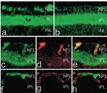
Patient characteristics are summarized in the Table. COX-2 immunoreactivity was observed in all eyes in the retinal pigment epithelial cells, in the outer segment of the photore-ceptors and to some degree in the inner plexiform layer (data not shown). In all diabetic subjects, COX-2 immunoreactivity was also detected in the nerve fiber layer (NFL) (Figure 1a, cases 1 to 7); this COX-2 immunostaining was granular and discontinuous. COX-2 immunolocalized mostly with GFAP indicative of astrocytes (Figure 1c through 1e); similar results were obtained using 2 different COX-2–specific antibodies (see Methods). A comparable pattern was found in the eye of a nondiabetic subject who had suffered from severe carotid artery obstruction (case 12). In contrast, in the other nondia-betic patients, COX-2 immunoreactivity was not observed in the NFL (Figure 1b and 1f through 1h, cases 8 to 11).
COX-2 Expression and Localization, and PGE2Concentrations in Experimental
Ischemic Retinopathy
COX-2 expression is altered by ischemia in neural tissue.24
We explored the involvement of COX-2 in a murine model of retinopathy of prematurity. Equivalent COX-2 mRNA
ex-pression was observed in room air and after hyperoxia exposure at P12, P14, and P17 (Figure 2A). Immunolocaliza-tion of COX-2 was studied in mouse retina and was found to be similar to that in humans. In normoxic mice at P14, COX-2 protein was robustly expressed in retinal pigment epithelial cells both in the outer photoreceptor segment and in the inner plexiform layer (data not shown). In the post– hyperoxia-exposed retinas, COX-2 expression was also detected in the NFL at P14 (Figure 3a) predominantly in astrocytes (GFAP positive) (Figure 3e and 3f).
COX-2 protein expression was evaluated in membrane fractions of retina extracts from hyperoxia-exposed rats. At the end of the hyperoxic period (P1 through P7), COX-2 immunoreactivity diminished and tended to increase in the early hours after resuming exposure to room air (24 hours after hyperoxia; Figure 2C). COX-1 exhibited the reverse pattern, and consequently whole retina concentrations of PGE2, a mediator involved in angiogenesis,25 were
untered during the posthyperoxia period (Figure 2B); al-though this does not exclude a local paracrine effect of this autacoid. Consistent with these observations, primary ret-inal astrocyte cultures exposed for 24 hours to relative
hypoxia (2% O2, 5% CO2, 93%) exhibited increased
COX-2 expression (Figure 2D) and PGE2 levels (Figure
2E) compared with those in normoxia (95% air, 5% CO2),
whereas COX-1 expression remained steady. Specific COX-2 inhibitors, APHS and etodolac, markedly
dimin-Figure 1. COX-2 immunohistochemistry on human retinas of
diabetic (a and c through e) and nondiabetic (b and f through h) subjects. Shown is colocalization of GFAP and COX-2 in nondi-abetic (f through h) and dinondi-abetic (c through e) eyes. Panel heights are 240m (a and b) and 90 m (c through h). NFL indicates nerve fiber layer; INL and ONL, inner nuclear layer and outer nuclear layer, respectively.
ished PGE2levels, whereas COX-1–selective SC-560 only
caused a small decrease in PGE2 concentrations (Figure
2E) even at highest dose tested (5 mol/L), suggesting a
dominant role for COX-2 in PGE2 generation during
hypoxia.
In age-adjusted normoxic whole retinas, COX-1 and -2 expression did not change. Immunolocalization of COX-2 in rat retina was similar to that observed in mouse (data not shown).
COX Inhibition in Ischemic Proliferative Retinopathy
The impact of COX inhibition on neovascularization was tested using specific COX-2 (APHS and etodolac) and COX-1 (SC-560) inhibitors; concentrations corresponded to effective ones on astrocyte PGE2levels (Figure 2E). In vivo,
posthyperoxia administration of the preferential COX-2 in-hibitor APHS did not affect the degree of capillary-free area
or the intraretinal revascularization of the ischemic retina studied 4 days after the first dose (P17) (Figure 4A and 4B). However, intravitreal neovascularization (revealed by intrav-itreal vascular nuclear counts) was dose-dependently dimin-ished by APHS, whereas the selective COX-1 inhibitor SC-56015was ineffective (Figure 4C). PGE
2levels in whole
retina were reduced by 65% 24 hours after APHS treatment. Moreover, intravitreal injection of PGE2 after the hyperoxic
period led to a significant (albeit mild) increase in intravitreal neovascularization.
To confirm the role of COX-2 in retinal neovasculariza-tion, its involvement was tested using a distinct selective COX-2 inhibitor, etodolac,16as well as a different species, the
rat model of ischemic proliferative retinopathy. Etodolac caused a marked decrease in retinal neovascularization (stud-ied at 5 days after the first injection) (Figure 5A and 5B). This effect was reversed by PGE2.
To further explore the PGE2 pathway, PGE2 receptor
expression and its changes during the posthyperoxia period were studied. Hyperoxia caused a slight decrease in EP4
(Figure 5C), whereas EP1was undetectable (not shown). In
contrast, EP2and to a greater extent EP3receptor
expres-sion was significantly decreased by hyperoxia and in-creased substantially during the posthyperoxia period (Fig-ure 5C), coincidental with COX-2 changes (Fig(Fig-ure 2C). Moreover, addition of the EP2 agonist butaprost and the
EP3 agonist M&B2876717 reversed in part or exacerbated
the inhibitory effects of etodolac on retinal neovascular-ization. EP3receptor expression also increased in the same
phase in that of the murine eye, predominantly localized in retinal endothelium (lectin griffonia–positive cells) (Fig-ure 3f through 3h).
Figure 2. COX expression and PGE2levels in retina of models of ischemic proliferative retinopathy. Mice were exposed to 75% O2from P5 to P12, and rats to cycling O2concentrations of 10% to 80% from P1 to P7. A, COX-2 mRNA expression in mice. B, Retinal PGE2concentrations in mice. C, COX-1 and -2 immunoreactivity in rats. D, COX-1 and -2 immunoreactivity in cultured astrocytes in 21% or 2% O2for 24 hours (see Meth-ods). E, PGE2levels in media of cultured astrocytes in normoxic (20% O2) and hypoxic (2% O2) conditions with or without 1-hour treatment with APHS (1mol/L), etodolac (1 mol/L), and/or SC-560 (0.2mol/L). Histogram bar values are mean⫾SEM. *P⬍0.5 compared with other corresponding values.
Figure 3. COX-2 (a through c, e) and EP3(f and h) immunohis-tochemistry of posthyperoxic (ischemic; a, c through e, f through h) and normoxic mouse retina at P14 (as described in Figure 2); shown are GFAP immunoreactivity (astrocyte marker, d) and lectin griffonia simplicifolia labeling (endothelium marker, g). Panel height, 130m (a and b); 95 m (c through e); and 48 m (f through h). NFL indicates nerve fiber layer; INL and IPL, inner nuclear layer and inner plexiform layer, respectively.
Effects of COX-2 Inhibition and EP3Stimulation
on Expression of Modulators of Angiogenesis
Involvement of COX-2 and EP3on pro- and antiangiogenic
factors, notably VEGF, VEGFR2, TSP-1, and CD36,26,27was
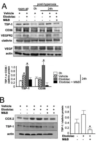
studied in retina of models of proliferative retinopathy. Twenty-four hours after removal from hyperoxia, there was an increase in TSP-1 expression. The COX-2 inhibitor etodolac induced a substantial increase in both TSP-1 and CD36, and addition of the EP3agonist M&B28767 reversed
this effect. In contrast, effects of COX-2 on neovasculariza-tion could not be explained by the VEGF pathway; VEGF expression, although as anticipated it increased during the posthyperoxic period, was marginally affected by etodolac and M&B28767 (Figure 6A), and VEGFR2 remained unal-tered. In accord with in vivo observations, etodolac caused a slight increase in TSP-1 expression in neuroretinovascular endothelial cells, and this effect was reversed by M&B28767 (Figure 6B), consistent with EP3expression on retinal
endo-thelium (Figure 3f).
Discussion
Inflammatory mediators contribute to the pathogenesis of ischemic proliferative retinopathy.3,4 In a related manner,
COX-2 has been implicated in angiogenesis, especially of tumors.6Although COX inhibition diminishes retinal
neovas-cularization in ischemic models,28the specific involvement of
COX-2 and the identity of its products in this process have not been elucidated. Our findings disclose an important role for this immediate-early gene product in proliferative retinop-athy. COX-2 expression increased coincidentally with that of EP2and EP3during the ischemic (posthyperoxic in models)
phase associated with proliferation, particularly localized, respectively, in astrocytes and in endothelial cells of the NFL. COX-2 contributed to the preretinal neovascularization in different models of ischemic retinopathies, which seems to be mediated by PGE2acting via EP2and to a greater extent EP3
receptors, and these in turn modulate the antiangiogenic factor TSP-1 and its receptor CD36 on target endothelial cells.
COX-2 was abundantly present in synaptic regions of the retina of all species studied (human, mouse, and rat) as seen in the brain.29In addition, COX-2 was induced in the NFL of
retinas of humans with diabetes and vascular obstruction (case 12) and in animals after hyperoxia, hence during hypoxic-ischemic phases; in these instances, COX-2 was mostly induced in astrocytes (but also in endothelium [Figure
Figure 4. Effects of COX-1 and -2 inhibition on retinal vascular
density (A and B) and intravitreal neovascularization in murine model of ischemic proliferative retinopathy at P17. Animals were prepared as described in Methods and injected intravitreally at P13 and at P15 with APHS, SC560, or PGE2; estimated effective vitreal concentrations are indicated.15,16B, Arrows on retinal flat mounts in panels 1 and 2 point to avascular zone and in panel 3 to intravitreal neovessels. Panel heights, 5 mm (B1 and B2) and 240m (B3 and B4). Values in histograms are mean⫾SEM; *P⬍0.01 compared with vehicle; †P⬍0.05 compared with other corresponding values for APHS (ANOVA).
Figure 5. Effects of COX-2 inhibitor etodolac and PGE2analogs in rat model of ischemic proliferative retinopathy. Rats were exposed to hyperoxia from P1 through P7 (as defined in Figure 2 legend) and injected intravitreally on P7 and P9 with etodolac with or without PGE2, butaprost, or M&B28767 (M&B) for esti-mated effective16,17vitreal concentrations, respectively, of 1, 0.1, 10, and 0.1mol/L; retinas were isolated on P12. A, Retinal flat mounts; 1, vehicle treated; 2, etodolac treated; 3,
etodolac⫹PGE2treated; and 4, etodolac⫹M&B28767 treated. Arrows indicate neovascular buds. Panel height, 1.1 mm. B, EP2, EP3, and EP4protein expression in retinas. Values in histo-grams are mean⫾SEM; *P⬍0.05 compared with all other corre-sponding values.
6B]) and seemed to account for a large portion of PGE2
generation (Figure 2E). Consistent with the time-dependent and propitious localization of COX-2, molecularly distinct inhibitors of this enzyme (APHS and etodolac15,16) markedly
diminished preretinal neovascularization (Figures 4 and 5), whereas COX-1 inhibition was ineffective. Moreover, admin-istration of the well-recognized proangiogenic COX product PGE22,5,10 (Figure 4) reversed the antiangiogenic effects of
COX-2 inhibitors in the retina (Figures 4 and 5). In view of the significant COX-1 expression even during the hypoxic phase (Figure 2C and 2D), one would have predicted some effect of COX-1 inhibition on neovascularization. However, COX-2 seems to be coregulated and biochemically coupled with the inducible gene product microsomal PGE2synthase, a
dominant generator of PGE2 under certain inflammatory
conditions,30whereas COX-1 appears to be mostly coupled to
the constitutive cytosolic PGE2synthase. This inference may
explain at least in part the differences observed between COX-1 and COX-2 inhibitors. Of note, we found augmented microsomal PGE2synthase during the posthyperoxic period
(data not shown) and, accordingly, a major role for COX-2 in PGE2generation during hypoxia (Figure 2E).
The angiogenic effect of PGE2seemed to be mediated via
its EP2and especially EP3receptors, as specific stimulation of
these receptors reversed the effects of etodolac on
neovascu-larization (Figure 5); selective antagonists to these receptors are not readily available. Of interest, EP2 and EP3 have
recently been reported to participate in colorectal tumor angiogenesis,31,32 and these effects may be mediated via
VEGF, a major factor in ischemic proliferative retinopathy26;
similarly, COX-2 inhibition downregulated VEGF in colon tumor endothelial cells.8But in other endothelial cells (breast
tumor and cornea), COX-2 inhibition affected the basic fibroblast growth factor pathway,9which plays a minor role
in ischemic proliferative retinopathy.33Endothelial cells are
not homogeneous throughout tissues, and in retina the effect of COX-2 inhibition was largely unrelated to the VEGF pathway (Figure 6). Thus, COX-2 inhibition may interfere with pathways that are independent of specific growth fac-tors. Plausible candidate pathways are through TSP-1 and its receptor CD36, which inhibit angiogenesis via p38 mitogen-activated protein kinase and caspase 3.34This conjecture is
supported by an upregulation of TSP-1 and CD36 by COX-2 inhibitor and reversal of these effects by EP3 stimulation
(Figure 6). Although a prostaglandin D2 metabolite–
depen-dent peroxisome proliferator-activated receptor-␥–mediated induction of TSP-1 and CD36 has been documented,35 an
EP3-evoked one as we observed (Figure 6) discloses a
previously undescribed mode of regulation of these antian-giogenic factors.
In summary, COX-2 contributes markedly to preretinal neovascularization in ischemic retinopathies, and this effect seems to be PGE2mediated mostly via EP3receptors
impli-cating a new interaction through TSP-1 and CD36. Results suggest that selective COX-2 inhibitors could be used for the control of pathological vitreal neovascularization in ischemic proliferative retinopathy. More specifically, EP3 (and
possi-bly EP2) antagonists may be more selective by sparing the
potentially physiologically desirable effects of the various COX-2 products.
Acknowledgments
This work was supported by grants from the Canadian Institutes of Health Research, Fond de la Recherche en Santé du Québec (Réseau Vision), March of Dimes, and Heart and Stroke Foundation of Québec. Drs Sennlaub and Beauchamp are supported by fellowships from the Deutscher Akademischer Austauschdienst and the Canadian Institutes of Health Research, respectively. Dr Chemtob holds a Canada Research Chair. We thank Hendrika Fernandez for skillful technical assistance.
References
1. Lee P, Wang CC, Adamis AP. Ocular neovascularization: an epidemi-ologic review. Surv Ophthalmol. 1998;43:245–269.
2. Tolentino MJ, Adamis AP. Angiogenic factors in the development of diabetic iris neovascularization and retinopathy. Int Ophthalmol Clin. 1998;38:77–94.
3. Barouch FC, Miyamoto K, Allport JR, et al. Integrin-mediated neutrophil adhesion and retinal leukostasis in diabetes. Invest Ophthalmol Vis Sci. 2000;41:1153–1158.
4. Sennlaub F, Courtois Y, Goureau O. Inducible nitric oxide synthase mediates the change from retinal to vitreal neovascularization in ischemic retinopathy. J Clin Invest. 2001;107:717–725.
5. Sennlaub F, Courtois Y, Goureau O. Inducible nitric oxide synthase mediates retinal apoptosis in ischemic proliferative retinopathy.
J Neurosci. 2002;22:3987–3993.
6. Dubois RN, Abramson SB, Crofford L, et al. Cyclooxygenase in biology and disease. FASEB J. 1998;12:1063–1073.
Figure 6. A, TSP-1, CD36, VEGFR2, and VEGF expression in
rats exposed to room air and after hyperoxia (80% O2). Rats were prepared as described in Figure 2. B, COX-2 and TSP-1 immunoreactivity in cultured retinovascular endothelial cells (see Methods) untreated or treated with etodolac (1mol/L) in pres-ence of M&B28767 (M&B, 0.1mol/L) or not. Values in histo-grams are mean⫾SEM. *P⬍0.01 compared with all other corre-sponding values.
7. Degi R, Thore C, Bari F, et al. Ischemia increases prostaglandin H synthase-2 levels in retina and visual cortex in piglets. Graefes Arch Clin
Exp Ophthalmol. 2001;239:59 – 65.
8. Tsujii M, Kawano S, Tsuji S, et al. Cyclooxygenase regulates angio-genesis induced by colon cancer cells. Cell. 1998;93:705–716. 9. Masferrer JL, Leahy KM, Koki AT, et al. Antiangiogenic and antitumor
activities of cyclooxygenase-2 inhibitors. Cancer Res. 2000;60: 1306 –1311.
10. Leahy KM, Ornberg RL, Wang Y, et al. Cyclooxygenase-2 inhibition by celecoxib reduces proliferation and induces apoptosis in angiogenic en-dothelial cells in vivo. Cancer Res. 2002;62:625– 631.
11. Liu XH, Kirschenbaum A, Lu M, et al. Prostaglandin E2 induces hyp-oxia-inducible factor-1␣ stabilization and nuclear localization in a human prostate cancer cell line. J Biol Chem. 2002;277:50081–50086. 12. Jones MK, Wang H, Peskar BM, et al. Inhibition of angiogenesis by
nonsteroidal anti-inflammatory drugs: insight into mechanisms and impli-cations for cancer growth and ulcer healing. Nat Med. 1999;5: 1418 –1423.
13. Smith LE, Wesolowski E, McLellan A, et al. Oxygen-induced retinopathy in the mouse. Invest Ophthalmol Vis Sci. 1994;35:101–111.
14. Holmes JM, Duffner LA. The effect of postnatal growth retardation on abnormal neovascularization in the oxygen exposed neonatal rat. Curr
Eye Res. 1996;15:403– 409.
15. Kalgutkar AS, Crews BC, Rowlinson SW, et al. Aspirin-like molecules that covalently inactivate cyclooxygenase-2. Science. 1998;280: 1268 –1270.
16. Riendeau D, Percival MD, Boyce S, et al. Biochemical and pharmaco-logical profile of a tetrasubstituted furanone as a highly selective COX-2 inhibitor. Br J Pharmacol. 1997;121:105–117.
17. Abramovitz M, Adam M, Boie Y, et al. The utilization of recombinant prostanoid receptors to determine the affinities and selectivities of prosta-glandins and related analogs. Biochim Biophys Acta. 2000;1483:285–293. 18. Beauchamp MH, Marrache AM, Hou X, et al. Platelet-activating factor in
vasoobliteration of oxygen-induced retinopathy. Invest Ophthalmol Vis
Sci. 2002;43:3327–3337.
19. Hou X, Gobeil F Jr, Peri K, et al. Augmented vasoconstriction and thromboxane formation by 15-F(2t)-isoprostane (8-iso-prostaglandin F(2␣)) in immature pig periventricular brain microvessels. Stroke. 2000; 31:516 –525.
20. Dehouck MP, Meresse S, Delorme P, et al. An easier, reproducible, and mass-production method to study the blood-brain barrier in vitro. J
Neu-rochem. 1990;54:1798 –1801.
21. Marrache AM, Gobeil F Jr, Bernier SG, et al. Proinflammatory gene induction by platelet-activating factor mediated via its cognate nuclear receptor. J Immunol. 2002;169:6474 – 6481.
22. Hardy P, Abran D, Li DY, et al. Free radicals in retinal and choroidal blood flow autoregulation in the piglet: interaction with prostaglandins.
Invest Ophthalmol Vis Sci. 1994;35:580 –591.
23. Benjamin LE, Hemo I, Keshet E. A plasticity window for blood vessel remodelling is defined by pericyte coverage of the preformed endothelial network and is regulated by PDGF-B and VEGF. Development. 1998; 125:1591–1598.
24. Iadecola C, Niwa K, Nogawa S, et al. Reduced susceptibility to ischemic brain injury and N-methyl-D-aspartate-mediated neurotoxicity in cyclo-oxygenase-2-deficient mice. Proc Natl Acad Sci USA. 2001;98: 1294 –1299.
25. Form DM, Auerbach R. PGE2and angiogenesis. Proc Soc Exp Biol Med.
1983;172:214 –218.
26. Aiello LP, Pierce EA, Foley ED, et al. Suppression of retinal neovascu-larization in vivo by inhibition of vascular endothelial growth factor (VEGF) using soluble VEGF-receptor chimeric proteins. Proc Natl Acad
Sci U S A. 1995;92:10457–10461.
27. Suzuma K, Takagi H, Otani A, et al. Expression of thrombospondin-1 in ischemia-induced retinal neovascularization. Am J Pathol. 1999;154: 343–354.
28. Takahashi K, Saishin Y, Mori K, et al. Topical nepafenac inhibits ocular neovascularization. Invest Ophthalmol Vis Sci. 2003;44:409 – 415. 29. Kaufmann WE, Worley PF, Pegg J, et al. COX-2, a synaptically induced
enzyme, is expressed by excitatory neurons at postsynaptic sites in rat cerebral cortex. Proc Natl Acad Sci U S A. 1996;93:2317–2321. 30. Brock TG, McNish RW, Peters-Golden M. Arachidonic acid is
preferen-tially metabolized by cyclooxygenase-2 to prostacyclin and prostaglandin E2. J Biol Chem. 1999;274:11660 –11666.
31. Seno H, Oshima M, Ishikawa TO, et al. Cyclooxygenase 2- and prosta-glandin E(2) receptor EP(2)-dependent angiogenesis in Apc(Delta716) mouse intestinal polyps. Cancer Res. 2002;62:506 –511.
32. Amano H, Hayashi I, Endo H, et al. Host prostaglandin E(2)-EP3 sig-naling regulates tumor-associated angiogenesis and tumor growth. J Exp
Med. 2003;197:221–232.
33. Ozaki H, Okamoto N, Ortega S, et al. Basic fibroblast growth factor is neither necessary nor sufficient for the development of retinal neovascu-larization. Am J Pathol. 1998;153:757–765.
34. Jimenez B, Volpert OV, Crawford SE, et al. Signals leading to apopto-sis-dependent inhibition of neovascularization by thrombospondin-1. Nat
Med. 2000;6:41– 48.
35. Feng J, Han J, Pearce SF, et al. Induction of CD36 expression by oxidized LDL and IL-4 by a common signaling pathway dependent on protein kinase C and PPAR-␥. J Lipid Res. 2000;41:688–696.