HAL Id: inserm-00614775
https://www.hal.inserm.fr/inserm-00614775
Submitted on 16 Aug 2011HAL is a multi-disciplinary open access archive for the deposit and dissemination of sci-entific research documents, whether they are pub-lished or not. The documents may come from teaching and research institutions in France or abroad, or from public or private research centers.
L’archive ouverte pluridisciplinaire HAL, est destinée au dépôt et à la diffusion de documents scientifiques de niveau recherche, publiés ou non, émanant des établissements d’enseignement et de recherche français ou étrangers, des laboratoires publics ou privés.
Ptf1a/Rbpj complex inhibits ganglion cell fate and
drives the specification of all horizontal cell subtypes in
the chick retina.
Elise Lelièvre, Monkol Lek, Henrik Boije, L. Houille-Vernes, Valérie Brajeul,
A. Slembrouck, Jérôme Roger, José-Alain Sahel, Jean-Marc Matter, Florian
Sennlaub, et al.
To cite this version:
Elise Lelièvre, Monkol Lek, Henrik Boije, L. Houille-Vernes, Valérie Brajeul, et al.. Ptf1a/Rbpj complex inhibits ganglion cell fate and drives the specification of all horizontal cell subtypes in the chick retina.: Ptf1a in chick retinal development. Developmental Biology, Elsevier, 2011, 358 (2), pp.296-308. �10.1016/j.ydbio.2011.07.033�. �inserm-00614775�
1
Ptf1a/Rbpj complex inhibits ganglion cell fate and drives the specification of all horizontal cell subtypes in the chick retina.
E.C. Lelièvre1,2,3,4,5, M. Lek6, H. Boije6, L. Houille-Verne s2,4,5, V. Brajeul2,4,5, A.
Slembrouck2,4,5, J.E. Roger4, J. Sahel2,4,5, J.M. Matter7, F. Sennlaub1,2,3, F. Hallböök6, O. Goureau2,4,5* and X. Guillonneau1,2,3
1-Centre de Recherche des Cordeliers, INSERM UMR S872, 75006 Paris, France *.
2-Université Pierre et Marie Curie, 75006 Paris, France 3-Université Paris Descartes, 75006, Paris, France
4-Institut de la Vision, INSERM UMR S968, 75012 Paris, France 5- CNRS UMR 7210, 75012, Paris, France
6- Department of Neuroscience, Biomedical Centre, Uppsala University, Husargatan 3, Uppsala, Sweden. 7- Department of Biochemistry, Sciences II, University of Geneva, 1211 Genève 4, Switzerland.
*Corresponding authors
Xavier Guillonneau
Centre de Recherche des Cordeliers, UMR S872, Equipe 21 15, Rue de L’Ecole de Médecine, 75006 Paris, France. Tel: (33) 1.44.27.80.87 Fax: (33) 1.44.27.40.92 Email: [email protected]
Olivier Goureau
Institut de la Vision, UMR S869, Equipe 3 12, Rue Moreau, 75012 Paris, France.
Tel : (33) 1.53.46.25.32 Fax : (33) 1.53.46.26.01 Email : [email protected]
2
During development, progenitor cells of the retina give rise to six principal classes of neurons and the Müller glial cells found within the adult retina. The pancreas transcription factor 1 subunit a (Ptf1a) encodes a basic-helix-loop-helix transcription factor necessary for the specification of horizontal cells and the majority of amacrine cell subtypes in the mouse retina. The Ptf1a-regulated genes and the regulation of Ptf1a activity by transcription cofactors during retinogenesis have been poorly investigated. Using a retrovirus-mediated gene transfer approach, we reported that Ptf1a was sufficient to promote the fates of amacrine and horizontal cells from retinal progenitors and inhibit retinal ganglion cell and
photoreceptor differentiation in the chick retina. Both GABAergic H1 and non-GABAergic H3 horizontal cells were induced following the forced expression of Ptf1a. We describe Ptf1a as a strong, negative regulator of Atoh7 expression. Furthermore, the Rbpj-interacting
domains of Ptf1a protein were required for its effects on cell fate specification. Together, these data provide a novel insight into the molecular basis of Ptf1a activity on early cell
specification in the chick retina.
ABSTRACT
3
The vertebrate neural retina is a laminar structure composed of six types of neurons and one major type of glial cells, the Müller cells. The seven cell types are derived from a common pool of multipotent retinal progenitor cells (RPC) that differentiate in a conserved chronological order. Retinal ganglion cells, cones, horizontal (HC) and amacrine (AC) cells are produced first, whereas rods, Müller glial cells and bipolar cells are generated last (Prada et al., 1991; Young, 1985). The RPC differentiation pathway choice is determined by cell-intrinsic, i.e., transcription factors, and cell-extrinsic factors (Livesey and Cepko, 2001; Marquardt and Gruss, 2002; Ohsawa and Kageyama, 2008).
INTRODUCTION
Ptf1a encodes a basic-helix-loop-helix (bHLH) transcription factor that drives undifferentiated cells in the foregut to differentiate into a pancreatic lineage during pancreas development (Kawaguchi et al., 2002; Krapp et al., 1998). Ptf1a is also expressed during the development of many structures of the central nervous system (Glasgow et al., 2005; Zecchin et al., 2004). In the cerebellum and the spinal cord, Ptf1a is expressed in the precursors of GABAergic neurons, and the loss of Ptf1a induced these GABAergic neurons to adopt a glutamatergic phenotype (Glasgow et al., 2005; Hoshino et al., 2005; Pascual et al., 2007). In the retina, Ptf1a acts as an HC and AC fate determination factor at the expense of ganglion cells in mice, zebrafish and Xenopus (Dong et al., 2008; Dullin et al., 2007; Fujitani et al., 2006; Nakhai et al., 2007) and of photoreceptors in zebrafish and Xenopus (Dong et al., 2008; Dullin et al., 2007). These studies have thoroughly characterized the phenotypic modifications induced by Ptf1a loss- and gain-of-function in the retina, but the molecular mechanisms underlying Ptf1a activity are not as well understood. Loss-of-function studies reported a change in the expression of a set of retinogenic factors in the Ptf1a-null retinas (Fujitani et al., 2006; Nakhai et al., 2007). Nevertheless, because some of these factors are persistently
4
transcriptional regulations or qualitative changes in the cell populations expressing these factors.
Unlike other class II bHLH transcription factors, Ptf1a functions in the pancreas as part of a trimeric complex. The pancreas transcription factor 1 complex (PTF1) includes Ptf1a, a ubiquitous class I bHLH transcription factor (E-protein) and the mammalian Suppressor of Hairless protein, Rbpj (PTF1-J complex) or its paralog, Rbpjl (PTF1-L complex) (Beres et al., 2006; Masui et al., 2007; Obata et al., 2001). The Ptf1a-Rbpj
interaction is also required for Ptf1a to specify GABAergic cells in the dorsal spinal cord and the cerebellum (Hori et al., 2008). However, the requirement of the Ptf1a-Rbpj interaction in the specification of retinal cell types remains to be elucidated.
The chick retina has proven to be a powerful model to study retinal differentiation and its genetic regulation. Moreover, in contrast to mice possessing only one axon-bearing HC subtype (Peichl and Gonzalez-Soriano, 1994), the chick retina has both less and axon-bearing HC subtypes, as found in the majority of vertebrate retinas (Gallego, 1986; Genis-Galvez, 1979). The chick retina contains H1 axon-bearing HCs, H2 axon-less “stellate” HCs and H3 axon-less “candelabrum” HCs (Edqvist et al., 2008; Tanabe et al., 2006). Therefore, this model is more representative of Ptf1a activity on HC subtype determination in
vertebrates.
This study aimed to gain a better understanding of the molecular regulation underlying the Ptf1a activity during retinal development using the chick retina model. In this study, we showed that the forced expression of Ptf1a leads to a massive disorganization of the
differentiated retina and changes in retinal cell representation that complement the retinal phenotype of Ptf1a-null mice: an increase of ACs and all HC subtypes and a decrease of ganglion and photoreceptor cells. Using this model, we identified several retinogenic factors that were rapidly regulated by Ptf1a overexpression and reported that ectopic Ptf1a strongly
5
downregulated Atoh7 expression in the chick retina. Finally, our study demonstrated that the interaction between Ptf1a and Rbpj cofactors was required for Ptf1a activity in the developing retina.
MATERIALS AND METHODS
Gallus gallus white leghorn embryos were obtained from Haas (France). The animal
experimentation was conducted in accordance with the Association for Research in Vision and Ophthalmology (ARVO) statement on the use of animals in Ophthalmic and Vision Research and a protocol approved by our local animal care committee.
Animals
Chick genome sequences (V2.0 and V3.0) were obtained from The Genome Institute at Washington University (http://genome.wustl.edu/). The expressed sequence tags (accession numbers: BU487258 and BU347629) were ordered from Source Bioscience and sequenced (MWG-Operon). The alignments were performed using Basic Local Alignment Search Tool available at NCBI (http://blast.ncbi.nlm.nih.gov/) and VNTI software (Invitrogen).
DNA sequences
The mouse Ptf1a was subcloned using specific CDS primers into the pDONR221 vector (Invitrogen) using BP clonase (Invitrogen). The Ptf1a mutants were generated using
Polymerase Chain Reaction (PCR)-based mutagenesis. The pDONR221-Ptf1a plasmid was recombined in the presence of LR recombinase (Invitrogen) into either RCAS-BP(A)-NHY, which allowed for the fusion of a HA-tag with the N-terminal part of the Ptf1a protein (gift from Dr. Loftus) or the pCIG gateway vectors (Roger et al., 2006). The RCAS viral stocks Retroviral stock and plasmid production
6
with titers > 1x10⁸ colony forming units per milliliter (cfu/ml) were prepared by transfecting DF1 cells with viral DNA constructs using FuGENE6 Reagent (Roche Diagnostics) (Yang, 2002). Viruses were concentrated by centrifugation using 100K Centrifugal Filters Amicon Ultra (Millipore). All embryos were injected into the right optic vesicle at embryonic day 2 (E2). The openings in the eggs were sealed with scotch tape and further incubated at 37.5°C. Parental RCAS-BP(A) viruses (Hughes et al., 1987) served as controls in the viral infection experiments.
The eyes were fixed in 4% paraformaldehyde (PFA) and incubated in 30% sucrose (Sigma) in phosphate-buffered saline (PBS) overnight followed by 1 hour incubation at 37°C in PGS (PBS, 7.5% gelatin (Sigma), and 10% sucrose). Eyes were embedded in PGS, frozen at -50°C in isopentane and stored at -80°C. Ten micrometer-thick cryosections were collected.
Cryosection
The retinas were collected in HBSS without Ca Cell dissociation
2+
/Mg2+, trypsinized (1 mg/ml) (Sigma) and incubated for 10 minutes at 37°C. The digestion was stopped with HBSS with Ca2+/Mg2+ containing trypsin inhibitor (Sigma) (1 mg/ml), and the cells were mechanically dissociated in the presence of DNaseI (Sigma) (0.1 mg/ml). For flow cytometry, the suspended cells were washed with PBS, fixed for 15 minutes with 2% PFA at room temperature (RT) and washed in PBS before immunostaining. For the manual counting, 1x10⁵ cells/ml were seeded on Poly-L-Lysine (Sigma) coated plates. After two hours, adherent cells were fixed 10 minutes with 2% PFA at RT before immunostaining.
7
Mouse anti-gag (3c2), anti-Ap2α (3B5), Islet1 (39.4D5), Lim1/2 (4F2), and anti-Visinin (7G4) antibodies were purchased from the Developmental Studies Hybridoma Bank. Mouse Brn3a (MAB1585), mouse Glutamine Synthetase (MAB302), mouse anti-Prox1 (MAB5652), and rabbit anti-PhosphoHistone3 (07-145) antibodies were obtained from Millipore, rabbit anti-gag (p27)from Charles River, rabbit anti-Protein Kinase Cα (PKCα) (Sc-208) from Santa Cruz, and rabbit anti-Prox1 (DP3501P) from Acris. The rabbit anti-Ptf1a antibody was a gift from Dr. Edlund (Umeå University), and the rabbit anti-TrkA was a gift from Pr. Lefcort (Montana State University). Primary antibodies were detected using AlexaFluor488-, AlexaFluor594-, AlexaFluor633- (Invitrogen) or Phycoerythrine (PE)- (Beckman Coulter) conjugated goat antibodies.
Immunostaining was performed as previously described (Roger et al., 2006). Retinal section counterstaining was performed with 4',6'-diamidino-2-phenylindole (DAPI) (1:1200).
Apoptotic cells were detectedby terminal deoxynucleotidyl transferase (TdT)-mediated dUTP nick end labeling (TUNEL) labeling using the In Situ Cell Death Detection Kit (Roche
Diagnostics). For S-phase cell labeling, 5 µg (E7 embryos) or 10 µg (E9 embryos) of 5-ethynyl-2’-deoxyuridine (EdU) were injected into the intravitreal space two hours before being sacrificed. The Click iT
Immunostaining and in situ hybridization
TM
Digoxigenin-labeled Rbpj and cAtoh7 probes were generated by cloning template DNA (the full length coding sequence of cAtoh7 and the cRbpj coding sequence from 123 to 873 bp) into pCRII-TOPO vector (Invitrogen). In situ hybridization was performed as previously described (Roger et al., 2006).
EdU Imaging kit (Invitrogen) was used to visualize EdU-positive cells.
8
Images were captured with a DM5500 microscope (Leica) equipped with an ORCA ER Hamamatsu camera or a LSM710 confocal microscope (Zeiss) and analyzed with MetaMorph software (Molecular Devices).
Dissociated cells were incubated in blocking buffer (PBS, 10% fetal calf serum (FCS), 2% goat serum, and 0.1% saponin) one hour at RT. Primary antibodies were diluted in blocking buffer, applied two hours at RT, and washed. The cell pellets were then incubated for one hour with 5 µl of PE- and/or AlexaFluor633-conjugated secondary antibodies (1:10) at RT. The data were collected using an LSRII cytometer (BD Biosciences) and analyzed using BD FACS DIVA software. For cell sorting, a total of 1x10
Flow cytometry and cell sorting
5
GFP-positive cells were collected in lysis buffer for RNA extraction using Vantage Sorter (BD Biosciences).
The eyes were collected at E5, and the pigmented epithelium was removed from the retina. Whole retinas were positioned in an electroporation chamber (CUY520P5, Sonidel,
Napagene, Japan) filled with PBS with Ca Electroporation
2+
/Mg2+containing plasmids (0.5 μg/μl). Electroporation was performed using a CUY215C square wave generator (Sonidel) and consisted of 5 pulses at 30 V with 50 ms duration, 1 second interval and repeated twice. Whole retinas were then cultured as floating explants at 37°C in DMEM, 10% FCS, and 1% Penicillin/Streptomycin. For confocal microscopy, the retinas were fixed 20 minutes in 4% PFA and rinsed for 24 hours in PBS. For inclusion, the fixed retinas were cryoprotected and embedded in PGS.
9
Total RNA was extracted using the Nucleospin RNAII Kit (Macherey Nagel).
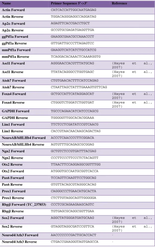
Retrotranscription was performed as described previously (Roger et al., 2006). Real-time PCR was performed using 7300 Real-Time PCR System (Applied Biosystems) in a 20 µl final volume with Power SYBR Green PCR Master Mix (Applied Biosystems) and 0.25 µM primers. All samples were run in triplicate. Primers used for real-time PCR analysis are listed in Table S1 (See Supplementary Materials).
RNA isolation, reverse-transcription and quantitative PCR.
Protein extracts were collected in hypotonic buffer (20 mM HEPES pH 7.9, 1 mM Na Cell fractionation
3VO4, 1 mM NaGlycerophoshate, 5 mM EDTA pH 7.5, 1 mM EGTA pH 7.5, 1 mM DTT, and protease inhibitors (Calbiochem, Merck4Biosciences)) plus 0.2% Nonidet P-40, incubated on ice for 15 minutes and centrifuged (20 seconds at 16000 g). For nuclear extracts, pellets were resuspended in saline buffer (120 mM NaCl and 20% glycerol in hypotonic buffer) supplemented with protease inhibitor cocktail, incubated for 30 minutes at 4°C and
centrifuged for 20 minutes at 16000 g to collect the supernatant. For cytosolic extracts, NaCl (120 mM) was added to the supernatant of the first centrifugation, and the extract was
centrifuged for 20 minutes at 16000 g to remove debris. Glycerol (10%) was then added to the supernatant.
Western Blotting was conducted as described previously (Roger et al., 2006) using the following primary antibodies: mouse anti-HA (MMS 101R, Covance), goat anti-LaminB (sc-6216, Santa Cruz), mouse Actin (Sigma), goat Ptf1a (ab62818, Abcam), and rat anti-Rbpj (T6719,Institute of Immunology, Tokyo, Japan).
10
Infected DF1 cells were lysed in immunoprecipitation buffer (50 mM Tris/HCl pH 8.0, 120 mM NaCl, 0.5% Nonidet P-40, and protease inhibitors) (Rodolosse et al., 2009). The lysates were incubated overnight and centrifuged for 20 minutes at 16000 g. Protein extracts (250 µg) were incubated with anti-HA antibody (Covance, 1/100) for 2 hours at RT to
immunoprecipitate the HA-tagged proteins. The immune complex was then captured by incubating for 2 hours at RT with 40 µl of Protein-G Sepharose beads (Fast Flow, GE Healthcare). The complexes were pelleted by gentle centrifugation, washed four times with immunoprecipitation buffer and eluted in the loading buffer before western blotting.
Co-immunoprecipitation
The quantifications are expressed as the mean ± standard error of the mean (s.e.m). Analyses were conducted using the Student’s t-test assuming equal variances (two groups) or one-way analysis of variance followed by Tukey’s multiple comparison tests (three or more groups) using Prism 5.0 (Graphpad software). (*) p<0.05, (**) p<0.01, (***) p<0.001, (ns) not significant. p
Statistical analyses
anova, p-value of the one-way analysis of variance; pTukey, p-value of the Tukey post-hoc test.
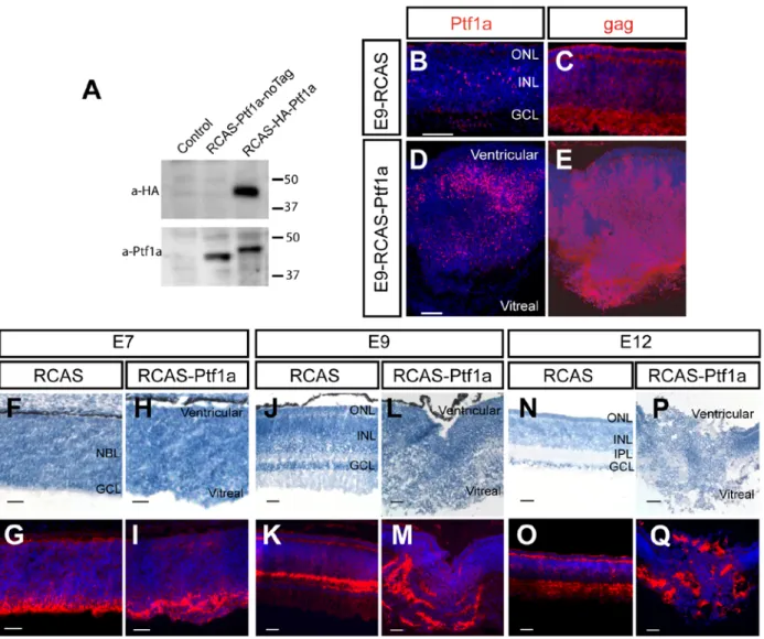
Forced expression of Ptf1a results in a loss of retinal lamination. RESULTS
The Ptf1a spatiotemporal expression pattern has been studied during retinogenesis (Boije et al., 2008; Fujitani et al., 2006; Nakhai et al., 2007). Ptf1a mRNA was detected from embryonic day 3 (E3) to E15 in the developing chick retina. Early, it is expressed in the inner neuroblastic layer (NBL) and then becomes restricted to the inner nuclear layer (INL) (Boije et al., 2008). To gain insight into the function of Ptf1a during chick retinal development, a replication-competent retrovirus, RCAS, was used to drive ectopic and overexpression of
11
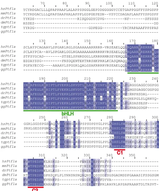
Ptf1a in the retina. The chick Ptf1a gene sequence is not fully known. We have characterized its C-terminal using the available information at The Genome Institute and two ESTs. It contains the highly conserved bHLH DNA-binding and Rbpj/Rbpjl-interaction domains (Beres et al., 2006) (Fig. S1). However, the N-terminal of the chick Ptf1a gene has remained uncharacterized in recent chicken genome assemblies. In contrast, the mouse Ptf1a sequence is fully known, and the Rbpj/Rbpjl-interaction domains have been extensively studied (Beres et al., 2006). Moreover, previous studies have reported that mouse Ptf1a blocks the
differentiation of dILB excitatory neurons and promotes dILA
During any developmental stage, the plexiform and nuclear layers had formed
properly in the empty-RCAS (RCAS) control infected patches (Fig. 1F-G,J-K,N-O). At E7, no major structural defects were observed in the RCAS-Ptf1a-infected patches other than the presence of a thicker retina and a decreased cell density (Fig. 1H-I). In contrast, at E9 (Fig. 1L-M) and, more extensively, at E12 (Fig. 1P-Q), the lamination of the RCAS-Ptf1a-infected patches was severely disturbed with disorganized plexiform and nuclear layers. No changes in
inhibitory neuron specification in the chick spinal cord, a phenotype that was consistent with Ptf1a loss of function studies in mouse (Glasgow et al., 2005; Hori et al., 2008; Mizuguchi et al., 2006; Wildner et al., 2006). These results suggest a conserved activity of mouse Ptf1a in avian and rodent models. Therefore, the mouse Ptf1a cDNA was chosen to allow for misexpression experiments of Ptf1a in the chick retina and for genetic manipulations of the Ptf1a coding sequence. Effective wild type Ptf1a protein mis/overexpression from RCAS-HA-Ptf1a (RCAS-Ptf1a) was verified by western blotting for the HA-tagged proteins in DF1 cells (Fig. 1A) and by
immunohistochemistry in E9 retinas (Fig. 1D). In the chick retina, viral injections resulted in a widespread infection. Retinal patches infected with the different RCAS viruses and infected retinal dissociated cells could be detected using either the p27 or 3c2 anti-gag antibodies (Fig. 1C,E).
12
the retinal structure were observed outside of the RCAS-Ptf1a-infected areas (data not shown).
Forced expression of Ptf1a affects progenitor cell proliferation and migration.
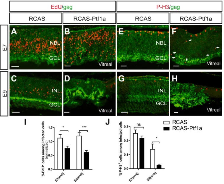
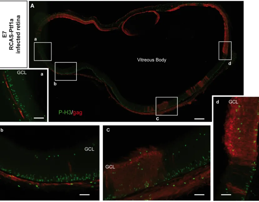
These defects in retinal structure and lamination might arise from disturbed migration or modification of the cell cycle. Therefore, S-phase cells were pulse-labeled with 5-ethynyl-2’-deoxyuridine (EdU), and mitotically active progenitors were immunostained using a Phospho-Histone3 (P-H3) antibody. At E7, in the control retinas, EdU-positive cells were located in the NBL (Fig. 2A), whereas P-H3 positive cells were primarily found in the
ventricular side (Fig. 2E), which was consistent with interkinetic nuclear movements (IKNM) of progenitor nuclei and free-cell migration (Baye and Link, 2008; Boije et al., 2009; Hinds and Hinds, 1979). P-H3-positive (Fig. 2F) and numerous EdU-positive cells (Fig. 2B) were detected within the RCAS-Ptf1a-infected patches at E7. The EdU-positive cells were
mislocalized in the ventricular half of the retina (Fig. 2B), and an increased number of ectopic P-H3 positive cells were detected in the NBL and in the vitreal side of the RCAS-Ptf1a-infected patches (Fig. 2F, arrows and Fig. S2). These results suggest that the movement of progenitors was altered in the RCAS-Ptf1a-infected retina at E7 and might partially account for the observed structural defects.
The EdU-positive and P-H3-positive cells were scored using flow cytometry. The percentage of EdU-positive cells among the infected cells was normalized by the percentage of EdU-positive cells among non-infected cells. At E7, the number of EdU-positive cells was significantly decreased following the forced expression of Ptf1a (p=0.031) (Fig. 2I). At E9, fewer EdU- (p=0.001) (Fig. 2D) and P-H3-positive cells (Fig. 2H) (0.14 ± 0.04%, s.e.m, in the RCAS-infected cells and 0.02 ± 0.01%, s.e.m, in the RCAS-Ptf1a-infected cells,
13
suggest that the forced expression of Ptf1a induced progenitors to withdraw from the cell cycle.
Ptf1a mis/overexpression is sufficient to induce a change in retinal cell type representation.
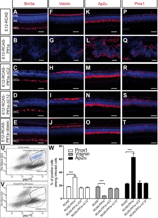
To investigate whether the structural and cell cycle changes were paired with alterations in the proportion of major retinal cell types, using immunocytochemistry, we analyzed the markers of the different cell classes in the infected areas of E12 retinas, the age when most cells have initiated their differentiation. Concomitantly, the number of ACs, HCs and photoreceptor cells among the gag-positive infected cells was scored by flow cytometry. At E12, immunoreactivity for Ap2α was found in most AC nuclei in the INL (Fig. 3K) (Edqvist et al., 2006; Fischer et al., 2007). Prox1 strongly labeled HCs and a small subset of ACs (Fig. 3P). The forced expression of Ptf1a induced a three-fold increase of Ap2α-positive cells compared to control cells (22.7 ± 2.2% in the RCAS-infected population and 63.0 ± 12.9% in the RCAS-Ptf1a-infected population, n=5) and a two-fold increase of Prox1-positive cells (18.0 ± 1.3% in the RCAS-infected population and 32.6 ± 4.7% in the RCAS-Ptf1a-infected population, n=5) (Fig. 3L,Q,W). Conversely, Brn3a labeling for ganglion cells was severely reduced in the RCAS-Ptf1a-infected patches (Fig. 3B), and the number of Visinin-positive photoreceptors was decreased from 17.7 ± 2.6% in the RCAS-infected cells to 4.2 ± 0.5% in the RCAS-Ptf1a-infected cells (n=5) (Fig. 3G,U-W). For late born retinal cell subtypes, the number of Glutamine Synthetase (GS)-positive Müller glial cells was significantly decreased from 3.2 ± 0.2% in the RCAS-infected cells to 1.8 ± 0.3% in the RCAS-Ptf1a-infected cells (p=0.0083, n=4) (Fig. S3B,E), but Protein Kinase Cα
14
among the RCAS-infected cells and 3.9 ± 0.8% among the RCAS-Ptf1a-infected cells, p=0.2651, n=4) (Fig. S3D,E).
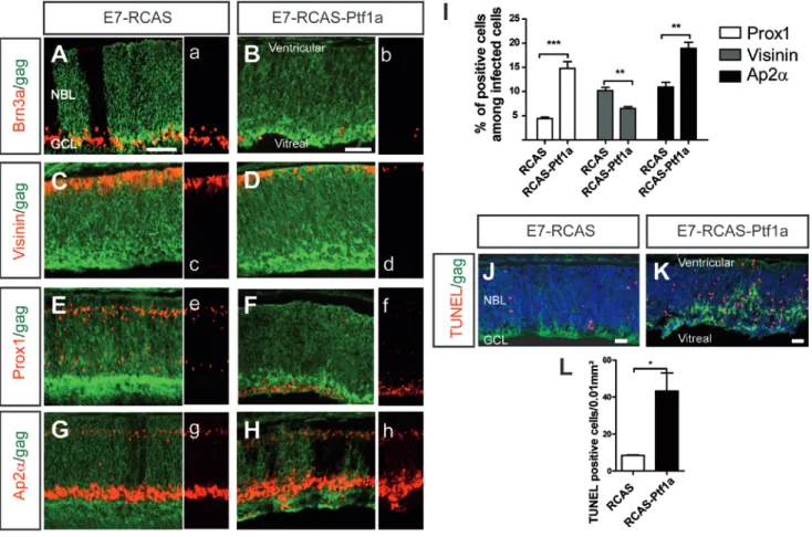
Retinal lamination defects at E9 and later stages may result in a secondary alteration of cell type representation. Therefore, we conducted the same analysis prior to the structural defects, at E7. At this age, Ptf1a mis/overexpression also led to a significant increase of Prox1- (4.4 ± 0.3% among the RCAS-infected cells and 14.7 ± 1.5% among the RCAS-Ptf1a-infected cells, p=0.0004, n=4) (Fig. 4F,I) and Ap2α-positive cells (10.9 ± 0.1% among the RCAS-infected cells and 18.9 ± 1.2% among the RCAS-Ptf1a-infected cells, p=0.001, n=5) (Fig. 4H,I) at the expense of ganglion cells (Fig. 4B) and photoreceptor cells (10.2 ± 0.7% in the RCAS-infected retinas and 6.5 ± 0.4% in the RCAS-Ptf1a-infected retinas, p=0.0014, n=5) (Fig. 4D,I). These results indicated that the Ptf1a mis/overexpression effects on early-born retinal cell types occurred prior to the defects in retinal lamination and argued against the notion that the loss of photoreceptors and ganglion cells was secondary to the retinal
lamination defects. Interestingly, a few Ap2α-positive cells (Fig. 4H) and the majority of Prox1-positive cells (Fig. 4F) were ectopically located in the vitreal side of the retina.
We performed TUNEL labeling to assess if the reduction of some cell subtypes could be due to early cell death. A significantly higher number of apoptotic cells, located throughout the entire thickness of the NBL, was detected in the RCAS-Ptf1a patches (43.3 ± 9.8 cells per area) compared to the patches infected with the control virus (8.5 ± 0.2 cells per area, n=3, p=0.0305) (Fig. 4J-L).
Ptf1a mis/overexpression is sufficient to induce all HC subtype specification.
Our results showed that Ptf1a mis/overexpression increased the size of the HC population. The chick HC population is composed of at least three HC subtypes that can be molecularly distinguished by the expression of Prox1, Lim1, Islet1, TrkA and GABA
15
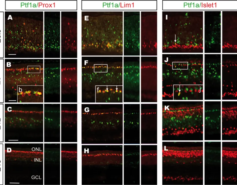
(Edqvist et al., 2008; Fischer et al., 2007). All three subtypes express Prox1. The H1 subtype (50% of all HCs) has GABA, is Lim1-positive, Islet1-negative, and TrkA-negative. The H2 (approximately 10% of all HCs) also has GABA, is Lim1-negative, Islet1-positive but TrkA-negative, and the H3 (approximately 40% of all HCs) does not have GABA, is Lim1-negative and Islet1/TrkA double-positive. We first investigated the normal Ptf1a expression in the HC subtypes in the developing chick retina. The retina was analyzed with respect to Ptf1a, Prox1, Lim1 and Islet1 immunoreactivity in E6.5 retinas when HCs migrated bi-directionally and, in E9, E12 and E16 retinas when the HC layer (HCL) was established (Boije et al., 2009; Edqvist and Hallbook, 2004). At E6.5, after the onset of Prox1 expression in HCs, 31 ± 5% (n=3) of the Ptf1a-immunoreactive cells were also Prox1-positive (Fig. 5A). The Ptf1a/Prox1 double-positive cells were localized on the vitreal side of the retina but also scattered in the prospective INL, which was consistent with the location of migrating HCs at this age. At E9, all Ptf1a/Prox1 double-positive cells were located in the developing HCL. Of the Prox1-positive cells in the HCL, 64 ± 5% (n=3) were Ptf1a-Prox1-positive HCs (Fig. 5B). This result implies that not all HCs expressed Ptf1a at this age. The Lim1-positive cells were Ptf1a-positive, and this overlap was found at both E9 (96 ± 1%, n=3) in the HCL and at earlier ages on the vitreal side (Fig. 5E,F). In contrast, only a few Ptf1a/Islet1 double-positive cells could be seen at E6.5 and at E9 (5 ± 0.6%, n=3) (Fig. 5I,J).
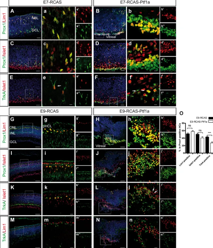
To assess whether Ptf1a was able to direct the development of all HC subtypes, we next analyzed the presence of HC subtype markers in the RCAS-Ptf1a-infected patches. At E7, an accumulation of Prox1/Lim1 double-positive and Prox1/Islet1 double-positive cells (Fig. 6B,D) were found in the normal location of the GCL in the RCAS-Ptf1a-infected patches compared to the control patches, suggesting that both the H1 and H3 subtypes had been generated following Ptf1a mis/overexpression. These results were strengthened by the TrkA immunoreactivity of Islet1-positive cells in the RCAS-Ptf1a-infected areas (Fig. 6F).
16
Furthermore, the accumulation of some HCs in the vitreal side suggested that HC migration was either arrested or delayed by Ptf1a mis/overexpression. These results were similar at E9 when all HCs had normally migrated back to the HCL. Supernumerary Prox1/Lim1 double-positive (Fig. 6H), Prox1/Islet1 double-double-positive (Fig. 6J), and Islet1/TrkA double-double-positive cells (Fig. 6L) were observed in the RCAS-Ptf1a-infected retinas. Islet1-positive and TrkA-negative cells were found within the clusters of Prox1-positive cells of the RCAS-Ptf1a-infected patches (Fig. 6l, arrows), suggesting the presence of H2 HCs. Moreover, TrkA and Lim1 were never expressed in the same cell, indicating that H1 and H3 HCs were correctly specified (Fig. 6N). Wefound that the proportion of non-GABAergic TrkA-positive cells among the Prox1-positive HCs decreased significantly from 43.3 ± 1.9% in the RCAS-infected patches to 27.1 ± 2.8% in the RCAS-Ptf1a-RCAS-infected areas (p=0.0007), whereas a slight, but not significant, increase of Lim1-positive HCs was observed (51.9 ± 2.2% in the RCAS-infected patches and 58.9 ± 2.1% in the RCAS-Ptf1a-infected patches, p=0.0898) (Fig. 6O). Moreover, the back-migration of ectopic HCs to the HCL was inhibited, and ectopic HCs remained on the vitreal side of the E9 RCAS-Ptf1a-infected retinas.
Ptf1a overexpression induces changes in retinogenic factor expression.
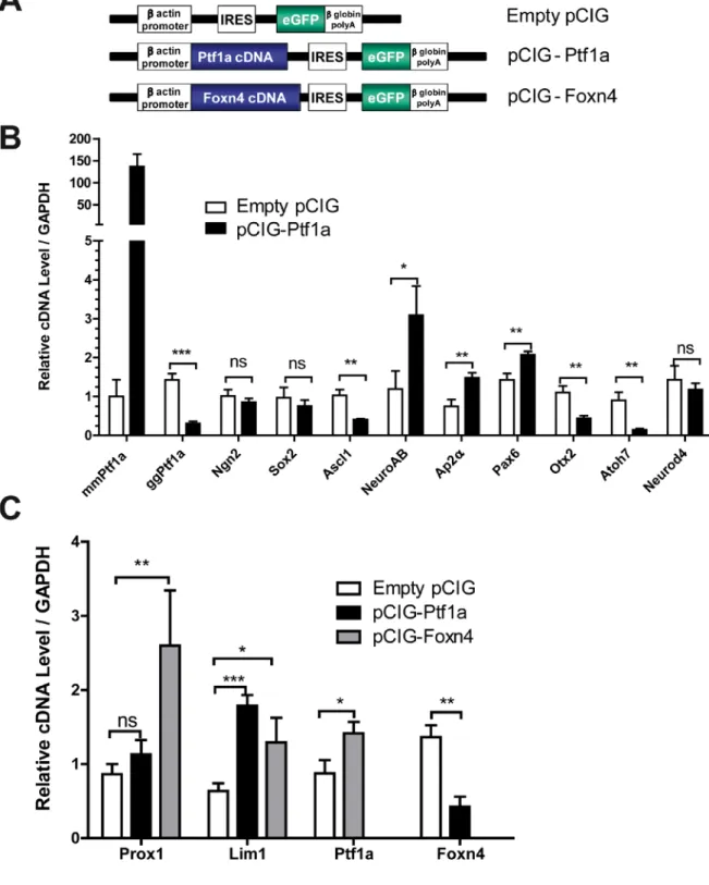
To gain knowledge about the molecular mechanisms underlying the changes in retinal cell specification, we studied early changes in the expression of a selected set of genes involved in retinal differentiation.The E5 whole chick retinas were electroporated ex vivo with pCIG-Ptf1a-GFP plasmids, which drive the green fluorescent protein (GFP) expression (Roger et al., 2006) (Fig. 7A). The GFP-positive cells were isolated by fluorescence activated cell sorting as early as 36 hours after electroporation to study changes in gene expression that were as independent as possible from cell differentiation. The empty pCIG-GFP plasmids were used as controls. First, our quantification demonstrated that electroporation of
pCIG-17
Ptf1a in the chick retinas resulted in a strong expression of mouse Ptf1a mRNA, compared to the reference level (the non-specific amplification in pCIG-electroporated retinal cells) (Fig. 7B). Surprisingly, endogenous chick Ptf1a mRNA levels were significantly decreased in the mouse Ptf1a-overexpressing cells (p=0.0002) (Fig. 7B). No effects were observed on either
Ngn2, a direct target of Ptf1a in the spinal cord and the cerebellum (Henke et al., 2009), or Sox2. We found that Pax6 and Ap2α, coding for two transcription factors highly expressed in
ACs (Belecky-Adams et al., 1997; de Melo et al., 2003; Edqvist et al., 2006), were
upregulated in Ptf1a-overexpressing cells (p=0.009) (Fig. 7B). A significant 2.6-fold increase of NeuroAB mRNA, a transcription factor suggested to be involved in GABAergic AC development (Ohkawara et al., 2004), was also observed in Ptf1a-overexpressing cells
(p=0.0167). Interestingly, the expression levels of Ascl1, a pro-amacrine transcription factor in chick retina (Mao et al., 2009), was downregulated (p=0.0037), suggesting that Ptf1a might act downstream of Ascl1 and inhibit its expression in the retina as described in the spinal cord (Hori et al., 2008). The expression level of Otx2, a key regulatory gene for photoreceptor development (Nishida et al., 2003), was decreased by 2.5-fold in Ptf1a-overexpressing cells (p=0.0076), and Atoh7, a transcription factor involved in ganglion cell specification (Liu et al., 2001; Matter-Sadzinski et al., 2005), was strongly repressed by 6.4-fold by Ptf1a
overexpression (p=0.0071) (Fig. 7B). Furthermore, Atoh7 mRNA was nearly undetectable by
in situ hybridization in RCAS-Ptf1a-infected patches (Fig. S5C). Consistently, ex vivo, ectopic
Ptf1a downregulated the activity of the chick Atoh7 promoter. Indeed, a significant decrease in the number of GFP-positive cells was observed in retinas co-electroporated with RCAS-Ptf1a and pAtoh7-GFP, a plasmid that drives GFP expression under the control of the chick
Atoh7 promoter, compared to RCAS and pAtoh7-GFP co-electroporated retinas (Fig.
18
We further focused our analysis on transcription factors involved in HC genesis. The mRNA levels of Neurod4 (Fig. 7B) and Prox1 (Fig. 7C), two factors involved in HC
development (Dyer et al., 2003; Inoue et al., 2002), were not altered, whereas Lim1, which is necessary for H1 specification in the chick retina (Suga et al., 2009), was induced following Ptf1a overexpression (Fig. 7C). Foxn4 is upstream of Ptf1a during AC/HC specification in mouse retina (Fujitani et al., 2006; Li et al., 2004). Interestingly, Ptf1a overexpression induced a two-fold decrease of Foxn4 expression levels (p=0.0030) (Fig. 7C). To investigate whether Foxn4 conversely regulates Ptf1a in the chick retina, we electroporated E5 retinas with pCIG-Foxn4-GFP vectors (Fig. 7A). Foxn4 overexpression resulted in a significant increase in Ptf1a expression (p=0.0498) and Lim1 and Prox1 mRNA expression levels (Fig. 7C).
Ectopic Ptf1a requires its interaction with endogenous RBP-J
Ptf1a forms the PTF1 complex with any one of the E-proteins and Rbpj (PTF1-J) or Rbpjl (PTF1-L) (Beres et al., 2006). Ptf1a interacts with Rbpjl through its C1 domain and with Rbpj through its C2 and C1 domains (Fig. S4K). The C2 domain deletion was sufficient to abolish the formation of the PTF1-J complex (Beres et al., 2006). In situ hybridization revealed that Rbpj mRNA was expressed throughout the NBL at early stages and in the INL at later stages up to E12 (see Fig. S6A-J). Conversely,Rbpjl mRNA was neither detected during
chick retinal development (data not shown) nor in mature retinas by quantitative PCR (Fig. S6K). To test the requirement for Rbpj proteins for Ptf1a activity in the chick retina, we generated Ptf1a-∆C2 and Ptf1a-∆C1∆C2 (Ptf1a-∆C12) mutant forms of Ptf1a that interact with E-proteins but are unable to interact with Rbpj (Beres et al., 2006) (Fig. S4L). The Ptf1a-∆C2 and Ptf1a-∆C12 proteins were expressed at levels equivalent to Ptf1a full-length proteins (Fig. S4N). Moreover, subcellular fractionation revealed that deletion of the C1 and C2 domains did not prevent Ptf1a-∆C12 nuclear importation (Fig. S4O). The injection of
RCAS-19
Ptf1a-∆C2 and RCAS-Ptf1a-∆C12 in the optic cup did not lead to gross structural defects of the retina (Fig. 3 and Fig. S4E-H). However, the outer plexiform and nuclear layers were disrupted with rosette-like structures (Fig. 3H,I). Early-born cell types were analyzed by immunochemistry and flow cytometry in the RCAS-Ptf1a-∆C2- and RCAS-Ptf1a -∆C12-infected population of E12 retinas, but no alteration of the cell type distribution was observed following the forced expression of these two Ptf1a mutant forms (Fig. 3C-D,H-I,M-N,R-S,W). The requirement of Rbpj-Ptf1a interaction for gene regulation downstream of ectopic Ptf1a was assessed by evaluating the ability of ectopic Ptf1a-∆C12 to regulate the Atoh7 promoter activity. Co-electroporation of pAtoh7-GFP with the RCAS-Ptf1a-∆C12 plasmids did not decrease the number of GFP-positive cells compared to co-electroporation with the control RCAS vectors (Fig. S5G,H ).
Ectopic Ptf1a activity is independent from Rbpj pool depletion
Rbpj is a major downstream effector of the Notch pathway. Previous studies have suggested that the Ptf1a activity might be downregulated by sequestering Rbpj from Ptf1a (Cras-Meneur et al., 2009) following Notch activation. Therefore, we examined if ectopic Ptf1a might function in the chick retina by conversely sequestering Rbpj. We generated a Ptf1a-∆basic mutant where the highly conserved RER-R amino acids (168-172) (Fig. S4L,M) in the basic domain were replaced by AVA-A. These mutations were shown to abolish MyoD and bHLHa15/Mist1 bHLH transcription factor binding to DNA (Lemercier et al., 1998; Skowronska-Krawczyk et al., 2005). The Ptf1a-∆basic protein was still imported into the nucleus (See Fig. S4O) and retained its ability to interact with Rbpj (See Fig. S4P). The in
vitro reporter assay confirmed that the transcriptional activity of Ptf1a-∆basic was strongly
decreased (data not shown). No alteration of retinal structure and cell type representation (Fig. 3E,J,O,T and Fig. S4I-J) was detected within the E12 RCAS-Ptf1a-∆basic-infected patches.
20
This result argues against the hypothesis that the activity of wild-type ectopic Ptf1a might be completely mediated by a disruption of Rbpj function.
Our study demonstrated that Ptf1a was sufficient to affect the chick retinal cell
composition in an Rbpj-dependent manner. Our “candidate-gene” approach showed that Ptf1a overexpression selectively upregulated NeuroAB, Ap2α and Lim1 while it downregulated
Otx2 and Atoh7. This study provides a new molecular basis for Ptf1a activity in specifying the
ACs and all HC subtypes and inhibiting photoreceptor and ganglion cell genesis.
DISCUSSION
Cellular and molecular mechanisms underlying ectopic Ptf1a activity during chick retinal development.
Several hypotheses may explain the changes in cell type representation induced by Ptf1a.
First, cell proliferation defects may change the final retinal cell composition. Indeed, early cell cycle exit has been shown to favor early-born retinal cell types. Conversely, late cell cycle exit biases progenitor cells toward later neuronal fates (Austin et al., 1995; Dorsky et al., 1997; Henrique et al., 1997; Jadhav et al., 2006; Livesey and Cepko, 2001; Nelson et al., 2007; Ohnuma et al., 2002; Silva et al., 2003; Yaron et al., 2006). We found that the forced expression of Ptf1a in the chick retina induced a premature cell-cycle exit, but led to a dramatic decrease of early-born ganglion and cone photoreceptor cells. Thus, it is unlikely that cell proliferation defects alone account for the effect of ectopic Ptf1a activity in chick retinogenesis. How Ptf1a induced cell cycle withdrawal remains to be defined. The cell cycle exit could be linked to the defects in the IKNM of retinal progenitors observed following Ptf1a mis/overexpression. Indeed, in the chick retina, pharmacological perturbation of IKNM
21
was shown to promote premature neurogenesis (Murciano et al., 2002) through a reduction of Notch-mediated lateral inhibition (Del Bene et al., 2008; Murciano et al., 2002). Consistent with this model, the expression of Hes5, a target of the Notch pathway in the retina (Nelson et al., 2006), was significantly decreased in electroporated Ptf1a-overexpressing cells (data not shown). Moreover, the expression level of cyclin D1 that is required for RPC proliferation (Fantl et al., 1995; Godbout and Andison, 1996; Sicinski et al., 1995) was also significantly downregulated in Ptf1a-overexpressing cells (data not shown), suggesting that ectopic Ptf1a might regulate the core of the cell cycle machinery.
Second, Ptf1a might bias progenitor cells toward the AC and HC fates. We found that the expression of Atoh7 (Brown et al., 2001; Wang et al., 2001) and Otx2 (Nishida et al., 2003), which specify ganglion cells and photoreceptors, respectively, were inhibited in cells that overexpressed Ptf1a. In contrast, Lim1, involved in HC specification (Suga et al., 2009), and NeuroAB, which is presumably involved in AC genesis (Ohkawara et al., 2004), were upregulated. These transcriptional regulations support a mechanism where ectopic Ptf1a might act cell-autonomously to drive retinal progenitors towardthe AC and HC fates at the expense of ganglion cells and photoreceptors by regulating their transcriptional program. The effects of ectopic Ptf1a on cell fate might also be influenced by cell proliferation regulation. Indeed, early cell cycle exit was shown to enhance the activity of proneural factors (Atoh7, Ngn1, and Neurod4) that promote early cell fates (Ohnuma et al., 2002). Recently, the majority of Ptf1a-expressing cells were found to originate from Atoh7-positive progenitor cells, and Ptf1a expression drove their differentiation toward ACs/HCs at the expense of ganglion cells in the zebrafish retina (Jusuf et al., 2011). Our overexpression study showed that ectopic Ptf1a was a negative regulator of Atoh7. Therefore, it would be interesting to assess whether endogenous Ptf1a, which is turned on after Atoh7 in the Atoh7-positive
22
lineage, is able to downregulate Atoh7 expression during retinogenesis, how it might regulate
Atoh7, and if this regulation is involved in the fate switch toward ACs/HCs.
Third, Ptf1a might also selectively induce the death of ganglion and photoreceptor cells, modifying the cell representation in favor of ACs/HCs. The number of apoptotic cells was increased in Ptf1a-infected retinas at E7, but the precise determination of their identity was difficult to assess because many apoptotic cells have lost their differentiation markers. To our knowledge, no report has demonstrated a direct induction of cell death either by Ptf1a or by the misexpression of transcription factors without apoptotic regulatory properties. Dullin et al. (Dullin et al., 2007) did not detect any apoptosis induction following Ptf1a overexpression in Xenopus retinas, suggesting that Ptf1a has no pro-apoptotic function per se. Secondary cell death is a common feature of transcription factor gain- and loss-of-functions and has often been associated with defects in cell trans-commitment in the retina (Fujitani et al., 2006; Mao et al., 2009; Qiu et al., 2008; Zheng et al., 2009). Therefore, the increase in cell death is likely a secondary event that occurs following the late re-specification of already committed
precursors. Alternatively, a lack of crucial trophic support and inappropriate cell-cell interactions may lead to the elimination of prematurely differentiated cells.
Transcriptional regulation upstream and downstream of Ptf1a
Our results showed that Foxn4 upregulated Prox1, Lim1 and Ptf1a in the chick retina, suggesting that Foxn4 might be sufficient to induce HC fate in the developing chick retina, which is in contrast to mice (Li et al., 2004). Together with the upregulation of endogenous
Ptf1a by Foxn4, these data confirm that Ptf1a functions downstream of Foxn4 in the
transcriptional cascade leading to HC specification as proposed in mice (Fujitani et al., 2006). Interestingly, we discovered that Ptf1a downregulated Foxn4, indicating that Ptf1a would be required to finely regulate Foxn4 expression in HC and AC precursors. In line with this
23
hypothesis, Li et al. reported that in Foxn4LacZ knock-in mice, β−galactosidase (LacZ) expression was upregulated and persisted longer than in wild type retinas (Li et al., 2004). This Foxn4 locus deregulation could result, at least partially, from the loss of Ptf1a expression in Foxn4
-/-An increase in photoreceptor cells was reported in addition to the decrease of ACs/HCs following Foxn4 and Ptf1a inactivation (Dong et al., 2008; Dullin et al., 2007; Fujitani et al., 2006; Li et al., 2004; Nakhai et al., 2007). Consistently, our misexpression study demonstrated that Ptf1a inhibited photoreceptor cell production and decreased Otx2 expression. Inversely, the loss of Otx2 in the mouse retina decreased the number of
photoreceptors in favor of ACs (Nishida et al., 2003). These gain- and loss-of-function studies highlight a possible connection between the production of HCs/ACs on the one hand and photoreceptors and ganglion cells on the other. This balance may rely, to some extent, on Notch-mediated lateral inhibition. Notch signaling was shown to specifically inhibit mouse photoreceptor specification (Jadhav et al., 2006; Yaron et al., 2006). In various structures of the central nervous system, Ascl1 and Foxn4 have been implicated in the regulation of the Delta-like/Notch signaling pathway. Notably, in the ventral spinal cord, Foxn4 functions upstream of Ascl1, Delta-like and Notch expression to regulate the diversification of
excitatory and inhibitory V2 cells by lateral inhibition (Del Barrio et al., 2007). In the retina (Nelson et al., 2009; Nelson and Reh, 2008) and mouse dorsal spinal cord (Mizuguchi et al., 2006), Ascl1 activates Delta-like and Ptf1a expression. Thus, similar to the spinal cord, Foxn4 and/or Ascl1 might initiate lateral inhibition among a subset of retinal progenitors. These factors would then be required to sustain Notch signaling in AC/HC precursors that were activated by Delta-expressing neighboring cells that were prone to differentiate as photoreceptor cells. Ascl1 and Foxn4 expression would also contribute to turning off the photoreceptor and ganglion cell specification programs in AC/HC precursors through Ptf1a
24
activation. Further studies will be needed to determine to what extent Foxn4 and Ascl1 function together to regulate Notch signaling and Ptf1a expression in the chick retina. Ptf1a alone might not sustain Notch signaling because Hes5 was downregulated in
Ptf1a-overexpressing cells (data not shown).
Surprisingly, endogenous chick Ptf1a mRNA levels were significantly decreased in mouse Ptf1a-overexpressing cells. These results were in contrast with the positive
autoregulation of Ptf1a that occurs in thepancreas, spinal cord and cerebellum (Masui et al., 2008; Meredith et al., 2009). Recently, Jusuf et al. (Jusuf et al., 2011) reported that a feedback loop originating from HCs/ACs operated to limit the number of cells initiating Ptf1a
expression in the zebrafish retina. Thus, supernumerary ACs/HCs might regulate the expression of pro-amacrine factors, such as Foxn4 and Ptf1a, in autocrine or paracrine feedback loops. Alternatively, our results demonstrated that Foxn4 was downregulated in mouse Ptf1a-overexpressing cells and that Foxn4 activated endogenous chick Ptf1a. Chick
Ptf1a expression would not be triggered in mouse Ptf1a-overexpressing cells as a result of the
absence of its upstream activator, Foxn4.
PTF1-J complex during retinogenesis
Previous studies have demonstrated that the Ptf1a/Rbpj/E-protein (PTF1-J) complex is the functional endogenous PTF1 complex required for GABAergic cell specification in mouse cerebellum and spinal cord (Hori et al., 2008). Nonetheless, the formation of this functional trimeric complex during retinogenesis has not yet been assessed. In this study, we showed that the loss of the Rbpj-binding domains within the Ptf1a protein was sufficient to abolish the Ptf1a effects on chick retinal cell differentiation, indicating that ectopic Ptf1a was dependent on the Rbpj cofactors in the chick retina.
25
Does the endogenous Ptf1a factor require Rbpj for the specification of ACs and HCs? Several points suggest that this functional interaction occurs. First, we showed that Rbpj was expressed in the retina during retinogenesis, notably in the INL (Fig. S6A-J). Second, recent conditional loss-of-function studies (Riesenberg et al., 2009; Zheng et al., 2009) reported that retinas null for Rbpj exhibited a phenotype that mimics some features of the Ptf1a-null retinas (Fujitani et al., 2006; Nakhai et al., 2007) and complement our gain-of-function study. Indeed, the loss of Rbpj in the retina induced an increase of ganglion cells together with an
upregulation of Atoh7 (Riesenberg et al., 2009; Zheng et al., 2009). Moreover, the loss of Rbpj in Chx10-positive retinal cells induced a significant decrease of ACs and HCs in P21 mouse retinas (Zheng et al., 2009). The ganglion cell increase was attributed to Notch3 signaling inhibition (Riesenberg et al., 2009). We found that the Ptf1a-Rbpj interaction was necessary to inhibit ganglion cell specification following Ptf1a overexpression. Based on our study, the increase in ganglion cell number in Rbpj-null mice might result from either Notch3 signaling inhibition or from endogenous PTF1-J complex inactivation. The increase of ACs/HCs was not observed following Rbpj inactivation in the alpha-positive peripheral retinal cells (Riesenberg et al., 2009).The discrepancies between the two conditional Rbpj knock-out mouse models might result from the inactivation of Rbpj in different progenitor pools or a genetic compensation by Rbpjl because Rbpj was shown to compensate for the loss of Rbpjl in the Rbpjl-/-
It is noteworthy that, in humans, mutations of the Rbpj-interaction domain of Ptf1a protein caused optic nerve hypoplasy in addition to a cerebellar and pancreatic agenesis (Sellick et al., 2004). The same phenotype was reported in Rbpj-null mice where ectopic ganglion cells underwent cell death during late developmental stages. Even if the late specific cell death of the supernumerary ganglion cells remained to be assessed in Ptf1a-deficient
mouse pancreas (Masui et al., 2010). They could also be due to the use of different amacrine cell markers.
26
retina, this supports a highly conserved Ptf1a-Rbpj functional interaction during vertebrate retinal development.
Horizontal cell subtype specification by Ptf1a
It has been shown that Ptf1a is required to specify GABAergic neurons over
glutamatergic neurons in the mouse dorsal spinal cord (Glasgow et al., 2005) and cerebellum (Hoshino et al., 2005). However, Ptf1a controls the specification of glutamatergic climbing fiber neurons of the inferior olivary nucleus (Yamada et al., 2007), suggesting that Ptf1a is a neuronal fate determinant in this structure. In the retina, two hypotheses were formulated: either Ptf1a is involved in all AC/HC specifications (Fujitani et al., 2006; Jusuf and Harris, 2009), or Ptf1a favors only the GABAergic cell subtypes (Dullin et al., 2007). We found in this study that the number of both H1 GABAergic cells (and probably H2 cells) and H3 non-GABAergic HCs were increased in the RCAS-Ptf1a-infected patches.Thus, Ptf1a is likely to act primarily as a general HC determination factor.
We detected only a few Islet1-positive HCs (H2 or H3 cells) that expressed
endogenous Ptf1a during chick retinogenesis, which argues against a direct exclusion between Islet1 and Ptf1a. One hypothesis is that H3 HCs, which are the majority of the Lim1-negative and Islet1-positive, may not express Ptf1a and that the few Ptf1a/Islet1 double-positive cells in the HCL were H2 HCs. Alternatively, Ptf1a expression could be transiently required for the specification of H2/H3 cells and turned off at the onset of expression of H2 and H3 markers in the late-born Islet1-positive HCs (Edqvist et al., 2008). Consistent with this hypothesis in the zebrafish retina containing both Lim1-positive and Islet1-positive HC subtypes (Hallböök, unpublished), the knock-down of Ptf1a expression caused a decrease of Prox1/Islet1-positive HCs (Dong et al., 2008).
27
We found that Lim1-positive HCs expressed Ptf1a and that Ptf1a remained during bidirectional migration and once the cells stopped their migration in the HCL, indicating that Ptf1a might be involved in several steps of H1 cell specification/differentiation beyond its implication in all HC fate. In contrast, we found that Ptf1a downregulated Islet1 expression, suggesting that Ptf1a should be turned off for Islet1 to be expressed (data not shown). Therefore, secondary to the specification for all HCs, differential regulation of Ptf1a expression in the HC subtypes (sustained expression in H1 and downregulation in H2/H3) might be necessary to achieve their proper differentiation. In the context of forced and persistent Ptf1a expression, this regulation might fail and result in a decrease of the H3 proportion among HCs.
Acknowledgments: We thank Pr. Lefcort for the TrkA antibody, Pr. Edlund for the
anti-Ptf1a antibody, Pr. Real for the anti-Ptf1aK200R construct and Pr. Loftus for the RCAS-BP(A)-NHY plasmid. We are grateful to Prs. Martinerie, Laurent and Chiodini for their helpful support. We acknowledge Drs. Sadzinski-Matter, Thomasseau and Sandlung for technical assistance. This work was financed by INSERM, Retina France, EU (LSHG-CT-2005-512036, ERC-StG-210345), French ANR (ANR-Geno-031-03, ANR-08-MNPS-003), Swedish Research Council, Swedish SSMF and SSF.
28
REFERENCES
Austin, C. P., Feldman, D. E., Ida, J. A., Jr., Cepko, C. L., 1995. Vertebrate retinal ganglion cells are selected from competent progenitors by the action of Notch. Development. 121, 3637-50.
Baye, L. M., Link, B. A., 2008. Nuclear migration during retinal development. Brain Res. 1192, 29-36.
Belecky-Adams, T., Tomarev, S., Li, H. S., Ploder, L., McInnes, R. R., Sundin, O., Adler, R., 1997. Pax-6, Prox 1, and Chx10 homeobox gene expression correlates with
phenotypic fate of retinal precursor cells. Invest Ophthalmol Vis Sci. 38, 1293-303. Beres, T. M., Masui, T., Swift, G. H., Shi, L., Henke, R. M., MacDonald, R. J., 2006. PTF1 is
an organ-specific and Notch-independent basic helix-loop-helix complex containing the mammalian Suppressor of Hairless (RBP-J) or its paralogue, RBP-L. Mol Cell Biol. 26, 117-30.
Boije, H., Edqvist, P. H., Hallbook, F., 2008. Temporal and spatial expression of transcription factors FoxN4, Ptf1a, Prox1, Isl1 and Lim1 mRNA in the developing chick retina. Gene Expr Patterns. 8, 117-23.
Boije, H., Edqvist, P. H., Hallbook, F., 2009. Horizontal cell progenitors arrest in G2-phase and undergo terminal mitosis on the vitreal side of the chick retina. Dev Biol. 330, 105-13.
Brown, N. L., Patel, S., Brzezinski, J., Glaser, T., 2001. Math5 is required for retinal ganglion cell and optic nerve formation. Development. 128, 2497-508.
Cras-Meneur, C., Li, L., Kopan, R., Permutt, M. A., 2009. Presenilins, Notch dose control the fate of pancreatic endocrine progenitors during a narrow developmental window. Genes Dev. 23, 2088-101.
de Melo, J., Qiu, X., Du, G., Cristante, L., Eisenstat, D. D., 2003. Dlx1, Dlx2, Pax6, Brn3b, and Chx10 homeobox gene expression defines the retinal ganglion and inner nuclear layers of the developing and adult mouse retina. J Comp Neurol. 461, 187-204. Del Barrio, M. G., Taveira-Marques, R., Muroyama, Y., Yuk, D. I., Li, S., Wines-Samuelson,
M., Shen, J., Smith, H. K., Xiang, M., Rowitch, D., Richardson, W. D., 2007. A regulatory network involving Foxn4, Mash1 and delta-like 4/Notch1 generates V2a and V2b spinal interneurons from a common progenitor pool. Development. 134, 3427-36.
Del Bene, F., Wehman, A. M., Link, B. A., Baier, H., 2008. Regulation of neurogenesis by interkinetic nuclear migration through an apical-basal notch gradient. Cell. 134, 1055-65.
Dong, P. D., Provost, E., Leach, S. D., Stainier, D. Y., 2008. Graded levels of Ptf1a
differentially regulate endocrine and exocrine fates in the developing pancreas. Genes Dev. 22, 1445-50.
Dorsky, R. I., Chang, W. S., Rapaport, D. H., Harris, W. A., 1997. Regulation of neuronal diversity in the Xenopus retina by Delta signalling. Nature. 385, 67-70.
Dullin, J. P., Locker, M., Robach, M., Henningfeld, K. A., Parain, K., Afelik, S., Pieler, T., Perron, M., 2007. Ptf1a triggers GABAergic neuronal cell fates in the retina. BMC Dev Biol. 7, 110.
Dyer, M. A., Livesey, F. J., Cepko, C. L., Oliver, G., 2003. Prox1 function controls progenitor cell proliferation and horizontal cell genesis in the mammalian retina. Nat Genet. 34, 53-8.
Edqvist, P. H., Hallbook, F., 2004. Newborn horizontal cells migrate bi-directionally across the neuroepithelium during retinal development. Development. 131, 1343-51.
29
Edqvist, P. H., Lek, M., Boije, H., Lindback, S. M., Hallbook, F., 2008. Axon-bearing and axon-less horizontal cell subtypes are generated consecutively during chick retinal development from progenitors that are sensitive to follistatin. BMC Dev Biol. 8, 46. Edqvist, P. H., Myers, S. M., Hallbook, F., 2006. Early identification of retinal subtypes in the
developing, pre-laminated chick retina using the transcription factors Prox1, Lim1, Ap2alpha, Pax6, Isl1, Isl2, Lim3 and Chx10. Eur J Histochem. 50, 147-54.
Fantl, V., Stamp, G., Andrews, A., Rosewell, I., Dickson, C., 1995. Mice lacking cyclin D1 are small and show defects in eye and mammary gland development. Genes Dev. 9, 2364-72.
Fischer, A. J., Stanke, J. J., Aloisio, G., Hoy, H., Stell, W. K., 2007. Heterogeneity of horizontal cells in the chicken retina. J Comp Neurol. 500, 1154-71.
Fujitani, Y., Fujitani, S., Luo, H., Qiu, F., Burlison, J., Long, Q., Kawaguchi, Y., Edlund, H., Macdonald, R. J., Furukawa, T., Fujikado, T., Magnuson, M. A., Xiang, M., Wright, C. V., 2006. Ptf1a determines horizontal and amacrine cell fates during mouse retinal development. Development. 133, 4439-50.
Gallego, A., 1986. Chapter 7. Comparative studies on horizontal cells and a note on microglial cells. Prog. Retin. Res. 5. 165-206.
Genis-Galvez, J. M., Prada, F., Armengol, J.A, 1979. Evidence of three horizontal cells in the chick retina. Jpn.J.Ophtamol. 23, 378-387.
Glasgow, S. M., Henke, R. M., Macdonald, R. J., Wright, C. V., Johnson, J. E., 2005. Ptf1a determines GABAergic over glutamatergic neuronal cell fate in the spinal cord dorsal horn. Development. 132, 5461-9.
Godbout, R., Andison, R., 1996. Elevated levels of cyclin D1 mRNA in the undifferentiated chick retina. Gene. 182, 111-5.
Hayes, S., Nelson, B. R., Buckingham, B., Reh, T. A., 2007. Notch signaling regulates regeneration in the avian retina. Dev Biol. 312, 300-11.
Henke, R. M., Savage, T. K., Meredith, D. M., Glasgow, S. M., Hori, K., Dumas, J.,
MacDonald, R. J., Johnson, J. E., 2009. Neurog2 is a direct downstream target of the Ptf1a-Rbpj transcription complex in dorsal spinal cord. Development. 136, 2945-54. Henrique, D., Hirsinger, E., Adam, J., Le Roux, I., Pourquie, O., Ish-Horowicz, D., Lewis, J.,
1997. Maintenance of neuroepithelial progenitor cells by Delta-Notch signalling in the embryonic chick retina. Curr Biol. 7, 661-70.
Hinds, J. W., Hinds, P. L., 1979. Differentiation of photoreceptors and horizontal cells in the embryonic mouse retina: an electron microscopic, serial section analysis. J Comp Neurol. 187, 495-511.
Hori, K., Cholewa-Waclaw, J., Nakada, Y., Glasgow, S. M., Masui, T., Henke, R. M., Wildner, H., Martarelli, B., Beres, T. M., Epstein, J. A., Magnuson, M. A., Macdonald, R. J., Birchmeier, C., Johnson, J. E., 2008. A nonclassical bHLH Rbpj transcription factor complex is required for specification of GABAergic neurons independent of Notch signaling. Genes Dev. 22, 166-78.
Hoshino, M., Nakamura, S., Mori, K., Kawauchi, T., Terao, M., Nishimura, Y. V., Fukuda, A., Fuse, T., Matsuo, N., Sone, M., Watanabe, M., Bito, H., Terashima, T., Wright, C. V., Kawaguchi, Y., Nakao, K., Nabeshima, Y., 2005. Ptf1a, a bHLH transcriptional gene, defines GABAergic neuronal fates in cerebellum. Neuron. 47, 201-13.
Hughes, S. H., Greenhouse, J. J., Petropoulos, C. J., Sutrave, P., 1987. Adaptor plasmids simplify the insertion of foreign DNA into helper-independent retroviral vectors. J Virol. 61, 3004-12.
Inoue, T., Hojo, M., Bessho, Y., Tano, Y., Lee, J. E., Kageyama, R., 2002. Math3 and NeuroD regulate amacrine cell fate specification in the retina. Development. 129, 831-42.
30
Jadhav, A. P., Mason, H. A., Cepko, C. L., 2006. Notch 1 inhibits photoreceptor production in the developing mammalian retina. Development. 133, 913-23.
Jusuf, P. R., Almeida, A. D., Randlett, O., Joubin, K., Poggi, L., Harris, W. A., 2011. Origin and determination of inhibitory cell lineages in the vertebrate retina. J Neurosci. 31, 2549-62.
Jusuf, P. R., Harris, W. A., 2009. Ptf1a is expressed transiently in all types of amacrine cells in the embryonic zebrafish retina. Neural Dev. 4, 34.
Kawaguchi, Y., Cooper, B., Gannon, M., Ray, M., MacDonald, R. J., Wright, C. V., 2002. The role of the transcriptional regulator Ptf1a in converting intestinal to pancreatic
progenitors. Nat Genet. 32, 128-34.
Krapp, A., Knofler, M., Ledermann, B., Burki, K., Berney, C., Zoerkler, N., Hagenbuchle, O., Wellauer, P. K., 1998. The bHLH protein PTF1-p48 is essential for the formation of the exocrine and the correct spatial organization of the endocrine pancreas. Genes Dev. 12, 3752-63.
Lemercier, C., To, R. Q., Carrasco, R. A., Konieczny, S. F., 1998. The basic helix-loop-helix transcription factor Mist1 functions as a transcriptional repressor of myoD. EMBO J. 17, 1412-22.
Li, S., Mo, Z., Yang, X., Price, S. M., Shen, M. M., Xiang, M., 2004. Foxn4 controls the genesis of amacrine and horizontal cells by retinal progenitors. Neuron. 43, 795-807. Liu, W., Mo, Z., Xiang, M., 2001. The Ath5 proneural genes function upstream of Brn3 POU
domain transcription factor genes to promote retinal ganglion cell development. Proc Natl Acad Sci U S A. 98, 1649-54.
Livesey, F. J., Cepko, C. L., 2001. Vertebrate neural cell-fate determination: lessons from the retina. Nat Rev Neurosci. 2, 109-18.
Mao, W., Yan, R. T., Wang, S. Z., 2009. Proneural gene ash1 promotes amacrine cell production in the chick retina. Dev Neurobiol. 69, 88-104.
Marquardt, T., Gruss, P., 2002. Generating neuronal diversity in the retina: one for nearly all. Trends Neurosci. 25, 32-8.
Masui, T., Long, Q., Beres, T. M., Magnuson, M. A., MacDonald, R. J., 2007. Early
pancreatic development requires the vertebrate Suppressor of Hairless (RBPJ) in the PTF1 bHLH complex. Genes Dev. 21, 2629-43.
Masui, T., Swift, G. H., Deering, T., Shen, C., Coats, W. S., Long, Q., Elsasser, H. P., Magnuson, M. A., MacDonald, R. J., 2010. Replacement of Rbpj with Rbpjl in the PTF1 complex controls the final maturation of pancreatic acinar cells.
Gastroenterology. 139, 270-80.
Masui, T., Swift, G. H., Hale, M. A., Meredith, D. M., Johnson, J. E., Macdonald, R. J., 2008. Transcriptional autoregulation controls pancreatic Ptf1a expression during
development and adulthood. Mol Cell Biol. 28, 5458-68.
Matter-Sadzinski, L., Matter, J. M., Ong, M. T., Hernandez, J., Ballivet, M., 2001. Specification of neurotransmitter receptor identity in developing retina: the chick ATH5 promoter integrates the positive and negative effects of several bHLH proteins. Development. 128, 217-31.
Matter-Sadzinski, L., Puzianowska-Kuznicka, M., Hernandez, J., Ballivet, M., Matter, J. M., 2005. A bHLH transcriptional network regulating the specification of retinal ganglion cells. Development. 132, 3907-21.
Meredith, D. M., Masui, T., Swift, G. H., MacDonald, R. J., Johnson, J. E., 2009. Multiple transcriptional mechanisms control Ptf1a levels during neural development including autoregulation by the PTF1-J complex. J Neurosci. 29, 11139-48.
32
Rodolosse, A., Campos, M. L., Rooman, I., Lichtenstein, M., Real, F. X., 2009. p/CAF modulates the activity of the transcription factor p48/Ptf1a involved in pancreatic acinar differentiation. Biochem J. 418, 463-73.
Roger, J., Brajeul, V., Thomasseau, S., Hienola, A., Sahel, J. A., Guillonneau, X., Goureau, O., 2006. Involvement of Pleiotrophin in CNTF-mediated differentiation of the late retinal progenitor cells. Dev Biol. 298, 527-39.
Sellick, G. S., Barker, K. T., Stolte-Dijkstra, I., Fleischmann, C., Coleman, R. J., Garrett, C., Gloyn, A. L., Edghill, E. L., Hattersley, A. T., Wellauer, P. K., Goodwin, G., Houlston, R. S., 2004. Mutations in PTF1A cause pancreatic and cerebellar agenesis. Nat Genet. 36, 1301-5.
Sicinski, P., Donaher, J. L., Parker, S. B., Li, T., Fazeli, A., Gardner, H., Haslam, S. Z., Bronson, R. T., Elledge, S. J., Weinberg, R. A., 1995. Cyclin D1 provides a link between development and oncogenesis in the retina and breast. Cell. 82, 621-30. Silva, A. O., Ercole, C. E., McLoon, S. C., 2003. Regulation of ganglion cell production by
Notch signaling during retinal development. J Neurobiol. 54, 511-24.
Skowronska-Krawczyk, D., Chiodini, F., Ebeling, M., Alliod, C., Kundzewicz, A., Castro, D., Ballivet, M., Guillemot, F., Matter-Sadzinski, L., Matter, J. M., 2009. Conserved regulatory sequences in Atoh7 mediate non-conserved regulatory responses in retina ontogenesis. Development. 136, 3767-77.
Skowronska-Krawczyk, D., Matter-Sadzinski, L., Ballivet, M., Matter, J. M., 2005. The basic domain of ATH5 mediates neuron-specific promoter activity during retina
development. Mol Cell Biol. 25, 10029-39.
Suga, A., Taira, M., Nakagawa, S., 2009. LIM family transcription factors regulate the subtype-specific morphogenesis of retinal horizontal cells at post-migratory stages. Dev Biol. 330, 318-28.
Tanabe, K., Takahashi, Y., Sato, Y., Kawakami, K., Takeichi, M., Nakagawa, S., 2006. Cadherin is required for dendritic morphogenesis and synaptic terminal organization of retinal horizontal cells. Development. 133, 4085-96.
Wang, S. W., Kim, B. S., Ding, K., Wang, H., Sun, D., Johnson, R. L., Klein, W. H., Gan, L., 2001. Requirement for math5 in the development of retinal ganglion cells. Genes Dev. 15, 24-9.
Wildner, H., Muller, T., Cho, S. H., Brohl, D., Cepko, C. L., Guillemot, F., Birchmeier, C., 2006. dILA neurons in the dorsal spinal cord are the product of terminal and non-terminal asymmetric progenitor cell divisions, and require Mash1 for their development. Development. 133, 2105-13.
Yamada, M., Terao, M., Terashima, T., Fujiyama, T., Kawaguchi, Y., Nabeshima, Y., Hoshino, M., 2007. Origin of climbing fiber neurons and their developmental dependence on Ptf1a. J Neurosci. 27, 10924-34.
Yang, X. J., 2002. Retrovirus-mediated gene expression during chick visual system development. Methods. 28, 396-401.
Yaron, O., Farhy, C., Marquardt, T., Applebury, M., Ashery-Padan, R., 2006. Notch1 functions to suppress cone-photoreceptor fate specification in the developing mouse retina. Development. 133, 1367-78.
Young, R. W., 1985. Cell differentiation in the retina of the mouse. Anat Rec. 212, 199-205. Zecchin, E., Mavropoulos, A., Devos, N., Filippi, A., Tiso, N., Meyer, D., Peers, B.,
Bortolussi, M., Argenton, F., 2004. Evolutionary conserved role of ptf1a in the specification of exocrine pancreatic fates. Dev Biol. 268, 174-84.
Zheng, M. H., Shi, M., Pei, Z., Gao, F., Han, H., Ding, Y. Q., 2009. The transcription factor RBP-J is essential for retinal cell differentiation and lamination. Mol Brain. 2, 38.
Fig. 1: Mis/overexpression of Ptf1a affects retinal structure.
(A) Lysates from DF1 cells transfected with the RCAS-Ptf1a plasmids or not (Control) were harvested, and the expression of exogenous Ptf1a proteins was monitored by western blotting using anti-Ptf1a and anti-HA antibodies. RCAS-Ptf1a-noTag: RCAS-Ptf1a without HA-Tag,
RCAS-HA-Ptf1a: RCAS-Ptf1a with HA-tag. (B-E) The RCAS-infected (B-C) and RCASPtf1a-infected (D-E) retinal sections were stained with anti-Ptf1a (B,D) or anti-gag (p27) (C,E) antibodies at E9. (F-Q) The RCAS-infected (F-G,J -K,N-O) and RCAS-Ptf1a-infected (H-I,L-M,P-Q) retinal sections were co-stained with hemalun (F,H,J,L,N,P) and anti-gag (3c2) antibodies (G,I,K,M,O,Q). In B-E,G,I,K,M,O,Q, retinal sections were counterstained with DAPI. ONL, outer nuclear layer; INL, inner nuclear layer; IPL, inner plexiform layer; GCL, ganglion cell layer; NBL, neuroblastic layer. Bars: 50 μm (B-E in B and D), 25 μm (F-Q).
Fig. 2: Effects of Ptf1a mis/overexpression on retinal progenitor cell proliferation. (A-H) Sections from the RCAS-infected (A,C,E,G) and the RCAS-Ptf1a-infected (B,D,F,H)
retinas were stained with EdU and anti-gag (3c2) antibodies (A-D) or with anti-P-H3 and antigag antibodies (E-H) at E7 and E9. In F, arrows point to P-H3 positive cells in an ectopic
location in the RCAS-Ptf1a-infected retinas. (I-J) Quantitative analysis of EdU-positive (I) or P-H3-positive cells (J) among the RCAS- and RCAS-Ptf1a-infected cells at E7 and E9.
Values represent the mean ± s.e.m. The percentage of EdU-positive cells among the infected cells was normalized by the percentage of EdU-positive cells among the non-infected cells for each embryo. NBL, neuroblastic layer; GCL, ganglion cell layer; INL, inner nuclear layer.
Fig. 3: Effects of wild-type and mutant forms of Ptf1a on retinal cell differentiation. (A-T) The RCAS-infected (A,F,K,P), RCAS-Ptf1a-infected (B,G,L,Q), RCAS-Ptf1a-ΔC2-infected (C,H,M,R), RCAS-Ptf1a-ΔC12-RCAS-Ptf1a-ΔC2-infected (D,I,N,S) and RCAS-Ptf1a-Δbasic-RCAS-Ptf1a-ΔC2-infected (E,J,O,T) patches from E12 retinas were immunostained using anti-Brn3a (A-E), anti-Visinin (F-J), anti-Ap2α (K-O), and anti-Prox1 (P-T) antibodies. For clarity, the gag labeling is not represented. Retinal sections were counterstained with DAPI. (U-V) Representative flow cytometry analysis after staining the RCAS-infected (U) and RCAS-Ptf1a-infected (V) dissociated cells with anti-gag (p27) and anti-Visinin antibodies at E12. (W) Quantitative analysis of Visinin-, Ap2α- and Prox1-positive cells among the infected cells at E12. Values represent the mean ± s.e.m of at least four separate eye counts. ONL, outer nuclear layer; INL, inner nuclear layer; GCL, ganglion cell layer; PE, Phycoerythrin; Alexa633, AlexaFluor633. Bars: 50 μm
Fig. 4: Retinal cell type representation is modified in E7 retinas prior to lamination defects.
(A-H) The E7 RCAS-infected (A,C,E,G) and RCAS-Ptf1a-infected (B,D,F,H) retinal patches
were immunostained using an anti-gag antibody and either anti-Brn3a (A-B), anti-Visinin (CD), anti-Prox1 (E-F) or anti-Ap2α (G-H) antibodies. Cell type-specific labeling in panels A-H
was represented without gag labeling in panels a-h, respectively. (I) Quantitative analysis of Visinin-, Ap2α- or Prox1-positive cells among the infected cells. The values represent the
mean ± s.e.m of at least four separate retinal counts and are representative of two independent injections. (J-L) Cells undergoing apoptosis were TUNEL-labeled at E7 in the RCASinfected (J) and RCAS-Ptf1a-infected (K) patches detected using anti-gag (p27) antibody.
Sections in J and K were counterstained with DAPI. (L) Quantitative analysis of the number of apoptotic cells per area in the RCAS- and RCAS-Ptf1a-infected patches at E7. The values represent the mean ± s.e.m of at least three separate retinas. NBL, neuroblastic layer; GCL, ganglion cell layer. Bars: 50 μm (A,C,E, and G in A and B,D,F, and H in B), 25 μm (J,K).
Fig. 5: Ptf1a expression in subtypes of developing horizontal cells.
Epifluorescence micrographs show Ptf1a, Prox1 (A-D), Ptf1a, Lim1 (E-H), and Ptf1a, Islet1 (I-L) double-labeling of E6.5, E9, E12 and E16 chick retinas, and the corresponding split fluorescence images are to the right of each panel. Insets in b, f, and j are higher magnifications of the boxes in B, F and J. Arrows point at double-labeled cells. GCL, ganglion cell layer; INL, inner nuclear layer; ONL, outer nuclear layer. Bars: 25 μm.