HAL Id: hal-02342494
https://hal.archives-ouvertes.fr/hal-02342494
Submitted on 31 Oct 2019HAL is a multi-disciplinary open access archive for the deposit and dissemination of sci-entific research documents, whether they are pub-lished or not. The documents may come from teaching and research institutions in France or abroad, or from public or private research centers.
L’archive ouverte pluridisciplinaire HAL, est destinée au dépôt et à la diffusion de documents scientifiques de niveau recherche, publiés ou non, émanant des établissements d’enseignement et de recherche français ou étrangers, des laboratoires publics ou privés.
Poly(ethylene glycol) Chains on Iron Oxide Surfaces
Nicoletta Giamblanco, Giovanni Marletta, Alain Graillot, Nicolas Bia, Cedric
Loubat, Jean-François Berret
To cite this version:
Nicoletta Giamblanco, Giovanni Marletta, Alain Graillot, Nicolas Bia, Cedric Loubat, et al.. Serum Protein-Resistant Behavior of Multisite-Bound Poly(ethylene glycol) Chains on Iron Oxide Surfaces.
ACS Omega, ACS Publications, 2017, 2 (4), pp.1309-1320. �10.1021/acsomega.7b00007�.
Serum protein resistant behavior of multisite-‐bound
poly(ethylene glycol) chains onto iron oxide surfaces
Nicoletta Giamblanco1*, Giovanni Marletta1*, Alain Graillot2, Nicolas Bia2, Cédric Loubat2and Jean-‐François Berret3*
1Laboratory for Molecular Surface and Nanotechnology (LAMSUN), Department of Chemical Sciences, University of
Catania and CSGI, Viale A. Doria 6, 95125, Catania, Italy
2Specific Polymers, ZAC Via Domitia, 150 Avenue des Cocardières, 34160 Castries, France
3Matière et Systèmes Complexes, UMR 7057 CNRS Université Denis Diderot Paris-‐VII, Bâtiment Condorcet, 10 rue
Alice Domon et Léonie Duquet, 75205 Paris, France.
Abstract: Recent surveys have shown that the number of nanoparticle-‐based formulations actually used at the clinical level is significantly lower than expected a decade ago. One reason for this is that the nanoparticle physicochemical properties fall short for handling the complexity of biological environments and for preventing nonspecific protein adsorption. In this study, we address the issue of the interactions of plasma proteins with polymer coated surfaces. To this aim, we use a non-‐covalent grafting-‐to method to functionalize iron oxide sub-‐ 10 nm nanoparticles and iron oxide flat substrates, and compare their protein responses. The functionalized copolymers consist in alternating poly(ethylene glycol) (PEG) chains and phosphonic acid grafted on the same backbone. Quartz Crystal Microbalance with dissipation was used to monitor the polymer adsorption kinetics and to evaluate the resistance to protein adsorption. On flat substrates, functionalized PEG copolymers adsorb and form a brush in the moderate or in the highly stretched regimes, with density between 0.15 and 1.5 nm-‐2.
PEG layers using phosphonic acid as linkers exhibit excellent protein resistance. In contrast, layers prepared with carboxylic acid as grafting agent exhibit mitigated protein responses and layer destructuration. The present study establishes a correlation between the long-‐term stability of PEG coated particles in biofluids and the protein resistance of surfaces coated with the same polymers.
keywords: QCM-‐D, iron oxide nanoparticles, PEGylated coating, phosphonic acid, protein adsorption
Corresponding author: jean-‐francois.berret@univ-‐ paris-‐diderot.fr
I -‐ Introduction
In nanomedicine, the possibility to use engineered nanoparticles for medical imaging and therapy has attracted much interest during the last 15 years. Recent surveys have shown however that nanotechnology-‐based formulations have not been as successful as initially thought.1 The number of
nanoparticle carriers actually used to improve patient outcomes at the clinical level is significantly lower than expected a decade ago. Today most therapeutic drug-‐carrying particles are in the form of liposomes, lipid-‐based complexes, or biodegradable polymer/drug combinations. More complex nano-‐ formulations, e.g. including inorganic particles have been barely exploited or are still in clinical trials. One of the reasons of these mixed results is related to the difficulty to match the bare physicochemical properties of nanoparticle carriers to the constraints of the biological environments, and in particular to prevent the ubiquitous nonspecific protein adsorption. For a vast majority, particles administered in vivo are recognized by plasma proteins and eliminated from the blood stream within a few minutes, leading to their accumulation in unrelated organs such as liver and kidneys.2,3
On the physicochemical side, it is now well established that for biological applications, nanoparticle surfaces need to be modified to prevent protein adsorption. A great deal of surface functionalization methods has been developed and assessed either in vitro or in vivo. The most advanced strategies have been based on modified or grafted polymers,4-‐11 although new fabrication
techniques using supported bilayer or biomembrane mimetics are currently evaluated.12 With polymers,
surface functionalization is achieved either by physical adsorption methods (such as spin-‐coating, layer-‐by-‐layer assembly, solvent-‐casting).13,14 – or by
chemical bonding methods based on surface-‐ initiated polymerization10,11 or on surface activation
by means of radiation treatments.15-‐17 Surface
functionalization takes advantage of the extended library of polymer architectures (linear chains, copolymers, stars, dendrimers) and of chelating agents that were developed in polymer and coordination chemistry.18-‐24 Hydrosoluble neutral
polymers such as poly(ethylene glycol) (PEG), poly(acrylamide) and some polysaccharides show an improved protein resistance (as compared to ion-‐ containing chains) and were incorporated into nanomedicine formulation synthesis. Among the polymers tested, PEG is the most studied bioresis-‐ tant polymer.3,21,23-‐33 Apart from being inexpensive
and approved by regulatory and control agencies, poly(ethylene glycol) offers many advantages. Made of a sequence of –CH2–CH2–O– monomers, PEG is a
flexible macromolecule and can be synthesized with narrow molecular weight dispersity. Moreover, PEG was also found to follow accurately polymer dynamics predictions34,35 and by so doing it allows
quantitative evaluation of the chain conformation, in view of the importance of this factor in determining the adsorption protein behavior.
For polymers at curved or flat interfaces, chain conformations may be strikingly different. Alexander and de Gennes were the first to describe theoretically the conformational behavior of polymers at interfaces, and specifically polymers tethered by one extremity and having the remaining part of the chain dangling in the solvent.36,37 For flat
surfaces, two main regimes were predicted.37,38 At
low polymer density 𝜎𝜎 such 𝜎𝜎 < 1/𝜋𝜋𝑅𝑅!!, where 𝑅𝑅! is the gyration radius of the chain in good solvent conditions, the polymers adopt a so-‐called mushroom configuration. In this first case, the adlayer thickness is twice the gyration radius. At higher densities, 𝜎𝜎 > 1/𝜋𝜋𝑅𝑅!!, monomer-‐monomer excluded volume interactions induce a stretching of the chains, which then enter into the brush regime. In this configuration, the height increases and varies as ℎ ~ 𝜎𝜎!𝑁𝑁 , where 𝑁𝑁 is with the degree of polymerization and 𝜈𝜈 a coefficient between 1/3 and 1 that depends on the solvent quality.38-‐40 It is
commonly admitted that the soft interfaces represented by hydrosoluble polymer brushes are excellent protein repellent.6,15,21,25,27,30,41-‐43 Polymer
adlayers or brushes are generally regarded as steric repulsive barriers. Recent studies have shown
however that protein interactions with soft interfaces are far more complex and that the brushes act as kinetic barriers rather than efficient prevention of adsorption.42
The present report aims to establish a correlation between the stability of PEGylated particles in biological environments, and the protein resistance of PEGylated surfaces coated with the same polymers. To this end, we used a non-‐covalent grafting-‐to method to deposit functionalized PEG copolymers on iron oxide substrates under environmentally friendly conditions, i.e. in aqueous media and at room temperature. Copolymers studied consist in alternating PEG chains (of molecular weight 1000, 2000 and 5000 g mol-‐1) and
acidic moieties grafted on the same backbone. The deposition on iron oxide is driven by the acid groups, which are of two kinds, carboxylic acid and phosphonic acid. Phosphonic acid is known to have a higher affinity towards metals or metal oxides compared to sulfates and carboxylates, and it is anticipated that these residues will build stronger links with the surface.21-‐23,32,41,44,45 This research
follows up recent pharmacokinetics studies showing that sub-‐10 nm iron oxide nanoparticles coated with the above polymers circulate in the blood pool for over than 2 hours without being recognized by the mononuclear phagocytic system.24 The circulation
time was about 50 times larger than that of non-‐ PEGylated probes and benchmarks, a feature that was unequivocally attributed to the coating. To study the interaction of PEGylated polymers and proteins with iron oxide substrates, Quartz Crystal Microbalance with Dissipation (QCM-‐D) was carried out with a twofold objective: i) monitor the polymer adsorption kinetics and derive the adlayer structure, and ii) evaluate the protein resistance of the built-‐up layers. By varying parameters such as the acid-‐base conditions, the polymer molecular weight and the nature of the grafting agent, different protein resistance performances were obtained, with values ranging typically from 65% to 99 % of repulsion efficacy. Interestingly, we have found a positive correlation between the strength of the PEG-‐ substrate linkage, the stability of PEGylated nanoparticles and the protein resistance determined by QCM-‐D.
II – Experimental Section
Polymer synthesis and characterizationSynthesis: Poly(ethylene glycol methacrylate-‐co-‐ dimethyl(methacryoyloxy) methyl phosphonic acid), abbreviated phosphonic acid PEG copolymer in the following was synthesized by Specific Polymers®,
France (http://www.specificpolymers.fr/). The syn-‐ thesis was carried out by free radical polymerization from PEG-‐methacrylate (PEGMA, SP-‐43-‐3-‐002, CAS: 26915-‐72-‐0) and dimethyl(methacryoyloxy)methyl phosphonate (MAPC1, SP-‐41-‐003, CAS: 86242-‐61-‐7) monomers, leading to the poly(PEGMA-‐co-‐MAPC1) statistical polymer (Supporting Information S1). PEGMA and MAPC1 monomer conversion rates were determined during synthesis and showed similar time dependences, indicating that the copolymers have the same number of PEGs and of phosphonic acid groups (Supporting Information S2). For this synthesis, the molecular weights of the PEG pending side-‐chains were 1000, 2000 and 5000 g mol-‐1, refereed to as PEG
1K, PEG2K and PEG5K in the
sequel of the paper (Fig. 1a). Poly(poly(ethylene glycol) methacrylate-‐co-‐methacrylic acid), in short poly(PEGMA-‐co-‐MAA) was synthesized by free radical polymerization from PEG-‐methacrylate and from methacrylic acid (MAA) (CAS: 79-‐41-‐4, Acros Organics) monomers (Supporting Information S2). The resulting statistical copolymer is abbreviated carboxylic acid PEG copolymer. For this synthesis the molecular weight of PEG pending side chains studied is 2000 g mol-‐1.
Figure 1: a) Chemical formulae of the statistical
copolymers used in this work. Four copolymers were investigated, three with phosphonic acid and PEG side chains 1000, 2000 and 5000 g mol-‐1, and one with carboxylic acid and PEG 2000 g mol-‐1. b) Schematic
representation of the statistical copolymers
Characterization by light scattering: The polymer weight-‐averaged molecular weight was determined by static light scattering measurements (NanoZS Zetasizer, Malvern). Toluene was used for calibration. The polymers solutions were prepared
with 18.2 MΩ MilliQ water, filtered with 0.2 µm cellulose filters and their 𝑝𝑝𝑝𝑝 was adjusted to 8 by addition of sodium hydroxide. The scattering intensity was found to vary linearly with the concentration between 0 and 1 wt. % (Supporting Information S2). The refractive index increment of the different polymer solutions was obtained from refractometry (Arago, Cordouan Technologies), and it was used to calculate the polymer scattering contrast. The molecular weights were derived from the Zimm representation and from the zero-‐ concentration extrapolated scattering, as detailed in Ref.21,32. The weight-‐averaged molecular weights of
the 4 polymers studied are provided in Table 1. They are in good agreement with the targeted ones. For the characterization of the iron oxide particles, dynamic light scattering was performed using the NanoZS at the wavelength of 633 nm and in a close-‐ to-‐backscattering configuration, i.e. with a scattering angle of 173°. From the time dependence of the scattered intensity, the second-‐order autocorrela-‐ tion function of the light was calculated and analy-‐ zed using the cumulant method and the CONTIN algorithm. Both procedures gave consistent values for the hydrodynamic diameter 𝐷𝐷!. For bare nano-‐ particles the hydrodynamic diameters were found to be larger than those obtained by TEM, the reason being attributed to the particle size distribution and to the fact that the scattered intensity varies as the sixth power of the particle diameter.
Table 1: Structural parameters of the phosphonic and
carboxylic PEG copolymers used in this work. 𝑀𝑀!(PEG)
denotes the PEG molecular weight of the pendant side chains, 𝑀𝑀! and 𝑀𝑀! the number and mass averaged
molecular weights of the statistical copolymers as determined by light scattering. The molar equivalent of acid groups per gram (milli eq g-‐1) of polymer was
determined from 1H and 31P NMR, leading to the number of functionalized groups per chain.
Acid-‐base titration: To study the role of the acid functionality on grafting, poly(PEGMA-‐co-‐MAPC1) and poly(PEGMA-‐co-‐MAA) adsorptions were performed at two pH values, pH 2.0 and pH 7.4. pH
2.0 corresponds to the conditions for coating iron oxide nanoparticles.21,24,46 Acid-‐base titration curves
have shown the presence of two pKA’s (pKA1 = 2.7
and pKA2 = 7.8) for the phosphonic acid (Supporting
Information S3). For carboxylic acid, the pKA was
found at 5.5. For iron oxides, the point of zero charge (PZC) is observed around pH 8.0. Below, the surface is positively charged due to Fe-‐OH2+ groups
whereas above the surface bear Fe-‐O-‐ negative
charges.47,48 It should be mentioned here that the
iron oxide nanoparticles tested are made from maghemite γ-‐Fe2O3, whereas the substrate is
magnetite, Fe3O4. As shown by Jolsterå et al. using
high precision potentiometric titrations,49 the
acid/base properties of magnetite are similar to those of maghemite, except for the difference in surface site density, estimated at 1.50 nm-‐2 for
magnetite, and 0.99 nm-‐2 for maghemite. For this
reason, we assume that the two iron oxide substrates behave similarly with respect to the acidic residues.
Characterization by size exclusion chromatography: The molar-‐mass dispersity for poly(PEGMA-‐co-‐ MAPC1) and poly(PEGMA-‐co-‐MAA) was determined from size exclusion chromatography on PolyPore column using THF as eluent and polystyrene standards. For PEG2K side chains, it was found at
1.81 and 1.78 respectively.21
Characterization by NMR: Phosphonic acid PEG copolymers were characterized by 1H NMR and 31P
NMR using a Bruker Avance 300 spectrometer operating at 300 MHz (Supporting Information S4). From the molar equivalent of acid groups per gram obtained by NMR, the average number of functional moieties and PEG side chains per chain was derived. It was found to be between 3 and 7 depending on the PEG side chain molecular weight (Table 1). These findings confirm the existence of multiple functional groups on the same polymer backbone (Fig. 1b).
Iron oxide nanoparticles: Iron oxide nanocrystals with diameter 6.8 nm were synthesized by co-‐ precipitation of iron(II) and iron(III) salts in aqueous media and by further oxidation of the magnetite (Fe3O4) into maghemite (γ-‐Fe2O3).46,50,51 As-‐prepared
particles are positively charged and have nitrate counterions at their surfaces. The resulting interparticle interactions are repulsive, and provide long-‐term excellent colloidal stability to the dispersion. The particle size distribution was
determined from dynamic light scattering and transmission electron microscopy. Their crystalline cubic structure was assessed by electron beam microdiffraction (Supporting Information S5).52
Nanoparticle coating: For nanoparticles coating, we used a protocol established in 200832,46 and later
applied for the development of MRI contrast agents for in vivo experimentations.24 Dispersions of
particles and PEGylated copolymers were prepared in the same conditions of pH (pH 2.0) and concentration, and then mixed at different volume ratios 𝑋𝑋. The choice of pH was dictated by the fact the uncoated iron oxide particles are aggregating in neutral or alkaline conditions. Following the mixing, the polymers adsorb spontaneously at the particle surfaces due to the acid groups complexing the iron hydroxide sites on the magnetite surface,32 resulting
in an increase of the particle hydrodynamic diameter (Fig. 2a). With the PEGylated copolymers studied here, no precipitation or particle aggregation was observed following the mixing at this pH. The pH of the mixed solution was then raised to pH 8.0 by sodium hydroxide addition, leading to two distinct behaviors. Below a critical mixing ratio 𝑋𝑋!, well-‐dispersed coated particles were obtained, again with a 𝐷𝐷! slightly larger than that of bare particles. Above 𝑋𝑋!, particles precipitate and form large clumps in solution, which eventually sediment. Detailed investigations on polymer coverage as a function of the mixing ratio are discussed in Refs.21,24
Serum proteins: Phosphate buffer solution (PBS) was prepared by dissolving one tablet (Sigma-‐Aldrich) in 200 mL of de-‐ionized water (Millipore 18.2 MΩ resistivity), resulting in a solution ionic strength of 10 mM for the phosphate salts, 2.7 mM for potassium chloride, and 137 mM for sodium chloride (pH 7.4 at 25 °C). Fetal bovine serum (FBS) from Gibco invitrogen, with nominal composition 23.9 g L-‐1 of BSA, α-‐globulin 13.2 g mL-‐1, β-‐globulin
4.5 g L-‐1 and γ-‐globulin 0.155 g L-‐1 was used. The FBS
was diluted with PBS at concentration of 10 vol. %, and used without further modification.
Nanoparticle stability in physiological and culture media: For the evaluation of the particle stability, the following protocol was applied.46,53 A few
microliters of a concentrated dispersion were poured and homogenized rapidly in 1 mL of the solvent to be studied, and simultaneously the
scattered intensity 𝐼𝐼! and diameter 𝐷𝐷! were measured by light scattering. After mixing, the measurements were monitored over a 2-‐hour period, and subsequent measurements were made after 24 hours and 1 week. Nanoparticles are considered to be stable if their hydrodynamic diameter 𝐷𝐷! in a given solvent remains constant as a function of the time and equal to its initial value. Solvents surveyed here are DI-‐water (pH 7.4), phosphate buffer saline and cell culture medium (Dulbecco’s Modified Eagle’s Medium, DMEM) with or without fetal bovine serum. Stability assays were performed on bare and coated 6.8 nm iron oxide nanoparticles. Examples of temporal behaviors in physiological and cell culture media together with the particle stability diagram are shown in Fig. S6.
Quartz Crystal Microbalance
A Quartz Crystal Microbalance with dissipation monitoring equipment (Q-‐Sense E1 system, Biolin Scientific, Sweden) was used to follow the adsorption kinetics of phosphonic acid and carboxylic acid PEG copolymers on iron oxide substrate. AT-‐cut quartz crystal sensors coated by a thin film of magnetite (Fe3O4) (Biolin Scientific,
Sweden), with a fundamental resonance frequency of 4.95 MHz, were cleaned by 10 min sonication and exposed to an ultraviolet (UV)/ozone cleaner for 10 minutes. QCM-‐D experiments were carried out at 25 ± 0.02 °C in an exchange mode at a flow rate of 100 μL min-‐1. Injected polymer solutions were prepared
at concentration 0.1 wt. % and at pH 2.0 and pH 7.4 adjusted with addition of sodium hydroxide or hydrochloric acid. At least 0.5 mL of the sample solution was delivered into the chamber containing the crystal sensor (of internal volume 40 μL) to ensure a complete liquid exchange. In a typical experiment the crystal was excited at its fundamental resonance frequency (𝑓𝑓!) through a driving voltage applied across the gold electrodes. Any material adsorbing or desorbing onto the crystal surface induces a decrease or an increase of the resonance frequency ∆𝑓𝑓! = 𝑓𝑓!− 𝑓𝑓! of the nth-‐ overtone. ∆𝑓𝑓! is related to the adsorbed mass per unit area (ng cm-‐2) through the Sauerbrey equation:
∆𝑚𝑚 = −𝐶𝐶 ∆𝑓𝑓! 𝑛𝑛 (1)
where 𝐶𝐶 is the Sauerbrey constant (17.7 ng s cm-‐2
for a 5 MHz quartz sensor) and 𝑛𝑛 = 1, 3, 5, 7, 9, 11 and 13 is the overtone number. An indication of frictional losses due to viscoelastic properties of the
adsorbed layer is provided by changes in dissipation ∆𝐷𝐷! = 𝐸𝐸!(𝑛𝑛) 2𝜋𝜋𝜋𝜋!(𝑛𝑛), where 𝐸𝐸!(𝑛𝑛) is the energy stored in the sensor crystal and 𝐸𝐸!(𝑛𝑛) is the energy dissipated by the viscous nature of the surrounding medium for the nth overtone.54,55 The combination of
dissipation measurement with the frequency monitoring allows the determination the adsorbed mass as well as layer viscoelastic properties.56 The
Sauerbrey equation mentioned previously assumed that the adsorbed film is laterally homogeneous, evenly distributed and thin, and that the change in resonance frequency is solely due to the adsorbed mass, including water hydrodynamically trapped in the film. However, polymer films are soft, i.e. they may exhibit viscoelasticity, and therefore the Voigt model is more appropriate for data treatment. The Voigt model uses frequency and dissipation data from multiple overtones to calculate the thickness, the shear elastic modulus and the shear viscosity of the adsorbed film.57-‐59 The model assumes in
addition that i) the bulk solution above the layer is purely viscous and Newtonian, ii) the film is uniform, iii) the viscoelastic properties of the layer are frequency independent in the range 5 – 65 MHz, and iv) there is no slip between the adsorbed layer and the crystal during shearing.57 In this work, results at
overtones n° 3, 5, 7, 9, 11 and 13 were adjusted to the Voigt model using the QTools software (Biolin Scientific AB, Sweden). The measurements were performed after mounting the crystals in the flow module and establishing a baseline with water. The water was exchanged with PEGylated polymer solution pumped into the chamber. The adsorption behavior of different PEG copolymers at various concentrations on the Fe3O4 was checked one after
another, and DI-‐water water at pH 7.4 was used to rinse the layer surface between each deposition step. The adsorption behavior of fetal bovine serum (FBS) 10 vol. % on the PEG brush was also studied using the same procedures.
III -‐ Results and Discussion
III.1 – Stealth phosphonic acid PEG coated nanoparticles
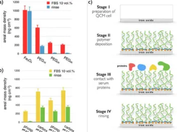
In this work, 6.8 nm iron oxide nanocrystals were coated with phosphonic and with carboxylic acid PEG copolymers using a formulation pathway described in the Materials and Methods section.50-‐52
In brief, dispersions of particles and of PEGylated copolymers were prepared in the same conditions of
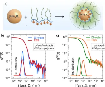
pH (pH 2.0) and concentration (𝑐𝑐 = 0.2 wt. %), and mixed at different volume ratios 𝑋𝑋. The pH of the mixed solution was raised to pH 8.0 by sodium hydroxide addition. It was found that below the critical mixing ratio 𝑋𝑋!, here equal to 1.5 for both polymers well-‐dispersed coated particles were obtained, with a 𝐷𝐷! about 5 nm larger than that of bare particles (Fig. 2a). Above 𝑋𝑋! in contrast, particles form large aggregates and precipitate in solution (as uncoated particles do), indicating an incomplete surface coverage from the polymers. The dispersions studied here were prepared at 𝑋𝑋 = 0.2, i.e. with a large excess of polymers to ensure that positive surface charges were all complexed by acid groups. The dispersions were then dialyzed against deionized water to remove the excess polymers (cut-‐off membrane 50 kD and 100 kD). Dynamic light scattering was used to measure the thickness of the polymer layer. For dispersions that are not stable, light scattering also allows estimating the aggregation kinetics.46,53 Figs. 2b and 2c display the
second autocorrelation function of the scattered intensity 𝑔𝑔(!) (t) for iron oxide coated with phosphonic and carboxylic acid PEG2K copolymers in
de-‐ionized water, respectively. The data exhibit a quasi-‐exponential decay associated with a unique relaxation mode. Derived from the second cumulant coefficient ( 𝑍𝑍!"#), the hydrodynamic diameters were 23.8 and 21.8 nm respectively, with dispersity indexes of 0.08 and 0.18. These 𝐷𝐷!-‐values are 9.8 and 7.8 nm larger than that of the bare particles (𝐷𝐷! = 14 nm24), and were ascribed to the layer thickness
noted ℎ!" to distinguish it from the polymer layer thickness on flat substrate ℎ!! defined in the next section. We found here ℎ!" = 4.5 ± 0.5 nm. With zeta potentials of -‐2 to -‐6 mV, electrokinetic measurements confirmed that the PEGylated particles are globally neutral. For PEG5K, the polymer
thickness was also determined and found at 8 ± 1 nm. The values for ℎ!" are consistent with stretched PEG chains forming a polymer brush.38,60 As shown
in the insets, the associated intensity distributions are characterized by a single population of particle size. When dispersed in 10 vol. % fetal bovine serum, the autocorrelation function and intensity distribution remains unchanged for particles with phosphonic acids (𝐷𝐷! = 21.8 nm, 𝑝𝑝𝑝𝑝𝑝𝑝 = 0.21). This result ascertains that PEGylated particles are stable in a serum rich medium and devoid of protein corona. A comprehensive characterization study has also shown that this stability is being maintained in cell culture media without serum and for extended
period of time (> weeks).21,24 For particles coated
with carboxylic acid functionalized polymers, the 𝑔𝑔(!)(t) relaxation is shifted to longer decay times, and the intensity distribution is now peaked at 𝐷𝐷! = 85.3 nm (𝑝𝑝𝑝𝑝𝑝𝑝 = 0.21), indicating a modification of the particle structures. The size increase could be due to protein adsorption on the PEG layer, a scenario that would be compliant with the corona model,3,31,53 or to particle aggregation induced by
the PEG layer depletion. From light scattering measurements, it is concluded that phosphonic acid PEG copolymer is an efficient coating agent compared to its carboxylic acid counterpart. Although the impact of the polymer type on particle stability is clear, the nature of the interactions of plasma proteins with a PEG coating layer remains open to question. To answer this question, a series of QCM-‐D experiments were performed using iron oxide substrates grafted with PEG copolymers.
Figure 2: a) Schematic representation of an iron oxide
particle, of a multi-‐phosphonic or carboxylic acid PEG copolymer and of the resulting nanostructure made from the two species. b) Autocorrelation function 𝑔𝑔(!)(t) of the
scattered light obtained from iron oxide nanoparticles coated with phosphonic acid PEG2K copolymers in DI-‐
water and in fetal bovine serum (FBS) containing medium. Inset: intensity distribution corresponding to the correlograms. c) Same as in Fig. 2b for carboxylic acid PEG2K copolymer coating. The data show the
destabilization of the dispersion and the particle agglomeration.
III.2 – Polymer adsorption on Fe3O4 substrate
Effect of PEG molecular weight
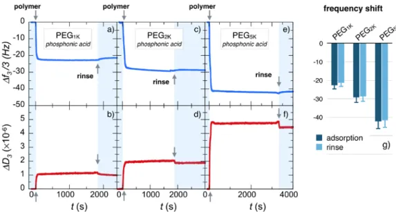
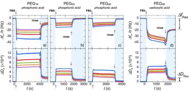
Fig. 3a and 3b display the adsorption profiles obtained by means of QCM-‐D technique for the
third overtone frequency ∆𝑓𝑓! 3 and the related dissipation ∆𝐷𝐷! of a 0.1 wt. % solution of phosphonic acid PEG1K copolymers at pH 2.0 and
room temperature, as for nanoparticle coating.24
Indeed, at this pH Fe3O4 is positively charged, with
an estimated density of active Fe-‐OH2+ sites of about
1.50 nm-‐2.47,49 Upon solution injection (arrow at t =
0), the frequency exhibits a rapid decrease and then a fast saturation at ∆𝑓𝑓! 3 = -‐22.7 Hz. Similarly, ∆𝐷𝐷! increases rapidly and reaches a plateau at 1.1×10⁶.
By increasing PEG molecular weights as in Fig. 3c and 3d for PEG2K and in Fig. 3e and 3f for PEG5K, the
time dependent profiles remain basically the same but the ∆𝑓𝑓! 3, in absolute values, increases to -‐29.4 Hz (with a dissipation ∆𝐷𝐷! = 2.0×10-‐6) and -‐43.3 Hz for PEG2K and -‐43.3 Hz (with ∆𝐷𝐷! = 4.8×10-‐6) for PEG5K. These adsorption kinetics are consistent with
those reported by QCM-‐D on the deposition of polymers on various substrates.8,41,42,61-‐64 The
polymer binding curves corresponding to the different overtones ( 𝑛𝑛 = 3, 5, 7, 9 and 11) are provided in Supplementary Information S7. Fig. 3g summarizes the frequency shift data, i.e. the adsorbed mass (including the solvation water) for the three molecular weights, indicating that increasing masses are bound to the substrate depending on chain length.65 After the deposition,
rinsing with DI-‐water at pH 7.4 has a little effect on the adsorbed layer for the considered polymers. The relative frequency losses after rinsing are 5%, 2% and 1.4% for PEG1K, PEG2K and PEG5K, respectively,
indicating that the polymer layers are firmly attached to the substrate, and that stability is enhanced for longer chains.
Figure 3: Real-‐time binding curves for frequency ∆𝑓𝑓! 3 and dissipation ∆𝐷𝐷! during the adsorption of phosphonic
acid poly(ethylene glycol) copolymers on Fe3O4 substrates at pH 2.0. PEG pending side-‐chains have molecular
weight of 1000 g mol-‐1 (a,b), 2000 g mol-‐1 (c,d) and 5000 g mol-‐1 (e,f). The data are those of the 3rd overtone of the QCM-‐D acoustic device. In each panel, the first arrow at t = 0 denotes the time at which the polymer solution (concentration 0.1 wt. %) is injected. The second arrow denotes the time at which DI-‐water (pH 7.4) is introduced for rinsing. g) Histogram for the steady state frequencies upon polymer adsorption and rinsing.
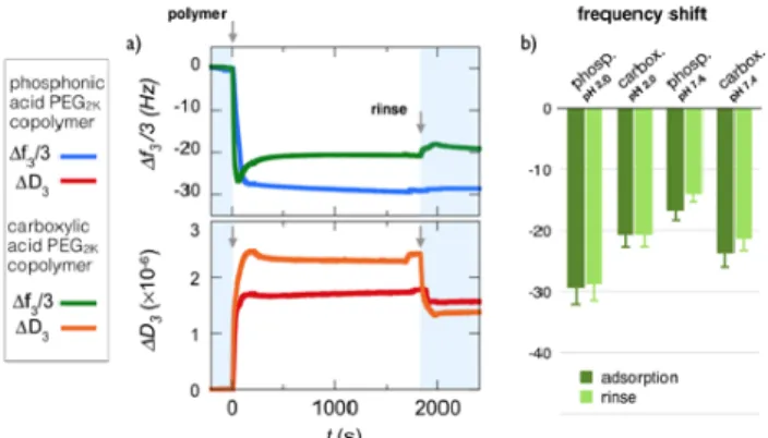
Effect of acid groups
The study of layer formation of PEG layers, respectively with phosphonic acid and carboxylic acid as linkers, at pH 2.0 sheds light on the role of acidic groups of different strength in the adsorption process. At this acidic pH, indeed phosphonic acid groups are negatively charged, carboxylic acid group are uncharged and the Fe3O4 substrate is positively
charged.47-‐49 Fig. 4a compares the frequency and
dissipation binding curves obtained for PEG2K
copolymers functionalized with phosphonic acid and
carboxylic acid moieties at pH 2.0 and 25 °C. It can be seen that the carboxylic acid PEG copolymer undergoes a strikingly different adsorption process compared to its phosphonic acid counterpart. In particular, a well defined undershoot behavior is observed and suggests fast adsorption/desorption processes due to conformational rearrangement of the adsorbent on oversaturated surfaces.66,67
Furthermore, the adsorbed mass at saturation is 30% lower than for phosphonic acid (∆𝑓𝑓! 3 = -‐ 20.6 Hz versus -‐29.4 Hz), while the related dissipation is
slightly higher, globally indicating that the less carboxylic acid PEG copolymers are more loosely bound to the Fe3O4 substrate than are the
phosphonic acid functionalized chains. Accordingly, upon rinsing, the carboxylic acid containing polymer shows a slight decrease and an apparent compaction of the bound mass, which are not observed for with phosphonic acid.
Figure 4: a) Binding curves for frequency ∆𝑓𝑓! 3 and
dissipation ∆𝐷𝐷! during the adsorption of phosphonic acid
and carboxylic poly(ethylene glycol) copolymers on Fe3O4
substrates at pH 2.0. Poly(ethylene glycol) side-‐chains are 2000 g mol-‐1 for both copolymers. In each panel, the first
arrow at t = 0 denotes the time at which the polymer solution (concentration 0.1 wt. %) is injected. The second arrow denotes the time at which DI-‐water (pH 7.4) is introduced for rinsing. b) Histogram for the steady state frequencies upon polymer adsorption and rinsing at the two 𝑝𝑝𝑝𝑝 values, pH 2.0 and pH 7.4.
To further assess the role of the charges on adsorption, experiments were performed at pH 7.4, i.e. in conditions where the phosphonic and carboxylic acids are both negatively charged and where the Fe3O4 substrate is neutral.48,49 Degrees of
ionization estimated from 𝑝𝑝𝑝𝑝𝑝𝑝 ’s values are 1.3 (indicating that 30% of the phosphonic acid groups bear two negative charges) and 0.8, respectively. Fig. 4b summarizes the frequency shifts for the two acid functionalized PEGs at the two pHs (see Supporting Information S8 for complete adsorption profiles). In particular, the phosphonic acid PEG copolymer suffers a drastic reduction (of about 50%) of the adsorbed mass from pH 2.0 to pH 7.4, while the carboxylic acid functionalized PEG mass is adsorbed in comparable amount at both pHs. Again, rinsing has a negligible effect on the adsorbed masses. Data from Fig. 4b suggest that for phosphonic acid containing polymers, electrostatics
is an important driving force for binding iron oxide surface, as adsorption is related to oppositely charge pairing and complexation.41,63 The lower adsorption
levels exhibited by carboxylic acid functionalized PEG may result from the fact that the acid groups and the substrate are only weakly charged. In this later case, other binding mechanism, including H-‐ bonding might be relevant.10,68,69 In overall, the data
indicate that the most efficient coating and binding to the Fe3O4 substrate occurred in acidic conditions
with phosphonic acid groups interacting with protonated Fe-‐OH2+ groups. The two different acidic
groups then interact with a strikingly different efficiency, depending on the relative pKa’s of phosphonic and carboxylic acid residues.
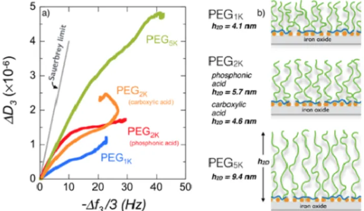
III.3 – Polymer brush structure
Fig. 5a shows the relationship between the variation of the dissipation ∆𝐷𝐷! and the resonance frequency shift, − ∆𝑓𝑓! 3, during the adsorption process of phosphonic acid PEG1K, PEG2K and PEG5K and
carboxylic acid PEG2K at pH 2.0. Previous studies
have shown that the slope of the 𝐷𝐷 − 𝑓𝑓 plot reflects the layer viscoelasticity, depending upon processes such as conformational changes, compaction or hydration/dehydration of the macromolecules at the surface.55,62 For phosphonic acid containing
polymers, the ratio – ∆𝐷𝐷!/ ∆𝑓𝑓! 3 is found in the range (0.6 − 1.7)×10!! Hz-‐1 and remains below the
Sauerbrey limit 4×10!! Hz-‐1 typical of
homogeneous and rigid films.54,55 For the carboxylic
acid PEG2K copolymer, the undershoot observed in
Fig. 4a translates into a change of regime with a negative slope 5 minutes after injection. The Sauerbrey equation (Eq. 1) is thus used to derive the areal mass density of the film, which in the present conditions includes both polymer adsorbate and solvent. With increasing molecular weight, from PEG1K to PEG5K, the areal mass density increases
from 400 to 750 ng cm-‐2 for the deposition step at
pH 2.0 (Table 2 and Supporting Information S9), and from 380 to 740 ng cm-‐2 for the rinsing step at pH7.4.
Values in the range 200 to 1000 ng cm-‐2 are usual for
polymers adsorbing spontaneously at interfaces either via physisorption or via grafting-‐to mechanisms.41,42,61-‐63
The hydrodynamic film thickness ℎ!! was estimated using the Voigt viscoelastic model.54,55 In the
deposition step, the thickness increases from 4.1, 5.7 to 9.4 nm for phosphonic acid containing PEG1K,
PEG2K and PEG5K polymers respectively (Fig. 5b). The
thickness of carboxylic acid functionalized PEG2K is
4.6 nm. After rinsing with DI-‐water at pH 7.4, ℎ!!-‐ values are lowered by 3% – 10%, the reduction being strong for PEG1K. Values for PEG2K and PEG5K brushes
are in good agreement with those obtained by Nalam et al. using poly(L-‐lysine)-‐graft-‐PEG63 and by
Emilsson et al. using thiol-‐terminated PEGs.42 Note
also that the thickness of the PEG2K and PEG5K films
on flat Fe3O4 substrates compares well with that of
the nanoparticle coating layer (Section III.1). From light scattering, spherical brush thicknesses were found at ℎ!" = 4.5 ± 0.5 nm and 8.0 ± 1.5 nm for PEG2K and PEG5K, in excellent agreement with the
ℎ!! = 5.7 nm and ℎ!! = 9.4 nm measured in QCM-‐D. The slight difference between the two determinations may arise from curvature effects.60
Figure 5: a) Plot of the dissipation ∆𝐷𝐷! as a function of the
frequency shift, − ∆𝑓𝑓! 3, during the adsorption process of
phosphonic acid PEG1K, PEG2K and PEG5K and carboxylic
acid PEG2K at pH 2.0. The straight line represents the
Sauerbrey limit (slope 4×10!! Hz-‐1) valid for
homogeneous and rigid films.54,55 b) Schematic representation of PEGylated layers deposited on iron oxide substrates obtained for the 4 polymers in a). Also indicated are the values of the brush thicknesses estimated from the Voigt viscoelastic model.54
To estimate the PEG density at the surface, it is assumed that the layer structure obeys the polymer brush theory38,60 in the moderate and high surface
density regimes, and that the density 𝜎𝜎!"# and the height ℎ!! are linked through the relation:39,40
ℎ!! = 𝜎𝜎!"# 3 !/ ! 𝑏𝑏!/! 𝑎𝑎𝑎𝑎 (2)
where 𝑎𝑎 = 0.28 nm and 𝑏𝑏 = 0.72 nm are respectively the chemical monomer and Kuhn lengths for
poly(ethylene glycol)29,42 and 𝑁𝑁 the degree of
polymerization of the chains. The derived PEG densities are 1.55, 0.57 (0.28) and 0.15 nm-‐2 for
PEG1K, PEG2K and PEG5K, the value in parenthesis
being that of carboxylic acid functionalized copolymers. A commonly used parameter for quantitative characterization of polymer brushes is the reduced tethered density Σ = 𝜋𝜋𝜎𝜎!"#𝑅𝑅!!, where 𝑅𝑅! is the gyration radius of the chain in the bulk phase.34 For poly(ethylene glycol), we adopt recent
small-‐angle neutron scattering results from Le Coeur and coworkers,70 who found a dependence of the
form: 𝑅𝑅! 𝑀𝑀! = 7.32×10!!𝑀𝑀!!.!!" . From the QCM-‐D data collected from the different polymers, Σ is found in the range 3.8 to 11.7 (Table 2), corresponding to grafting densities in the moderate (carboxylic acid-‐PEG2K, phosphonic acid-‐PEG5K,
1 < Σ < 5) and in the highly stretched regimes (phosphonic acid-‐PEG1K and PEG2K, Σ > 5 ).39 This
criterion to evaluate polymer stretching is similar to that found in parallel studies focusing on the ratio 𝐿𝐿/2𝑅𝑅!, where 𝐿𝐿 is the distance between tethered points (𝐿𝐿 = 𝜎𝜎!"#!!/!).30,71 A decrease in 𝜎𝜎!"# by a factor 10 between PEG1K and PEG5K (Tab. II) is
attributed to excluded volume interaction and steric repulsion between chains during deposition. The already adsorbed chains act as a barrier to the incoming ones, a mechanism that is more effective for longer chains. As a result, the brush stretching and morphology are different: dense and solid-‐like for PEG1K and soft and viscoelastic for PEG5K.42 These
differences in structure appear also in the different spreading of the overtones measured during deposition (S7, S9).55 The Voigt viscoelastic model
also allows estimating the adsorbed mass from the layer thickness ℎ!! .72 Table S1 in Supporting Information compares the areal mass densities for phosphonic acid PEG copolymers obtained from the Sauerbrey equation (Eq. 1) and from the Voigt model, leading to a good agreement between the two determinations.
In conclusion, phosphonic acid PEG copolymers are shown to adsorb spontaneously at Fe3O4 interfaces
and acidic pH. The copolymer backbone attaches to the surface via multisite binding, and the PEG side-‐ chains organize themselves into moderate-‐density or highly stretched brushes of a few nanometers. The QCM-‐D results confirm the data obtained in the bulk phase with iron oxide nanoparticles.21