Multidrug-Resistant MCF-7
Cells: An Identity Crisis?
Two recent correspondences pub-lished in the Journal (1,2) labeled the MCF-7/ADR cell line—a multidrug-resistant (MDR) human breast cancer MCF-7 subline—as having a non-MCF-7 origin, which led to a change in the nomenclature of this cell line to NCI/ ADR. We believe the original nomen-clature of MCF-7/ADR should be re-tained.
Although the two MDR MCF-7 sub-lines (MCF-7/ADR and MCF-7 TH) used by the investigators whose work prompted the nomenclature change were independently established, they showed several genotypic and phenotypic simi-larities. Both contained a full-length functional caspase-3 protein (2), despite complete loss of this protein in the pa-rental MCF-7 cells because of a 47-base-pair deletion in exon 3 of the CASP-3 gene (3). Interestingly, several features of the MCF-7/DOX subline es-tablished in our laboratory several years ago (4) were identical to those of the MCF-7/ADR and MCF-7 TH cells but different from those of the parental MCF-7 cells (5). For example, similar to MCF-7/ADR and MCF-7 TH cells, the MCF-7/DOX cells showed high expres-sion levels of P-glycoprotein (P-gp) and of a protein cross-linking enzyme, tissue transglutaminase; they also contained the full-length caspase-3 protein (6). We thus sought to determine whether the de-velopment of drug resistance in MCF-7 cells represents selective selection and expansion of an inherently resistant 1652 CORRESPONDENCE Journal of the National Cancer Institute, Vol. 94, No. 21, November 6, 2002
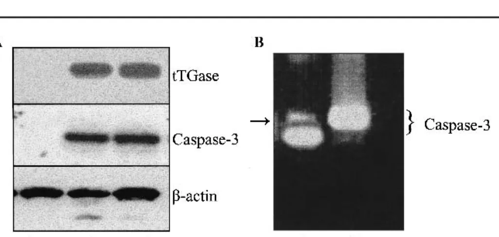
clone in the parental MCF-7 cell popu-lation. We purchased MCF-7 cells from the American Type Culture Collection (lot 2015862; Manassas, VA) and treated them with doxorubicin at 1 g/ mL. More than 99% of the MCF-7 cells died within 1 week, but a few colonies (an average of two colonies per T-75 flask were observed to grow in the pres-ence of doxorubicin) of cells survived about 3 weeks of continuous culture in the presence of the drug. We expanded these colonies and, to our surprise, the newly established MCF-7 cell subline (MCF-7/WT/DOX) exhibited several biochemical features similar to those of the MCF-7/DOX and MCF-7/ADR cells but different from those of the parental MCF-7 cells. The MCF-7/WT/DOX cells showed high expression levels of both P-gp and tissue transglutaminase and contained full-length functional caspase-3 protein (Fig. 1, A). Karyo-typic analysis of the MCF-7, MCF-7/ DOX, and MCF-7/WT/DOX cell lines revealed unique features that were highly conserved in the drug-resistant sublines but were quite distinct in the parental MCF-7 cells (5).
MCF-7 cells express a truncated iso-form of caspase-3 transcript, whereas drug-resistant sublines express full-length caspase-3 transcript. Thus, we amplified the complementary DNA (cDNA) from MCF-7 and MCF-7/WT/ DOX cells by polymerase chain reaction (PCR) using primers specific for full-length caspase-3 cDNA to further
vali-date the presence of a drug-resistant clone in the parental MCF-7 cells. The resulting PCR products were analyzed for the presence of caspase-3 transcripts by agarose gel electrophoresis and ethidium bromide staining. The PCR products of the MCF-7 cells contained a predominant band corresponding to truncated caspase-3 transcript (Fig. 1, B); a minor band corresponding to full-length caspase-3 transcript became evi-dent after many PCR cycles on the am-plified cDNA (Fig. 1, B, arrow). The PCR-amplified products from the MCF-7/DOX cells showed only a single band, corresponding to the full-length cas-pase-3 transcript. These results con-firmed the presence of one or more inherently resistant subclones in the pa-rental MCF-7 cells that harbor the full-length CASP-3 gene and are likely to propagate into drug-resistant cell lines in the presence of MDR-related drugs. This possibility was further supported by our inability to establish any doxoru-bicin-resistant cell lines from two MCF-7 single-cell clones. These results appeared to be consistent with the find-ings that caspase-3-deficient MCF-7 cells, when reconstituted with caspase-3 cDNA, become more susceptible to che-motherapy-induced apoptosis (6). It is likely that caspase-3-adequate MCF-7 subclones that propagate into drug-resistant sublines have additional path-ways that confer selective resistance of these cells to chemotherapeutic agents. Despite their high resistance to
doxoru-bicin, these cells are exquisitely sensi-tive to certain apoptosis-inducing agents. For example, Leoni et al. (7) ob-served that MCF-7/ADR cells were more sensitive to indanone-induced ap-optosis than were the drug-sensitive MCF-7 cells. In our experience, the MCF-7/DOX cells were much more sensitive to a staurosporine-induced ap-optosis than were the parental MCF-7 cells (5).
Our research has demonstrated that drug-resistant MCF-7 cell lines result from parental MCF-7 cells that harbor the full-length CASP-3 gene. In view of these results, we suggest that the origi-nal nomenclature of MCF-7/ADR for MCF-7-derived drug-resistant sublines be retained to reveal the fact that various clones in a given tumor population can be extremely diverse in terms of their genotype and phenotypic characteristics. KAPILMEHTA ESWARANDEVARAJAN JACKCHEN ASHAMULTANI SENPATHAK
R
EFERENCES(1) Scudiero DA, Monks A, Sausville EA. Cell line designation change: multidrug-resistant cell line in the NCI anticancer screen. J Natl Cancer Inst 1998;90:862.
(2) Pirnia F, Breuleux M, Schneider E, Hochmeis-ter M, Bates SE, Marti A, et al. Uncertain identity of doxorubicin-resistant MCF-7 cell lines expressing mutated p53. J Natl Cancer Inst 2000;92:1535–6.
(3) Janike RU, Sprengart ML, Wati MR, Porter AG. Caspase-3 is required for DNA fragmen-tation and morphological changes associated with apoptosis. J Biol Chem 1998;273: 9357–60.
(4) Mehta K. High levels of transglutaminase ex-pression in doxorubicin-resistant human breast carcinoma cells. Int J Cancer 1994;58:400–6. (5) Devarajan E, Chen J, Multani AS, Pathak S, Sahin AA, Mehta K. Human breast cancer MCF-7 cell line contains inherently drug-resistant subclones with distinct genotypic and phenotypic features. Int J Oncol 2002;20: 913–20.
(6) Yang XH, Sladek TL, Liu X, Butler BR, Fro-elich CJ, Thor AD. Reconstitution of caspase-3 sensitizes MCF-7 breast cancer cells to doxorubicin- and etoposide-induced apop-tosis. Cancer Res 2001;61:348–54.
(7) Leoni LM, Hamel E, Genini D, Shih H, Car-rera CJ, Cottam HB, et al. Indanocine, a mi-crotubule-binding indanone and a selective in-ducer of apoptosis in multidrug-resistant can-cer cells. J Natl Cancan-cer Inst 2000;92:217–24.
N
OTESEditor’s note: Dr. Scudiero declined to com-ment.
Fig. 1. Tissue transglutaminase (tTGase) and caspase-3 expression in MCF-7, MCF-7/WT/DOX, and
MCF-7/DOX cell lines. A) tTGase and caspase-3 protein expression levels were examined by western blot analysis in the parental MCF-7 cells (purchased from American Type Culture Collection; lane 1), in drug-resistant MCF-7/WT/DOX cells (obtained by continuous culture of the parental MCF-7 cells in doxorubicin at 1g/mL; lane 2), and in MCF-7/DOX cells (obtained from Dr. Ken Cowan, National Cancer Institute; lane 3). B) Caspase-3 transcripts in MCF-7 cells (lane 1) and MCF-7/WT/DOX cells (lane 2), as determined after many polymerase chain reaction cycles of the complementary DNA from the respective cell lines. Arrow indicates the presence of full-length caspase-3 transcript in MCF-7 cells.
Affiliation of authors: K. Mehta, E. Devarajan, J. Chen (Department of Bioimmunotherapy), A. Multani, S. Pathak (Department of Cancer Biol-ogy), The University of Texas M. D. Anderson Cancer Center, Houston.
Correspondence to: Kapil Mehta, Ph.D., Divi-sion of Cancer Medicine, Box 422, The University of Texas M. D. Anderson Cancer Center, 1515 Holcombe Blvd., Houston, TX 77030 (e-mail: [email protected]).
RESPONSE
We read with great interest the cor-respondence by Mehta et al. These au-thors propose the hypothesis that the various doxorubicin (ADR)-selected MCF-7 sublines may result from the se-lective expansion of inherently resistant subclones that contaminate the parental MCF-7 cell line and that harbor the full-length CASP-3 gene. Although this theory is quite interesting, we have some caveats (1). Because the CASP-3 gene is deleted in parental MCF-7 (1), it is difficult to understand how this
geno-type could have been reverted in the hypothetically inherently resistant sub-clones (2). In our original descrip-tion, we assessed the nonidentity of MCF-7 and MCF-7 TH by DNA finger-printing (2). This method is more reli-able than phenotypic karyotyping (3). When they claim that an inherently re-sistant subclone contains functional caspase-3, Mehta et al. somehow con-tradict the finding that caspase-3 re-stores chemotherapy sensitivity in MCF-7 cells (3).
Most importantly, Mehta et al. ex-plain their hypothesis with a contamina-tion of the parental MCF-7 cell line. Thus, we strongly caution against retain-ing the original nomenclature—MCF-7/ ADR or MCF-7 TH—for these sublines of unknown origin. Good research can-not be performed with tools of uncertain identity.
FARZANEHPIRNIA MARKUSM. BORNER
R
EFERENCES(1) Janicke RU, Sprengart ML, Wati MR, Porter AG. Caspase-3 is required for DNA fragmen-tation and morphological changes associated with apoptosis. J Biol Chem 1998;273: 9357–60.
(2) Pirnia F, Breuleux M, Schneider E, Hochmeis-ter M, Bates SE, Marti A, et al. Uncertain identity of doxorubicin-resistant MCF-7 cell lines expressing mutated p53. J Natl Cancer Inst 2000;92:1535–6.
(3) Yang XH, Sladek TL, Liu X, Butler BR, Froelich CJ, Thor AD. Reconstitution of caspase-3 sensitizes MCF-7 breast cancer cells to doxorubicin- and etoposide-induced apoptosis. Cancer Res 2001;61: 348–54.
N
OTESAffiliation of authors: F. Pirnia, M. M. Borner, Institute of Medical Oncology, Inselspital, Bern, Switzerland.
Correspondence to: Markus M. Borner, M.D., University of Bern, Institute of Medical Oncology, Inselspital, CH-3010 Bern, Switzerland (e-mail: [email protected]).