Case report
Bronchogenic cyst of the left lower lobe associated with severe
hemoptysis
Didier Lardinois*, Matthias Gugger, Hans-Beat Ris
Department of Thoracic and Cardiovascular Surgery, Division of Pneumology, University of Bern, 3010 Bern, Switzerland Received 28 March 1999; received in revised form 31 May 1999; accepted 15 June 1999
Abstract
Bronchogenic cysts result from congenital disorders, are often asymptomatic at diagnosis, but complications are not uncommon. We report the case of a 19-year-old woman with severe hemoptysis. This rare presentation of an intrapulmonary bronchogenic cyst should be considered as differential diagnosis in patients with cavernous lesion of a lobe. Surgery was performed as a diagnostic and therapeutic measure. q 1999 Elsevier Science B.V. All rights reserved.
Keywords: Bronchogenic cyst; Hemoptysis
1. Case report
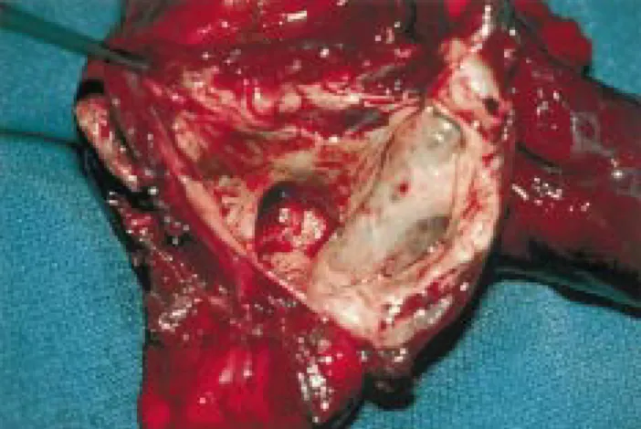
A 19-year-old nurse from Sri Lanka presented with acute, massive hemoptysis with hypotension and collapse. She lived in Switzerland since 1986. The medical history revealed a pneumonia on the right side treated with antibio-tics in 1985 but no tuberculosis or other infectious diseases. The history of her family was also uneventful. The patient had neither fever, cough, sweating nor dyspnea. On the chest X-ray, a sharply de®ned, solitary cavernous lesion of the left lower lobe was found. The white blood count and blood chemistry showed no pathological ®ndings, especially no signs of infection. The hemoglobin value fell from 13.7 to 10.1 g/dl. The patient was transferred to the intensive care unit with suspicion of active tuberculosis. An antitubercu-lous chemotherapy with four drugs was started. CT scans showed a 6 £ 5 £ 4 cm large, thin-walled cavernous lesion of the left lower lobe without any parenchymal in®ltrations (Fig. 1); a bronchoscopy showed no pathological ®ndings, except blood clots in the left lower lobe bronchus. A thor-acotomy with resection of the left lower lobe was performed on the next day to prevent bleeding recurrence (Fig. 2). There were no postoperative complications and the patient was discharged 8 days later. Histology revealed a cyst communicating with the bronchial tree. The wall of the cyst contained smooth muscle, bronchial mucus glands and cartilage. No epithelioid granulomas and no abscesses
were found. Cultures showed no fungi, no mycobacteriae, no parasites.
2. Discussion
Bronchogenic cysts are lesions of congenital origin derived from the primitive foregut [1]. Fifty to 80% of the patients are symptomatic on presentation but an incidental ®nding on radiographic examination is not rare. They are slightly more common in men and are mainly discovered in the third or fourth decade of life [1,2]. The most frequent symptoms are pain, cough, fever or dyspnea, but severe hemoptysis is rarely reported [3,4]. Although more commonly located in the mediastinum, intrapulmonary cysts represent about 15±20% of all the bronchogenic cysts and usually occur in the lower lobes [5]. Most of them have no patent communication with the tracheobron-chial tree. Most of the complications of bronchogenic cysts result from compression of mediastinal structures, infection, or rarely, changing into a malignant tumor [6,7]. The differ-ential diagnosis of the parenchymal form includes lung abscess, hydatid cyst, fungal diseases, tuberculosis, infec-tion with nocardia, infected bulla, vascular malformainfec-tion and neoplasm [8]. In our case, the preoperative chest X-ray showed no calci®cation (hydatid cyst) and no air ¯uid level (abscess). Contrast ¯uid did not ®ll the lesion (vascular malformation). The location in the lower lobe, the thin wall of the cyst without surrounding parenchymal pathology, and the absence of acid-fast bacilli in the bronchial secretions were atypical for tuberculosis. However, because of the
European Journal of Cardio-thoracic Surgery 16 (1999) 382±383
1010-7940/99/$ - see front matter q 1999 Elsevier Science B.V. All rights reserved. PII: S1010-7940(99)00226-2
www.elsevier.com/locate/ejcts
* Corresponding author. Tel.: 2330; fax: 141-31-632-2327.
origin of the patient and the increased prevalence of tuber-culosis in immigrants, a mycobacteriosis could not be excluded completely. CT scan revealed a homogeneous content of a part of the cavernous lesion, suggesting blood, but the content was indistinguishable from tubercu-lous pus or aspergilloma. It is well known that aspergillomas can produce a massive hemorrhage, making this diagnosis possible as well. The resection was mainly performed to prevent a second episode with massive hemoptysis, but also to obtain a de®nitive diagnosis. In the case of tubercu-losis or an aspergilloma with severe hemoptysis, the opera-tive procedure seems to be the therapy of choice. In our case, an intrapulmonary bronchogenic cyst was histologi-cally diagnosed without isolation of pathogens. A review of the literature reveals that surgery represents the therapy in symptomatic bronchogenic cysts and some authors recom-mend the resection of all bronchogenic cysts, asymptomatic or not [1,3,9]. The resection must be complete because recurrence has been observed after incomplete surgical removal. On the basis of the size of the lesion, a segmen-tectomy was not possible and the whole lower lobe had to be resected. The therapy of asymptomatic bronchogenic cysts remains controversial but the majority of authors seem to advocate a surgical approach to obtain a diagnosis and to prevent complications, which occur in up to 80% in patients with symptomatic cysts [1,3].
In conclusion, severe hemoptysis is a rare complication of intrapulmonary bronchogenic cysts and may be the only symptom at presentation time. Bronchogenic cysts should
be considered in the differential diagnosis of patients with severe hemoptysis, especially because a de®nitive con®dent preoperative diagnosis is not always possible on the basis of standard investigations.
References
[1] Aktogu S, Yuncu G, Halil CËolar H, Ermete S, Buduneli T. Broncho-genic cysts: clinicopathological presentation and treatment. Eur Respir J 1996;9:2017±2021.
[2] Jones JC, Almond CH, Snyder HM, Meyer BW. Congenital pulmonary cysts in infants and children. Ann Thorac Surg 1967;3:297±306. [3] Patel S, Meeker D, Biscotti C, Kirby T, Rice T. Presentation and
management of bronchogenic cysts in the adult. Chest 1994;106:79± 85.
[4] Liam CK. Intrapulmonary bronchogenic cyst presenting with haemop-tysis. Med J Malaysia 1994;49:404±405.
[5] St-Georges R, Deslauriers J, Duranceau A, et al. Clinical spectrum of bronchogenic cysts of the mediastinum and lung in the adult. Ann Thorac Surg 1991;52:6±13.
[6] Avital A, Udassin R, Bar-Ziv J, Godfrey S, Springer C. Bronchogenic cyst associated with left lower lobe bronchiectasis. Pediatr Pulmonol 1993;16:323±325.
[7] Yim APC, Lam HCK, Ho JKS. Thoracoscopic lobectomy for an infected intrapulmonary bronchogenic cyst. Surg Endosc 1996;10:439±440.
[8] Suen H, Mathisen D, Grillo H, Moncure A, Hilgenberg A. Surgical management and radiological characteristics of bronchogenic cysts. Ann Thorac Surg 1993;55:476±481.
[9] Cuypers Ph, De Leyn P, Cappelle L, Demedts M, Deneffe G. Broncho-genic cysts: a review of 20 cases. Eur J Cardio-thorac Surg 1996;10:393±396.
D. Lardinois et al. / European Journal of Cardio-thoracic Surgery 16 (1999) 382±383 383
Fig. 1. CT scans with thin-walled cavernous lesion of the left lower lobe.