Multiple immune abnormalities in tumor
necrosis factor and lymphotoxin-ot
double-deficient mice
Hans-Pietro Eugster, Matthias Muller, Urs Karrer
1, Bruce D. Car
2, Bruno Schnyder,
Vicki M. Eng
3, Gaetane Woerly, Michel Le Hir, Franco di Padova
4, Michel Aguet
5,
Rolf Zinkernagel
1, Horst Bluethmann
6and Bernhard Ryffel
Swiss Federal Institute of Technology, Institute of Toxicology, Schorenstrasse 16, 8603 Schwerzenbach, Switzerland
institute for Experimental Immunology, Schmelzbergstrasse 12, 8091 Zurich, Switzerland 4Sandoz Ltd, Preclinical Research, 4002 Basel, Switzerland
Pharmaceutical Research Gene Technology, F. Hoffmann-LaRoche Ltd, 4002 Basel, Switzerland 2Present address- Du Pont Merck, Research and Development, Stine-Haskell Research Center, PO Box 30, Building 320, Newark, DE 19714-0030, USA
3Present address: University Laboratory Animal Resources, University of Pennsylvania, Philadelphia, PA 19104-6021, USA
5Present address: Genentech Inc., 460 Point San Bruno Boulevard, South San Francisco, CA 94080, USA
Keywords alymphoplasia, endotoxic shock, isotype switch, Listena, lymphocytosis, Peyer's patches
Abstract
To investigate the roles of tumor necrosis factor (TNF) and lymphotoxin (LT)-a in the development and function of the immune system, the Tnf and /.forgeries were simultaneously inactivated in mice by homologous recombination. These mutant mice are highly susceptible to Listeria
monocytogenes infection and resistant to endotoxic shock induced by the combined administration of D-galactosamine (D-GaIN) and lipopolysaccharide (LPS). Their splenic
microarchitecture is disorganized, characterized by the loss of the clearly defined marginal zone, ill defined T and B cell areas, and absence of MAdCAM-1 and reduced ICAM-1, VCAM-1 and Mac-1 expression. They are devoid of peripheral lymph nodes and Peyer's patches, and show a strong reduction of lgA+ plasma cells in the intestinal lamina propria. The alymphoplasia is accompanied
by a marked B lymphocytosis and reduced basal Ig levels. Ig depositions in the renal glomerulus and a strong up-regulation of MHC class I antigen expression on endothelial cells of different tissues are observed. The primary humoral immune response towards sheep red blood cells reveals a defective IgG isotype switch, while that against vescicular stomatitis virus is normal. The cytotoxic T cell responses are attenuated, although still effective, against vaccinia, lymphocytic choriomeningitis virus (LCMV-ARM) and LCMV-WE. In conclusion, the combined inactivation of Tnf and Lta confirms their essential role in the normal development and function of the immune system.
Introduction
Tumor necrosis factor (TNF) is a proinflammatory cytokme involved in host defence and pathogenesis of various diseases (1,2). Soluble trimenc TNF hgands bind to TNF receptor 1 (TNFR1) and TNFR2, leading to receptor homotrimer formation (3) and consecutive triggering of various biological responses such as proliferation, cytotoxicity and apoptosis in target cells. Lymphotoxin (LT)-a, which like TNF belongs to the growing
family of TNF-like ligands (4), can bind to the same receptors as TNF. Consequentely, administration of TNF and LT-cc leads to similar biological responses in vivo and in vitro. In mice, TNF is mainly expressed in macrophages and T cells, whereas LT-a expression is confined to T and B cells. The redundancy in receptor binding and the overlapping expression pattern of TNF and LT-a renders a clearcut dissection of their function Correspondence to- H.-P. Eugster
rather difficult. Therefore, the importance of both ligands to TNF receptor-mediated effects tends to be obscured in single gene knockout mice, because the loss of one ligand may be compensated by the remaining one. As both genes are very closely linked on chromosome 17, single mutations cannot be crossed together. Rather, mice deficient for both ligands have to be generated by simultaneous targeting of both genes. Prominent functions of TNF include central roles in endotoxic shock, host defence and tumor (or parasite) induced cachexia In addition TNF plays an important, yet undefined role in the pathogenesis of autoimmune diseases (5-9). LT-a deficiency has been shown to lead to aberrant development of the spleen and absence of peripheral lymph nodes (LN) (10) Recently a third ligand, LT-(J, was cloned and characterized as present on the cell surface as a LT-a/ 2LT-P heterotrimer, able to bind to the newly described LT-(3-specific receptor (11-13).
To investigate the consequences of the combined defi-ciency of TNF and LT-a on the immune system we generated TNF/LT-a double-deficient mice by homologous recombination in mouse embryonic stem cells (ES) We demonstrate that the TNF/LT-a double-deficient mice are viable and fertile, but show profound structural and functional defects of the immune system
Methods
Reagents
Geneticin (G418) was obtained from Calbiochem (La Jolla, CA), FIAU [1-(2-deoxy, 2-fluoro-|3-D-arabinofuranosyl)-5-lodouracil] from Bristol Myers (Squibb Pharmaceutical Research Institute, Wallingford, CT), D-galactosamine (D-GalN) from Carl Roth (Karlsruhe, Germany), Concanavalm A (Con A, C5275) and lipopolysaccharide (LPS; Escherichia
coli, serotype O111B4) from Sigma (St Louis, MO).
Biotin-conjugated isotype-specific goat anti-mouse IgGi, lgG2a, lgG2b, lgG3, IgM and IgA were from Southern Biotechnology Associates (Birmingham, AL), and alkaline phosphatase (AP)-conjugated streptavidin was from Jackson Immunoresearch (West Grove, PA). IgA-specific goat anti-mouse antibody (Chemicon IG108, a chain specific), rabbit anti-goat-Cy3 antibody (Jackson) and goat anti-rat-Cy3 antibody (Jackson) were purchased from Milan Analytica (LaRoche, Switzerland) Rat anti-mouse MHC class I and class II antibodies were from ATCC clones M1/42 and M5/114 respectively. The anti MAdCAM-1 antibody was a generous gift from B. Holzmann
Construction of targeting vector
A 0.8 kb Hinc\\-Dra\ fragment from a TNF genomic clone (14) (provided by Dr C. V. Jongeneel) was inserted in a clockwise orientation as the 3' target into the HincW site of a modified pBLSK+ plasmid lacking sites between C/al and Eag\ A
Xho\-Xba\ fragment carrying the pgk-neo cassette from pPNT (15)
provided by Dr G Veres was inserted into the respective sites, placing the 3' target beside the neo cassette As a 5' target, a PCR product (5' primer- 5'-GCGGTACCCAGTCAC-GACGTTGTAAAAC-3'; 3' primer 5'-GGTCTAGACGGAAGA-CAGACCTTACCTC-3') encompassing the complete 5' region of the genomic clone (14) up to bp 1655 was digested with Asp718l and Xba\ and subcloned into the respective sites,
placing the neo cassette between the genomic 3' and 5' targets. Subcloning of a EcoRI-/-//ndlll blunt-ended fragment from pPNT, carrying the thymidine kinase cassette, into the unique, blunt-ended Asp718l site resulted in the final targeting construct which was linearized by restriction with Not\.
Generation of TNF/LT-a double-deficient mice
Briefly, 3X10⁷ GS1 ES cells (16) were transfected with 10 ng linearized targeting vector. Recombinant clones were selected in media containing G418 and FIAU (17) Mutant ES cell clones were identified by PCR analysis. Further characterization by Southern blot analysis revealed a deletion from Lta exon 2 to the middle of Tnf exon 4 thus completely inactivating both genes (Fig. 1A). Mutant ES cell clones were injected into C57BL/6 blastocysts and implantation led in one case to chimeras and eventual germline transmission of the mutation. All mice were maintained under specific pathogen-free condi-tions. Permission for animal experimentation was obtained from local authorities and performed according to institutional guidelines All mice used in experiments were 6- to 8-week-old females.
Genetic analysis of the mutant locus
Genomic DNA was isolated from tail biopsies (18) Digested DNA (10 u.g) was subjected to agarose gel electropheresis and subsequently transferred to nylon membranes (Hybond-N+, Amersham, Zurich, Switzerland) according to standard procedures. A genomic probe (Probe A, a Stu\~BamH\ gen-omic fragment, see Fig. 1A) was random labeled with [a-32P]dCTP (Amersham) and used for hybridization. Filters were prehybndized for 15 min at 65°C in 1% BSA, 0.5 M sodium phosphate, pH 7 2, 20 mM EDTA, 15% deionized formamide and 7% SDS. Then the denatured probe was added to the prehybridization mix and hybridization was carried out for 2.5 h. Filters were rinsed twice in 2xSSC, 1% SDS at 65°C, followed by two 30 min washes at 65°C in 2xSSC, 1% SDS and 0.2xSSC, 1% SDS respectively, and exposed to Hyperfilm-MP (Amersham).
LPS/D-GaIN induced shock
Mice (6-8 weeks old) were injected i.p. with a combination of 20 mg D-GaIN and varying doses of LPS (0.1, 1, 10 or 100 |ig; E. coli, serotype O111:B4) in saline solution (0.9%). The mice were monitored for hepatic failure and lethality. Blood was drawn 6 h after treatment from mice anesthetized with methoxyflurane (Metofane; Pitman-Moore, Mundelein, IL) to analyze alanine and aspartate aminotransferases using a COBAS phara kit (F. Hoffmann-LaRoche, Basel, Switzerland) Mice demonstrating severe signs of endotoxemia were killed
Flow cytometry
Cell suspensions from thymus and spleen were prepared by gentle squeezing of organ pieces at 4°C in DMEM, 2% FCS through a wire mesh sieve. Cells were washed and resuspended in PBS, 2% FCS at 4°C and stained with directly labeled antibodies. The following antibodies were used: anti-mouse CD4-phycoerythrin (Becton Dickinson), anti-anti-mouse CD8-FITC (Becton Dickinson), anti-mouse B220-FITC (CD45R, clone RA3-6B2; PharMingen, San Diego, CA) and anti-mouse CD3-biotin (PharMingen). As secondary reagent
Xpgk-neo~|x
wt-B i
I
LTa
BTNF
2.9 kb
probe A4.1 kb
B
4.1 kb
- 2.9 kb
Fig. 1. Inactivation of Tnf and Lta by homologous recombination. (A)
Targeted mutation strategy showing the gene replacement vector On top, the targeting vector, in the middle the Tnf/Lta locus and the predicted structure of the mutant locus at the bottom The length of the wild-type and mutated fragments detectable in the Southern blot with probe A are given. The position of the diagnostic PCR primers are indicated by arrows (B) Southern blot analysis of BstEM (B) digested genomic DNA from tail biopsies of a litter from a heterozygous intercross showing wild-type, homozygous mutant and heterozygous pattern of the TNF/LT-o locus ( + / + , wild-type, +/-, heterozygous, -/-, homozygous mutant).
streptavidm-Cy-Chrome (PharMingen) was used. Cells from peripheral blood were prepared by standard procedures and labeled as described above. Cells were analyzed with a FACScan flow cytometer (Becton Dickinson) using Lysys II software
Analysis of naive Ig levels and induced humoral immune response
For the determination of isotype levels, Nunc Immuno plates were coated with 5 ng/ml goat anti-mouse lgG1, lgG2a, lgG2b, lgG3, IgM and IgA. Bound Ig of diluted serum samples was detected with AP-coupled isotype-specific goat anti-mouse Ig antibodies. Serum Ig concentrations were calculated using mAb of the various isotypes as standards. Mice were immunized i.p. with sheep red blood cells (SRBC, 108 SRBC in 0.9% pyrogen free saline), and bled on days 3, 9 and 15 after immunization. Anti-SRBC serum isotype levels were determined by a sandwich ELISA Maxisorb microtiter plates
(Nunc, Roskilde, Denmark) were coated with 50 u.l of a solubihzed extract (3 jig/ml) from SRBC prepared according to Kelly et al. (19) Plates were blocked with PBS/BSA 2% for 2 h at 37°C and incubated overnight with serially diluted samples. Biotinylated goat anti-mouse Ig isotype was added for 4 h Subsequently, AP-conjugated streptavidin was added for 45 min each The reaction was stopped 30 min after substrate addition with NaOH (1.5 M). Absorbance was read at 405 nm in a Titerteck Multiskan spectrophotometer (Flow Laboratories) The titer of a serum sample is expressed as the reciprocal value of the dilution showing an optical density of 0.1 over background. Neutralizing IgM and IgG titers against vesicular stomatitis virus (VSV) were determined at different time points after i v. infection with 2x10⁶ p f u VSV (Indiana strain) according to established protocols (20).
Infection with Listeria monocytogenes
Mice were infected i.v with 1.2x10³ c f u of L
monocytog-enes Animals were killed after 3 days, and hsterial titers of
spleen and liver were determined by plating serial dilutions of organ homogenates onto Trypticase-soy agar (21) Lethality was monitored twice daily in animals infected as described for the determination of organ titers
In vitro and in vivo anti-viral responses
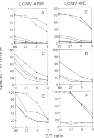
For the assay of vaccinia virus (VV-WR; WR strain) specific cytotoxic T lymphocytes (CTL), mice were infected i.v. with 2X10⁶ p f u. and primary ex vivo CTL activity was tested 6 days after infection using MC57 target cells infected with VV-WR (20). For the lymphocytic choriomeningitis virus (LCMV)-specific primary CTL response, mice were infected i.v. with 2X10³ p f u. LCMV-ARM (Armstrong isolate) or 2X10² p.f.u. LCMV-WE, and primary ex vivo CTL activity was tested 8 days after infection using MC57 target cells infected with either LCMV-ARM or LCMV-WE For the LCMV-specific sec-ondary CTL response, splenocytes from mice infected i.v. with LCMV-ARM (2X10³ p.fu.) or LCMV-WE (2X10² p f u ) were re-stimulated for 5 days in vitro after 8 days with LCMV-ARM or LCMV-WE infected macrophages and CTL activity was assessed as described above.
Foot pad swelling and corresponding secondary CTL were induced by injecting either 3X10³ LCMV-ARM or 3X10³ p f u. LCMV-WE into the hind foot pad (i.f.p.). Splenocytes were re-stimulated in vitro after 23 days for 5 days with LCMV-ARM-or LCMV-WE-infected macrophages and CTL activity was assessed as described above.
Histology
Sections were from freshly fixed tissue (buffered 4% formalin in PBS), paraffin embedded, cut at 5 u.m and stained with hematoxylin & eosin. Tissues for immunofluorescence analysis were snap frozen in isopentane, supercooled in liquid nitro-gen. Primary antibodies were applied overnight at 4°C to acetone fixed cryosections cut at 5 u.m. After two washes (PBS, 1% BSA), secondary antibodies were applied for 1 h at room temperature. Slides were mounted in Shandon Immunomount and tissues were photographed in confocal mode with a Laser Scan Microscope 320 (Carl Zeiss Micro-scope Systems, Zurich, Switzerland) utilizing the fluorescence excitation wavelength of 543 A. Where tissues from positive
and negative mice are presented together, identical contrast and brightness settings were employed. Negative controls (not shown) were uniformly negative
2000 100 0.1 1 10 100 LPS (ng) 0.1 1 10 LPS (ng) 100
Fig. 2. (A) Liver transaminase activity in serum and (B) mortality of
mutant and wild-type mice in the LPS/n-Gal sensitization model Data for liver transammases are presented as mean units/I (± SD) Four animals were used for the two lower dose of LPS and six animals were used for the two higher doses of LPS
Results
Combined genetic inactivation of Tnf and Lta
Simultaneous inactivation of 7n/and Lta genes was achieved in GS1 ES cells (16) with a linearized replacement vector encompassing essential parts of both genes (Fig. 1A). The null mutation of both genes was introduced into the mouse germline by blastocyst injection using established methods. Targeting of Tnf and Lta loci was verified in the offspring at the genomic level by Southern blot analysis from tail biopsy derived DNA (Fig 1B). In contrast to control mice, neither transcripts nor bioactive TNF could be demonstrated from LPS and Con A stimulated BMDM and splenocytes respect-ively (not shown). The generated TNF/LT-a double-deficient mice represent the first mice devoid of TNF- and LT-a-dependent signaling. The macroscopic phenotype of TNF/LT-a double-deficient mice consists of TNF/LT-a slightly reduced birth weight and reduced body weight in adult mice and absence of LN Homozygous mutant mice are viable and have normal litter size
Protection from LPS/D-GalN-induced hepatic failure and endo-toxic shock
TNF has been shown to be a central mediator of endotoxemia and neutralizing antibodies to TNF could protect mice from endotoxic shock (22) We therefore assessed our TNF/LT-a double-deficient mice in the LPS/D-GaIN model While wild-type mice succumbed to cardiovascular shock and acute hepatic failure at 1 ng LPS, in the presence of 20 mg D-GaIN,
Thymus Spleen Blood
CO 8. Q " O * 3.5% 8 4 %
1
3.3% *~y* '**.""*. • ': (». 83% 10.7% 56%If
Si
* * > ! * 3 2 % 10° 10' 10s CD4 63% :' . ^ | K 22%P
H191
65% ]-• ;v.'-'^fp
: 28%
10" 10s CD3 CD3TNF/LT-oc double-deficient mice survived a 100 times higher dose of LPS (Fig. 2). No toxicity was observed after administra-tion of D-GaIN alone. These results confirm a key role of TNF in this endotoxic shock model.
Abnormalities of secondary lymphatic organs and lympho-cytosis
It has been shown that administration of TNF antibodies caused atrophy of thymus and LN (23). We therefore
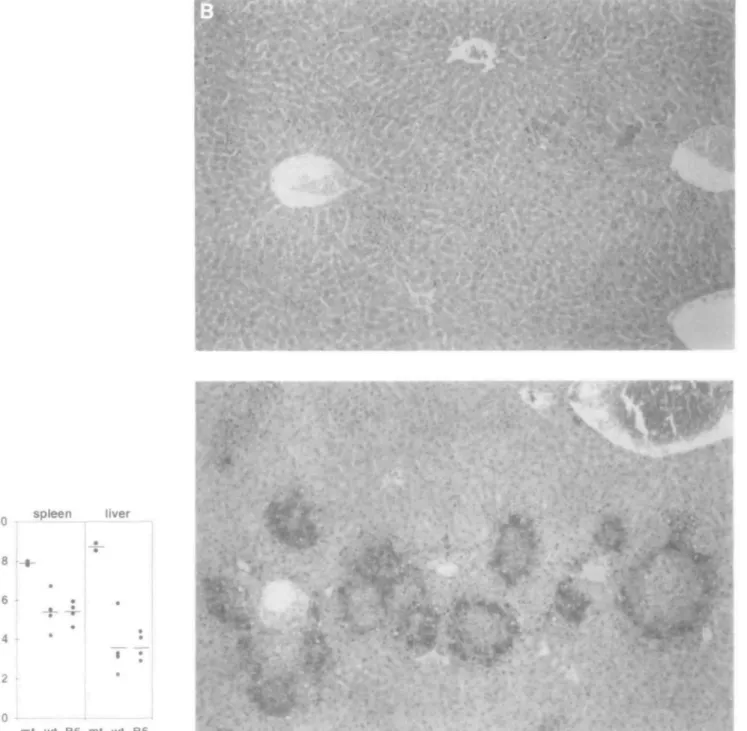
investi-Fig. 4. Altered microarchitecture in mutant spleen. Hematoxylin & eosin stains of (A) wild-type (X200) and (B) mutant (X100) spleen showing the absence of a clearly defined marginal zone in the mutant spleen.
gated whether these alterations also occurred in our TNF/LT-a-deficient mice. The size and microscopic structure of mutant thymi, however, were normal. In addition the percentage of single CD4+, CD8+ and CD4+CD8+ double-positive thymo-cytes was not altered in mutant mice (Fig 3). On the other hand, mutant mice were completely devoid of LN and PP. Microscopic analysis of the tissues at the sites of LN revealed the presence of lymphatic vessels, but no anlage of lymphoid organs were detectable (not shown). Hence, the only peri-pheral lymphoid organ present in mutant mice is the spleen, which has a normal weight, size and cellularity, but exhibits
a slightly altered lymphocyte composition (Fig 3) and an altered microarchitecture (Fig. 4). The splenic microarchitec-ture is characterized by a loss of clearly defined T cell zones and the absence of MAdCAM-1 expression in the ill-defined marginal zone (Fig 5). The expression of other adhesion molecules such as ICAM-1, VCAM-1 and Mac-1 is remarkably reduced in the marginal zone and red pulp area of mutant mice (not shown). We further asked whether the leukocyte counts in peripheral circulation were increased in this context. The hemogram showed indeed a 4-fold increase of total leukocyte counts (Table 1). Cytofluorometric analysis revealed
Fig. 5. Immunofluorescent analysis of spleen. Distribution of CD3+ T cells (A) in wild-type and (B) mutant spleen, showing the dissolved T cell
zone in mutant spleen. Distribution of B220+ cells (C) in wild-type and (D) mutant spleen showing the dissolved B cell zone in the mutant
spleen. MAdCAM-1 expression in (E) wild-type and (F) mutant spleen respectively. (A) and (C) as well as (B) and (D) show areas of consecutive sections (X320).
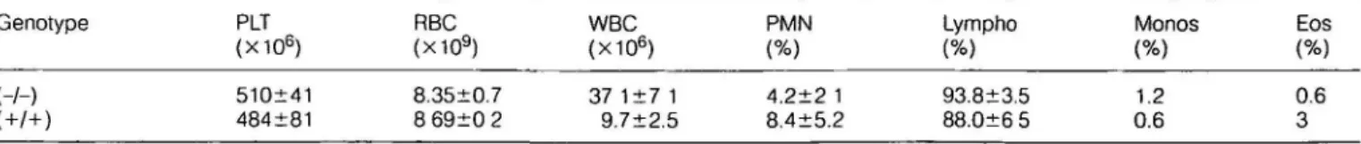
Table 1. Hematology of TNF/LT-a-deficient and control mice Genotype (-/-)
(+/+)
PLT (X106) 510±41 484±81 RBC (X109) 8.35±0.7 8 69±0 2 WBC (X106) 3 7 1 + 7 1 9.7±2.5 PMN (%) 4.2±2 1 8.4±5.2 Lympho (%) 93.8+3.5 88.0±6 5 Monos (%) 1.2 0.6 Eos (%) 0.6 3 Values are given as mean±SD (n = 5).Fig. 6. Decreased incidence of intestinal IgA-specific plasma cells, renal glomerular Ig deposits and aberrant MHC class I expression in mutant mice. (A) lleum from a wild-type mouse showing intensely lgA+ plasma cells in the lamina propria. The epithelial cells are weakly lgA+
(X450) (B) Duodenum of a mutant mouse with dramatic reduction of lgA+ plasma cells and no fluorescence in the epithelium (X450). (C)
Renal cortex of a wild-type mouse with background lgG2a-specific glomerular deposits (X400). (D) Renal cortex of a mutant mouse showing increased amount of lgG2a-specific glomerular deposits (X400). (E) MHC class I expression in myocard of wild-type mice is below detection level. (F) Marked MHC class I up-regulation is apparent in intermyocardial fiber capillary endothelia (X400) in mutant myocard.
Table 2. Basal isotype levels of TNF/LT-oc-deficient and control mice
Genotype IgM lgG1 lgG2a lgG2b lgG3 IgA
370±46 253+17 144±39 28±5.6 95 ±39 27±5 4 382+114 28+10 66+23 13+2 8 63±13 1 4±0 4
Values are given as mean±SD (n = 5)
65 ± 4% B220+ B cells in mutant mice compared with 44 ± 2% in wild-type mice and a relative decrease of CD3+ T cells from 45 ± 3% in wild-type to 25 ± 3% in mutant mice (Fig. 3). Considering the absolute lymphocyte counts, mutant mice have significantly increased peripheral lymphocyte counts with a 6-fold increase of B cells and a 2-fold increase in T cells. No evidence of increased B lymphopoiesis could be found in the bone marrow (data not shown).
PP represent the adequate environment for differentiation of precursors of lgA+ plasma cells (24) Immunohistochemical analysis revealed dramatically reduced numbers of lgA+ plasma cells in ileum (Fig 6A and B) and duodenum (not shown) of the mutants Collectively, TNF/LT-a-deficient mice show an altered splenic architecture and a complete loss of other peripheral lymphoid organs with a significant lymph-ocytosis
Ig deposits in renal mesangium and MHC class I up-regulation in different organs
Since TNF has been reported to play an important role in autoimmunity (25), we investigated MHC expression in differ-ent organs and the kidney for Ig deposits. Immunohistological analysis of the kidney of 3-month-old mutant mice revealed granular mesangial Ig deposits (Fig. 6C and D) of the IgM, lgG1, lgG2a and IgA isotypes in the absence of complement and any signs of active renal disease. Moreover, TNF/LT-a-deficient mice show an unexpected distinct up-regulation of MHC class I expression on capillary endothelial cells in the myocardium (Fig. 6E and F), lung, thymic medulla and kidney (not shown) compared with control animals. MHC class II expression was normal. Despite this alteration in class I expression and glomerular Ig deposits, TNF/LT-a mice show no evidence of an autoimmune disorder.
Ig levels and antibody response
We then asked to what extent the basal Ig synthesis and the humoral immune response was affected in the context of alymphoplasia, disorganized spleen and B lymphocytosis. We found a distinct reduction of IgM and all IgG isotypes; and in accordance with the strong reduction of mucosal IgA producing plasma cells, very low IgA levels in the serum (Table 2) The antibody response was measured after immun-ization with VSV or SRBC. Neutralizing Ig titers against VSV were determined at different time points, on day 4 for the T-cell-independent IgM response and on days 8 and 12 for the strictly T-cell-dependent IgG response. Surprisingly, no differences between mutant and wild-type neutralizing IgM and IgG titers against VSV were detectable and the antibody response correlated with the clearance of VSV (not shown). However, the SRBC-specific antibody response was markedly
Table 3. SRBC-specific immune response
Day Genotype IgM lgG1
15 8640+1880 32,940±9650 111,780±30,280 35,400±9540 9900±1800 38,700± 10,950 113±22 176±35 3940±1170 232 ±90 66,700±21,890 1425 ±880
Values are given as mean±SEM
altered. Mutant mice were unable to mount a primary IgG response against SRBC (Table 3, only lgG1 titers are shown) Thus, despite the observed B cell lymphocytosis, basal Ig levels are reduced and the response to a T-cell-dependent antigen is largely abrogated.
Anti-viral activity
TNF has been shown to exert variable effects on anti-viral activity in wVoand in vitro (26-30). Wild-type and mutant mice were therefore tested for their CTL responses against VV-WR, LCMV-ARM and LCMV-WE The primary in vivo CTL response from mutant mice infected i.v. with VV was slightly reduced (not shown) but strongly reduced in the case of LCMV-ARM and LCMV-WE (Fig. 7A and B). Nevertheless, secondary CTL responses (day 8) against LCMV-ARM and LCMV-WE are present, although still reduced compared with control mice (Fig. 7C and D). These CTL present were able to clear the virus from spleen and liver by day 20 (not shown) If mice were infected into the foot pad (3000 p f.u.), secondary CTL were present with LCMV-ARM, but not with LCMV-WE (Fig. 7E and F), demonstrating the lack of LCMV-WE-specific memory CTL. In addition, footpad swelling was absent after infection with both viral strains in contrast to control mice (not shown).
Anti-listerial activity
TNF plays an important role in the defence against intracellular bacteria such as L monocytogenes and Mycobacterium
bovis (17,31,32). Therefore, mutant and wild-type mice were
infected with low titers of L monocytogenes. In contrast to wild-type mice, mutant mice could not control this dose of L.
monocytogenes, and eventually die from listeriosis
character-ized by strongly increased titers of L monocytogenes in liver and spleen (Fig. 8A) and large necrotic lesions with boundaries of heavily infected hepatocytes (Fig. 8B).
LCMV-ARM
LCMV-WE
(0I
0) O8
Q. in 80 27 00' 80; 60! 40 20 n \ \ \v;
9V
3c
100 100 100E/T ratio
Fig. 7. Primary and secondary CTL responses against LCMV-ARM
and LCMV-WE Filled symbols represent wild-type data, open symbols represent data from mutant mice. Each curve represent results from one mouse. All data points are mean values of duplicate assays. Primary CTL responses (day 8) after i.v. priming with (A) 2X10³
pf.u LCMV-ARM and (B) 2X10² p.f.u LCMV-WE Secondary CTL
responses (day 8) after i v priming with (C) 2x 10³ p.f.u LCMV-ARM
and (D) 2x 10² p f.u. LCMV-WE. Secondary CTL responses after i f.p.
injection of (E) 3X10³ LCMV-ARM and (F) 3X10³ p.fu LCMV-WE
Spontaneous lysis of all used target cells in absence of effector cells was <15%. Unspecific lysis of unmfected target cells was <20% in all assays.
Discussion
Simultaneous deletion of the closely linked and homologous genes coding for TNF and LT-a resulted in viable and fertile homozygous mutant animals which allowed us to investigate the specific phenotypic alterations in the immune system caused by the lack of TNF and LT-a.
Investigations using TNF neutralizing approaches revealed a central role for TNF as a mediator of endotoxic shock (22,33-35). Genetic inactivation of TNFR1 (17,31), but not TNFR2 (36), provides resistance towards LPS-induced endo-toxic shock after sensitization with D-GalN. The fact that T cells represent the main source of LT-a in adult mice and that SCID mice, devoid of T and B cells, are responsive in the LPS/D-GaIN model of shock (37), precludes an important role of LT-a in the pathogenesis of LPS-induced endotoxic shock. Our data, which show complete protection of
TNF/LT-a-deficient mice from LPS/o-GalN-induced hepatic failure and endotoxic shock (Fig. 2), confirm the central role of TNF in this endotoxic shock model.
TNF and LT-a are expressed in the adult thymus (38) and TNF neutralization during gestation and the first 19 days of life lead to a marked atrophy of thymus, spleen and LN, and lymphopenia (23). In vitro, TNF induces thymocyte apoptosis (39) and either ligand mediates proliferation of immature and mature T cells in vitro (40). Together, these findings suggest an important role of TNF and/or LT-a in T cell maturation and the development of primary and secondary lymphatic organs. In our TNF/LT-a-deficient mice, however, thymus morphology as well as thymocyte development was normal, as it was reported for TNFR1- and TNFR2-deficient mice (31,36). How-ever, we see alterations of spleen morphology and a complete absence of LN and PP. As TNFR1- and TNFR2-deficient mice do not exhibit these abnormalities, signaling through the newly discovered LT-p" receptor is likely to be responsible for morphogenesis of LN. LT-a-deficient mice revealed similar alterations in spleen microarchitecture and absence of LN (10). Together, this defines the LT-a/LT-f3-receptor interaction as the cognate ligand-receptor signaling necessary for proper spleen and LN development. Systemic absence of LN and PP and presence of ill defined splenic follicles have also been reported for the a/y mouse (41) These overlapping phenotypic traits suggest that the aly gene product is involved in the LT-a/LT-P-receptor signaling pathway.
The distinct lymphocytosis observed in TNF/LT-a-deficient mice is also found in the LT-a-deficient mice (10), but not in
aly mice (41). Therefore the lack of LN and PP cannot be
invoked as the explanation for this observation, as all three mutants are devoid of these peripheral lymphatic organs Adhesion molecules play a critical role in leukocyte emigration (42), and it has been shown that TNF can induce MAdCAM-1, ICAM-1 and VCAM-1 (43). The complete absence of splenic MAdCAM-1 expression combined with the marked reduction of ICAM-1, VCAM-1 and Mac-1 expression in the marginal zone and splenic follicles of mutant mice might be responsible for the reduced margination of lymphocytes and as a con-sequence for the observed lymphocytosis. The fact that also ICAM-1 knockout mice present a similar leukocytosis (44) supports a causal relationship between leukocytosis and decreased adhesion molecule expression in the TNF/LT-a-deficient mouse.
A mucosal IgA deficiency was also seen in IL-6-deficient and in aly mice (41,45) In aly and the TNF/LT-a-deficient mice, this deficiency is likely due to the absence of PP and might lead to decreased mucosal immunity as shown in IL-6-deficient mice (46). The coincidence of IgA deficiency and glomerular Ig deposits of IgM, IgG and IgA isotypes is intriguing, since in man, IgA deficiency is associated with the presence of auto-antibodies, including anti-lgA antibodies (47). IgA deficiency is the most common form of immuno-deficiency in man, and its incidence is increased in patients with allergies, autoimmune and gastrointestinal tract dis-eases (47).
The aberrant expression of MHC class I molecules on the endothelial cells of different organs suggests a negative regulatory function of basal TNF or LT-a levels for MHC class I expression. TNF has been shown to induce MHC class I
expression in vitro via an NFicB-like activity (48,-49), and TNF and LT-a transgenic mice showed increased expression of MHC class I on islet cells (8). In contrast, TNF induced c-Jun in 3T3-L1 pre-adipocytes and osteoblastic MC3T3-E1 cells (50,51), and c-Jun has been shown to inhibit class I expression in murine L cells (52). These data illustrate the different action of TNF and/or LT-a on the expression of class I genes which are mediated, on the one hand, by an NFicB-like activity and,
on the other hand, by the c-Jun/AP-1 transcription factor. A possible explanation for the aberrant class I expression might be that the lack of basal TNF or LT-a levels leads to a down-regulation of c-Jun/AP-1 and to the loss of its negative regulatory effect on class I transcription resulting in increased basal class I expression. Whether this aberrant MHC class I expression is reversible by reintroduction of a TNF or LT-a transgene and if the aberrant class I expression might promote
mt wt B6 mt wt B6
Fig. 8. (A) Liver and spleen listerial titers of mutant (mt), wild-type (wt) and C57BL/6 (B6) mice 3 days after i.v. infection with 300 c.f.u. of L. monocytogenes and (B) liver sections of wild-type (top) and mutant mice (bottom) showing extensive necrosis in mutant livers at day 6 after infection (x90).
any kind of pathology in a non-specific pathogen-free environ-ment is currently under investigation Since not only TNF, but also other cytokmes have been shown to be able to induce MHC class I expression, serum cytokine levels for IFN-y, IL-1, IL-12 and transforming growth factor-p were determined, but no difference in cytokine levels could be demonstrated in the serum of mutant mice (not shown), which, however, does not exclude changes in the local production of these cytokines.
The general reduction of basal Ig levels is likely due to the absence of LN and PP The reduction in the basal Ig levels and the defective IgG isotype class switch is reminiscent of that seen in aly CD40 and CD40L-deficient mice (41,53,54), and suggests a common defect in all three mutant mice provoked by different mutations in the same or convergent signaling pathways of isotype switching. The fact that CD40 ligation has been shown to induce surface LT-a on human B cells (55) and that TNF/LT-a-deficient mice show an isotype switch deficiency with the T-cell-dependent antigen SRBC, strongly suggests a co-stimulatory function of LT-a in T-B cell interaction necessary for efficient T-cell-dependent isotype switching
TNF and LT-a can protect against infections with RNA and DNA viruses (26-29). The anti-viral responses are either humoral and/or cytotoxic In our experiments we used VSV to investigate the humoral and VV and LCMV viruses to test the virus-specific cytotoxic T cell responses. While the antibody response to SRBC revealed a specific IgG class switch defect, the neutralizing IgM and IgG response against VSV was normal in mutant mice The humoral response against a broad range of viral infection in mice is lgG2a restricted (56), an isotype response which might be conserved in the mutant mouse
In contrast to the present neutralizing immune response, the primary MHC class I restricted ex vivo CTL responses are strongly reduced for LCMV-ARM and LCMV-WE In compa-rision, the CTL responses in TNFR1-deficient mice were normal (17) and no abnormal CTL response was reported for TNFR2-deficient mice (36). This suggests that signaling via the LT-p-receptor might be important for the primary ex vivo CTL responses. It has been shown that TNF is not involved in the acute (short-term) target cell damage induced by CTL and a reason for this seems to be the down-regulation of TNFR1 expression preceding TNF and LT-a secretion in the effector phase (57,58). The reduction observed in our mutant mice might stem from the inefficient activation of T^ cells or precursor CTL due to the absence of a LT-a/LT-p-receptor mediated co-stimulatory signal or due to low IL-2R expression of activated precursor CTL. Observations supporting the latter assumption are the IL-2R a chain induction by TNF and the suppression of the in vivo priming for TNP-specific CTL by anti-TNF antibodies (59,60). The interpretation of these in vitro data is, however, related to the finding that IL-2-deficient mice exhibit only a marginal reduction of the primary LCMV-specific CTL response, but clear the virus similar to the TNF/LT-a-deficient mice (61). The fact that secondary CTL responses are present, but still reduced compared with control mice, for both LCMV strains suggests the presence of virus-specific precursor CTL, which, however, require prolonged activation
to become competent effector cells. Analysis of secondary CTL 13 and 20 days after i.v. priming resulted in loss of LCMV-WE-specific CTL from mutant mice, which is probably the result of virus-specific CTL exhaustion due to fast replica-tion within the lymphatic and non-lymphatic environment (62). In any case, mutant mice were still able to clear the viruses from spleen and liver. Infection of LCMV-ARM as well as LCMV-WE into the footpad did not provoke footpad swelling, which reflects the absence of CTL-mediated immunopath-ology. Nevertheless, footpad injection of LCMV-ARM lead to a potent secondary CTL response in contrast to LCMV-WE. The lack of the secondary CTL response towards LCMV-WE after footpad infection is likely due to CTL exhaustion. The fact that footpad infection with LCMV-ARM lead to an efficient secondary CTL response demonstrates that a central response after a peripheral challenge is possible also in the absence of LN. We suggest that the lack of footpad swelling, which is a delayed type hypersensitivity reaction, must be due to impaired recirculation of memory CTL. This impairment is likely due to the aberrant splenic microarchitecture and might therefore be the reason for the absence of peripheral immunopathology
Infection with the Gram-positive, facultative intracellular bacterium L monocytogenes has been shown to induce cell-mediated immunity and efficient clearance of this pathogen critically depends on proper T cell and macrophage function (63,64) Genetic inactivation of TCRa, TCRp, MHC class I and II, IFN-yR, TNFR1 and IL-6 (reviewed in 65) has defined a critical network of cytokines which play a crucial role in macrophage activation, extravasation and granuloma forma-tion. The sensitivity towards low titer infection with L
monocy-togenes seen in the TNF/LT-a-deficient mice is comparable
with that of TNFR1-deficient mice and confirms a crucial role for TNF in the defence against L monocytogenes infections The fact that perform (66) and MHC class I deficiency (67) does not lead to a drastic increase in sensitivity to primary L
monocytogenes infection shows that other cell populations
than T cells, like neutrophils and macrophages (64,68), are critical for an efficient primary host defence against L.
monocy-togenes. The anti-listerial activity of mutant macrophages
might be impaired at least at two levels. First, the lack of TNF, which is an important co-stimulator of IL-12 for IFN-y production by NK cells, might lead to a decreased activation of macrophages and a reduced development of an efficient Th1 response (69,70). Second, phagocytosed L.
monocytog-enes might not be killed efficiently. The second assumption
is supported by the fact that IFN-y stimulated BMDM from mutant mice showed reduced in vitro killing of phagocytosed
L. monocytogenes compared with control macrophages
(not shown).
In conclusion, the combined genetic inactivation of TNF and LT-a leads to distinct morphological and functional defects of the peripheral immune system whereby absence of TNF and LT-a markedly affects innate and acquired immunity, respectively. The described phenotypic features render these mice interesting tools to investigate in more detail the roles of both ligands for the development and function of the immune system. The interchangability of TNF and LT-a still leaves open questions, mainly about the functions of the
soluble form of LT-cc, which might be answered if a TNF knockout mouse would become available.
Acknowledgements
H -P Eugster and M. Muller contributed equally to this work We thank P Koebel, N Schultze, Y. Lang, G Stark, U Steckholzer, C. Haas and A. Althage for their expert technical assistance, and C V Jongeneel for the genomic clone This work was supported by the Swiss National Science Foundation grant 32-33966 92.
Abbreviations CTL D-GaIN ES LPS LCMV LT LN PP SRBC TNF VV cytotoxic T lymphocytes D-galactosamine embryonic stem cells lipopolysaccharide
lymphocytic chonomeningitis virus lymphotoxin
lymph nodes Peyer's patches sheep red blood cells tumor necrosis factor Vaccinia virus
References
1 Aggarwal, B B and vllcek, J 1992 Tumor Necrosis Factor, Structure, Function, and Mechanism of Action Marcel Dekker, New York
2 Beutler, B 1992 Tumour Necrosis Factor the Molecules and their Emerging Role In Medicine Raven Press, New York
3 Tartaglia, L A., Ayres, T. M., Wong, G H and Goeddel, D V 1993 A novel domain within the 55 kDa TNF receptor signals cell death. Cell74845.
4 Beutler, B and van Huffel, C 1994 Unraveling function in the TNF ligand and receptor families Science 264667.
5 Jacob, C. O , Aiso, S., Michie, S A., McDevitt, H. O and Acha-Orbea, H. 1990. Prevention of diabetes in nonobese diabetic mice by tumor necrosis factor (TNF)- similarities between TNF-alpha and interleukin 1 Proc Natl Acad. Sci. USA 87:968 6 Ruddle, N. H., Bergman, C. M., McGrath, K. M., Lingenheld, E
G., Grunnet, M L, Padula, S J and Clark, R B. 1990 An antibody to lymphotoxin and tumor necrosis factor prevents transfer of experimental allergic encephalomyelitis. J. Exp. Med.
1721193.
7 Picarella, D. E , Kratz, A., Li, C. B., Ruddle, N. H. and Flavell, R A 1992 Insulitis in transgenic mice expressing tumor necrosis factor beta (lymphotoxin) in the pancreas Proc Natl Acad Sci.
USA 8910036
8 Picarella, D. E., Kratz, A., Li, C B., Ruddle, N H and Flavell, R. A. 1993 Transgenic tumor necrosis factor (TNF)-alpha production in pancreatic islets leads to insulitis, not diabetes. Distinct patterns of inflammation in TNF-alpha and TNF-beta transgenic mice J. Immunol. 150:4136.
9 Guerder, S., Picarella, D. E., Linsley, P. S. and Flavell, R. A. 1994. Costimulator B7-1 confers antigen-presenting-cell function to parenchimal tissue and in conjugation with tumor necrosis factor a leads to autoimmunity in transgenic mice. Proc. Natl Acad Sci. USA 91:5138.
10 De Togni, P., Goellner, J., Ruddle, N. H., Streeter, P R., Fick, A., Mariathasan, S., Smith, S. C , Carlson, R., Shornick, L. P., Strauss-Schoenberger, J., Russel, J H , Karr, R. and Chaplin, D. D. 1994. Abnormal development of peripheral lymphoid organs in mice deficient in lymphotoxin. Science 264.703.
11 Androlewicz, M. J., Browning, J. L and Ware, C. F. 1992. Lymphotoxin is expressed as a heteromeric complex with a distinct 33-kDa glycoprotem on the surface of an activated human
T cell hybridoma. J. Biol Chem 267:2542.
12 Crowe, P. D , VanArsdale, T. L, Walter, B. N , Dahms, K M and Ware, C. F 1994 Production of lymphotoxin (LT alpha) and a soluble dimeric form of its receptor using the baculovirus expression system. J Immunol. Methods 168:79
13 Baens, M., Chaffanet, M., Cassiman, J J , van-den-Berghe, H. and Marynen, P 1993 Construction and evaluation of a hncDNA library of human 12p transcribed sequences derived from a somatic cell hybrid. Genomics 16 214.
14 Semon, D., Kawashima, E , Jongeneel, C V, Shakhov, A. N. and Nedospasov, S. A. 1987 Nucleotide sequence of the murme TNF locus, including the alpha (tumor necrosis factor) and TNF-beta (lymphotoxin) genes. Nucleic Acids Res. 15:9083. 15 Tybulewicz, V. L., Crawford, C. E., Jackson, P. K , Bronson, R. T
and Mulligan, R. C. 1991. Neonatal lethality and lymphopenia in mice with a homozygous disruption of the c-abl proto-oncogene Cell 65:1153
16 Heuchel, R., Radtke, F, Georgiev, O , Stark, G., Aguet, M. and Schaffner, W. 1994. The transcription factor MTF-1 is essential for basal and heavy metal-induced methallothionein gene expression EMBO J. 2870
17 Rothe, J., Lesslauer, W., Lotscher, H., Lang, Y, Koebel, P, Kontgen, F, Althage, A., Zinkernagel, R., Steinmetz, M and Bluethmann, H 1993 Mice lacking the tumour necrosis factor receptor 1 are resistant to TNF-mediated toxicity but highly susceptible to infection by Listena monocytogenes. Nature 364.798.
18 Laird, P W, Zijderveld, A , Linders, K., Rudnicki, M A , Jaenisch, R and Berns, A 1991 Simplified mammalian DNA isolation procedure Nucleic Acids Res 19 4293
19 Kelly, B. S., Levy, J G and Sikora, L 1979 The use of the enzyme-linked immunosorbent assay (ELISA) for the detection and quantification of specific antibody from cell cultures Immunology 37:45.
20 Bachmann, M F and Kundig, T. M. 1994 In vivo versus in vitro assays for assessment of T- and B-cell function. Curr. Opm Immunol. 6.320.
21 Blanden, R. V. and Langman, R E. 1972. Cell-mediated immunity to bacterial infection in the mouse. Thymus-derived cells as effectors of acquired resistance to Listena monocytogenes. Scand. J. Immunol. 1.379
22 Tracey, K J , Fong, Y, Hesse, D G , Manogue, K R , Lee, A. T, Kuo, G C , Lowry, S. F and Cerami, A. 1987. Anti-cachectm/ TNF monoclonal antibodies prevent septic shock during lethal bacteraemia. Nature 330.662
23 de-Kossodo, S., Grau, G E., Daneva, T, Pointaire, P, Fossati, L., Ody, C , Zapf, J., Piguet, P F, Gaillard, R C and Vassalli, P 1992. Tumor necrosis factor alpha is involved in mouse growth and lymphoid tissue development J. Exp. Med 176'1259. 24 Brandtzaeg, P. 1989. Overview of the mucosal immune system.
Curr Topics Microbiol Immunol 14613.
25 Jacob, C. O. and McDevitt, H O 1988. Tumour necrosis factor-alpha in murine autoimmune 'lupus' nephritis. Nature 331:356. 26 Wong, G. H. and Goeddel, D. V. 1986. Tumour necrosis factors
alpha and beta inhibit virus replication and synergize with interferons. Nature 323819.
27 Kohase, M , Hennksen-DeStefano, D., May, L T, vllcek, J. and Sehgal, P B. 1986. Induction of beta 2-interferon by tumor necrosis factor: a homeostatic mechanism in the control of cell proliferation. Cell 45.659.
28 Mestan, J., Digel, W., Mittnacht, S., Hillen, H., Blohm, D , Moller, A., Jacobsen, H. and Kirchner, H. 1986. Antiviral effects of recombinant tumour necrosis factor in vitro. Nature 323:816. 29 Sambhi, S. K, Kohonen-Corish, M. R. and Ramshaw, I. A.
1991 Local production of tumor necrosis factor encoded by recombinant vaccinia virus is effective in controlling viral replication in vivo. Proc. Natl Acad. Sci. USA 88:4025
30 Leist, T. P. and Zinkernagel, R. M. 1990. Treatment with anti-tumor necrosis factor alpha does not influence the immune pathological response against lymphocytic choriomeningitis virus Cytokine 2:29.
31 Pfeffer, K., Matsuyama, T., Kundig, T M , Wakeham, A., Kishihara, K , Shahinian, A., Wiegmann, K., Ohashi, P. S., Kronke, M and Mak, T. W. 1993. Mice deficient for the 55 kDa tumor necrosis factor receptor are resistant to endotoxic shock, yet succumb to L. monocytogenes infection. Cell 73:457.
32 Kindler, V., Sappino, A. P., Grau, G. E , Piguet, P F. and Vassalli, P. 1989. The inducing role of tumor necrosis factor in the development of bactericidal granulomas during BCG infection. Cell 56:731
33 Lucas, R., Heirwegh, K., Neirynck, A , Remels, L, Van-Heuverswyn, H and De-Baetseher, P. 1990 Generation and characterization of a neutralizing rat anti-rmTNF-alpha mono-clonal antibody Immunology 71 218
34 Ashkenazi, A., Marsters, S. A , Capon, D J , Chamow, S. M , Ftgan, I S , Pennica, D , Goeddel, D V, Palladmo, M A. and Smith, D H. 1991 Protection against endotoxic shock by a tumor necrosis factor receptor immunoadhesin Proc Natl Acad Sci USA 8810535
35 Lesslauer, W., Tabuchi, H , Gentz, R., Brockhaus, M., Schlaeger, E J , Grau, G , Piguet, P F, Pointaire, P, Vassalli, P and Loetscher, H 1991. Recombinant soluble tumor necrosis factor receptor proteins protect mice from lipopolysacchande-induced lethality Eur J Immunol 21 2883
36 Enckson, S L , de-Sauvage, F J , Kikly, K , Carver-Moore, K , Pitts-Meek, S, Gillett, N., Sheehan, K C , Schreiber, R. D., Goeddel, D. V. and Moore, M W 1994. Decreased sensitivity to tumour-necrosis factor but normal T-cell development in TNF receptor-2-deficient mice Nature 372 560.
37 Miethke, T, Wahl, C , Heeg, K., Echtenacher, B , Krammer, P H and Wagner, H 1992 T cell-mediated lethal shock triggered in mice by the superantigen staphylococcal enterotoxin B: critical role of tumor necrosis factor. J. Exp. Med. 175.91
38 Giroir, B. P., Johnson, J H , Brown, T, Allen, G L and Beutler, B. 1992. The tissue distribution of tumor necrosis factor biosynthesis during endotoxemia. J Clin Invest 90 693
39 Hernandez-Caselles, T and Stutman, O. 1993. Immune functions of tumor necrosis factor I Tumor necrosis factor induces apoptosis of mouse thymocytes and can also stimulate or inhibit IL-6-induced proliferation depending on the concentration of mitogenic costimulation J Immunol 1513999
40 Tartaglia, L A., Goeddel, D. V., Reynolds, C , Figan, I S , Weber, R F., Fendly, B M. and Palladmo, M , Jr 1993 Stimulation of human T-cell proliferation by specific activation of the 75-kDa tumor necrosis factor receptor J Immunol 151 4637.
41 Miyawaki, S , Nakamura, Y., Suzuka, H., Koba, M., Yasumizu, R., Ikehara, S and Shibata, Y 1994 A new mutation, aly that induces a generalized lack of lymph nodes accompanied by
immunodeficiency in mice Eur J. Immunol. 24 429
42 Berlin, C , Bargatze, R F, Campbell, J. J , von-Andrian, U. H., Szabo, M. C , Hasslen, S. R., Nelson, R D., Berg, E L , Erlandsen, S. L. and Butcher, E C. 1995. Alpha 4 integrins mediate lymphocyte attachment and rolling under physiologic flow. Cell 80:413.
43 Sikorski, E. E., Hallmann, R., Berg, E. L and Butcher, E. C. 1993. The Peyer's patch high endothehal receptor for lymphocytes, the mucosal vascular addressin, is induced on a murine endothelial cell line by tumor necrosis factor-alpha and IL-1 J Immunol 151 5239.
44 Xu, H., Gonzalo, J. A., St-Pierre, Y, Williams, I. R , Kupper, T. S., Cotran, R. S., Springer, T. A. and Gutierrez-Ramos, J. C. 1994 Leukocytosis and resistance to septic shock in intercellular adhesion molecule 1-deficient mice. J. Exp. Med. 180:95 45 Ramsay, A. J., Husband, A. J., Ramshaw, I. A., Bao, S., Matthei,
K. I., Koehler, G. and Kopf, M. 1994. The role of interleukin-6 in mucosal IgA antibody responses in vivo. Science 264:561. 46 Kopf, M., Baumann, H , Freer, G., Freudenberg, M., Lamers, M.,
Kishimoto, T, Zinkemagel, R, Bluethmann, H. and Kohler, G. 1994 Impaired immune and acute-phase responses in interleukin-6-deficient mice. Nature 368:339.
47 Burks, A., Jr. and Steele, R. W. 1986. Selective IgA deficiency Ann. Allergy 57:3.
48 Israel, A , Le-Bail, O., Hatat, D., Piette, J , Kieran, M , Logeat, F, Wallach, D , Fellous, M and Kourilsky, P. 1989. TNF stimulates expression of mouse MHC class I genes by inducing an NF kappa B-like enhancer binding activity which displaces constitutive factors. EMBO J 8:3793.
49 David-Watine, B., Israel, A. and Kourilsky, P 1990. The regulation and expression of MHC class I genes. Immunol. Today 11 286 50 Stephens, J M., Butts, M., Stone, R., Pekala, P. H. and Bernlohr,
D. A. 1993. Regulation of transcription factor mRNA accumulation during 3T3-L1 preadipocyte differentiation by antagonists of adipogenesis. Mol. Cell. Biochem 12363
51 Hanazawa, S , Takeshita, A , Amano, S , Semba, T., Nirazuka, T, Katoh, H. and Kitano, S. 1993. Tumor necrosis factor-alpha induces expression of monocyte chemoattractant JE via fos and jun genes in clonal osteoblastic MC3T3-E1 cells. J. Biol Chem 268.9526
52 Howcroft, T K, Richardson, J C and Singer, D S. 1993 MHC class I gene expression is negatively regulated by the proto-oncogene, c-jun. EMBO J 123163
53 Castigh, E., Alt, F W, Davidson, L, Bottaro, A , Mizoguchi, E., Bhan.A K and Geha, R. S 1994 CD40-deficient mice generated by recombination-activating gene-2-deficient blastocyst complementation Proc Natl Acad Sci LISAS'! 12135
54 Renshaw, B. R , Fanslow, W. 3., Armitage, R J , Campbell, K. A , Liggitt, D , Wright, B., Davison, B. L. and Mahszewski, C R 1994. Humoral immune responses in CD40 hgand-deficient mice J Exp Med 180 1889
55 Worm, M and Geha, R S 1994. CD40 ligation induces lymphotoxin alpha gene expression in human b cells. Int. Immunol 6 1883
56 Couteher, J P, van-der-Logt, J. T., Heessen, F. W, Warnier, G and Van-Snick, J 1987 lgG2a restriction of murine antibodies elicited by viral infections. J. Exp. Med. 16564
57 Ratner, A and Clark, W R 1993 Role of TNF-alpha in CD8+
cytotoxic T lymphocyte-mediated lysis. J Immunol. 150:4303 58 Ware, C F, Crowe, P D, Vanarsdale, T. L, Andrews, J L,
Grayson, M H , Jerzy, R., Smith, C A. and Goodwin, R. G. 1991 Tumor necrosis factor (TNF) receptor expression in T lymphocytes. Differential regulation of the type I TNF receptor during activation of resting and effector T cells. J Immunol. 1474229
59 loannides, C G , Fisk, B , Tomasovic, B , Pandita, R , Aggarwal, B. B. and Freedman, R. S 1992. Induction of interleukin-2 receptor by tumor necrosis factor alpha on cultured ovarian tumor-associated lymphocytes. Cancer Immunol. Immunother. 35 83. 60 Bromberg, J S., Chavin, K D. and Kunkel, S L. 1992 Anti-tumor necrosis factor antibodies suppress cell-mediated immunity in vivo. J. Immunol 1483412
61 Kundig, T M , Schorle, H , Bachmann, M F, Hengartner, H , Zinkemagel, R M and Horak, I 1993. Immune responses in interleukin-2-deficient mice Science 262 1059
62 Moskophidis, D , Lechner, F, Hengartner, H. and Zinkemagel, R. M. 1994. MHC class I and non-MHC-linked capacity for generating an anti-viral CTL response determines susceptibility to CTL exhaustion and establishment of virus persistence in mice. J. Immunol. 152:4976
63 Mombaerts, P., Arnoldi, J , Russ, F., Tonegawa, S. and Kaufmann, S H. 1993. Different roles of alpha beta and gamma delta T cells in immunity against an intracellular bacterial pathogen. Nature 365.53.
64 Beckerman, K. P., Rogers, H. W., Corbett, J. A., Schreiber, R. D., McDaniel, M L. and Unanue, E. R. 1993. Release of nitric oxide during the T cell-independent pathway of macrophage activation. Its role in resistance to Listena monocytogenes. J Immunol. 150:888.
65 Kaufmann, S H and Ladel, C. H. 1994 Application of knockout mice to the experimental analysis of infections with bacteria and protozoa. Trends Microbiol 2:235.
66 Kagi, D., Ledermann, B , Burki, K., Hengartner, H. and Zinkemagel, R. M. 1994. CD8+ T cell-mediated protection against
an intracellular bacterium by perforin-dependent cytotoxicity. Eur. J. Immunol. 24:3068
67 Ladel, C H , Flesch, I E., Arnoldi, J and Kaufmann, S. H. 1994. Studies with MHC-deficient knock-out mice reveal impact of both MHC I- and MHC ll-dependent T cell responses on Listens monocytogenes infection J. Immunol. 153:3116
68 Rogers, H. W and Unanue, E R. 1993 Neutrophils are involved in acute, nonspecific resistance to Listens monocytogenes in mice. Infect. Immun 61 5090.
69 Tripp, C. S., Wolf, S F and Unanue, E R 1993 Interleukin 12
and tumor necrosis factor alpha are costimulators of interferon gamma production by natural killer cells in severe combined immunodeficiency mice with hstenosis, and interleukin 10 is a physiologic antagonist Proc NatlAcad Sci USA 903725 70 Hsieh, C S , Macatoma, S E , Tripp, C S , Wolf, S. F, O'Garra,
A. and Murphy, K M 1993. Development of TH1 CD4+ T cells
through IL-12 produced by Listena-mduced macrophages [see comments] Science 260:547