Surface Reduction in Monoclinic BiVO
4for Photocatalytic Applications
Marta D Rossell1, Andreas Borgschulte2 and Rolf Erni11 Electron Microscopy Center, Empa, Swiss Federal Laboratories for Materials Science and Technology,
CH-8600 Dübendorf, Switzerland
2 Laboratory for Hydrogen & Energy, Empa, Swiss Federal Laboratories for Materials Science and
Technology, CH-8600 Dübendorf, Switzerland
Bismuth vanadate (BiVO4, short BVO) is a promising photoelectrochemically active candidate for water
splitting used for hydrogen generation. Compared to TiO2, i.e. the most widely used and investigated
photocatalyst, BVO uses visible instead of ultraviolet light for its photoelectrochmical activity. Amongst the three polymorphs of BVO, the monoclinic clinobisvanite BVO (m-BVO) phase exhibits the highest photocatalytical activity. This is due to both, the favorable band gap energy of ~2.4 eV and the valence band position, which is suitable for driving water oxidation under irradiation. Despite the fact that the conduction band is reported to be too low relative to the proton reduction potential for hydrogen evolution [1], the water splitting reaction can be promoted by simultaneously using a hydrogen evolution catalyst, such as a noble- or transition-metal catalyst [2]. Apart from the low charge carrier mobility of m-BVO [3], which can be improved by using various dopants [2,4], another crucial point, which affects m-BVO’s photocatalytic activity, concerns oxygen vacancies. These vacancies, which have been recognized to play a key role in the photocatalytic performance of m-BVO, are the dominant intrinsic defects in m-BFO [5] and might act as harmful photogenerated carrier traps [6]. However, little is known about the concentration and the distribution of oxygen vacancies in m-BVO.
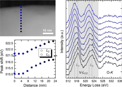
In this contribution we present a study based on atomic-resolution scanning transmission electron microscopy (STEM) and electron energy-loss spectroscopy (EELS) which aims at investigating the distribution and concentration of oxygen vacancies in pristine m-BVO. High-resolution imaging of grains of BVO rules out the presence of relevant structural defects or structural modifications that could account for oxygen vacancies. However, spatially resolved EELS measurements reveal a distinct shift of the vanadium L3,2 edge towards lower energies when the electron probe is positioned near the surface of
the particles. Indeed, in a surface shell of about 5 nm the vanadium L3,2 edge is shifted by about 1 eV
towards lower energy compared with the corresponding bulk measurement. Comparing this shift with the location of the vanadium L-edge measured in three vanadates of different oxidation state, namely V2O3, VO2, V2O5, allows for explaining this shift in terms of a surface reduction in m-BVO. Within a
surface area of about 5 nm thickness, the oxidation state of vanadium is reduced from +5 to about +4. The reduction of the vanadium oxidation state demands for a concentration of oxygen vacancies above 10%. The rather high amount of oxygen vacancies might question the stability of m-BVO phase. However, m-BVO has the scheelite structure, which is known to easily accommodate cations with various oxidation states, and to tolerate cation and oxygen vacancies [7]. In order to confirm the STEM/EELS results, X-ray photoelectron spectroscopy (XPS) was carried out on pellets of powder samples. The XPS measurements unambiguously confirmed the surface reduction in m-BVO ruling out effects to the electron irradiation in STEM for this electron-sensitive material.
We thus conclude that oxygen vacancies in m-BVO are accumulated in a distinct surface layer of about 5 nm thickness. The occurrence of this surface reduction shell must be considered in the optimization of the photochemical activity of m-BVO and in possible dopant strategies.
436
doi:10.1017/S1431927614003900 © Microscopy Society of America 2014Microsc. Microanal. 20 (Suppl 3), 2014
https:/www.cambridge.org/core/terms. https://doi.org/10.1017/S1431927614003900
References:
[1] A. Kudo, K. Ueda, H. Kato, and I. Mikami, Catal. Lett. 53 (1998) 229. [2] W. Luo et al., Energy Environ. Sci. 4 (2011) 4046.
[3] K. Sayama et al., J. Phys. Chem. B 110 (2006) 11352. [4] K. Zhang et al., Phys. Chem. Chem. Phys. 14 (2012) 11119. [5] F. F. Abdi et al., J. Phys. Chem. Lett. 4 (2013) 2752.
[6] Y. K. Kho et al., ACS Appl. Mater. Interfaces 3 (2011) 1997. [7] V. Babin et al., J. Lumin. 124 (2007) 113.
Figure 1. High-angle annular dark-field STEM micrograph revealing the structural intactness at the
surface of m-BVO, despite of an oxygen concentration that is roughly 10% lower than in the bulk.
Figure 2. Line scan of EEL spectra of the vanadium L3,2 edge, the location of the measurements and the
plot of the peak shifts of the L3 and L2 line.
437 Microsc. Microanal. 20 (Suppl 3), 2014
https:/www.cambridge.org/core/terms. https://doi.org/10.1017/S1431927614003900