M A J O R A R T I C L E
Acute Muscular Sarcocystosis: An International
Investigation Among Ill Travelers Returning
From Tioman Island, Malaysia, 2011
–2012
Douglas H. Esposito,1August Stich,2Loïc Epelboin,3,4Denis Malvy,5Pauline V. Han,1Emmanuel Bottieau,6
Alexandre da Silva,7Philipp Zanger,8Günther Slesak,9Perry J. J. van Genderen,10Benjamin M. Rosenthal,11 Jakob P. Cramer,12,13Leo G. Visser,14José Muñoz,15Clifton P. Drew,16Cynthia S. Goldsmith,16Florian Steiner,17
Noémie Wagner,18Martin P. Grobusch,19D. Adam Plier,20Dennis Tappe,13Mark J. Sotir,1Clive Brown,1Gary W. Brunette,1 Ronald Fayer,11Frank von Sonnenburg,21Andreas Neumayr,22,23and Phyllis E. Kozarsky1,24; for the Tioman Island
Sarcocystosis Investigation Teama
1Division of Global Migration and Quarantine, National Center for Emerging and Zoonotic Infectious Diseases, Centers for Disease Control and Prevention
(CDC), Atlanta, Georgia;2Department of Tropical Medicine, Medical Mission Hospital, Würzburg, Germany;3Assistance Publique–Hôpitaux de Paris,
Infectious and Tropical Diseases Department, Groupe Hospitalier Pitié-Salpêtrière,4Université Pierre et Marie Curie, Paris, and5Division of Tropical
Medicine and Clinical International Health, University Hospital Center, Bordeaux, France;6Department of Clinical Sciences, Institute of Tropical Medicine,
Antwerp, Belgium;7Division of Parasitic Diseases and Malaria, Center for Global Health, CDC, Atlanta, Georgia;8Institute of Tropical Medicine, Eberhard
Karls University, and9Tropenklinik Paul-Lechler-Krankenhaus, Tübingen, Germany;10Institute for Tropical Diseases, Harbor Hospital, Rotterdam, The
Netherlands;11US Department of Agriculture, Beltsville Agricultural Research Center, Maryland;12Section Tropical Medicine, I. Department of Internal
Medicine, University Medical Center Hamburg-Eppendorf, and13Bernhard Nocht Institute for Tropical Medicine, Hamburg, Germany;14Department of
Infectious Diseases, Leiden University Medical Centre, The Netherlands;15Barcelona Centre for International Health Research Hospital Clínic, Universitat de
Barcelona, Spain;16Division of High-Consequence Pathogens and Pathology, National Center for Emerging and Zoonotic Infectious Diseases, CDC, Atlanta,
Georgia;17Institute of Tropical Medicine and International Health, Charité–Universitätsmedizin Berlin, Germany;18Department of Pediatrics, Children’s
Hospital, University Hospitals of Geneva, Switzerland;19Centre of Tropical Medicine and Travel Medicine, Department of Infectious Diseases, Academic
Medical Centre, University of Amsterdam, The Netherlands;20Gorgas Center for Geographic Medicine, Division of Infectious Diseases, University of Alabama
at Birmingham;21Department of Infectious Disease and Tropical Medicine, University of Munich, Germany;22TropNet and the Swiss Tropical and Public Health
Institute, and23University of Basel, Switzerland; and24Department of Medicine, Division of Infectious Diseases, Emory University, Atlanta, Georgia
Background. Through 2 international traveler-focused surveillance networks (GeoSentinel and TropNet), we iden-tified and investigated a large outbreak of acute muscular sarcocystosis (AMS), a rarely reported zoonosis caused by a protozoan parasite of the genus Sarcocystis, associated with travel to Tioman Island, Malaysia, during 2011–2012.
Methods. Clinicians reporting patients with suspected AMS to GeoSentinel submitted demographic, clinical, itin-erary, and exposure data. We defined a probable case as travel to Tioman Island after 1 March 2011, eosinophilia (>5%), clinical or laboratory-supported myositis, and negative trichinellosis serology. Case confirmation required histologic observation of sarcocysts or isolation of Sarcocystis species DNA from muscle biopsy.
Results. Sixty-eight patients met the case definition (62 probable and 6 confirmed). All but 2 resided in Europe; all were tourists and traveled mostly during the summer months. The most frequent symptoms reported were myalgia (100%), fatigue (91%), fever (82%), headache (59%), and arthralgia (29%); onset clustered during 2 distinct periods: “early” during the second and “late” during the sixth week after departure from the island. Blood eosinophilia and elevated serum creatinine phosphokinase (CPK) levels were observed beginning during thefifth week after departure. Sarcocystis nesbitti DNA was recovered from 1 muscle biopsy.
Conclusions. Clinicians evaluating travelers returning ill from Malaysia with myalgia, with or without fever, should consider AMS, noting the apparent biphasic aspect of the disease, the later onset of elevated CPK and eosinophilia, and
Received 21 April 2014; accepted 24 July 2014; electronically published 4 August 2014.
a
Members of the Tioman Island Sarcocystosis Investigation Team are listed in the Appendix.
Correspondence: Douglas H. Esposito, MD, MPH, Division of Global Migration and Quarantine, Travelers’ Health Branch, National Center for Emerging and Zoonot-ic Infectious Diseases, Centers for Disease Control and Prevention, 1600 Clifton Rd NE, MS E-03, Atlanta, GA 30333 (hgj4@cdc.gov).
Clinical Infectious Diseases® 2014;59(10):1401–10
Published by Oxford University Press on behalf of the Infectious Diseases Society of America 2014. This work is written by (a) US Government employee(s) and is in the public domain in the US.
the possibility for relapses. The exact source of infection among travelers to Tioman Island remains unclear but needs to be determined to prevent future illnesses.
Keywords. infectious disease outbreak; sarcocystosis; parasitic disease; Malaysia; travel.
On 25 October 2011, GeoSentinel, the surveillance program of the International Society of Travel Medicine (ISTM) and the US Centers for Disease Control and Prevention (CDC) [1], was no-tified of a cluster of ill German patients recently returned from Tioman Island, off the eastern coast of peninsular Malaysia (Figure 1) [2, 3]. These patients had an unusual clinical
presentation: All of them reported fever and significant muscle pain, had blood eosinophilia and elevated serum creatinine phosphokinase (CPK) levels, and tested seronegative for trichi-nellosis and toxoplasmosis. A muscle biopsy from one of these patients was diagnostic for acute muscular sarcocystosis (AMS), a rarely reported zoonotic infection. The GeoSentinel
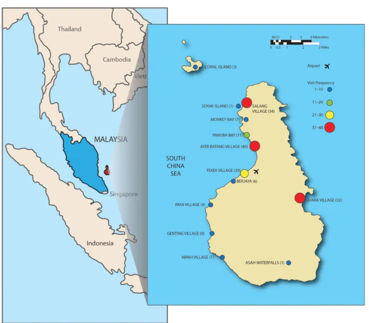
Figure 1. Tioman Island, Malaysia, and locations visited by 68 case patients with acute muscular sarcocystosis, 2011–2012. Marker color represents the frequency of visitation to each village or attraction for the 61 case patients with these data available; each patient may have visited multiple locations. Other locations visited but not mapped include“south of Tekek” (n = 4), “between Ayer Batang and Salang” (n = 3), “between Ayer Batang and Genting” (n = 1), “Tekek to Juara” (n = 1), “northwest of island” (n = 3), and a snorkeling trip circumnavigating the island by boat with multiple stops (n = 1). Tioman Island is not drawn to scale relative to the mainland.
(available at:http://www.istm.org/geosentinelandhttp://www. istm.org/eurotravnet) and TropNet (available at:http://www. tropnet.net) networks contacted their members and alerted rel-evant public health authorities to conduct additional case find-ing. Within days of the network alerts, additional patients were identified, prompting further investigation [2,4].
Human muscular sarcocystosis is caused by an intracellular protozoan parasite of the genus Sarcocystis (available at:
http://www.cdc.gov/dpdx/sarcocystosis/index.html). First de-scribed in 1843, approximately 130 species of Sarcocystis have been identified from a variety of wild and domestic mammals, birds, and reptiles [5]. These organisms have an obligatory 2-host life cycle, alternating between predator and prey (the de-finitive–intermediate hosts). Humans are the definitive host for Sarcocystis hominis and Sarcocystis suihominis, acquired by eat-ing undercooked sarcocyst-containeat-ing beef or pork, respective-ly. Definitive host infection is limited to the intestine and may cause acute gastroenteritis, although most infections are proba-bly asymptomatic [6]. Humans can also become the accidental dead-end intermediate host for an unknown number of other Sarcocystis species, presumably acquired by ingesting sporo-cyst-containing food or water contaminated with feces from in-fected carnivores. In the intermediate host, generations of reproduction occur in the vasculature, ultimately leading to the formation of characteristic cysts (sarcocysts) within myo-cytes of skeletal, cardiac, and, infrequently, smooth muscle.
Until recently, <100 cases of human muscular sarcocystosis were reported in the literature, particularly from Malaysia [7– 9]. The majority of these cases were diagnosed incidentally in asymptomatic persons or in patients whose symptoms were not clearly related to their Sarcocystis infection. At most, 10 of these cases had symptomatic AMS [10–13]; an additional recent report describes an outbreak involving 89 patients with AMS acquired on Pangkor Island, off the west coast of peninsular Malaysia [14]. Effective treatment for this disease has not been determined.
The principal objectives of this investigation were to describe the clinical, laboratory, and epidemiologic characteristics of this outbreak of AMS. We also sought to identify possible sources of infection among the ill travelers, and to alert clinicians to con-sider AMS when evaluating ill returned travelers.
METHODS
Epidemiologic Investigation
GeoSentinel is a global provider-based, traveler-focused sentinel surveillance network established collaboratively by ISTM and the CDC [1]. The 57 GeoSentinel sites in 24 countries consist of travel and tropical medicine clinics that actively monitor trav-el-related morbidity. TropNet is a European travel and tropical medicine research and surveillance network [15]. Together,
these networks encompass >110 travel and tropical medicine sites and >225 participating affiliated sites worldwide.
After thefirst patients were reported, members of these net-works were notified of the outbreak through e-mail alerts and other informal channels. In addition, the networks communi-cated to the larger infectious diseases community through ProMED Mail postings [16–19] and published outbreak alerts [2,4]. Relevant global public health authorities were also noti-fied. All were encouraged to report patients suspected of having AMS to GeoSentinel. Clinicians were asked to complete 2 struc-tured questionnaires: one for demographic and clinical data and one for travel and exposures. They were also asked to document the date of onset for a list of symptoms that were based on our experience with thefirst reported patients, as well as a literature review. This outbreak investigation was determined to be public health response, and thus institutional review board review was not required. All patients gave informed consent.
We report only those patients meeting an intentionally spe-cific outbreak case definition of probable or confirmed AMS. A probable case required travel to Tioman Island after 1 March 2011, with myositis, eosinophilia >500 cells/µL, and negative trichinellosis serology. Myositis required at least 1 of the follow-ing: a complaint of muscle pain and a CPK level >200 IU/L; muscle tenderness documented on physical examination; or histologic evidence of myositis in a muscle biopsy. Case confir-mation required histologic observation of intramuscular cysts compatible with sarcocysts or the isolation of Sarcocystis species DNA from a muscle biopsy.
Laboratory Analysis
Diagnostic testing was at the discretion of the clinician but could include complete blood counts and differentials, serum biochem-ical testing, electrocardiography, echocardiography, imaging studies, and electromyography. Some patients were tested for spe-cific diseases such as malaria, intestinal parasites, toxocariasis, filariasis, and dengue, chikungunya, and Epstein-Barr virus infec-tion. Serum samples were requested, and testing at the CDC Parasitic Diseases Reference Laboratory for trichinellosis, toxo-plasmosis, and strongyloidiasis was performed on available sam-ples not previously tested as part of the initial clinical evaluation. Sera were also used by the CDC for developing a serological assay for human sarcocystosis using both whole digested merozoites and recombinant surface peptides of Sarcocystis neurona.
At the discretion of the clinician, some patients underwent muscle biopsy. All biopsies were examined histologically by a pathologist at the institution where collected. When possible, tissue blocks, frozen tissue, and extracted DNA cryoprecipitate were sent to the CDC Infectious Diseases Pathology Branch for additional examination, including histopathology, polymerase chain reaction (PCR) detection, and DNA sequencing analysis of Sarcocystis species 18S ribosomal RNA (rRNA) amplicons.
For histopathologic examination, 3-µm sections were cut from formalin-fixed, paraffin-embedded muscle biopsy specimens and stained with hematoxylin and eosin. For electron microsco-py (EM), a paraffin section was embedded in Epon-Araldite epoxy resin, removed from the slide and glued to a blank EM block, sectioned and stained, and examined in an FEI Tecnai Spirit microscope. PCR amplification was performed with a primer pair designed to amplify a fragment of approximately 800 base pairs from the 18S rRNA gene of different species of Sarcocystis [20]. Amplicons were sequenced by using BigDye version 3.1 chemistry (Applied Biosystems), and sequence data were analyzed as previously described [21].
Statistical Analysis
Descriptive statistics were estimated using SAS software, version 9.2 (SAS Institute).
RESULTS
Epidemiologic Characteristics of Case Patients Demographic and Travel Characteristics
From October 2011 through April 2013, 99 ill persons who returned home after travel to Tioman Island were reported to GeoSentinel as having suspected AMS. Overall, 68 (69%)
patients met the case definition and were included in the anal-ysis; 62 were probable and 6 were confirmed cases. The median age of these patients was 34 years (range, 4–72 years); 39 (57%) were female. Of the 31 who did not meet the case definition, 24 did not satisfy criteria for myositis and 5 did not have eosino-philia, trichinellosis testing, or both.
More than three-quarters of the case patients resided in ei-ther Germany (43%) or France (34%). Fourteen (21%) lived elsewhere in Europe (6 in the Netherlands, 3 in Switzerland, 2 each in Belgium and Spain, and 1 in Italy); the 2 non-Europeans resided in Canada and Singapore. Most traveled to Tioman Island during the European summer months (Figure2); all were tourists. The median stay was 5 days (range, 3–16 days), with 49 (72%) of the travelers staying <7 days.
Exposures
Complete travel and exposure data were submitted from 61 (90%) case patients. Most lodging, eating, and recreational activities were concentrated on the northwest coast of Tioman Island; no single village or attraction was visited by >40 (68%, data missing for 2) case patients (Figure1). All travelers report-ed eating at restaurants and lodging at resorts, hotels, huts, or bungalows; no single eating or lodging establishment was visit-ed by >22 (36%, Table1). All case patients visited a beach, and Figure 2. Date of departure from Tioman Island, Malaysia, by week, of 68 travelers with probable and confirmed acute muscular sarcocystosis, 2011– 2012. The date of departure from Tioman Island reflects the last possible point of exposure of the traveler to the Sarcocystis species parasite. The earliest departure date was 1 May 2011, and the latest was 5 September 2012.
all but 1 (98%) reported swimming in the ocean. All but 1 pa-tient reported consuming drinks with ice, and most ate fresh produce that might have been washed with unsafe water and/ or brushed their teeth using tap water. Cats were frequently ob-served in tourist areas. Reptiles (including snakes and lizards) and monkeys were infrequently contacted.
Clinical and Laboratory Findings
Case patientsfirst became ill a median of 11 days (range, 0–40 days) after departing from Tioman Island. The most frequent symptoms reported among the 68 case patients were myalgia (100%), fatigue (91%), fever (82%), headache (59%), and ar-thralgia (29%); other symptoms were less frequently reported (Figure3). Patients werefirst evaluated by the reporting clini-cian at different points during the course of their illness;
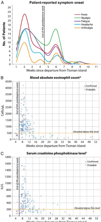
physical examination data were collected only on thatfirst visit, which was a median of 44 days (range, 12–79 days) after departing from the island. Twenty-nine of 55 (53%) case pa-tients were febrile (temperature≥38°C); 19 of these had a tem-perature≥39°C. Muscle tenderness was present among 47 of 64 (73%) and was described as moderate or severe in 32 (68%). Thirty-eight of 64 (88%) had tenderness involving their extrem-ities; 1 reported tenderness of the masseter muscle of the face and another of the tongue. Skin rash, lungfindings, and lymph-adenopathy were each found in≤3 patients. One patient report-ed subcutaneous nodules on the chin and neck that resolvreport-ed prior to presentation, and another had muscle fasciculations in-volving all 4 extremities. One patient had a thrombosis of the left sigmoidal and transverse sinuses followed by pulmonary emboli; no clotting abnormality was found, and a sarcocysto-sis-related vasculitis could not be ruled out. One patient was pregnant at the time of infection; she experienced no complica-tions. No patient was known to have an underlying medical condition (such as immunosuppression) that would have been expected to have affected the infection. At least 7 case patients (10%) were hospitalized; none required intensive care. Onset of the 5 most frequent symptoms appeared to cluster during 2 distinct periods: early during the second and late dur-ing the sixth week after departure (Figure4A). No other symp-tom showed this pattern. To account for persons presenting at different times during the course of their illness, we also restrict-ed the analysis to those who sought care after the fourth post-departure week; the biphasic symptom-onset pattern persisted. For 25 (37%) patients, the illness was described as phasic, inter-mittent, or waxing–waning in character, with periods of symp-toms separated by a period of relative improvement.
Although few absolute eosinophil counts or serum CPK levels were performed during thefirst 4 postdeparture weeks, elevations in both were noted most frequently during the late period, after the fourth week postdeparture (Figure4B and4C). Of 10 case patients with a mildly elevated CPK-myocardial band (MB) frac-tion, 8 had a normal electrocardiogram (ECG), echocardiogram, or troponin, and none were thought to have cardiac pathology. One patient with a normal CPK-MB fraction, troponin, and ECG on presentation had an echocardiogram showing mild dila-tation of right ventricular outflow and was thought clinically to have developed mild myocarditis. Eighteen (26%) case patients had positive Toxoplasma immunoglobulin G, but all those tested were immunoglobulin M negative (6 unknown). Strongyloides se-rology was positive in 4 (6%, 2 unknown). Attempts to develop and validate a serologic assay for human Sarcocystis species infec-tion were unsuccessful due to inconsistent reactivity of sera from patients with confirmed sarcocystosis and cross-reactivity of sera from patients with known toxoplasmosis.
Patients were treated with a range of antiparasitic agents, in-cluding albendazole, as well as oral steroids. Some clinicians Table 1. Exposures Reported by 61 of 68 Case Patients With
Acute Muscular Sarcocystosis and With Data Available, Tioman Island, Malaysia, 2011–2012
Travel Exposures (n = 61) Frequency % Transportation to island
By ferry only 42 69 By air only 8 13 Both ferry and air 11 18 Activity exposures
Ate at restaurantsa 61 100 Stayed at resort, hotel, hut, or bungalowa 61 100 Visited a beach 61 100 Swam in the ocean 60 98 Swam in freshwater 12 20
Snorkeled 53 87
Scuba dived 18 30 Food/water exposuresb
Drank beverages with ice 60 98 Ate fresh vegetables or fruits 47 77 Brushed teeth with tap water 40 66 Animal exposures
Cats seen in restaurant kitchens, dining rooms,
beachesc 37 61
Touched or fed a cat or kitten 43 72 Touched or fed a monkey 8 14 Touched or fed a lizard 8 14 Touched or fed a dog or puppy 2 4 Touched or fed a snake or rodent 0 0
Missing values: Swam in fresh water (1), cats seen in restaurant kitchens, dining rooms, beaches (1), touched or fed a monkey (2), touched or fed a lizard (4), touched or fed a dog or puppy (4).
a
No single eating or lodging establishment was visited by >22 (36%) of travelers.
b
A single patient lacked any of these potential exposures. c
Unsolicited information spontaneously reported in the questionnaire narrative; similar mention was not made for any other animals, including reptiles and monkeys.
reported symptom improvement on oral steroids, although we could not determine whether improvement was due to the med-ication or the natural course of the illness. The patient with mild myocarditis was treated with oral steroids and improved, al-though causality could not be established.
Histopathology and Species Identification
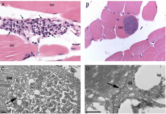
Fifteen patients had a muscle biopsy performed a median of 60 days postdeparture (range, 39–92 days). All tissue samples were examined histologically by a pathologist where collected. The CDC received tissue from a total of 14 (93%) biopsied patients, as tissue blocks, extracted DNA cryoprecipitate, frozen tissue, and/or prepared slides. Photographs of representative histopa-thology were received from 8 patients, 1 from the patient from whom tissue was not available. In all samples and photo-graphs examined, histopathology showed inflammation that was predominantly perivascular within the endomysium and perimysium with a wide range of severity, from rare foci of pre-dominantly lymphohistiocytic inflammation to more frequent foci of mixed inflammation (Figure5A). Sarcocysts were ob-served histologically in the muscle of only 6 patients (40%), de-spite intensive searches including examination of >60 sections from a single muscle biopsy sample in which a lone sarcocyst was observed by a pathologist at the point where collected. When present, intramyocytic sarcocysts were characterized by
single, large (up to 100 µm in width and several hundred micrometers in length), thin-walled, septated cysts containing innumerable, crescent-shaped bradyzoites that were approxi-mately 4 × 8 µm (Figure5B); no inflammation was observed immediately adjacent to any sarcocyst. By EM, large cysts were found embedded in skeletal muscle; they contained numerous, approximately 3.5 µm bradyzoites (Figure 5C). The cyst wall demonstrated short, undulating type 1 protrusions (Figure5D) [5].
Ten patients had PCR testing for Sarcocystis species DNA; 1 biopsy sample, also with sarcocysts visible on histopathology, was positive. The sequencing analysis of the amplified fragment showed 100% similarity with the 18S rRNA gene from Sarcocystis nesbitti (GenBank accession number HF544323).
DISCUSSION
We report 68 travelers with AMS acquired while vacationing on Tioman Island, Malaysia. The spectrum of AMS in humans is broad: from asymptomatic, which may characterize most infect-ed patients [7–9], to severe and relapsing illness lasting years [11]. Comparable with other reports, the majority of the Tio-man Island patients experienced muscle pain, fatigue, fever, and headache. In contrast, we did notfind cough, rash, lymph-adenopathy, swelling, facial tenderness, subcutaneous nodules, Figure 3. Symptoms among 68 patients with acute muscular sarcocystosis, Tioman Island, Malaysia, 2011–2012. Shown are the proportions of patients experiencing each symptom at any time during the course of their illness. Symptoms reported in <5% of the case patients are not shown and include throat pain* (3%), loss of appetite (3%), and stiff neck* (1%). *Unsolicited symptom spontaneously reported in the questionnaire narrative.
or cardiac abnormalities to be prominent, although patients were not examined at standardized intervals [10–14].
Onset of symptoms occurred in 2 distinct phases: early (be-ginning within the second week postdeparture) and late (begin-ning within the sixth week postdeparture). As others have reported, more than one-third of our patients experienced wax-ing and wanwax-ing of symptoms [11,14]. We found that the late phase corresponded with a rise in the serum CPK level and blood eosinophil count, possibly reflecting the onset of an im-mune-mediated myositis. All 15 muscle biopsies showed histo-logic evidence of varying degrees of diffuse multifocal myositis; all were done on or after the 39th day after departure from the island, and none had evidence of inflammation immediately ad-jacent to any observed sarcocyst. As few CPK determinations and no biopsies were done within 5 weeks of departure from the island, earlier development of myositis cannot be ruled out. The lack of clinical suspicion, reflected by the paucity of serum CPK and biopsy data in the early weeks, however, sug-gests that this is unlikely.
The phasic nature of AMS in humans reported here is well characterized in animals with experimentally induced interme-diate-host infections [5]. Signs and symptoms of disease in animals reflect the stages of development and the migratory tra-jectories of the parasite as it passes from the intestine via the vascular endothelium to itsfinal destination within the myo-cyte. The exact time course, however, depends on the specific species of infecting Sarcocystis and the specific host species that is infected [5]. Given the patterns observed among the Tio-man Island patients, we hypothesize that disease in huTio-mans parallels that seen in animals. More work is needed to charac-terize the pathophysiology of disease in humans infected by Sar-cocystis species as the intermediate host.
Despite the presence of swimming beaches and lodging across the island, the ill travelers concentrated most of their ac-tivities on the northwest portion; however, no obvious source of infection could be identified through epidemiologic analyses, al-though nearly all case patients reported potential exposure to untreated water. Cats were ubiquitous and were frequently ob-served in tourist areas, with nearly three-quarters of the case pa-tients reporting having either touched or fed a cat. Although long-tailed macaques [22] and numerous reptile species, includ-ing water monitors and a variety of snakes [23], inhabit Tioman Island, few patients reported contacts. Interestingly, an environ-mental evaluation on the island conducted by the Malaysia Figure 4. Timing of specific elements of acute muscular sarcocystosis.
A, Onset of the 5 most frequently reported symptoms of acute muscular sarcocystosis relative to the number of weeks since departing from Tioman Island; onset clustered during the second (early) and the sixth (late) post-departure weeks. More than 1 onset date for at least 1 of these symptoms was reported by 15 patients (myalgia [n = 10], fever [n = 7], headache [n = 2], fatigue [n = 1]; none for arthralgia). For 25 (37%) patients, the illness was described as phasic, intermittent, or waxing–waning in character, with periods of symptoms separated by a period of relative improvement. B and C, All blood absolute eosinophil counts and serum creatinine phos-phokinase (CPK) levels, respectively, for all patients relative to the number
Figure 4 continued. of weeks since departing Tioman Island. Each case patient may have had >1 of each laboratory determination. One absolute eosinophil count of 6200 cells/µL and 1 CPK level of 3900 U/L, each from a different patient, is not included in B and C, respectively. *Based on the case definition, >500 cells/µL is considered elevated.†Based on the case definition, >200 U/L is considered elevated.
Ministry of Health (MMoH) in response to this outbreak isolat-ed Escherichia coli from almost all, and parasites from several, sampled water sources, although no Sarcocystis species were re-covered [24]. In addition, no Sarcocystis species sporocysts were observed in the animal feces that were sampled, including from cats. Although the MMoH isolated Toxoplasma gondii from sampled cats, none of the case patients identified during this outbreak had evidence of acute toxoplasmosis, as might be ex-pected if cats were the source of this outbreak. Notably, the MMoH did not sample any local rodent, bird, or reptile species during their survey of the island, and their investigation was limited in scope and conducted during the monsoon season, at a time when no infections among travelers were being report-ed [22–25]. Travelers to Tioman Island should be advised to practice proper precautions, including avoiding contact with animals, eating and drinking safe food and water, and washing hands frequently.
We identified S. nesbitti from the formalin-fixed muscle tis-sue of a patient with acute myositis and multiple sarcocysts ob-served on microscopic examination of histologic sections. This finding corroborates recent data showing that S. nesbitti was re-covered from the muscle of 2 of 89 acutely ill patients [14]. Sarcocystis nesbitti has been considered to be a potential cause
of human infections because it infects Southeast Asian species of nonhuman primates, including the long-tailed macaque, and because it resembles morphologically sarcocysts seen in humans [26–28]. Although the natural life cycle of this organism has not yet been definitively determined, evidence is mounting that S. nesbitti has a snake species as its natural definitive host [29–31]. An understanding of the natural life cycle of this zoo-notic parasite would aid the development of preventive mea-sures and messaging.
AMS among travelers is largely a clinical diagnosis that re-quires an understanding of symptoms and symptom progres-sion and the geographic risk distribution of the disease. Early diagnosis is made difficult by the nonspecific nature of the early phase of the disease and the absence of a simple and ac-curate diagnostic test, such as serology. Diagnosis in the later phase may be easier because relatively few infectious diseases are characterized by clinical myositis and eosinophilia, the most notable exception being trichinellosis. In addition, the bi-phasic nature of the illness may offer a hint to the clinician. Cur-rently, definitive diagnosis requires histologic observation of sarcocysts in or amplification of Sarcocystis species DNA by PCR from a muscle biopsy sample. However, we found that sar-cocysts are likely distributed diffusely and difficult to find. Figure 5. Histopathology and electron microscopy. A, Skeletal muscle (SM) with inflammation (arrows) adjacent to a small vessel (v). Inflammation com-posed of lymphocytes and macrophages with small numbers of plasma cells, eosinophils, and rarely neutrophils. Original magnification ×400, hematoxylin and eosin staining. B, SM with intramyocytic sarcocyst. Note the absence of inflammation surrounding the sarcocyst-containing myocyte. Original mag-nification 400×, hematoxylin and eosin staining. C, Portion of a cyst within SM, containing abundant bradyzoites (arrow). D, Higher magnification of the cyst wall, showing short protrusions (arrow) and a fairly thin granular layer (GL). Bars = 2 µm (C) and 500 nm (D). Abbreviations: GL, granular layer; SM, skeletal muscle; v, small vessel.
Although there is currently no treatment regimen that has been shown to be effective, anti-inflammatory agents may be helpful in the management of pain. The value of specific antiparasitic agents is an area for further exploration.
These data have several limitations. At the outset of this in-vestigation, AMS in humans was not well characterized and rel-evant data may have gone unrecorded. Data were gathered retrospectively and at different stages of disease, potentially af-fecting the accuracy of patient recall. Many clinicians from a va-riety of countries and practice settings participated in data collection, which may have introduced variability in data con-sistency. Finally, our methods likely identified more severely af-fected patients. Our data may not reflect disease among all patients infected with S. nesbitti, some of whom may have been asymptomatic.
Our data largely corroborate previous descriptions of human sarcocystosis and offer important new details on this emerging disease. Foremost among these is the suggestion of a 2-phase clinical presentation, commencing about 2 and 6 weeks after in-fection. Unfortunately, the outbreak investigation did not lend itself to determining what proportion of those infected remain asymptomatic, assessing the various therapeutic measures, or determining the typical duration of illness. Clinicians who see posttravel illness should add AMS to their differential diagnosis of the ill patient returning from Malaysia with myalgia, with or without fever, noting the apparent biphasic aspect of the dis-ease, the later onset of elevated CPK and eosinophilia, and the possibility for relapses. Although only 1 biopsy was positive by PCR, our work and the work of others suggest S. nesbitti as a cause of human AMS. Further studies should focus on con firm-ing the natural life cycle of this organism to develop preventive recommendations, following patients along the full course of their illnesses, and studying treatment options. To prevent fu-ture illnesses among travelers to Tioman Island, the exact source of the outbreak needs to be determined and transmission interrupted.
Notes
Acknowledgments. We are grateful to the patients and families who gave generously and energetically of their time to contribute data to this re-port. We thank David O. Freedman, MD, for his many contributions early in this investigation; Alice M. Spivey, MSTCP, for her expertise and effort in creating Figures 1 and 4; Kira Harvey, MPH, for her contribution to data entry; and Emily W. Lankau, DVM, Epidemic Intelligence Service Officer, for her contribution to the design and planning and the early collection and analysis of the data.
Disclaimer. Thefindings and conclusions in this report are the findings and conclusions of the authors and do not necessarily represent the official position of the Centers for Disease Control and Prevention (CDC).
Financial support. This work was supported by the CDC. Potential conflicts of interest. All authors: No reported conflicts. All authors have submitted the ICMJE Form for Disclosure of Potential Conflicts of Interest. Conflicts that the editors consider relevant to the con-tent of the manuscript have been disclosed.
APPENDIX
Tioman Island Sarcocystosis Investigation Team.Members of the investigation team who contributed data and background information were Erwin Van Den Enden (deceased) and Marjan Van Esbroeck, Department of Clinical Sciences, Institute of Tropical Medicine, Antwerp, Belgium; Wayne Ghesquiere, Vancouver General Hospital and Vancouver Island Health Authority, Vancouver and Victoria, British Columbia, Canada; Duc Nguyen and Marie-Catherine Receveur, Travel Clinics and Division of Tropical Medicine–Clinical Internation-al HeInternation-alth, Department of Infectious and TropicInternation-al Medicine, University Hospital Center, Bordeaux, France; François Peyron, Institut de Parasitologie et de Mycologie Médicale, Hôpital de la Croix-Rousse, Lyon, France; Philippe Parola, University Hospi-tal Institute for Infectious and Tropical Diseases, Aix-Marseille University and Assistance Publique Hôpitaux de Marseille, Marseille, France; Hélène Savini, Department of Tropical and Infectious Diseases, Laveran Military Teaching Hospital, Marseille, France; Eric Caumes, AP-HP, Infectious and Tropical Diseases Department, Groupe Hospitalier Pitié-Salpêtrière, Université Pierre et Marie Curie University, Paris, France; Alice Perignon, AP-HP, Infectious and Tropical Diseases De-partment, Groupe Hospitalier Pitié-Salpêtrière, Paris, France; Michel Develoux, Service de Maladies Infectieuses et Tropicales, APHP Hôpital Tenon, Université Pierre et Marie Curie, Paris, France; Christophe Rapp, Service de Pathologie Infectieuse et Tropicale, Hôpital d’Instruction des Armées Bégin, Saint-Mandé, France; Christian A. Keller, Bernhard Nocht Institute for Tropical Medicine, Hamburg, Germany; Martin Haditsch, Labor Hannover, Hannover, Germany; Wolfgang Güthoff and Ines Liebold, Klinik für Gastroenterologie und Infektiologie, Klinikum Ernst von Bergmann, Potsdam, Germany; Johannes Schäfer, Tropenklinik Paul-Lechler-Krankenhaus, Tübingen, Germany; Federico Gobbi, Centre for Tropical Diseases, Hospi-tal Sacro Cuore-Don Calabria, Negrar, Verone, IHospi-taly; Willemijn Kortmann and Gitte van Twillert, Medisch Centrum Alkmaar, Alkmaar, The Netherlands; Abraham Goorhuis, Vanessa Harris, Michèle van Vugt, and Kees Stijnis, Centre of Tropical Medicine and Travel Medicine, Department of Infectious Dis-eases, Academic Medical Centre, University of Amsterdam, Amsterdam, The Netherlands; Eleonora Aronica, Department of (Neuro)Pathology, Academic Medical Center, University of Amsterdam, Amsterdam, The Netherlands; Lisette van Liesh-out and Meta Roestenberg, Laboratory for Parasitology, Leiden University Medical Center, Leiden, The Netherlands; Jan van Wout, Bronovo Hospital, The Hague, The Netherlands; Timo-thy Barkham, Department of Laboratory Medicine, Tan Tock Seng Hospital, Singapore; Poh Lian Lim, Department of Infec-tious Diseases, Institute of InfecInfec-tious Diseases and Epidemiolo-gy, Tan Tock Seng Hospital, Singapore; Lee Kong Chian School
of Medicine, Singapore; Christoph Hatz, Swiss Tropical and Public Health Institute, Basel, Switzerland; Silvio D. Brugger and Hansjakob Furrer, Department of Infectious Diseases, Bern University Hospital, University of Bern, Bern, Switzerland; Franҫois Chappuis and Yann Michel, Division of International and Humanitarian Medicine, Geneva University Hospitals, Geneva, Switzerland; Sanya Choochumporn, Bangkok Hospital Medical Center, Thailand; Than Narkwiboonwong, Bangkok Samui Hospital, Ko Samui, Thailand; Matthew S. Dryden, Hampshire Hospitals Foundation Trust, Royal Hampshire County Hospital, Winchester, Hampshire, UK; Theresa Benedict, Sukwan Handali, and Patricia P. Wilkins, Division of Parasitic Diseases and Malaria, Center for Global Health, Centers for Disease Control and Prevention, Atlanta, Georgia; Wun-Ju Shieh and Sherif Zaki, Division of High-Consequence Pathogens and Pathology, National Center for Emerging and Zoonotic Infectious Diseases, Centers for Disease Control and Prevention, Atlanta, Georgia; Laura Kogelman, Division of Geographic Medicine and Infectious Diseases, Tufts Medical Center, Boston, Massachusetts; and Steven Hatch, Division of Infectious Disease and Immunology, University of Massachu-setts Medical School, Worcester.
References
1. Harvey K, Esposito DH, Han PV, et al. Surveillance for travel-related disease—GeoSentinel, United States, 1997–2011. MMWR Morb Mortal Wkly Rep2013; 62:1–23.
2. Esposito DH, Freedman DO, Neumayr A, Parola P. Ongoing outbreak of an acute muscular Sarcocystosis-like illness among travellers return-ing from Tioman Island, Malaysia, 2011–2012. Euro Surveill 2012; 17. pii:20310.
3. Tappe D, Ernestus K, Rauthe S, et al. Initial patient cluster andfirst pos-itive biopsyfindings in an outbreak of acute muscular Sarcocystis-like infection in travelers returning from Tioman island, peninsular Malay-sia, in 2011. J Clin Microbiol2013; 51:725–6.
4. Centers for Disease Control and Prevention. Notes from thefield: acute muscular sarcocystosis among returning travelers—Tioman Island, Malaysia, 2011. MMWR Morb Mortal Wkly Rep2012; 61:37. 5. Dubey JP, Speer CA, Fayer R. Sarcocystosis of animals and man. Boca
Raton, FL: CRC Press, Inc,1989.
6. Fayer R. Sarcocystis spp. in human infections. Clin Microbiol Rev2004; 17:894–902.
7. Beaver PC, Gadgil RK, Morera P. Sarcocystis in man: a review and report offive cases. Am J Trop Med Hyg 1979; 28:819–44.
8. Pathmanathan R, Kan SP. Three cases of human Sarcocystis infection with a review of human muscular sarcocystosis in Malaysia. Trop Geogr Med1992; 44:102–8.
9. Wong KT, Pathmanathan R. High prevalence of human muscle sarco-cystosis in south-east Asia. Trans R Soc Trop Med Hyg1992; 86:631–2. 10. Jeffrey HC. Sarcosporidiosis in man. Trans R Soc Trop Med Hyg1974;
68:17–29.
11. Arness MK, Brown JD, Dubey JP, Neafie RC, Granstrom DE. An out-break of acute eosinophilic myositis attributed to human sarcocystis parasitism. Am J Trop Med Hyg1999; 61:548–53.
12. Van den Enden E, Praet M, Joos R, Van Gompel A, Gigasse P. Eosin-ophilic myositis resulting from sarcocystosis. J Trop Med Hyg1995; 98:273–6.
13. Mehrotra R, Bisht D, Singh PA, Gupta SC, Gupta RK. Diagnosis of human sarcocystis infection from biopsies of the skeletal muscle. Pa-thology1996; 28:281–2.
14. Abubakar S, Teoh BT, Sam SS, et al. Outbreak of human infection with Sarcocystis nesbitti, Malaysia, 2012. Emerg Infect Dis 2013; 19:1989–91.
15. Bouchaud O, Mühlberger N, Parola P, et al. Therapy of uncomplicated falciparum malaria in Europe: MALTHER—a prospective observational multicentre study. Malar J2012; 11:212.
16. Freedman DO, Stich A, von Sonnenburg F. Sarcocystosis, human, Malaysia: Tioman Island. ProMed. 31 October 2011. Available at:
http://www.promedmail.org. Archive no. 20111031.3240. Accessed 11 August 2014.
17. Freedman DO, Caumes E. Sarcocystosis, human—Malaysia (02): Tioman Island. ProMed. 24 August 2012. Available at :http://www. promedmail.org. Archive no. 20120826.1262494. Accessed 11 August 2014.
18. Visser LG. Sarcocystosis, human—Malaysia (03): new cases, travel relat-ed. ProMrelat-ed. 21 October 2012. Available at:http://www.promedmail.org. Archive no. 20121021.1356457. Accessed 11 August 2014.
19. Nguyen D, Receveur MC, Albert O, Malvy D. Sarcocystosis—Malaysia: (Tioman Island) travel related,2012. ProMed. February 18 2013. Avail-able at:http://www.promedmail.org. Archive no. 20130309.1578678. Accessed 11 August 2014.
20. Oryan A, Sharifiyazdi H, Khordadmehr M, Larki S. Characterization of Sarcocystis fusiformis based on sequencing and PCR-RFLP in water buf-falo (Bubalus bubalis) in Iran. Parasitol Res2011; 109:563–1570. 21. de Almeida M, Steurer F, Koru O, Herwaldt B, Pieniazek NJ, da Silva AJ.
PCR amplification and sequencing of a fragment of the rRNA internal transcribed spacer 2 (ITS 2) for simultaneous diagnostic characteriza-tion of 10 Leishmania spp. An alternative approach for the laboratory diagnosis of leishmaniasis. J Clin Microbiol2011; 49:3143–9. 22. Lim BL, Lim KKP, Yong HS. The terrestrial mammals of Pulau Tioman,
peninsular Malaysia, with a catalog of specimens at the Raffles Museum, National University of Singapore. Raffles Bull Zool 1999; suppl 6: 101–23.
23. Lim KKP, Lim LJ. The terrestrial herpetofauna of Pulau Tioman, peninsular Malaysia. Raffles Bull Zool 1999; suppl 6:131–55. 24. Husna Maizura AM, Khebir V, Chong CK, Shah A, Hakim L.
Surveil-lance for sarcocystosis in Tioman Island, Malaysia. Malaysian J Public Health Med2012; 12:39–44.
25. Sodhi NS. An annotated checklist of the birds of Pulau Tioman, penin-sular Malaysia. Raffles Bull Zool 1999; suppl 6:125–30.
26. Yang ZQ, Wei CG, Zen JS, et al. A taxonomic re-appraisal of Sarcocystis nesbitti (Protozoa: Sarcocystidae) from the monkey Macaca fascicularis in Yunnan, PR China. Parasitol Int2005; 54:75–81.
27. Kan SP, Prathap K, Dissanaike AS. Light and electron microstructure of a Sarcocystis sp. from the Malaysian long-tailed monkey, Macaca fasci-cularis. Am J Trop Med Hyg1979; 28:634–42.
28. Wong KT, Pathmanathan R. Ultrastructure of the human skeletal muscle sarcocyst. J Parasitol1994; 80:327–30.
29. Tian M, Chen Y, Wu L, et al. Phylogenetic analysis of Sarcocystis nesbitti (Coccidia: Sarcocystidae) suggests a snake as its probable definitive host. Vet Parasitol2012; 183:373–6.
30. Lau YL, Chang PY, Tan CT, Fong MY, Mahmud R, Wong KT. Sarcocys-tis nesbitti infection in human skeletal muscle: possible transmission from snakes. Am J Trop Med Hyg2014; 90:361–4.
31. Lau YL, Chang PY, Subramaniam V, et al. Genetic assemblage of Sarco-cystis spp. in Malaysian snakes. Parasit Vectors2013; 6:257.