Endothelial Regulation of Vascular Tone
and Growth
Thomas F. Luscher and Felix C. Tanner
The endothelium regulates vascular tone by releas-ing factors involved in relaxation and contraction, in coagulation and thrombus formation, and in growth inhibition and stimulation. Endothelium-depend-ent relaxations are elicited by transmitters,
hormones, platelet substances, and the coagulation system, and by physical stimuli such as the shear stress from circulating blood. They are mediated by the endothelium-derived relaxing factor, recently identified as nitric oxide, which causes vasodilation and platelet deactivation. Other proposed endothe-lium-derived relaxing factors include a hyperpolar-izing factor, lipooxygenase products, and the cyto-chrome P450 pathway. Endothelium-derived con-tracting factors are produced by the cyclooxygenase pathway and by endothelial cells, which produce the peptide endothelin-1, a potent vasoconstrictor that under normal conditions circulates at low lev-els. The endothelium produces both growth inhib-itors—normally dominant—and growth stimuli. Denuded or dysfunctional endothelium leads to a proliferative response and intimal hyperplasia in the vessel wall; moreover, platelets adhere to the
site and release potent growth factors. Endothelial dysfunction has numerous causes: Aging is associ-ated with increased formation of contracting factor and decreased relaxing factor; denudation, such as by coronary angioplasty, impairs the capacities of regenerated endothelial cells; oxidized low-density lipoproteins and hypercholesterolemia interfere with nitric oxide production; hypertension morpho-logically and functionally alters the endothelium; and atherosclerosis markedly attenuates some endo-thelium-dependent relaxations. For patients with coronary bypass grafts, differences in endothelium-derived vasoactive factors between the internal mammary artery and the saphenous vein may be im-portant determinants of graft function, with the mammary artery having more pronounced relaxa-tions than the saphenous vein and thus a higher pat-ency rate. Am J Hypertens 1993;6:283S-293S
KEY WORDS: Endothelial regulation, endothelial
dys-function, vascular tone, endothelium-derived relax-ing factors, endothelium-derived contractrelax-ing factors, nitric oxide.
T
he endothelium takes part in the regulation of vascular tone1 by releasing relaxing and con-tracting factors both under basal conditions and when activated by neurotransmitters, hormones, autacoids, or physical stimuli. In addition, endothelial cells release factors involved in coagula-tion and thrombus formacoagula-tion and in growth inhibicoagula-tion and stimulation.Owing to its strategic anatomic position, the endothe-lium is a target organ for hypertension, diabetes, and hyperlipidemia.1 Alterations in endothelial function may contribute to the pathogenesis as well as to the
pro-gression and complications of hypertension and its se-quelae such as atherosclerotic vascular disease.
From the Department of Medicine, Division of Clinical Pharma-cology, and the Department of Research, Laboratory of Vascular Re-search, University Hospital, Basel, Switzerland.
Original research was supported by the Swiss National Research Foundation Grant 32-254.68.88, SCORE Grant 3231-025.150, the Swiss Cardiology Foundation, the Helmut Horten Foundation, and the Swiss Foundation for Nutrition Research.
Address correspondence and reprint requests to Thomas F. Luscher, MD, Division of Cardiology, University Hospital, 3010 Bern, Switzerland.
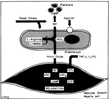
284S LUSCHER AND TANNER AJH-JULY 1993-VOL 6, NO. 7, PART 2 Shear S t r e s s <3m) P l a t e l e t s
<&m>
cGMP ^ A g o n i s t NO'J
Endothelium Nitric O x i d e ^HH TNFJL-1.LPS Libsig Vascular Smooth M u s c l e c e l l FIGURE 1. The L-Arginine pathway in the blood vessel wall:Endothelial cells form nitric oxide (NO) from L-arginine by the ac tivity of the constitutive nitric oxide synthase (NOsc), which can be
inhibited by analogs of the amino acid such as L-NG-monomethyl
arginine (L-NMMA). Nitric oxide activates soluble guanylyl cyclase (sGC) in vascular smooth muscle and platelets and causes increases in cyclic 3/,5/-guanosine monophosphate (cGMP), which
mediates relaxation and platelet inhibition, respectively. Shear stress and receptor-operated agonists (not shown) stimulate the re lease of nitric oxide. In addition, vascular smooth muscle cells can form nitric oxide by the activity of an inducible (by tumor necrosis factor, interleukin 1, and lipopolysaccharide) form of nitric oxide
synthase (NOSt).
ENDOTHELIUM-DEPENDENT REGULATION OF VASCULAR TONE
Relaxing Factors Endothelium-dependent relaxa tions occur both in vitro and in vivo2^; transmitters, hormones, substances derived from platelets, and the coagulation system can cause these responses.1 Fur thermore, physical stimuli, such as shear stress (ex erted by the circulating blood), elicit endothelium-de pendent vasodilation.5
Endothelium-derived Nitric Oxide The relaxations
are mediated by a diffusible substance with a half-life of a few seconds,6 the so-called endothelium-derived relaxing factor (EDRF),2 which has recently been iden tified as nitric oxide.7 Endothelium-derived nitric oxide (EDNO) has the same chemical characteristics as EDRF and is liberated in amounts sufficient to account for the vascular action of EDRF.7 Possibly, EDNO is not released as a free radical, but rather as a nitrosyl-ated compound, for instance, S-nitrosocysteine.8
EDNO is formed from L-arginine by oxidation of the guanidine-nitrogen terminal of L-arginine (Figure l).9Nitric oxide synthase has been cloned recently1 0; it is primarily a cytosolic enzyme requiring calmodulin, calcium, and NADPH, and has similarities to cyto chrome P450 enzymes.1 0 , 1 1 Several isoforms of the en zyme occur not only in endothelial cells, but also in platelets, macrophages, vascular smooth muscle cells, and in the brain.1 2 - 1 5 Endothelium-dependent relaxa tions to serotonin are inhibited by analogs of L-argin ine such as L-AP-monomethyl arginine (L-NMMA) and are restored by L-arginine, but not by
D-argin-With Endothelium Without Endothelium
120-, spons e 100-Φ 80-in t o f t (10 0 m
60-go
« ο 40-ο1
s c ο 20-δ 0 -• Control n=4 • L - N M M A n=4 120η tsuo d 100-Re s t o f th e )0 mM ) 80- 60-tion , Per c to KC I 40->ntra c 20" Ο 0 -• Control n=4 • L - N M M A n=4Norepinephrine (-logM) Norepinephrine (-logM)
FIGURE 2. Endothelium-dependent effects to the inhibitor of nitric oxide formation i-NG-monomethyl arginine (L-NMMA) in the
human internal mammary artery. In the preparation with endothelium, L-NMMA causes concentration-dependent contractions and augments those to norepinephrine. In contrast, in the preparation without endothelium, L-NMMA has no effect. From Yang et al3 with
2CH 4 0 Η 6 0
I
8 0 ^ c ioo Η3
1 2 0 ι 1 1 1 1 1 1 1 0 9 8 7 B r a d y k i n i n (-log M ) Endothelium.: » control with (n = 6) • control without (n = 6) 3 L-NNMA m4 Μ with (n = 6) 1 0 B r a d y k i n i n (-log M ) controlMethylene Blue ΙΟ"5 Μ with
Hemoglobin ΙΟ"5 Μ with
Endothelium: with (n=ll)
(n = 5)
(n = 6)
FIGURE 3. Effect of L-NG
-monom-ethyl arginine (L-NMMA), m-monom-ethylene blue, and hemoglobin on endothelium-dependent relaxations to bradykinin in epicardial coronary arteries. L-NMMA and methylene blue only cause a weak shift of the concentration-response curve to higher concentrations of the re laxing agonist, whereas hemoglobin also reduces the maximal relaxation to bradykinin. From Richard et al16 with
permission.
ine.3'1 6 In quiescent arteries, L-NMMA causes endothe lium-dependent contractions (Figure 2).3 When in fused in rabbits, it causes long-lasting increases in blood pressure that are reversible by L-arginine,1 7 de monstrating that the vasculature is in a constant state of vasodilation as a result of the continuous release of nitric oxide from the endothelium.
Relaxations to EDNO are associated with an in crease in cyclic 3',5'-guanosine monophosphate (cGMP) in vascular smooth muscle cells (Figure l ) .1 8 Methylene blue, an inhibitor of soluble guanylyl cyclase, prevents the production of cGMP and inhibits endothelium-dependent relaxations,1'1 8 an indication that EDNO causes relaxations by stimulating the en zyme and, in turn, the formation of cGMP. Soluble guanylyl cyclase is also present in platelets and is acti vated by EDNO (Figure l ) .1 9 Increased levels of cGMP in platelets are associated with reduced adhesion and aggregation. Because EDNO causes both vasodilata tion and platelet deactivation, it represents an impor tant antithrombotic feature of the endothelium.
Vascular Smooth Muscle-derived Nitric Oxide Al
though the media of the blood vessel wall normally does not produce nitric oxide, vascular smooth muscle cells (including those obtained from human vessels) can do so when stimulated by endotoxin and inter-leukin-1 (Figure l ) .2 0-2 3 Thus, it appears that there are at least two enzymes for the production of nitric oxide: The constitutive endothelial enzyme is calcium de pendent and produces picomoles of nitric oxide, whereas the inducible enzyme is calcium independent, produces nanomoles of nitric oxide, and is primarily expressed in smooth muscle cells and monocytes. Acti vation of the L-arginine pathway in smooth muscle
cells by endotoxin, interleukin-1, and tumor necrosis factor may play a role in septic shock and explain why the cardiovascular system becomes resistant to cat echolamines under these conditions. On the other hand, L-NMMA may provide a therapeutic tool as it can prevent nitric oxide formation in vascular smooth muscle cells stimulated with endotoxin; preliminary data indeed suggest that L-NMMA may be beneficial for patients in septic shock.2 4
Prostacyclin Prostacyclin increases cyclic
3',5'-ade-nosine monophosphate (cAMP) in smooth muscle and in platelets and, thus, causes relaxation and inhibition of platelet aggregation.1'2 5 However, the contribution of prostacyclin to endothelium-dependent relaxations is negligible.1 In human platelets, EDNO and prostacy clin synergistically inhibit aggregation.2 6
Other Endothelium-derived Relaxing Factors In the
porcine coronary circulation, L-NMMA inhibits endo thelium-dependent relaxations to serotonin, but not to bradykinin (Figure 3).1 6 As similar effects are obtained by other inhibitors of the action of EDNO, such as he moglobin and methylene blue, endothelial cells appear to release another relaxing factor distinct from nitric oxide. Prostacyclin can be excluded, as it is a weak vas odilator of porcine coronary arteries, and indometha cin does not affect the response to bradykinin. Several candidates for these responses have been proposed, including a hyperpolarizing factor, products of lipoox-ygenase, or the cytochrome P450 pathway.1
Acetylcholine causes not only an endothelium-de pendent relaxation, but also an endothelium-depend ent hyperpolarization of vascular smooth muscle.2 7'2 8 Although the relaxation to the muscarinic agonist is sustained, the hyperpolarization is transient. An
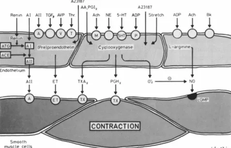
endo-286S LUSCHER AND TANNER AJH-JULY 1993-VOL 6, NO. 7, PART 2 S t r e t c h ADP Ach Bk R e n i n 4
iATGt"**"»prr
i^AC*E^ffvvv?vfvHte-: IAIIFIGURE 4. The endothelium is a source of
different contracting factors. Products of cy clooxygenase, such as thromboxane A2 (TX
A2) and prostaglandin H2 (PGH2) have a di
rect contractile effect, while superoxide ani ons (02) induce increases in tension by inac
tivating nitric oxide (NO). Endothelin (ET), as well as angiotensin II (A II), are also endo thelium-derived contracting factors, as they are both produced by endothelial cells.
S m o o t h m u s c l e c e l l s
thelium-dependent hyperpolarizing factor (EDHF) distinct from nitric oxide may be involved, although the latter was itself shown to have hyperpolarizing properties under certain conditions.2 7 , 2 9 The EDHF ap pears to activate ATP-sensitive potassium channels or sodium-potassium ATPase in smooth muscle, or both.2 7 , 3 0 The hyperpolarization may contribute to en dothelium-dependent relaxations or reduce the sensi tivity of vascular smooth muscle to vasoconstrictor stimuli.
Contracting Factors
Cyclooxygenase-dependent Endothelium-derived Contract ing Factor (EDCF) Exogenous arachidonic acid
evokes endothelium-dependent contractions that can be prevented by indomethacin (an inhibitor of cy clooxygenase).3 1 In the human saphenous vein, ace tylcholine and histamine evoke endothelium-depend ent contractions; in the presence of indomethacin, however, these relaxations are unmasked.3 The prod ucts of cyclooxygenase mediating the contractions are thromboxane A2 in the case of acetylcholine and en-doperoxides (prostaglandin H2) in that of histamine (Figure 4).3
The cyclooxygenase pathway is also a source of su peroxide anions, which can mediate endothelium-de pendent contractions either by the breakdown of nitric oxide or by direct effects on vascular smooth muscle (Figure 4 ) .3 2 Thus, the cyclooxygenase pathway pro duces a variety of endothelium-derived contracting factors; their release appears to be more prominent in veins than in arteries.1
Endothelin Endothelial cells produce the
21-amino-acid peptide endothelin (Figure 4).1 , 3 3 Among the three peptides—endothelin-1, endothelin-2, and endothelin-3—endothelial cells appear to produce en dothelin-1 exclusively.1
Translation of messenger RNA generates preproen-dothelin, which is converted to big endothelin3 3; the formation of endothelin-1 by the endothelin-convert-ing enzyme is then necessary for the development of full vascular activity.3 4 The expression of messenger RNA and the release of the peptide are stimulated by thrombin, transforming growth factor-β, interleukin-1, epinephrine, angiotensin II, arginine vasopressin, cal cium ionophore, and phorbol ester (Figure 4 ) .1 , 3 3 , 3 5
Endothelin-1 is a potent vasoconstrictor both in vitro and in vivo. In the coronary circulation and the human forearm circulation, endothelin-1 causes vaso dilation at lower concentrations and marked contrac tions at higher concentrations (Figure 5 ) .3 6 , 3 7 In human arterial and venous coronary bypass vessels, it causes marked contractions.3 8
The circulating levels of endothelin-1 are very low, however, suggesting that little of the peptide is formed under physiological conditions.3 9 This may be related
E n d o t h e l i n ( n g / m i n / 1 0 0 m l )
FIGURE 5. Effects of endothelin on blood flow in the human fore
arm circulation. low concentrations of endothelin (0.5 ng/min/100 mL) cause a slight increase, whereas higher concentrations of the peptide (25 and 50 ng/min/100 mL) induce a marked decrease of forearm blood flow. Modified from Kiowski et al57 with permission.
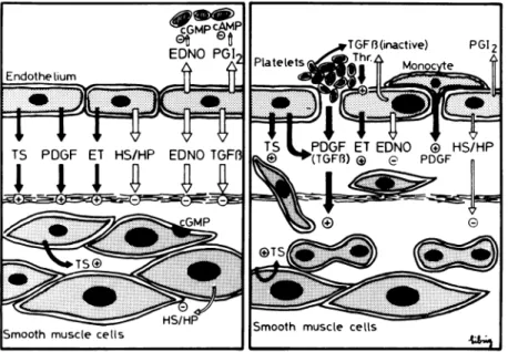
FIGURE 6. Endothelium and vascular growth.
The endothelium produces growth inhibitors such asheparin (HP), heparin sulphates (HS), trans forming growth factor beta (TGFB), and also ni tric oxide (EDNO). On the other hand, it releases growth promotors, such as platelet-derived growth factor (PDGF), thrombospondin (TS), and possibly endothelin (ET). At sites of damaged en dothelium the production of nitric oxide (EDNO) and prostacyclin (PGI2) is diminished, favoring
platelet adhesion and aggregation. PDGF is re leased by aggregating platelets and leads to prolif eration as well as migration of vascular smooth muscle cells into the intima. The endothelium most probably takes part in these structural changes of the vascular wall, at least indirectly, by inhibiting platelet aggregation and with that the release of growth-stimulating factors.
to the absence of stimuli or to the presence of potent inhibitory mechanisms, or to both, or it may be that the peptide is released preferentially toward vascular smooth muscle cells.3 5 , 4 0'4 1 Indeed, three inhibitory mechanisms regulating endothelin production have been delineated: 1) cGMP-dependent inhibition35; 2) cAMP-dependent inhibition42; and 3) an inhibitory fac tor produced by vascular smooth muscle cells.4 3 The cGMP-dependent mechanism can be activated by EDNO, nitroglycerine, 3-morpholino sydnominine (SIN-1),4 4 and atrial natriuretic peptide (which acti vates particulate guanylyl cyclase).4 5 Thus, after inhi bition of endothelial nitric oxide production, the thrombin-induced formation of endothelin is aug mented3 5; on the other hand, SIN-1 prevents the thrombin-induced endothelin release by a cGMP-de-pendent mechanism.4 4 Endothelin can also release ni tric oxide and prostacyclin from endothelial cells in what may represent a negative feedback mechanism.4 0
The contraction to endothelin is not related to direct activation of voltage-operated calcium channels on vas cular smooth muscle, as calcium antagonists do not pre vent its effects in most blood vessels.4 0'4 6 , 4 7 However, the peptide activates indirectly those channels in the porcine coronary artery where calcium antagonists at tenuate endothelin-induced vasoconstriction.48 In the human forearm circulation, endothelin-1 induces po tent contractions, which can be prevented by nifedipine and verapamil hydrochloride, unmasking the vasodila tor effects of the peptide.3 7 The vasodilator effects of en dothelin are related to the endothelial production of prostacyclin, whereas nitric oxide may contribute to the relaxation effects.49 Although endothelin is a secre-tagogue for atrial natriuretic factor, its release is not in volved in the vasodilator action of endothelin.50
EDNO also interacts with endothelin at the level of vascular smooth muscle. Indeed, the contractions to
the peptide are enhanced after endothelial removal, in dicating that basal production of EDNO reduces the response to the peptide.3 8 Stimulation of the formation of EDNO by acetylcholine reverses endothelin-in duced contractions in most blood vessels although this mechanism appears to be less potent in veins.3 8
ENDOTHELIAL REGULATION OF VASCULAR GROWTH
Removal of the endothelium is a procedure that invari ably leads to a proliferative response in the blood ves sel wall with intimal hyperplasia.5 1 Important growth inhibitors produced by the endothelium are heparin, heparin sulfates, transforming growth factor-β, and most likely nitric oxide.5 2 , 5 3 On the other hand, endo thelial cells can also produce growth factors such as basic fibroblast growth factor, platelet-derived growth factor (PDGF), and possibly also endothelin (Figure 6 ) .5 4 , 5 5 These factors contribute to proliferative re sponses, at least under certain conditions, whereas it has to be assumed that under normal conditions inhib itory stimuli prevail. At sites of endothelial denuda tion or dysfunction, platelets adhere and release po tent growth factors such as PDGF.5 6 Hence, the platelet inhibitory effect1 9 of nitric oxide and prostacyclin also contribute indirectly to growth inhibition (Figure 6).
In vascular smooth muscle cells from saphenous vein coronary bypass grafts exposed to pulsatile stretch, the [3H]thymidine incorporation is enhanced after 24 h of stretch, whereas the cell number is in creased after 6 days. In contrast, both [3H]thymidine incorporation and cell number remain constant in vas cular smooth muscle cells from internal mammary ar teries (Figure 7).5 7 Thus, the stimulation by pulsatile stretch of vascular smooth muscle cell proliferation in saphenous vein, but not in internal mammary artery, may contribute to the higher occlusion rate of venous
288S LUSCHER AND TANNER AJH-JULY 1993-VOL. 6, NO. 7, PART 2
Control Stretch Control Stretch Internal Mammary Artery Saphenous Vein
Control Stretch Control Stretc Internal Mammary Artery Saphenous Vein
FIGURE 7. Proliferative responses to pulsatile stretch (60
cy-cles/min) of human vascular smooth muscle cells from internal mammary artery and saphenous vein. After 24 h of pulsatile stretch the [3H]thymidine incorporation (top) was increased in
sa-phenous vein but not in mammary artery. After 6 days of stretch
(lower) the cell number was increased in the vein but not in the
artery. From Preded et al57 with permission.
coronary bypass grafts. As shear forces stimulate the release of nitric oxide and prostacyclin from the endo-thelium, it is possible that the growth responses of vas-cular smooth muscle cells are modulated by or inhib-ited in the presence of endothelial cells.
ENDOTHELIAL DYSFUNCTION
Because of its strategic anatomic position between the circulating blood and vascular smooth muscle, the
en-dothelium is a primary target organ for cardiovascular risk factors and mechanical forces, such as pressure and shear stress, particularly at branching sites where blood flow is nonlaminar.1
Aging Aging is one of the most important determi-nants of vascular disease. In the rat, aging is associated with an increased formation of the EDCF (prosta-glandin H2),5 8 as well as a mild decrease in the release of EDRF.5 9 In contrast, the responsiveness of vascular smooth muscle to nitric-oxide-forming compounds does not change under these conditions.5 8 In the human coronary microcirculation, the increase in cor-onary flow induced by intraarterial infusions of ace-tylcholine declines with aging.6 0
Endothelial Regeneration After mechanical denu-dation of the porcine coronary artery, the capacity of regenerated endothelial cells to release EDNO in re-sponse to platelet-derived serotonin is impaired be-cause of a defect of the G{ protein linked to the
endo-thelial 5HT1-serotonergic receptor.6 1 These functional alterations occurring during regeneration may con-tribute to the age-dependent impairments of endothe-lial function in the coronary circulation and may also play a role after percutaneous transluminal coronary angioplasty. Dysfunction of the endothelium, particu-larly in response to platelet-derived products, may in-crease platelet adhesion, thereby providing high local concentrations of platelet-derived growth factor and contributing to the proliferative response at sites of en-dothelial dysfunction.
Lipoproteins and Hypercholesterolemia Morpho-logically, the endothelium remains intact during the early stages of atherogenesis; functionally, however, pronounced alterations occur.1 Particularly, oxidized low-density lipoprotein (OX-LDL) is present in human atherosclerotic lesions.6 2 In the porcine coronary ar-tery, OX-LDL inhibits endotheUum-dependent relaxa-tions to platelets, serotonin, and thrombin.6 3 In con-trast, relaxations to the nitric oxide donor SIN-1 are well maintained, excluding a reduced responsiveness of vascular smooth muscle to EDNO. The inhibition is specific for OX-LDL, as it is not induced by compara-ble concentrations of native LDL. In the rabbit aorta the effect of OX-LDL is mimicked by lysolecithin (a characteristic component of OX-LDL).6 4'6 5
OX-LDL appears to activate an endothelial receptor distinct from the LDL receptor, such as the scavenger receptor (Figure 8)6 3'6 5; indeed, dextran sulfate, a com-petitive antagonist of modified LDL at this receptor, prevents the endothelial effects of OX-LDL.6 3 Because endotheUum-dependent relaxations to serotonin, but not to bradykinin, are inhibited by the lipoproteins, they specifically interfere with endothelial production of nitric oxide, whereas that of other EDRFs does not
Vascular smooth muscle cell
FIGURE 8. Schematic representation of the effects of oxidized low-density lipoprotein (Ox-LDL) on the endothelial L-arginine pathway
in the coronary circulation: Ox-LDL activates the endothelial scavenger receptor and interferes with the intracellular availability of L-arginine (L-ARG). This reduces the efficacy of the receptor-operated activation of the L-arginine pathway by serotonin (5-HT) and other mediators, and thus favors vascular contraction and platelet aggregation. Modified from Tanner et al65 with permission.
seem to be affected. Nitric oxide synthetase remains unaffected, however, as L-arginine evokes a full relaxa-tion in vessels treated with OX-LDL. Because pretreat-ment with L-arginine restores the response to sero-tonin in vessels treated with OX-LDL, it may be that OX-LDL interacts with the intracellular availability of L-arginine (Figure 8).6 3'6 5
This mechanism may also occur in vivo, as a similar inhibition of endothelium-dependent relaxation to se-rotonin and to bradykinin occurs in hypercholesterol-emic pigs as in coronary arteries exposed to OX-LDL.6 6 In humans with hypercholesterolemia, L-arginine infu-sion augments the blunted increase in coronary blood flow in response to acetylcholine67; in contrast, the loss of endotheUum-dependent vasodilation to ace-tylcholine in epicardial coronary arteries is unaffected by the amino acid, possibly because of the presence of fully developed atherosclerosis.
In addition to their effect on the L-arginine pathway, both native and OX-LDL inactivate nitric oxide.6 8 OX-LDL not only inhibits endothelium-dependent relaxa-tion, but also causes endothelium-dependent contrac-tion.69 In the rabbit femoral artery, OX-LDL potenti-ates contractions to potassium chloride as well as to receptor-operated vasoconstrictors.7 0
OX-LDL also induces the expression of messenger RNA for endothelin in cultured aortic endothelial cells
as well as the release of the peptide from the intact por-cine aorta (Figure 8).7 1 In this context it is of interest that threshold and low concentrations of endothelin, which by themselves evoke no appreciable vascular ef-fect, potentiate contractions induced by serotonin in the human coronary artery and by norepinephrine and serotonin in the human internal mammary ar-tery.7 2 Thus, OX-LDL inhibits endotheUum-dependent relaxations and promotes endotheUum-dependent as well as endothelium-independent contractions; the consequences are alterations in vascular tone leading to vasospasm and thrombus formation, both common events in patients with coronary artery disease.
Hypertension Hypertension is associated with morphological and functional alterations of the endo-thelium.1 In hypertensive blood vessels, endotheUal cells have an increased volume, bulging into the lumen, and fibrin and ceU deposition is increased in the subintimal space. Furthermore, the interaction of platelets and monocytes with the endothelium is in-creased in hypertensive vessels compared with normotensive controls.
Endothelium-dependent relaxations to ace-tylcholine are reduced in the aortic, cerebral, and pe-ripheral microcirculations of hypertensive rats.7 3 , 7 4 Similarly, the vasodilator effects of acetylcholine in the
290S LUSCHER AND TANNER AJH-JULY 1993-VOL. 6, NO. 7, PART 2 Τ
Τ
χ 0.6 ο. Sodium Nitroprusside ^g/min/100ml) I I Normotensives (n=8) 1.0 4.0 Acetylcholine Hypertensives (n=8)FIGURE 9. Effects of intraarterially infused acetylcholine on
forearm vascular resistance in normotensive subjects (open bars) and patients with essential hypertension (hatched bars). Note the reduced effects of acetylcholine in the hypertensive patients, n.s., not significant; * Ρ < .05 From hinder et al4 with permission.
human forearm of hypertensive subjects is blunted (Figure 9).4 In the spontaneously hypertensive rat, the
reduced response to acetylcholine is related to the pro duction of EDCF (ie, prostaglandin H2), whereas in most other forms of experimentally induced hyperten sion a reduced formation of EDNO predominates.7 4 In the mesenteric microcirculation, intraluminal (but not extraluminal) activation of the endothelium is dys functional, indicating a predominant alteration of that surface of the endothelium most exposed to high blood pressure.7 4
In contrast, the coronary circulation—at least as judged from work with the spontaneously hyperten sive rat model—appears less prone to hypertensive en dothelial dysfunction. Indeed, in epicardial coronary arteries of these rats, aging and hypertension only minimally reduce the response to acetylcholine,75 sug gesting that in the absence of hyperlipidemia, hyper tension exerts only a mild effect on the coronary endo thelium.
Atherosclerosis In porcine coronary arteries, endo thelium-dependent relaxations are severely impaired to serotonin and also reduced to bradykinin by estab lished atherosclerosis.6 6 Endothelium-independent re laxations to nitrovasodilators remain preserved, how ever, except in severely atherosclerotic arteries.1
In human coronary arteries, atherosclerosis attenu ates endothelium-dependent relaxations to substance P, bradykinin, aggregating platelets, and calcium iono-phore,7 6'7 7 and in vivo acetylcholine and serotonin cause paradoxical vasoconstriction.7 8 , 7 9
The nature of the mechanism responsible for the marked impairment or loss of endothelium-dependent relaxations in atherosclerosis is controversial. The re
lease of EDRF clearly is reduced in porcine coronary arteries with hypercholesterolemia and atherosclero sis.6 6 Direct measurements of nitric oxide in the rabbit aorta, however, suggest an increased formation of ni tric oxide with a concomitant massive breakdown of the endogenous nitrovasodilator.80 The latter observa tion suggests that atherosclerosis is associated with in creased formation of superoxide radicals and other products inactivating nitric oxide or with decreased activity of superoxide dismutase in the blood vessel wall, or both. It is conceivable that during the more de veloped stages of atherosclerosis, the marked invasion of monocytes and other blood cells induces nitric oxide synthase in the subintimal space and vascular smooth muscle cells. However, it is not known whether altera tions similar to those in the rabbit aorta occur in human coronary arteries.
Coronary Bypass Grafts In patients with coronary artery disease, surgical therapy involves implantation of an arterial or venous bypass graft using the internal mammary, gastroepiploic artery, or saphenous vein. The mammary artery has a remarkably higher patency rate than the saphenous vein.8 1 Endothelium-derived vasoactive factors may be important to graft function as they determine the antithrombotic properties and the regulation of blood flow. In addition, these factors may have antiproliferative and proliferative proper ties determining the late changes occurring in coro nary bypass grafts.
The mammary artery exhibits much more pro nounced endothelium-dependent relaxations than does the saphenous vein, because the release of EDNO by receptor-operated agonists and, in particular, by aggregating platelets is more efficient in the artery than in the vein.8 2 , 8 3 Particularly, the release of EDNO in response to platelet-derived adenosine diphosphate is an important antithrombotic property. The gas troepiploic artery releases amounts of EDNO compa rable with those released by the mammary artery, but exhibits more pronounced contractions.8 4 These differ ences in endothelial and vascular smooth muscle func tions of bypass graft vessels (Figures 6, 7) may play an important role in graft function and patency, and, hence, in the survival of patients undergoing coronary bypass surgery.
ACKNOWLEDGMENTS
The authors thank Amanda de Sola Pinto and Ber-nadette Weber-Libsig for their help in preparing the manuscript.
REFERENCES
1. Luscher TF, Vanhoutte PM: The Endothelium—Modu lator of Cardiovascular Function. Boca Raton, CRC Press, 1990, pp 1-215.
2. 3.
Furchgott RF, Zawadzki JV: The obligatory role of en-dothelial cells in the relaxation of arterial smooth mus-cle by acetylcholine. Nature 1980;299:373-376.
19.
16.
17
Richard V, Tanner FC, Tschudi M, Luscher TF: Differ-ent activation of L-arginine pathway by bradykinin, se-rotonin, and clonidine in coronary arteries. Am J Phys-iol 1990;259:H1433-1439.
22. Yang Z, von Segesser L, Bauer E, et al: Different activa- 20. tion of endothelial L-arginine and cyclooxygenase
path-way in human internal mammary artery and
saphe-nous vein. Circ Res 1991 ;68:52-60. 21 4. Linder L, Kiowski W, Buhler FR, Luscher TF: Indirect
evidence for release of endothelium-derived relaxing factor in human forearm circulation in vivo: blunted re-sponse in essential hypertension. Circulation 1990;81:1762-1767.
5. Rubanyi GM, Romero JC, Vanhoutte PM: Flow-in-duced release of endothelium-derived relaxing factor. Am J Physiol 1986;250:H1145-1149.
6. Rubanyi GM, Vanhoutte PM: Superoxide anions and 23. hyperoxia inactivate endothelium-derived relaxing factor. Am J Physiol 1986;250:H822-327.
7. Palmer RMI, Ferrige AG, Moncada S: Nitric oxide re-lease accounts for the biological activity of
endothe-lium-derived relaxing factor. Nature 1987;327:524-526. 24. 8. Myers PR, Minor RL Jr, Guerra R Jr, et al: Vasorelaxant
properties of the endothelium-derived relaxing factor more closely resemble S-nitrosocysteine than nitric 25. oxide. Nature 1990;345:161-163.
9. Palmer RMJ, Ashton DS, Moncada S: Vascular
endo-thelial cells synthesize nitric oxide from L-arginine. Na- 26. ture 1988;333:664-666.
10. Bredt DS, Hwang PM, Glatt CE, et al: Cloned and ex-pressed nitric oxide synthase structurally resembles
cy-tochrome P-450 reductase. Nature 1991;351:714-718. 27. 11. Bredt DS, Snyder SH: Isolation of nitric oxide
syn-thetase, a calmodulin-requiring enzyme. Proc Natl
Acad Sci USA 1990;87:682-685. 28. 12. Radomski MW, Palmer RMJ, Moncada S: An
L-argin-ine/nitric oxide pathway present in human platelets regulates aggregation. Proc Natl Acad Sci USA
1990;87:5193-5197. 29. 13. Hibbs JB, Traintor RR, Vavrin Z, Rachlin EM: Nitric
oxide: a cytotoxic activated macrophage molecule. Bio-chem Biophys Res Comm 1988;157:87-94.
14. Wood KS, Buga GM, Byms RE, Ignarro LJ: Vascular 3 0'
smooth muscle-derived relaxing factor (MDRF) and its close similarity to nitric oxide. Biochem Biophys Res Commun 1990;170:80-88.
15. Knowles RG, Palacios M, Palmer RMJ, Moncada S: For-mation of nitric oxide from L-arginine in the central nervous system: a transduction mechanism for stimula-tion of the soluble guanylate cyclase. Proc Natl Acad
Sci USA 1989;86:1^L 32. 33.
Rees DD, Palmer RMJ, Moncada S: Role of
endothe-lium-derived nitric oxide in the regulation of blood 34. pressure. Proc Natl Acad Sci USA 1989;86:3375-3378.
18. Rapoport RM, Murad F: Agonist-induced endothe-lium-dependent relaxation in rat thoracic aorta may be
mediated through cGMP. Circ Res 1983;52:352-357. 35.
Busse R, Luckhoff A, Bassenge E: Endothelium-derived relaxant factor inhibits platelet activation. Naunyn-Schrniedeberg's Arch Pharmacol 1987;336:566-571. Bernhardt J, Tschudi MR, Dohi Y, et al: Release of nitric oxide from human vascular smooth muscle cells. Bio-chem Biophys Res Commun 1991;180:907-912. Fleming I, Gray GA, Julou-Schaeffer G, et al: Incuba-tion with endotoxin activates the L-arginine pathway in vascular tissue. Biochem Biophys Res Commun 1990;171:562-568.
Rees DD, Cellek S, Palmer RMJ, Moncada S: Dexam-ethasone prevents the induction by endotoxin of a ni-tric oxide synthase and the associated effects on vascu-lar tone: an insight into endotoxin shock. Biochem Bio-phys Res Commun 1990;173:541-547
Schini VB, Junquero DC, Scott-Burden T, Vanhoutte PM: Interleukin-1 beta induces the production of an L-arginine-derived relaxing factor from cultured smooth muscle cells from rat aorta. Biochem Biophys Res Com-mun 1991;176:114-121.
Petros A, Bennett D, Vallance P: Effect of nitric oxide synthase inhibitors on hypotension in patients with septic shock. Lancet 1991;338:1557-1558.
Moncada S, Vane JR: Pharmacology and endogenous roles of prostaglandin endoperoxides, thromboxane A2
and prostacyclin. Pharmacol Rev 1979;30:293-331. Macdonald PS, Read MA, Dusting GJ: Synergistic inhi-bition of platelet aggregation by endothelium-derived relaxing factor and prostacyclin. Thromb Res 1988;49:437-449.
Feletou M, Vanhoutte PM: Endothelium-dependent hyperpolarization of canine coronary smooth muscle. Br J Pharmacol 1988;93:515-524.
Komori K, Lorenz RR, Vanhoutte PM: Nitric oxide, ace-tylcholine, and electrical and mechanical properties of canine arterial smooth muscle. Am J Physiol 1988;255:H207-212.
Tare M, Parkington HC, Coleman HA, et al: Hyperpo-larisation and relaxation of arterial smooth muscle caused by nitric oxide derived from the endothelium. Nature 1990;346:69-71.
Standen NB, Quayle JM, Davies NW, et al: Hyperpo-larising vasodilators activate ATP-sensitive K+
chan-nels in arterial smooth muscle. Science 1989;245:177-180.
Miller VM, Vanhoutte PM: Endothelium-dependent contractions to arachidonic acid are mediated by prod-ucts of cyclooxygenase in canine veins. Am J Physiol 1985;248:H432-437.
Katusic ZS, Vanhoutte PM: Superoxide anion is an en-dothelium-derived contracting factor. Am J Physiol 1989;257:H33-37.
Yanagisawa M, Kurihara H, Kimura S, et al: A novel potent vasoconstrictor peptide produced by vascular endothelial cells. Nature 1988;332:411-415.
Kimura S, Kasuya Y, Sawamura T, et al: Structure-ativity relationships of endothelin: importance of the c-terminal moiety. Biochem Biophys Res Commun 1988;156:1182-1186.
292S LUSCHER AND TANNER AJH-JULY 199^-VOL. 6, NO. 7, PART 2
the porcine aorta: inhibition by endothelium-derived nitric oxide. J Clin Invest 1990;85:587-590.
36. Kasuya Y, Ishikawa T, Yanagisawa M, et al: Mechanism of contraction to endothelin in isolated porcine coro nary artery. Am J Physiol 1989;257:H1828-1835. 37. Kiowski W, Luscher TF, Linder L, Buhler FR: Endothe
lin-1-induced vasoconstriction in man: Reversal by cal cium channel blockade but not by nitrovasodilators or endothelium-derived relaxing factor. Circulation 1991;83:469-475.
38. Luscher TF, Yang Z, Tschudi M, et al: Interaction be tween endothelin-1 and endothelium-derived relaxing factor in human arteries and veins. Circ Res 1990;66:1088-1094.
39. Suzuki N, Matsumoto H, Kitada C, et al: Immunoreac-tive endothelin-1 in plasma detected by a sandwich-type enzyme immunoassay. J Cardiovasc Pharmacol 1989;13(suppl 5):151-152.
40. Luscher TF, Boulanger CM, Dohi Y, Yang Z: Endothe lium-derived contracting factors. Hypertension 1992;19:117-130.
41. Yoshimoto S, Ishizaki Y, Mori A, et al: The role of cere bral microvessel endothelium in regulation of cerebral blood flow through production of endothelin-1 (abst). J Vase Med Biol 1990;2(4):178.
42. Yokokawa K, Kohno M, Yasunari K, et al: Endothelin-3 regulates endothelin-1 production in cultured human endothelial cells. Hypertension 1991;18:304-315. 43. Stewart DJ, Langleben D, Cernacek P, Cianflone K: En
dothelin release is inhibited by coculture of endothelial cells with cells of vascular media. Am J Physiol 1990;259:H1928-1932.
44. Boulanger CM, Luscher TF: Hirudin and nitric oxide donors inhibit the thrombin-induced release of endo thelin from the intact porcine aorta. Circ Res 1991;68:1768-1772.
45. Saijonmaa O, Ristimaki A, Fyhrquist F: Atrial natriu retic peptide, nitroglycerine, and nitroprusside reduce basal and stimulated endothelin production from cul tured endothelial cells. Biochem Biophys Res Commun 1990;173: 514-520.
46. Clozel M, Fischli W, Guilly C: Specific binding of endo thelin on human vascular smooth muscle cells in cul ture. J Clin Invest 1989;83:1758-1761.
47. Yang Z, von Segesser L, Bauer E, et al: Different mobili zation of calcium in endothelin-1-induced contractions in human arteries and veins: effects of calcium antago nists. J Cardiovasc Pharmacol 1990;16:654-660. 48. Goto K, Kasuya Y, Matsuki T, et al: Endothelin acti
vates the dihydropiridine-sensitive, voltage-depend ent Ca2 + channel in vascular smooth muscle. Proc Natl
Acad Sci USA 1989;86:3915-3918.
49. Dohi Y, Luscher TF: Endothelin-1 in hypertensive resis tance arteries: intraluminal and extraluminal dysfunc tion. Hypertension 1991;18:543-549.
50. Fukuda Y, Hirata Y, Yoshimi H, et al: Endothelin is a potent secretagogue for atrial natriuretic peptide in cultured rat atrial myocytes. Biochem Biophys Res Commun 1988;155:167-172.
51. Baumgartner HR, Studer A: Gezielte Ueberdehnung
der Aorta abdominalis am normo- und hyper-cholesteraemischen Kaninchen. Pathol Microbiol (Basel) 1963;26:129-148.
52. Hannan RL, Kourembanas S, Flanders KO, et al: Endo thelial cells synthesize basic fibroblast growth factor and transforming growth factor beta. Growth Factors 1988;1:7-18.
53. Garg UC, Hassid A: Nitric oxide-generating vasodila tors and 8-bromo-cyclic guanosine monophosphate in hibit mitogenesis and proliferation of cultured rat vas cular smooth muscle cells. J Clin Invest 1989;83:1774-1777.
54. DiCorleto PE, Fox PL: Growth factor production by en dothelial cells, in Ryan U (ed): Endothelial Cells. Boca Raton, CRC Press, 1990, volume Π, pp 51-62.
55. Dubin D, Pratt RE, Cooke JP, Dzau VJ: Endothelin, a potent vasoconstrictor, is a vascular smooth muscle mi togen. J Vase Med Biol 1989;1:13-17.
56. Ross R: The pathogenesis of atherosclerosis An update. Ν Engl J Med 1986;314:488^98.
57. Predel HG, Yang Z, Luscher TF: Pulsatile stretch stimu lates growth of saphenous vein but not mammary ar tery smooth muscle: Implications for coronary bypass graft disease. Lancet 1992;340:878-879.
58. Koga T, Takata Y, Kobayashi K, et al: Age and hyper tension promote endothelium-dependent contractions to acetylcholine in the aorta of the rat. Hypertension 1989;14:542-548.
59. Dohi Y, Luscher TF: Aging differentially affects direct and indirect actions of endothelin-1 in perfused mesen teric arteries of the rat. Br J Pharmacol 1990;100:889-893. 60. Vita J A, Treasure CB, Nabel EG: Coronary vasomotor response to acetylcholine relates to risk factors for coro nary artery disease. Circulation 1990;81:491-497. 61. Shimokawa H, Flavahan NA, Vanhoutte PM: Natural
course of the impairment of endothelium-dependent relaxations after balloon endothelium removal in por cine coronary arteries. Circ Res 1989;65:740-753. 62. Yla-Herttuala S, Palinski W, Rosenfeld ME, et al: Evi
dence for the presence of oxidatively modified low-density lipoproteins in atherosclerotic lesions of rabbit and man. J Clin Invest 1989;84:1086-1095.
63. Tanner FC, Noll G, Boulanger CM, Luscher TF: Oxi dized native low-density lipoproteins inhibit relaxa tions of porcine coronary arteries: role of scavenger re ceptor and endothelium-derived nitric oxide. Circula tion 1991;83:2012-2021.
64. Kugiyama K, Kerns SA, Morrisett JD, et al: Impairment of endothelium-dependent arterial relaxation by lysol-ecithin in modified low-density lipoproteins. Nature 1990;344:160-162.
65. Tanner FC, Tschudi MR, Luscher TF: Endothelium, lipoproteins and atherosclerotic vascular disease. Vase Med Rev 1991;2:161-176.
66. Shimokawa H, Vanhoutte PM: Impaired endothelium-dependent relaxation to aggregating platelets and re lated vasoactive substances in porcine coronary arter ies in hypercholesterolemia and in atherosclerosis. Circ Res 1989;64:900-914.
68.
69.
endothelial dysfunction in coronary microcirculation of hypercholesterolemic patients by L-arginine. Lancet 1991;338:1546-1550.
Galle J, Mulsch A, Busse R, Bassenge E: Effects of native and oxidized low-density lipoproteins on formation and inactivation of endothelium-derived relaxing fac tor. Arterio Thromb 1991;11:198-203.
81. Simon BC, Cunningham LD, Cohen RA: Oxidized
low-density lipoproteins cause contraction and inhibit en- 78. dothelium-dependent relaxation in the pig coronary artery. J Clin Invest 1990;86:75-79.
70. Galle J, Bassenge E, Busse R: Oxidized low-density lipoproteins potentiate vasoconstrictions to various ag- ^9. onists by direct interaction with vascular smooth mus
cle. Circ Res 1990;66:1287-1293.
71. Boulanger CM, Tanner FC, Bea ML, et al: Oxidized low-density lipoproteins induce mRNA expression ^0. and release of endothelin from human and porcine en
dothelium. Circ Res 1992;70:1191-1197.
72. Yang Z, Richard V, von Segesser L, et al: Threshold concentrations of endothelin-1 potentiate contractions to norepinephrine and serotonin in human arteries: a new mechanism of vasospasm? Circulation
1990;82:188-195. 82, 73. Luscher TF: Imbalance of endothelium-derived relax
ing and contracting factors: a new concept in hyperten sion? Am J Hypertens 1990;3:317-330.
74. Dohi Y, Thiel MA, Buhler FR, Luscher TF: Activation of endothelial L-arginine pathway in resistance arteries. Hypertension 1990;16:170-179.
75. Tschudi MR, Criscione L, Luscher TF: Effect of aging and hypertension on endothelium in rat coronary ar teries. J Hypertens 1991;9(suppl 6):164-165.
76. Bossaller C, Habib GB, Yamamoto H, et al: Impaired
muscarinic endothelium-dependent relaxation and cy clic guanosine 5'-monophosphate formation in athero sclerotic human coronary artery and rabbit aorta. J Clin Invest 1987;79:170-174.
77. Forstermann U, Mugge A, Alheid U, et al: Selective at tenuation of endothelium-mediated vasodilation in atherosclerotic human coronary arteries. Circ Res 1988;62:185-190.
Ludmer PL, Selwyn AP, Shook TL, et al: Paradoxical vasoconstriction induced by acetylcholine in athero sclerotic coronary arteries. Ν Engl J Med 1086;315:1046-1051.
Golino P, Piscione F, Willerson JT, et al: Divergent ef fects of serotonin on coronary-artery dimensions and blood flow in patients with coronary atherosclerosis and control patients. Ν Engl J Med 1991;324:641-648. Minor RL, Jr., Myers PR, Guerra R, Jr., et al: Diet-in duced atherosclerosis increases the release of nitrogen oxides from rabbit aorta. J Clin Invest 1990;86:2109-2116.
Loop FD, Lytle BW, Cosgrove DM, et al: Influence of the internal mammary artery graft on 10-year survival and other cardiac events. Ν Engl J Med 1986;314:1-6. Luscher TF, Diederich D, Siebenmann R, et al: Differ ence between endothelium-dependent relaxations in arterial and in venous coronary bypass grafts. Ν Engl J Med 1988;319:462-467.
83. Yang Z, Stulz P, von Segesser L, et al: Different interac tions of platelets with arterial and venous coronary by pass vessels. Lancet 1991;337:939-943.
84. Yang Z, Siebenmann R, Studer M, et al: Similar endo thelium-dependent relaxation, but enhanced contrac tility of the right gastroepiploic artery as compared with the internal mammary artery. J Thorac Cardiovasc Surg 1992;104:459-464.