A translocation breakpoint cluster disrupts the newly defined 3′ end of the SNURF-SNRPN transcription unit on chromosome 15
10
0
0
Texte intégral
(2) 202. Human Molecular Genetics, 2001, Vol. 10, No. 3. Figure 1. Cytogenetic analysis of the translocation (X;15)(q28;q12). (A and B) From left to right: der(15), chromosome 15, der(X) and chromosome X. (A) ISCN partial ideogram. (B) R-bands by BrdU incorporation and staining with acridine orange (RBA technique). The translocation breakpoints are marked by arrows. (C) DA/DAPI-stained chromosomes 15. The derivative chromosome 15 of the proband has a small fluorescent centromeric region similar to one chromosome of the father, whereas both chromosomes 15 of the mother show large brilliant fluorescence.. RESULTS Cytogenetic analysis High-resolution chromosome analysis demonstrated an apparently balanced reciprocal translocation between the telomeric region of the long arm of the X chromosome and the proximal long arm of chromosome 15. Breakpoints were assigned to chromosome bands Xq28 and 15q12: 46,X,t(X;15)(q28;q12) (Fig. 1A and B). By analysis of BrdU-incorporated metaphases, in 58% the normal X chromosome was inactivated, whereas in 42% the derivative X-chromosome was inactivated without visible spreading of the inactivation on chromosome 15. Parental chromosomes were normal, indicating that the translocation in the patient was de novo. Furthermore, analysis of DA/DAPIstained metaphases of the patient and the parents showed a paternal origin of the translocation chromosome 15 (Fig. 1C). Characterization of the translocation breakpoint We applied cytogenetic and molecular studies to characterize the breakpoint at 15q12 of the translocation patient in fine detail. Fluorescence in situ hybridization (FISH) analysis using several yeast artificial chromosome (YAC) clones of the PWS contig (16,17) resulted in the identification of a breakpoint spanning YAC 457B4, producing split signal on both derivatives,. the der(15) and the der(X) (data not shown). Further refinement followed first with a conventional probe spanning the SNURF-SNRPN locus (LSI SNURF-SNRPN; Vysis) and resulted in FISH signals on the der(15) only, indicating that SNURF-SNRPN is proximal to the t(X;15) breakpoint (data not shown). Three other cosmids, cosmids 125, 107 and 6, as well as four phage clones, λ48.7, λ48.14, λ48.34 and λ48.48, which have been mapped distal to SNURF-SNRPN (10,14; K. Buiting, unpublished data) were individually used as FISH probes. Cosmid 125, which contains PAR-5, produced signals on the der(15) only, proximal to the breakpoint. In contrast, cosmid 6 and phages λ48.7, λ48.14 and λ48.48 showed signals on the der(X) only, distal to the breakpoint. Cosmid 107 and phage clone λ48.34 span the breakpoint of the t(X;15) case, with signals present on both derivative chromosomes (Fig. 2). Interestingly, the phage clone was previously shown to span the breakpoint of another translocation patient t(2;15) with atypical PWS (14). Thus, the two translocation breakpoints must be located in the same 14 kb region. As cosmid 107 largely overlaps with cosmid 66 (10), which spans the t(9;15) translocation breakpoint (13), there appears to be a breakpoint cluster region 70–80 kb distal of SNURF-SNRPN exon 10 (Fig. 3). To characterize the breakpoint region, refined restriction mapping and partial sequence analysis on the overlapping cosmids 107 and 6 as well as of the two overlapping phage.
(3) Human Molecular Genetics, 2001, Vol. 10, No. 3. 203. used as FISH probes. With YAC clone ICRFy900C0183 (containing marker DXS304) split signals were observed on the der(X) and the der(15) (data not shown). FISH mapping of six cosmids (LJ1926, Qc9G10, Qc1G1, Qc7H3, LG0941, LC1230) from a contig spanning marker DXS304 on Xq28 (19) resulted in the identification of cosmid clone Qc7H3, which spans the Xq28 breakpoint (data not shown). Interestingly, cosmid Qc7H3 is located in close proximity to the myotubular myopathy gene (MTM1) (19–21). However, sequence analysis reported for cosmid Qc7H3 did not contain any bona fide expressed sequence (GenBank accession no. AF002223), suggesting that the Xq28 breakpoint is not responsible for the phenotype of the patient. DNA methylation analysis. Figure 2. FISH analysis with phage clone λ48.34. This probe spans the translocation breakpoint of the t(X;15) case. Signals (white) are present on the normal chromosome 15 and both derivative chromosomes, the der(15) and the der(X).. clones λ48.34 and λ48.14 was performed. We found that the four clones overlap as indicated in Figure 3. Based on the FISH results, the breakpoint in our patient must be located within the most proximal 7 kb of phage clone λ48.34. To characterize the reciprocal translocation breakpoint on the X chromosome, several YAC clones from a previously established YAC panel (for more information, see the Molecular Cytogenetics and Positional Cloning Center internet site at http://www.molgen.mpg.de ) (18) in the Xq28 region were. To exclude an imprinting defect in the patient, DNA methylation analysis was performed by Southern blot hybridization of HindIII + HpaII-digested DNA with PW71 (D15S63) and by methylation-specific PCR analysis of the SNURF-SNRPN exon 1 region. The translocation patient as well as her parents had a normal biparental methylation pattern at both loci (data not shown). Similar findings were reported in the other four patients with a balanced translocation involving 15q12 (11–14). Identification of novel downstream exons of the SNURFSNRPN gene To find out whether the translocation breakpoint t(X;15) on chromosome 15 affects transcribed sequences, we performed a BLASTN search using a genomic sequence derived from bacterial artificial chromosome (BAC) clone RP11–131I21 (GenBank accession no. AC009696) and found 17 overlapping expressed sequence tag (EST) clones (W80834, WE80693, AA188738, AA213872, AW274707, AI654337, AI339132, AI205486, AW138981, Z24969, AI205506, AI671100,. Figure 3. Physical map of the translocation breakpoint region. Genomic clones used for FISH analysis are shown as black bars above (YAC clones) or below (cosmid and phage clones) the map. The multiple copies of the C/D box snoRNA HBII-85 are marked by a bracket. Arrows, translocation breakpoints; arrowhead, the translocation described in this study; cos, cosmid..
(4) 204. Human Molecular Genetics, 2001, Vol. 10, No. 3. Figure 4. Overview of the known and novel SNURF-SNRPN exons and alternatively spliced transcripts represented by ESTs, cDNA clones and RT–PCR products. Exons 1–20 are shown at the top. Structure of EST sequences, cDNA clones and RT–PCR products are given below. RT-a, RT-c, RT-d and RT-e as well as cDNA clones 4.1, 4.8, 4.9, 4.14I and 4.15II have been reported before (7). Poly(A) sites are indicated by an arrow. The minimal translocation breakpoint region (doubleheaded arrow) is defined by the proximal end of phage 48.34 and the RT–PCR product RT-u (Fig. 5).. AI952746, AI623309, AI68470, AW593317 and AL120893) just proximal of phage clone λ48.34. By complete or partial sequencing of eight of these cDNA clones (W80834, AA188738, AA213872, AI339132, AI205486, AI205506, AI671100 and AL120893) and 11 RT–PCR products derived from fetal brain, adult kidney and testis, we obtained evidence for eight novel exons spanning ∼25 kb genomic sequence. Interestingly, the first 430 bp of clone AL120893 map 17 kb more centromeric within the overlap of the previously described SNURF-SNRPN downstream exon 12 (7) and the PAR-SN cDNA fragment (9). AL120893 uses a splice donor site in exon 12 and a splice acceptor site in exon 13, which is the most proximal of the novel exons. Complete sequencing of this 2.3 kb cDNA clone revealed that it contains all other seven novel exons and ends with a poly(A) tail 14 bp after a putative polyadenylation site in the most distal exon (designated exon 20) (Fig. 4). A connection between SNRPN exon 12 and the novel exons could also be demonstrated by sequence comparisons of two further ESTs, AW593317 and AI684700 (Fig. 4). In a previous study we were able to link the SNURF-SNRPN downstream exons 10a, 11 and 12 to SNURF-SNRPN exon 1 (7), indicating that these 3′ exons are part of an alternative transcript starting with SNURF-SNRPN exon 1. To find out whether the same is true for the novel exons, we performed exon-connection RT–PCR on fetal brain RNA with primers for SNURF-SNRPN exon 1 and one of the newly identified exons (exon 14). In this way, we obtained two RT–PCR products of 0.6 and 1.3 kb, which use different exons (Fig. 4, RT-L). Detailed analysis of all our cDNA clones and RT–PCR products revealed multiple alternative splice variants. As we did not find any significant open reading frame, the novel 3′ exons do not. appear to have coding potential. Also, we did not find any cross-hybridizing sequences in the mouse (data not shown). By BLASTN search we also identified EST clones distal to the translocation breakpoint (Z28671, AI634335, Z28671, AI634335, T07283, T17051, AI078815, AI74803, AA322334, AA413408, AW298315, BE82398 and M85944). By RT–PCR we could link these sequences to the ESTs proximal to the breakpoint as well as to the UniGene cluster Hs.22543 (data not shown), which was reported to be part of one non-coding, paternally expressed transcription unit (22). These data suggest that exon 20 extends over 15.8 kb and is disrupted by the translocation. Based on the presence of poly(A) tails in EST clones and a search for polyadenylation signals (AATAAA), we identified three putative polyadenylation sites; two centromeric to the translocation breakpoint and one telomeric to it, at the 3′ end of the UniGene cluster Hs.22543 (Fig. 4). Expression analysis Northern blots containing poly(A)+ RNA from 16 different adult and 4 fetal tissues were hybridized with two different probes (GenBank accession no. AA205486 and RT–PCR product RT-f). Both probes failed to detect a distinct signal. To investigate the imprinting status of the novel exons, we performed RT–PCR with primers NG3 and NG4 (RT-f), which anneal to exons 18 and 20. As a template we used lymphoblastoid cell line RNA from an Angelman syndrome (AS) patient who has a maternal deletion of 15q11–q13 and a PWS patient who has a paternal deletion of this region. As shown in Figure 5A, an RT–PCR product of the expected size of 280 bp was obtained from the AS RNA but not from the PWS RNA. These data indicate that these exons are expressed from the paternal chromosome only. This is in agreement with the previously.
(5) Human Molecular Genetics, 2001, Vol. 10, No. 3. 205. found expression of MKRN3 (formerly ZNF127), SNURFSNRPN exons 1–2, exons 4–10, exons 11–12 and PAR-5, all of which map centromeric to the translocation breakpoint. Exons and gene sequences distal to the translocation breakpoint [the recently identified C/D box small nucleolar RNA (snoRNA) gene cluster HBII-85 (23), IPW and PAR-1] were not expressed (Fig. 5). Similar findings were reported in the other two patients with a translocation breakpoint between PAR-5 and IPW (13,14). Based on our sequence data, exon 20 contains nearly the complete sequence of phage clone λ48.34, which spans the translocation breakpoint and, therefore, should be disrupted in the patient. Consequently, we investigated the expression of two different parts of this exon. RT–PCR with primers NG3 + NG4 (RT-f), which connect exon 18 and the most proximal part of exon 20, gave a product of expected size. Using primers NG45 + NG46 (RT-u), which anneal to the middle of exon 20, we obtained an RT–PCR product in control RNA, but not in the patient’s RNA (Fig. 5A). DISCUSSION. Figure 5. (A) Expression studies. RNA extracted from lymphoblastoid cells of the t(X;15) patient was positive for MKRN3, the SNURF-SNRPN exons 1–2, 4–10, 11–12, PAR-5 and exons 18–20 (RT-f), but negative for the telomeric part of exon 20 (RT-u), IPW and PAR-1. All 15q sequences tested were absent in RNA from a patient who had PWS and a paternal deletion 15q11–q13, but present in RNA from a patient who had AS and a maternal deletion 15q11–q13 as well as in RNA from a normal control. +RT, RT–PCR with reverse transcriptase; –RT, RT–PCR without reverse transcriptase; H2O, RT–PCR without RNA. The size of the RT–PCR products are given on the left. The integrity of the RNA samples was shown by amplification of a 265 bp transcript fragment from the GAPDH locus. (B) Northern blot analysis of the C/D box snoRNA gene HBII-85 showed lack of expression in the translocation patient, whereas expression could be shown in a control RNA sample. The integrity of the RNA samples is shown by hybridization with a 5.8S rRNA probe.. reported paternal expression of sequences represented by UniGene cluster Hs.22543 (22), which we have shown to be part of exon 20. To analyse whether the translocation affects the expression of SNURF-SNRPN and other paternally expressed transcripts, we performed RT–PCR and northern blot analysis on RNA isolated from a lymphoblastoid cell line RNA of patient E.H. RNA samples from a PWS patient, an AS patient and a normal individual served as controls. As shown in Figure 5A, we. Including the patient described here, five de novo balanced reciprocal translocations involving the paternal copy of 15q11–q13 have been reported (11–14 and this study). In each case, different partner chromosomes are involved. In two patients, who have been described as typical PWS, the 15q breakpoint is between exons 2 and 3 of the SNURF-SNRPN gene (11,12). In the other three patients, who exhibit an atypical PWS phenotype, the breakpoint is between PAR-5 and IPW, distal to SNURF-SNRPN (13,14 and this study). Here we show that the three breakpoints are tightly clustered in a region 70–80 kb distal to SNURF-SNRPN exon 1. Such a clustering of breakpoints could be indicative of the presence of direct or inverted repeats or an enrichment of repetitive sequences predisposing to structural rearrangements. However, we have not found any evidence for such sequences. Despite the clustering of breakpoints in the three cases, there appears to be a considerable phenotypic variation between the patients (Table 1). It should be noted, however, that these patients were not seen by one and the same clinician. The breakpoint cluster disrupts exon 20, the newly defined 3′ end of the SNURF-SNRPN gene. There are three canonical polyadenylation signals in this exon followed by a poly(A) stretch in several EST clones, suggesting that these positions are true alternative 3′ ends of the transcripts. The most 3′ end of exon 20, which covers a genomic sequence of ∼15.8 kb, has recently been reported to be part of a non-coding paternally expressed transcript (UniGene cluster Hs.22543) (22). The finding of eight additional SNURF-SNRPN exons confirms and extends our previous identification of alternative 3′ exons of this complex transcription unit (7). Similar to the situation at the 5′-end of SNURF-SNRPN (8), the 3′ exons occur in multiple alternatively spliced transcripts and we cannot exclude the presence of additional exons not represented in the EST clones and RT–PCR products. Multiple alternative splicing and/or the size of the transcripts may explain the failure to obtain distinct northern blot signals for the novel 3′ exons. The previously identified 3.4 kb northern blot signal with a probe for exons 10–12 (7) and PAR-SN (9) may represent an isoform which is preferentially expressed or a.
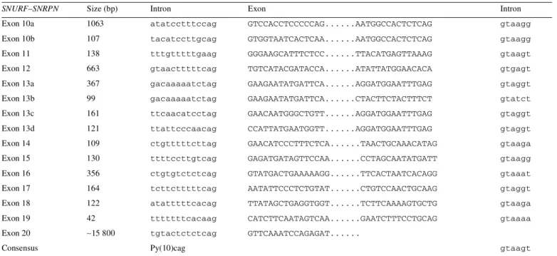
(6) 206. Human Molecular Genetics, 2001, Vol. 10, No. 3. Table 1. Clinical findings in the patients with a balanced translocation and breakpoints within the PWS region distal to SNURF-SNRPN [according to the diagnostic criteria (10)] Clinical findings. Schulze et al. 1996 (13). Conroy et al. 1997 (14). Present case. +. –. +/–. –. Major criteria Neonatal hypotonia, poor suck Feeding problems, failure to thrive. +. Obesity, beginning between ages 1 and 6 years. +. +. Characteristic facial features. Narrow bifrontal diameter. Narrow bifrontal diameter. –. Hypogonadism. Hypoplastic genitalia, delayed pubertal signs >16 years. –. Primary amenorrhoea, hypoplastic uterus. Mental retardation, developmental delay <6 years. +. +. +. Hyperphagia, food foraging, obsession with food. +. +. 46,XY,t(9;15)(q21;q12–q13). 46,XY,t(2;15)(q37.2;q11.2). 46,X,t(X;15)(q28;q12). 5 points. 7 points. 5 points. Characteristic behaviour problems. Aggressive outbursts, rigid personality, perseveration. Temper tantrums, aggressiveness. Temper tantrums, violent outbursts. Sleep disturbances or sleep apnoea. +. Deletion or abnormalities 15q11–q13. Minor criteria Decreased fetal movement, infantile lethargy, weak cry. -–. Short stature age 15 years. +. Hypopigmentation. +. – –. + –. Small hands and/or feet. –/+. –. –, short third finger. Eye abnormalities: oesotropia, myopia. Esotropia. Oesotropia. Leftsided oesotropia. Thick viscous saliva. +. Articulation defect. +. –. 3.5 points. 2 points. 1.5 points. High pain threshold. +. +. –. Decreased vomiting. +. Altered temperature sensitivity. +. Skin picking. +. –. Supportive findings. +, present; –, absent.. relatively stable degradation product derived from a larger transcript. As none of the novel 3′ exons has coding potential, their function remains unclear. We cannot exclude the formal possibility that these transcripts represent only transcriptional noise. However, some of these transcripts occur in considerable amounts. For example, the UniGene cluster Hs.22543 comprises 35 EST clones. It is possible that transcription 5′ and 3′ to the core SNURF-SNRPN gene serves to keep the paternal 15q11–q13 chromatin open. In this scenario, specific splicing of the primary transcript is functionally irrelevant and occurs at all possible sites. Some of these sites may be weak and predispose to exon skipping. Two splice donor sites are especially noteworthy in this regard (Table 2). As shown by three independent studies (7,9,10), >7 kb of DNA contiguous with exon 12 are expressed as RNA (the PAR-SN and PAR-5 cDNA fragments) and, to date, no 3′ end of these transcripts have been found. Similarly, we have observed expression of. >1 kb of DNA contiguous with exon 16 and the 3′ end of this transcript also remains elusive. The expression pattern of 15q11–q13 genes in our patient is similar to the patterns found in the other two patients who have a translocation breakpoint in the same region (Fig. 6). Whereas all tested loci centromeric to the breakpoint are expressed, HBII-85, a newly identified paternally expressed C/D box snoRNA (23), as well as IPW and PAR-1, are not expressed. The latter three genes or gene fragments are telomeric to the breakpoint and translocated to the X chromosome. The lack of expression of these sequences cannot be the result of X inactivation, because we have shown that X inactivation is random and does not spread into the chromosome 15 part of the derivative X chromosome. It is possible that the translocation breakpoints disrupt an as yet unknown gene and that this disruption may contribute to some aspects of PWS. Based on mouse data, it is likely that there is such a gene between SNURF-SNRPN and IPW. Whereas a mutation of the Snrpn open reading frame has no.
(7) Human Molecular Genetics, 2001, Vol. 10, No. 3. 207. Table 2. Exon–intron boundaries of SNURF-SNRPN exons 10–20 SNURF–SNRPN. Size (bp). Intron. Exon. Intron. Exon 10a. 1063. atatcctttccag. GTCCACCTCCCCCAG......AATGGCCACTCTCAG. gtaagg. Exon 10b. 107. tacatccttgcag. GTGGTAATCACTCAA......AATGGCCACTCTCAG. gtaagg. Exon 11. 138. tttgtttttgaag. GGGAAGCATTTCTCC......TTACATGAGTTAAAG. gtaagt. Exon 12. 663. gtaactttttcag. TGTCATACGATACCA......ATATTATGGAACACA. gtgagt. Exon 13a. 367. gacaaaaatctag. GAAGAATATGATTCA......AGGATGGAATTTGAG. gtaggt. Exon 13b. 99. gacaaaaatctag. GAAGAATATGATTCA......CTACTTCTACTTTCT. gtatct. Exon 13c. 161. ttcaacatcctag. GAACAATGGGCTGTT......AGGATGGAATTTGAG. gtaggt. Exon 13d. 121. ttattcccaacag. CCATTATGAATGGTT......AGGATGGAATTTGAG. gtaggt. Exon 14. 109. ctgtttttcttag. GAACATCCCTTTCTCA......TAACTGCAAACATAG. gtaaga. Exon 15. 130. ttttccttgtcag. GAGATGATAGTTCCAA......CCTAGCAATATGATT. gtaagg. Exon 16. 356. ctgtgtctctcag. GTATGACTGAAAAAGG......TTCACTAATCACAGG. gtaaat. Exon 17. 164. tcttctttttcag. AATATTCCCTCTGTAT......CTGTCCAACTGCAAG. gtaggt. Exon 18. 122. atatttttcacag. TTATAGCTGAGGTGGT......TCTTCAAAAGTGCTG. gtaaga. Exon 19. 42. tttttttcacaag. CATCTTCAATAGTCAA......GAATCTTTCCTGCAG. gtaaaa. Exon 20. ∼15 800. tgtactctctcag. GTTCAAATCCAGAGAT....... Consensus. Py(10)cag. gtaagt. For exons 10 and 13, different alternative splice variants have been identified, termed 10a and b and 13a–13d.. detrimental effect (24), a deletion of several hundreds of kilobases from Snurf-Snrpn to Ube3a leads to growth retardation, hypotonia and lethality in mice before weaning (25). Because deletions from IPW to Ube3a do not cause a PWS phenotype (26), the critical region must lie between SNRPN and IPW. We have shown that the breakpoint cluster disrupts an alternative 3′ exon of SNURF-SNRPN, but it is not easy to understand how the disruption of this alternative transcript might be related to the phenotype of the patients. It is more likely that the phenotype is caused by lack of the paternally expressed HBII-85 C/D box snoRNA (23), which lies between SNRPN and IPW, or an as yet unknown gene in this region. It is possible that SNRUF-SNRPN extends even more distally and is the host gene for HBII-85 or that the host gene, which remains to be identified, is indirectly affected by the translocation. On the other hand, IPW and PAR-1 appear to be under the control of an element between the breakpoint cluster region in SNURF-SNRPN intron 2 (patients 1 and 2) and SNURF-SNRPN exon 20 (patients 3–5), because both sequences are expressed in the first two patients, but not in the other three patients (Fig. 6). MATERIALS AND METHODS Clinical report The female proband E.H. was born in 1968 as the first child of an 18-year-old mother and a 21-year-old father. The parents were not related and were phenotypically normal, as were the patient’s two younger twin brothers. The family history was unremarkable. The pregnancy with the proband was normal; fetal movements were not decreased. Delivery occurred spontaneously at term; the cord was twisted around the neck,. without visible asphyxia of the newborn. Her birth weight was 2650 g, length was 51 cm and head circumference was not known. Because of an infection of the umbilicus at the third day of life, the baby stayed for 6–7 weeks at the paediatric University Clinic in Basel, Switzerland. The parents did not report any history of neonatal muscular hypotonia, poor suck, feeding problems or failure to thrive. A slight developmental delay was evident in the early periods of life: free sitting began at 9 months and first steps were at 14 months. Speech development was apparently normal, although articulation was indistinct. At the age of 4 years, the proband attended a kindergarten and at 7 years a school for mentally retarded children. She then attended a school for household management for 1 year with subsequent employment in a sanatorium. Leftsided esotropia was diagnosed during infancy and cured by conservative therapy. Obesity began at the age of 4–5 years. At that time she was a passionate eater and tended to hyperphagia and food foraging. As soon as the parents reduced her food supply, heavy altercations followed; she reacted with violent outbursts and temper tantrums. In 1988, at the age of 20 years, the patient was referred for cytogenetic investigation because of mental retardation, short stature, obesity, primary amenorrhea, hypogonadotropic hypogonadism with normal pubertal development and hypoplastic uterus. Height was 151 cm (3rd centile). Bone age was delayed (16–17 years). Weight was 72 kg (90–97 centile). She had brunette hair like her twin brothers and no sign of hypopigmentation. Small hands and feet were not present except a short middle finger bilaterally. Diabetes mellitus could not be demonstrated. Patient E.H. has several features characteristic of PWS including obesity and hyperphagia, together with food foraging, obsession with food, hypogonadism and mild mental.
(8) 208. Human Molecular Genetics, 2001, Vol. 10, No. 3. Figure 6. Overview of the expression patterns in the five translocation patients. Translocation breakpoints are indicated by an arrow. SNURF-SNRPN exons 1–20, IPW and PAR-1 are shown as black or grey shaded boxes. The multiple copies of the C/D box snoRNA HBII-85 are shown as vertical bars. –, not expressed; no entry, not done.. retardation. As minor symptoms she showed temper tantrums, violent outbursts, short stature and leftsided esotropia. Absent major symptoms included characteristic facial features and— according to the parents—neonatal hypotonia and failure to thrive (Table 1). According to the diagnostic criteria for PWS (27), five major and three minor criteria could be verified in the patient presented here. Thus, the patient reaches only 6.5 points and does not fulfil the minimal requested points of 8, necessary for diagnosis of PWS in the adulthood group.. standard nick translation procedure (18). Metaphase spreads were prepared using standard procedures and hybridization was performed for 45 h in a moist chamber at 37°C. Immunocytochemical detection of the hybridization probes was achieved as described elsewhere (18). Images were taken with a Zeiss epifluorescence microscope equipped with a thermoelectronically cooled charge coupled device camera (Photometrics CH250), which was controlled by an Apple Macintosh computer. Merging and pseudocolouring were accomplished using the ONCOR Image and Adobe Photoshop software.. Cytogenetic studies Chromosomal analysis in 1988–1990 was performed on whole-blood cultures according to standard methods. To obtain prometaphase chromosomes, the cultures were treated with methotrexate (0.05 µg/ml) after 48 h of incubation; bromodeoxyuridine (20 µg/ml) and fluorodeoxyuridine (1 µg/ ml) were added 6.5–7 h before being harvested. RBA, GTG, QFQ, CBG and DA/DAPI-banding of 250 metaphases of the proband were applied as well as RBA, QFQ and DA/DAPIbanding in both parents. FISH Standard FISH protocols were followed (28). YAC, cosmid or phage clones were labelled with biotin-14-dATP using a. DNA methylation Genomic DNA was purified from whole blood according to standard methods. Methylation at the D15S63 (PW71) locus was investigated by Southern blot analysis on HindIII + HpaIIdigested DNA using probe PW71C (29). Methylation at the SNURF-SNRPN locus was investigated by the recently described methylation-specific SNURF-SNRPN PCR test (30). RNA expression study Epstein–Barr virus (EBV)-transformed lymphoblastoid cell lines were established from the patient and her parents. Cells for RNA extraction were stored in liquid nitrogen. Total RNA was extracted by resuspending in 4 M guanidinium thiocyanate.
(9) Human Molecular Genetics, 2001, Vol. 10, No. 3. (31). RNA was treated with DNaseI to remove residual traces of genomic DNA. RT–PCR was performed using the GenAmp RNA PCR kit (Perkin Elmer). The primers were as follows: for MKRN3, RN153 and DD29 (32); for SNURF-SNRPN, exons 1–2, RN134 and RN175 (11); for SNURF-SNRPN, exons 4– 10, RN84 and RN97 (11); for KB, exons 2–3 (SNURF-SNRPN exons 11 and 12), c4.1p1 and 4p4 (7); for PAR-5 and PAR-1, see Sutcliffe et al. (10); for IPW, IPWA and IPWB, see Wevrick et al. (15) and for the novel exons NG3 + NG4 and NG45 + NG46 (Table 3). As a control for the integrity of the RNA, primers for GAPDH were used (10). RNA that was not reverse transcribed and a sample without cDNA template were included as negative controls. For northern blot analysis of the novel exons, human multiple tissue northern blots (Clontech) were hybridized according to the manufacturer’s protocol. The final wash was in 2× SSC, 0.1% SDS at 50°C for 10 min. As probes, the insert of cDNA clone AA339132 and the 280 bp RT–PCR product RT-L were used. To investigate the expression status of HBII85, northern blot analysis was performed. Total RNA was separated on an 8% denaturing polyacrylamide gel [7 M urea, 1× Tris–borate/EDTA (TBE) buffer] and transferred onto a nylon membrane (Qiabrane Nylon Plus; Qiagen) using the BioRad semi-dry blotting apparatus (Trans-blot SD). After immobilizing of RNAs using the Stratagene crosslinker, the nylon membrane was pre-hybridized for 15 min in 1 M sodium phosphate buffer (pH 6.2), 7% SDS. Oligonucleotide MBII-85 (5′-TTCCGATGAGAGTGGCGGTACAGA-3′), complementary to the mouse MBII-85 snoRNA, was end-labelled with [32P]ATP and T4 polynucleotide kinase and hybridization was carried out at 58°C in 1 M sodium phosphate buffer (pH 6.2), 7% SDS for 12 h. Final wash was carried out twice at room temperature for 15 min in 2× SSPE buffer (20 mM sodium phosphate, pH 7.4; 0.3 M NaCl; 2 mM EDTA), 0.1% SDS and subsequently for 1 min at 58°C in 0.1× SSPE, 0.5% SDS. Membranes were exposed to Kodak MS-1 film and autoradiographed for 12 h.. 209. Table 3. Primer sequences (5′→3′) Primer. Sequence. NG3. GCTGAGGTGGTACCAGTTTAAGA. NG4. TGCCCCTTTAGAAGCAAAGA. NG5. ACATCCCTTTCTCATGGAGTC. NG6. AAGCACAATGATTTGGATTGA. NG7. GACCTGTTGCAGTGCTTTCA. NG8. TGAGATCACAAGGGCTCAAA. NG11. CTGACCTGCAAATGGAATCA. NG12. TGTCATTCCAGCTCCTGAGA. NG13. GGCATTTGGTGAGATGGAAA. NG14. AGCACATGCTCTTTGTGAGA. NG16. ATTGCTGAGGGAACGAAGAA. NG17. GCGGTCAGTGACGCGATGGA. NG18. CCTGATGATTCCATTTGCAGGTCAGC. NG28. CTCTGCTTCTACCCCACAGC. NG29. GTATAAACTTGGCCCCCAGCA. NG30. AGTTAATCCTACCAACACAAGATTAAA. NG31. ACAAATTTTGCTACCTCTGTGTATTG. NG32. GTGTAAGGGAGAGCGAGACC. NG33. ATCAAGACAGGTTGGGCAAT. NG35. TTGAGCCCTTGTGATCTCATC. NG36. AAGTGTGTGATTAGGACACTGATCTT. NG37. CCATACCTACTTCCTAGGTTTGATAA. NG38. ATGGAGTTTCGCTCATCACC. NG41. ATGCCCATACCCTGAAGATG. NG42. GGCAATCACTGTGTTCCAAG. NG43. TTCCAACTGCTTTGGGTTTT. NG44. CCTTTCCCACAGCGATTAAG. NG45. TGTCTGAGACCATGCTTTGG. Sequence analysis and characterization of EST clones. NG46. CCATCCATTCCTTTCCTCAA. EST clones were provided by the Resource Centre of the Human Genome Project (RZPD), Berlin. Sequence analysis of the cDNA clones and the RT–PCR products was performed using vector- or sequence-specific primers. Sequencing reactions were performed using fluorescence-tagged dideoxynucleotides and the Taq cycle sequencing procedure (ABI). Sequences were analyzed on an ABI 377A DNA Sequencer. PCR products were purified with Microcon-100 microconcentrators (Amicon).. NG49. TCAGGTTTATTGGGAATGCAA. NG50. AGGCAGGAAGACTGCTTGAGT. NG53. GGTGATGAGCGAAACTCCAT. NG54. GGCCTTACTTCCATTTTCTCC. NG59. ATTACGGCAGGATTGCATTT. NG60. CACAATCCAACTCAACATGGTC. NG61. GGGGGAGAATGGCTGTAGAC. NG62. TCCCTTGCATTCCCAATAAA. Exon-connection PCR Exon-connection PCR was performed on Marathon ready cDNA of human fetal brain and testis (Clontech), and cDNA of a size-selected adult kidney cDNA library (L. Schomburg, Hannover) using the following primer combinations: NG3 + NG4 (RT-f); NG5 + NG6 (RT-g); NG7 + NG8 (RT-h); NG11 + NG12 (RT-i); NG13 + NG14 (RT-j); NG13 + NG16 (RT-k); NG17 + NG18 (RT-L); NG28 + NG29 (RT-m); NG30 + NG31 (RT-n); NG32 + NG33 (RT-o); NG35 + NG36 (RT-p); NG37 + NG38 (RT-q); NG53 + NG54 (RT-r); NG41 + NG42 (RT-s);. NG43 + NG44 (RT-t); NG45 + NG46 (RT-u); NG59 + NG60 (RT-v); NG61 + NG62 (RT-w); NG49 + NG50 (RT-x). Data deposition The sequences of the RT–PCR products RT-LI and RT-LII, as well as the complete sequence of the EST/cDNA clone AL120893, have been deposited in the GenBank database.
(10) 210. Human Molecular Genetics, 2001, Vol. 10, No. 3. (accession nos AF319522, AF319523 and AF319524, respectively). ACKNOWLEDGEMENTS We thank Maren Runte for helpful discussions, Frank Tschentscher for northern blot analysis and the proband and parents for helpful support. Parts of this work were supported by the Deutsche Forschungsgemeinschaft, the German Genome Programme/ Deutsche Forschungsanstalt für Luft- und Raumfahrt e.V. (4763), and the EU Commission (BMH4-CT97-2268) and by an IZKF grant (Teilprojekt F3, Münster) and the German Human Genome Project through the BMBF grant 01KW9966. REFERENCES 1. Özcelik, T., Leff, S., Robinson, W., Donlon, T., Lalande, M., Sanjines, E., Schinzel, A. and Francke, U. (1992) Small nuclear ribonucleoprotein polypeptide N (SNRPN), an expressed gene in the Prader–Willi syndrome critical region. Nature Genet., 2, 265–269. 2. Glenn, C.C., Saitoh, S., Jong, M.T.C., Filbrandt, M.M., Surti, U., Driscoll, D.J. and Nicholls, R.D. (1996) Gene structure, DNA methylation and imprinted expression of the human SNRPN gene. Am. J. Hum. Genet., 58, 335–346. 3. Nakao, M., Sutcliffe, J.S., Durtschi, B., Mutirangura, A., Ledbetter, D.H. and Beaudet, A.L. (1994) Imprinting analysis of three genes in the Prader– Willi/Angelman region: SNRPN, E6-associated protein and PAR-2 (D15S225E). Hum. Mol. Genet., 3, 309–315. 4. Reed, M. and Leff, S. (1994) Maternal imprinting of human SNRPN, a gene deleted in Prader–Willi syndrome. Nature Genet., 6, 163–167. 5. Gray T., Saitoh, S. and Nicholls, R.D. (1999) An imprinted, mammalian bicistronic transcript encodes two independent proteins. Proc. Natl Acad. Sci. USA, 96, 5616–5621. 6. Dittrich, B., Buiting, K., Korn, B., Poustka, A., Winterpacht, A., Zabel, B., Saitoh, S., Nicholls, R. and Horsthemke, B. (1996) Imprint switching on human chromosome 15 may involve alternative transcripts of the SNRPN gene. Nature Genet., 14, 163–170. 7. Buiting, K., Dittrich, B., Endele, S. and Horsthemke, B. (1996) Identification of novel exons 3′ of the human SNRPN gene. Genomics, 40, 132–137. 8. Färber, C., Dittrich, B., Buiting, K. and Horsthemke, B. (1999) The chromosome 15 imprinting centre (IC) region has undergone multiple duplication events and contains an upstream exon of SNRPN that is deleted in all Angelman syndrome patients with an IC microdeletion. Hum. Mol. Genet., 8, 337–343. 9. Ning, Y., Roschke, A., Christian, S.L., Lesser, J., Sutcliffe, J.S. and Ledbetter, D.H. (1996) Identification of a novel paternally expressed transcript adjacent to snRPN in the Prader–Willi syndrome critical region. Genome Res., 6, 742–746. 10. Sutcliffe, J.S., Nakao, M., Christian, S., Örstavik, K.H., Tommerup, N., Ledbetter, D.H. and Beaudet, A.L. (1994) Deletions of a differentially methylated CpG island at the SNRPN gene define a putative imprinting control region. Nature Genet., 8, 52–58. 11. Sun, Y., Nicholls, R.D., Butler, M.G., Saitoh, S., Hainline, B.E. and Palmer, C.G. (1996) Breakage in the SNRPN locus in a balanced 46,XY,t(15;19) Prader–Willi syndrome patient. Hum. Mol. Genet., 5, 517–524. 12. Kuslich, C.D., Kobori, J.A., Mohapatra, G., Gregorio-King, C. and Donlon, T.A. (1999) Prader–Willi syndrome is caused by disruption of the SNRPN gene. Am. J. Hum. Genet., 64, 70–76. 13. Schulze, A., Hansen, C., Skakkebaek, N.E., Brondum-Nielsen, K., Ledbetter, D.H. and Tommerup, N. (1996) Exclusion of SNRPN as a major determinant of Prader–Willi syndrome by a translocation breakpoint. Nature Genet., 12, 452–454. 14. Conroy, J.M., Grebe, T.A., Becker, L.A., Tsuchiya, K., Nicholls, R.D., Buiting, K., Horsthemke, B., Cassidy, S.B. and Schwartz, S. (1997) Balanced translocation 46,XY,t(2;15)(q37.2;q11.2) associated with atypical Prader–Willi syndrome. Am. J. Hum. Genet., 61, 388–394. 15. Wevrick, R., Kerns, J.A. and Francke, U. (1994) Identification of a novel paternally expressed gene in the Prader–Willi syndrome region. Hum. Mol. Genet., 3, 1877–1882.. 16. Kuwano, A., Mutirangura, A., Dittrich, B., Buiting, K., Horsthemke, B., Saitoh, S., Niikawa, N., Ledbetter, S.A., Greenberg, F., Chinault, A.C. et al. (1992) Molecular dissection of the Prader–Willi/Angelman syndrome region (15q11–13) by YAC cloning and FISH analysis. Hum. Mol. Genet., 1, 417–425. 17. Mutirangura, A., Jayakumar, A., Sutcliffe, J.S., Nakao, M., McKinney, M.J., Buiting, K., Horsthemke, B., Beaudet, A.L, Chinault, A.C. and Ledbetter, D.H. (1993) A complete YAC contig of the Prader–Willi/Angelman chromosome region (15q11–q13) and refined localization of the SNRPN gene. Genomics, 18, 546–552. 18. Wirth, J., Nothwang, H.G., van der Maarel, S., Menzel, C., Borck, G., Lopez-Pajares, I., Brondum-Nielsen, K., Tommerup, N., Bugge, M., Ropers, H.H. and Haaf, T. (1999) Systematic characterisation of disease associated balanced chromosome rearrangements by FISH: cytogenetically and genetically anchored YACs identify microdeletions and candidate regions for mental retardation genes. J. Med. Genet., 36, 271–278. 19. Kioschis, P., Rogner, U.C., Pick, E., Klauck, S.M., Heiss, N., Siebenhaar, R., Korn, B., Coy, J.F., Laporte, J., Liechti-Gallati, S. and Poustka, A. (1996) A 900 kb cosmid contig and 10 new transcripts within the candidate region for myotubular myopathy (MTM1). Genomics, 33, 365–373. 20. Hu, L.J., Laporte, J., Kress, W., Kioschis, P., Siebenhaar, R., Poustka, A., Fardeau, M., Metzenberg, A., Janssen, E.A., Thomas, N. et al. (1996) Deletions in Xq28 in two boys with myotubular myopathy and abnormal genital development define a new contiguous gene syndrome in a 430 kb region. Hum. Mol. Genet., 5, 139–143. 21. Kioschis, P., Wiemann, S., Heiss, N.S., Francis, F., Gotz, C., Poustka, A., Taudien, S., Platzer, M., Wiehe, T., Beckmann, G. et al. (1998) Genomic organization of a 225 kb region in Xq28 containing the gene for X-linked myotubular myopathy (MTM1) and a related gene (MTMR1). Genomics, 54, 256–266. 22. Lee, S. and Wevrick, R. (2000) Identification of novel imprinted transcripts in the Prader–Willi syndrome and Angelman syndrome deletion region: further evidence for regional imprinting control. Am. J. Hum. Genet., 66, 848–858. 23. Cavaille, J., Buiting, K., Kiefmann, M., Lalande, M., Brannan, C.I., Horsthemke, B., Bachellerie, J.-P., Brosius, J. and Hüttenhofer, A. (2000) Identification of imprinted, tissue-specific C/D box small nucleolar RNA genes in the Prader–Willi syndrome region. Proc. Natl Acad. Sci. USA, 97, 14311–14316. 24. Yang, T., Adamson, T.E., Resnick J.L., Leff, S., Wevrick, R., Francke, U., Jenkins, N.A., Copeland, N.G. and Brannan, C.I. (1998) A mouse model for Prader–Willi syndrome imprinting-centre mutations. Nature Genet., 19, 25. 25. Tsai, T.F., Jiang, Y.H., Bressler, J., Armstrong, D. and Beaudet, A.L. (1999) Paternal deletion from Snrpn to Ube3a in the mouse causes hypotonia, growth retardation and partial lethality and provides evidence for a gene contributing to Prader–Willi syndrome. Hum. Mol. Genet., 8, 1357–1364. 26. Nicholls, R.D. (1999) Incriminating gene suspects, Prader–Willi style. Nature Genet., 23, 132–134. 27. Holm, V.A., Cassidy, S.B., Butler, M.G., Hanchett, J.M., Greenswag, L.R., Whitman, B.Y. and Greenberg, F. (1993) Prader–Willi syndrome: consensus diagnostic criteria. Pediatrics, 91, 398–402. 28. Ward, D.C., Boyle, A. and Haaf, T. (1995) Fluorescence in situ hybridization techniques. Metaphase chromosomes, interphase nuclei, and extended chromatin fibers. In Verma, R.S. and Babu, A. (eds), Human Chromosomes. Principles and Techniques. McGraw-Hill, New York, NY, pp. 184–192. 29. Dittrich, B., Robinson, W., Knoblauch, H., Buiting, K., Schmidt, K., Gillessen-Kaesbach, G. and Horsthemke, B. (1992) Molecular diagnosis of the Prader–Willi and Angelman syndromes by detection of parent-oforigin specific DNA methylation in 15q11–13. Hum. Genet., 90, 313–315. 30. Zeschnigk, M., Lich, C., Buiting, K., Doerfler, W. and Horsthemke, B. (1997) A single tube PCR test for the diagnosis of Angelman and Prader– Willi syndrome based on allelic methylation differences at the SNRPN locus. Eur. J. Hum. Genet., 5, 94–98. 31. Chomczynski, P. and Sacci, N. (1987) Single-step method of RNA isolation by acid guanidinium thiocyanate-phenol-chloroform extraction. Anal. Biochem., 162, 156–159. 32. Jong, M.T.C., Carey, A.H., Caldwell, K.A., Lau, M.H., Handel, M.A., Driscoll, D.J., Stewart, C.L., Rinchik, E.M. and Nicholls, R.D. (1999) A novel imprinted gene, encoding a RING zinc-finger protein and overlapping antisense transcript in the Prader–Willi syndrome critical region. Hum. Mol. Genet., 8, 783–793..
(11)
Figure


+5
![Table 1. Clinical findings in the patients with a balanced translocation and breakpoints within the PWS region distal to SNURF-SNRPN [according to the diagnostic criteria (10)]](https://thumb-eu.123doks.com/thumbv2/123doknet/14924668.663733/6.892.60.827.185.782/clinical-findings-patients-balanced-translocation-breakpoints-according-diagnostic.webp)


Documents relatifs
To study the decay rate in case of non-exponential decay, we prefer to use the differential lifetime concept as suggested by Fuhs and Stuke [16] and is given
From the analytical results we hypothesize that stoichiometric high-Tc Nb3Ge can be formed through a heteroepitaxial process between the A-15 and the competing Nb5Ge3
From transmission electron microscopy and heat capacity measurements radiation induced da- mage in A-15 compounds was found to be inhomogeneous, consisting of samll disordered
Bouvet et al (1989) suggest that the absence of association between the sex bivalent and the autosomal trivalent could explain the normal spermatogenesis presented by
However, this translocation is not a major problem in Denmark today because the Danish BAQ farmers, who collaborate with our laboratory, systematically eradicate the
The bimodal distribution of the muscle tissue glycogen concentration (fig 1) supports the hypothesis of Le Roy et al (1994) that the RN locus is a major
Angelman Syndrome-Affected Individual with a Numerically Normal Karyotype and Isodisomic Paternal Uniparental Disomy of Chromosome 15 due to Maternal Robertsonian
We report here the draft genome sequences of 9 bacterial strains isolated from the intestinal microbiota of C57BL/6J specific-opportunistic-pathogen-free (SOPF) mice (Charles
![[PDF] Cour pour Débuter et avancer avec le logiciel GIMP | Cours informatique](data:image/gif;base64,R0lGODlhAQABAIAAAP///wAAACH5BAEAAAAALAAAAAABAAEAAAICRAEAOw==)