Adult triclabendazole-resistant
Fasciola
hepatica: morphological changes in the
tegument and gut following
in vivo
treatment with artemether in the rat model
J.F. O’Neill
1, R.C. Johnston
1, L. Halferty
1, G.P. Brennan
1, J. Keiser
2and I. Fairweather
1*
1
Parasite Proteomics and Therapeutics research Group, School
of Biological Sciences, Medical Biology Centre, The Queen’s University
of Belfast, 97 Lisburn Road, Belfast, Northern Ireland BT9 7BL:
2
Department of Medical Parasitology and Infection Biology, Swiss Tropical
Institute, PO Box, CH–4002 Basel, Switzerland
Abstract
A study has been carried out to determine the morphological changes to the adult liver fluke, Fasciola hepatica after treatment in vivo with artemether. Rats were infected with the triclabendazole-resistant Sligo isolate of F. hepatica, dosed orally with artemether at a concentration of 200 mg/kg and flukes recovered at 24, 48 and 72 h post-treatment (p.t.). Surface changes were monitored by scanning electron microscopy and fine structural changes to the tegument and gut by transmission electron microscopy. Twenty-four hours p.t., the external surface showed minor disruption, in the form of mild swelling of the tegument. The tegumental syncytium and sub-tegumental tissues appeared relatively normal. Forty-eight and seventy-two hours p.t., disruption to the tegumental system increased, with isolated patches of surface blebbing and reduced production of secretory bodies by the tegumental cells being the main changes seen. The gastrodermal cells showed a relatively normal morphology 24 h p.t. By 48 h, large numbers of autophagic vacuoles and lipid droplets were present. Autophagy increased in magnitude by 72 h p.t. and substantial disruption to the granular endoplasmic reticulum was observed. Results from this study show that flukes treated in vivo with artemether display progressive and time-dependent alterations to the tegument and gut. Disruption to the gut was consistently and substantially more severe than that to the tegument, suggesting that an oral route of uptake for this compound predominates. This is the first study providing ultrastructural information on the effect of an artemisinin compound against liver fluke.
Introduction
Fasciola hepatica is a parasite of ruminant animals prevalent in temperate regions across the world. It is a species of considerable agricultural and economic
importance, with losses resulting from infection estimated at US$2000–3000 million annually (Boray, 1994). In addition, fascioliasis has emerged as a major zoonotic disease in many countries (Mas-Coma et al., 2005), with an estimated 17 million people currently infected and up to 180 million at risk (WHO, 2007). During recent years, an upsurge in cases of fascioliasis in livestock has been noted (Mitchell, 2002), which is
*Fax: þ 44 28 90975877 E-mail: i.fairweather@qub.ac.uk
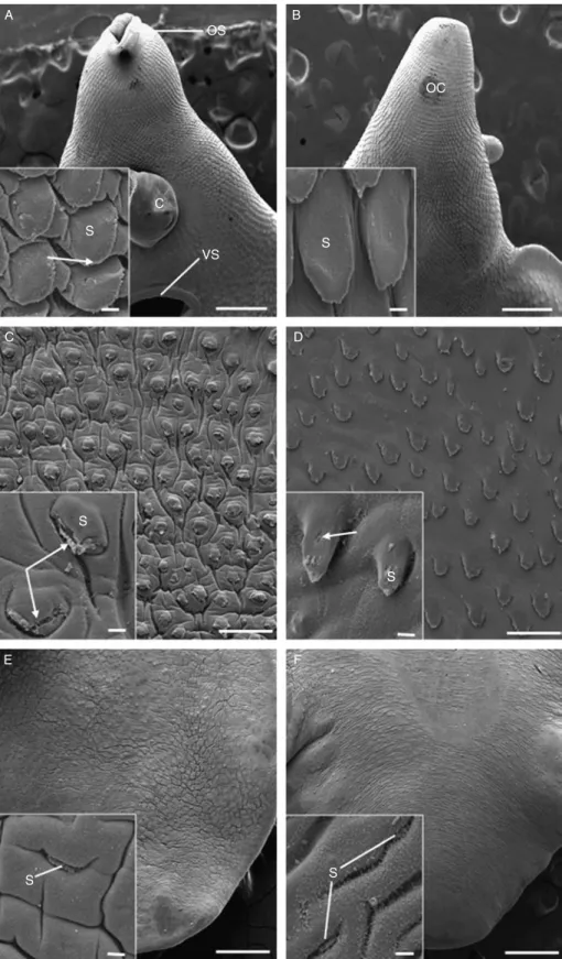
Fig. 1. A –F, Scanning electron micrographs of the tegumental surface of adult triclabendazole-resistant Fasciola hepatica, 24 h p.t. in vivo with 200 mg/kg artemether. (A) Ventral surface of the oral cone region, showing the oral sucker (OS), cirrus (C) and ventral sucker (VS). Scale bar ¼ 500 mm. Inset shows prominent spines (S) with apical spinelets (arrow). Scale bar ¼ 20 mm. (B) Dorsal surface of the oral cone
believed to be the result of climate-related changes in distribution of the intermediate snail host, Galba truncatula (Mitchell, 2002; Mas-Coma et al., 2005). Current control of fascioliasis relies heavily on the drug of choice, triclabendazole (TCBZ) (Fairweather, 2005, 2009; Keiser et al., 2005), due to its almost unique efficacy against both juvenile and mature flukes (Boray et al., 1983). In the continued absence of an effective vaccine, chemo-therapy will remain the mainstay of treatment for the foreseeable future.
Fluke populations demonstrating resistance to TCBZ have been well documented, with the first record on Australian farms in the mid-1990s (Overend & Bowen, 1995). Since then, resistant populations have been reported in Ireland, the Netherlands (references in Fairweather, 2005), Spain (A´ lvarez-Sa´nchez et al., 2006) and much of the UK, with mounting evidence suggesting that resistance is underestimated and spreading (Wolstenholme et al., 2004). The spread of resistance to the drug of choice creates a significant need for new strategies for fluke control. Such strategies should aim both at prolonging the life of currently available drugs (Wolstenholme et al., 2004) as well as addressing the need for effective novel compounds to be introduced. One family of potential candidates to be considered in the search for novel flukicides is the artemisinins, derived from the wormwood plant, Artemisia annua. The original parent compound, qinghaosu, extracted in the 1960s in China as part of a co-ordinated governmental effort to uncover novel antimalarial compounds, was later named artemisinin and, since this discovery, several semi- and fully-synthetic derivatives have been developed (Woodrow et al., 2005; Keiser & Utzinger, 2007). Today artemisinin derivatives and artemisinin combination treatments are the first-line drugs for uncomplicated falciparum malaria. In addition, artemisinin derivatives are known to be effective against juvenile stages of all major blood fluke species infecting humans (Utzinger et al., 2007). Artemisinin-type com-pounds have also been shown to be effective against different food-borne trematodes such as Opisthorchis viverrini (Keiser et al., 2006a) and Clonorchis sinensis in rodents (Keiser et al., 2006a, 2007b). A number of artemisinin compounds have been tested against F. hepatica: the semi-synthetic artemether and artesunate and the fully-synthetic trioxolane derivative, OZ78. They show high efficacy against adult and juvenile flukes in the rat model (Keiser et al., 2006b, c, 2007a). Recently, preliminary studies have reported promising fasciolicidal properties in sheep and even in humans (Hien et al., 2008; Keiser et al., 2008). Significantly, both artemether and OZ78 have shown substantial activity against the TCBZ-resistant Oberon isolate in the rat host (Keiser et al., 2007a).
The aim of the current study was to evaluate morphological changes to flukes belonging to another TCBZ-resistant isolate of F. hepatica, the Sligo isolate, following treatment in vivo in the rat model with 200 mg/kg artemether. Flukes were recovered at three time-points post-treatment (p.t.) (24, 48 and 72 h), in an attempt to determine a time-scale for drug action. Morphological observations were made using scanning and transmission electron microscopy and concentrated on the tegument and gut. These tissues provide the two main surfaces across which drugs can enter the fluke and were selected in the hope of gathering information on the route of uptake of artemether by the fluke. This paper is the first to present ultrastructural information on the effect of an artemisinin compound against the liver fluke.
Materials and methods
Fluke material
The Sligo isolate of Fasciola hepatica used for the current study was first reported in 1995 in county Sligo, Ireland (Fairweather, 2005). It has been characterized as showing resistance to the action of triclabendazole in vivo at both adult and juvenile stages (Coles et al., 2000; Coles & Stafford, 2001; McCoy et al., 2005; McConville et al., 2009a), as well as resistance to the TCBZ metabolite, triclabendazole sulphoxide in vitro (Robinson et al., 2002; Mottier et al., 2006a). Six male Sprague –Dawley rats under isoflurane anaesthesia were each orally infected, by means of a stomach tube, with 20 metacercarial cysts of the Sligo isolate of F. hepatica.
In vivo drug treatment
Five months post-infection, rats were weighed individually and dosed orally with 200 mg/kg body weight artemether. The stock solution used comprised 1.0 g of artemether dissolved in 7% (v/v) Tween 80 and 3% (v/v) ethanol. The dose used has been shown to effect a 100% adult worm burden reduction in the TCBZ-susceptible Cullompton isolate (Keiser et al., 2006b). Following dosing, flukes were recovered from the bile ducts at necropsy 24 h, 48 h and 72 h p.t. Eighteen flukes were recovered in total, each of which was washed briefly in NCTC 135 culture medium at 378C. Zero-hour untreated specimens were prepared as controls. Speci-mens were prepared for scanning electron microscopy (SEM) and transmission electron microscopy (TEM) as follows.
region (OC). Scale bar ¼ 100 mm. Inset shows the regular and uniform arrangement of the spines (S). Scale bar ¼ 20 mm. (C) Ventral surface, midbody region showing the slight swelling of the interspinal tegument. Scale bar ¼ 100 mm. Inset shows splitting of the tegument (arrows) at the tips of the spines (S). Scale bar ¼ 20 mm. (D) Dorsal surface of the midbody region. Scale bar ¼ 100 mm. Inset shows slight disruption (arrow) to the tegumental covering of the spines (S). Scale bar ¼ 20 mm. (E) Ventral surface of the tail region, showing swelling of the interspinal tegument. Scale bar ¼ 500 mm. Inset shows a spine (S) which appears to be submerged by the swollen tegument surrounding it. Scale bar ¼ 20 mm. (F) Dorsal surface of the tail region, showing swelling of the interspinal tegument. Scale bar ¼ 500 mm. Inset shows the roughened surface of the tegument. The spines (S) are barely visible, due to the swelling of the tegument
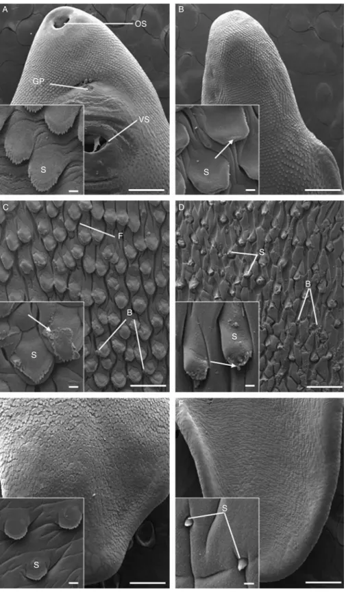
Fig. 2. A –F, Scanning electron micrographs of the tegumental surface of adult triclabendazole-resistant Fasciola hepatica, 72 h p.t. in vivo with 200 mg/kg artemether. (A) Ventral surface of the oral cone region, showing the oral sucker (OS), gonopore (GP) and ventral sucker (VS). Scale bar ¼ 500 mm. Inset shows well-defined spines (S) and smooth tegumental surface. Scale bar ¼ 20 mm. (B) Dorsal surface of the oral cone region. Scale bar ¼ 100 mm. Inset shows slight disruption to spines (S), as the spinelets are poorly defined (arrow).
Tissue preparation for SEM
Flukes were lightly flat-fixed for 1 h at room tempera-ture in 4% (w/v) glutaraldehyde in 0.1M sodium cacodylate buffer (pH 7.4). They were then free-fixed for a further 3 h using fresh fixative before being post-fixed in 1% osmium tetroxide for 1 h. Specimens were then washed several times with cacodylate buffer before being dehydrated in an ascending series of ethanol. Following dehydration, flukes were dried using hexamethyldisila-zane and mounted on aluminium stubs which were sputter-coated with gold –palladium. Samples were viewed using a FEI Quanta 200 scanning electron microscope operating at an accelerating voltage of 10 keV.
Tissue preparation for TEM
Flukes were lightly flat-fixed for 1 h at room tempera-ture in 4% (w/v) glutaraldehyde in 0.1M sodium cacodylate buffer (pH 7.4). Specimens were then dissected into oral cone, midbody and tail regions, which were further split into transverse slices approxi-mately 3 mm in thickness. These slices were free-fixed in fresh fixative for 3 h, before being washed several times in sodium cacodylate buffer. The samples were stored in buffer overnight.
Following washing, fluke samples were post-fixed for 1 h in 1% osmium tetroxide, washed in buffer and dehydrated through an ascending series of ethanol. Fluke material was then embedded using Agar 100 resin. Ultrathin sections (, 60–75 nm in thickness) were cut on a Reichert Ultracut E ultramicrotome and mounted on uncoated 200-mesh copper grids. The sections were double-stained with alcoholic uranyl acetate (9 min) and aqueous lead citrate (6 min) and viewed in a FEI CM100 transmission electron micro-scope at 100 keV.
Results
Visual observations
The flukes recovered from the untreated control rats were very active and possessed full gut contents. Twenty-four hours p.t. with artemether, recovered flukes showed normal activity and gut contents were present. No difference was observed between treated and control specimens. Forty-eight hours p.t. with artemether, flukes were highly active and gut contents were retained. Seventy-two hours p.t. although still motile, the flukes appeared sluggish in their movement. Gut contents were again retained in all specimens.
Scanning electron microscopy
The surface architecture of the control flukes appeared normal, matching the images presented by Bennett (1975) and Fairweather et al. (1999).
Twenty-four hours post-treatment
Little disruption was evident to the surface of the flukes in the oral cone region (fig. 1A and B). Tegumental spines were prominent, regularly arranged and separated by a smooth and uniform interspinal tegument (insets, fig. 1A and B). Individual spinelets were clearly visible on the spines (fig. 1A inset). The midbody region showed slight swelling of the interspinal tegument on the ventral surface (fig. 1C). At high magnification, disruption to the spinelets was observed and, in some cases, there was localized splitting of the tegument covering the spines (fig. 1C inset). In contrast, there was minimal swelling on the dorsal surface in this region, with a small number of spines showing minor disruption of their tegumental covering (fig. 1D and inset). In the tail region, the interspinal tegument was swollen both ventrally and dorsally, so that the spines appeared submerged (fig. 1E and F). On the dorsal surface, the interspinal tegument had a roughened and fibrous appearance (insets, fig. 1E and F).
Forty-eight hours post-treatment
For economy of space, micrographs for the 48 h treatment will not be included, but the results will be described. On examination, the surface morphology of the flukes was mostly normal, particularly in the oral cone region. The midbody region of these flukes showed isolated areas of tegumental swelling on the ventral surface, with slight tegumental furrowing. On the dorsal midbody surface there was mild swelling of the interspinal tegument. In the tail region, flukes exhibited substantial swelling of the interspinal tegument, causing the spines to appear submerged by the tegument surrounding them. This occurred to a similar extent on both surfaces and was greater than that observed in the 24 h-treated specimens.
Seventy-two hours post-treatment
Specimens recovered 72 h after dosing displayed no disruption to the ventral surface in the oral cone region (fig. 2A and inset). In contrast, the dorsal surface displayed a lack of spinelet definition due to minor swelling of the tegument covering the spines (fig. 2B and inset). In the midbody region, on both surfaces, swelling of the interspinal tegument was observed. On the ventral
Scale bar ¼ 20 mm. (C) Ventral surface of the midbody region, showing substantial disruption to the interspinal tegument which is furrowed (F) and bears blebs (B). Scale bar ¼ 100 mm. Inset shows the swelling of the interspinal tegument, as well as the loss of tegument (arrows) on one of the spines (S). Scale bar ¼ 20 mm. (D) Dorsal surface of the midbody region, demonstrating evidence of more widespread blebbing (B) and swelling to the tegument. S, spines. Scale bar ¼ 100 mm. Inset shows blebs (arrowed) forming at the tip of a spine (S). Scale bar ¼ 20 mm. (E) Ventral surface of the tail region. Scale bar ¼ 500 mm. Inset shows normal spine morphology (S), and only a slight swelling of the interspinal tegument is evident. Scale bar ¼ 20 mm. (F) Dorsal surface of the tail region. Scale bar ¼ 500 mm. Inset shows swelling of the interspinal tegument with the spines (S) appearing to be submerged by the surrounding tegument.
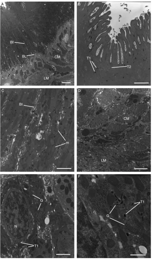
Fig. 3. A –F, Transmission electron micrographs of the tegument and sub-tegumental tissues of adult triclabendazole-resistant Fasciola hepatica, 24 h p.t. in vivo with 200 mg/kg artemether. (A) Tegument and sub-tegumental region, showing the full depth of the syncytium. The basal infolds (BI) and basal lamina (BL) retain a normal morphology. The circular (CM) and longitudinal (LM) muscle bundles also appear normal. Scale bar ¼ 3 mm. (B) A high magnification micrograph of the apical region of the tegument. Type 1 (T1) and type 2 (T2)
surface, this resulted in furrowing of the tegument and some isolated spines showed a partial loss of their tegumental covering (fig. 2C and inset). Swelling of the interspinal tegument on the dorsal midbody surface (fig. 2D) was coupled with disruption to the spine tips, as evidenced by the loss of spinelets (fig. 2D inset). On both dorsal and ventral surfaces there was a scattering of small blebs (fig. 2C and D and inset), although this was more evident on the dorsal surface (fig. 2D). The tail region of 72 h-treated specimens appeared reasonably unaffected, with only the dorsal surface displaying significant swelling of the interspinal tegument, so that the spines appeared to be partially submerged (fig. 2E and F). Transmission electron microscopy: tegument and sub-tegument
The ultrastructure of the tegumental system in the control specimens remained normal, as described by Threadgold (1963, 1967) and Fairweather et al. (1999). Twenty-four hours post-treatment
The tegumental syncytium of flukes appeared normal, with concentrations of tegumental secretory bodies at the apex of the syncytium (fig. 3A and B). Slight swelling of the mitochondria was visible and a few autophagic vacuoles were present (fig. 3C). The basal infolds appeared normal (fig. 3A and C) and the sub-tegumental musculature (both circular and longitudinal) retained a normal morphology (fig. 3D). Type 1 and type 2 tegumental cell bodies contained normal numbers of their secretory bodies (fig. 3E). At higher magnification, a number of Golgi complexes were observed (fig. 3F), many of which possessed dilated and irregularly stacked cisternae.
Forty-eight hours post-treatment
Again, no micrographs of this time period will be included. The tegumental syncytium of flukes recovered 48 h p.t. with artemether appeared normal, with the basal infolds and sub-syncytial basal lamina retaining a normal morphology. The mitochondria appeared swollen in a number of specimens, although no evidence of autophagy was observed. Tegumental secretory bodies were present in normal numbers and distribution within the syncy-tium. The sub-tegumental musculature appeared normal throughout, although the cytoplasmic connections from the tegumental cells that run between the muscle blocks appeared swollen and distended. In the tegumental cell bodies, a large proportion of the Golgi complexes contained dilated and irregularly stacked cisternae, although there were normal numbers of secretory bodies in the cells.
Seventy-two hours post-treatment
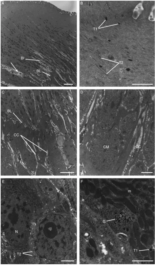
Flukes recovered from rats 72 h p.t. with artemether displayed changes to the tegumental syncytium and underlying tissues. In isolated areas of the syncytium, for example, there was substantial swelling of the mucopo-lysaccharide masses surrounding the basal infolds, although the basal infolds themselves appeared normal (fig. 4A). Both types of tegumental secretory body were present in normal numbers and distribution throughout the syncytium with a concentration at the apex (fig. 4B). Gross swelling and distension of the cytoplasmic connections was observed (fig. 4C and D), although the muscle bundles appeared normal. Tegumental cell bodies contained numerous Golgi complexes with irregularly stacked and dilated cisternae, together with swollen mitochondria (fig. 4F). The number of type 2 tegumental secretory bodies present in the cells appeared normal (fig. 4E), whereas there were few type 1 secretory bodies.
Transmission electron microscopy: gastrodermal cells The gastrodermal cells of the control specimens retained a normal morphology similar to that described by Robinson & Threadgold (1975).
Twenty-four hours post-treatment
Twenty-four hours p.t. with artemether, the gastro-dermal cells appeared relatively normal (fig. 5A). The number of secretory vesicles in the cells was normal, but they appeared somewhat elongated and irregularly shaped. The cisternae of the granular endoplasmic reticulum were swollen (fig. 5B(I) and B(II)).
Forty-eight hours post-treatment
Forty-eight hours p.t. large numbers of autophagic vacuoles and lipid droplets were observed in the cells (fig. 5C and D). The number of secretory vesicles present appeared normal, although they were small and oddly shaped. The cisternae of the granular endoplasmic reticulum were dilated (fig. 5D).
Seventy-two hours post-treatment
Specimens viewed 72 h p.t. with artemether showed a substantial increase in the level of autophagy and large lipid droplets were present throughout the tissue (fig. 5E). Dilation of the cisternae of the granular endoplasmic reticulum was visible, even at low magnification (fig. 5E). There were few secretory vesicles in the cells and they appeared small and irregularly shaped (fig. 5F).
tegumental secretory bodies are present in normal numbers. Scale bar ¼ 1 mm. (C) High-power image of the syncytium showing the presence of autophagic vacuoles (AV), as well as slight distension of the mitochondria (m). The basal infolds (BI) appear normal. Scale bar ¼ 1 mm. (D) The circular (CM) and longitudinal (LM) muscle bundles appear normal. Scale bar ¼ 2 mm. (E) Type 1 tegumental cell body, containing a Golgi complex (G) and the nucleus (N). T1, type 1 secretory body. Scale bar ¼ 2 mm. (F) High magnification view of the Golgi complex (G) within a type 1 tegumental cell body. The complex appears abnormal, with swelling and distension of its cisternae. T1,
Fig. 4. A –F, Transmission electron micrographs of the tegument and sub-tegumental tissues of adult triclabendazole-resistant Fasciola hepatica, 72 h p.t. in vivo with 200 mg/kg artemether. (A) Tegument and sub-tegumental region, showing the basal infolds (BI) and basal lamina (BL), which are normal. The mucopolysaccharide masses surrounding the basal infolds appear swollen and distended (arrow). Scale bar ¼ 3 mm. (B) High-power micrograph of the apical region of the tegument, showing the presence of normal numbers of type 1
Discussion
In the present study, changes to the tegumental system, the underlying musculature and gastrodermal cells were examined over a 72-h period, to monitor the response of the TCBZ-resistant fluke to treatment in vivo with artemether. While disruption to the tegument was relatively mild, the disruption to the gut became increasingly severe with time, with accumulations of lipid and extensive autophagic breakdown of the gastrodermal cells. The results will be discussed in terms of the time-course of drug action and the route of uptake of artemether.
The changes to the tegumental surface were relatively mild, not progressing beyond swelling and areas of scattered blebbing. There was no spine loss or tegumental sloughing, although there was some disruption and splitting of the tegumental covering of the spines. In previous SEM studies on F. hepatica involving artemisinins, instances of more severe blebbing, also spine loss and even sloughing have been described (Keiser et al., 2006b, c; Keiser & Morson, 2008a, b). Whether this is due to a different response of this particular isolate of F. hepatica to artemether is not known, although other studies on the fluke have shown that different isolates respond in different ways to the same drug (Walker et al., 2004; McKinstry et al., 2007, 2009). That is, while the changes seen are similar, the extent of those changes or degree of disruption differs, so that the sensitivity of an isolate to a particular drug varies. The present result may be related to the TCBZ-resistant status of the Sligo isolate, but this point needs to be examined further, with more detailed analysis of the efficacy of artemisinin compounds against different isolates of F. hepatica.
More dramatic changes to the fluke surface stem-ming from in vivo treatment have also been observed in studies with other flukicides: e.g. closantel (Skuce & Fairweather, 1990), clorsulon (Meaney et al., 2003, 2006), nitroxynil (McKinstry et al., 2003), TCBZ (Halferty et al., 2008) and the TCBZ derivative, compound alpha (McConville et al., 2008). The lack of serious disruption to the surface morphology of the fluke was reflected in the limited internal changes to the tegumental system observed by transmission electron microscopy. Transport of tegumental secretory bodies continued, as evidenced by the numbers of secretory bodies in the syncytium although, by 72 h p.t., production of type 1 secretory bodies had declined and there were few secretory bodies in the cells. This decline in production coincided with increasing disruption of the Golgi complexes responsible for their packaging. In contrast, production of type 2 secretory bodies remained normal and numbers present in the cells remained high.
The tegumental secretory bodies are known to be responsible for the maintenance of the apical tegumental membrane (Fairweather et al., 1999), so ultimately their decline would lead to severe disruption of the syncytium: that point was not reached at 72 h p.t.
The results provide some idea of the time-scale of drug action. For example, the reduction in motility that is evident at 72 h p.t. This will compound the autophagic disruption of the gut cells observed from 48 h p.t. onwards, as the fluke is unable to feed when it becomes inactive and essentially enters a state of starvation. The internal changes triggered by the degeneration of the gut are not manifested directly in any marked changes to the tegumental system because, although progressive changes to this tissue and to the external surface of the fluke were observed, they were not very severe. So, drug action is relatively slow, despite the relatively rapid attainment of peak plasma levels and the short half-life of artemether (Lefe`vre & Thomsen, 1999). Established pharmacokinetic data for artemether, detailing a low bioavailability when administered orally (19% in rats: Li et al., 1998), suggests that artemether undergoes extensive first-pass metabolism (Navaratnam et al., 2000). The delayed drug effect observed in this study is consistent with observations made in previous studies on F. hepatica following treatment with artemether, artesunate and OZ78. In those studies, the death and removal of flukes was seen to take 72–96 h to become apparent (Keiser et al., 2006b; Keiser & Morson, 2008a, b). The apparent contradiction between drug peaks, drug half-life and drug action has been seen in other drug studies on F. hepatica (e.g. Meaney et al., 2003; McConville et al., 2008, 2009b). For these drugs, which are relatively slow-acting, it may take some time before their action (and the morphological changes they induce) to become apparent; however, the effects are seemingly irreversible, as the drug continues to act following its elimination from the host. In contrast, the morphological changes induced by TCBZ more closely match its pharmacokinetics in the host (Halferty et al., 2008) and the data on nitroxynil fit this pattern, too (McKinstry et al., 2003, 2007).
The gut was more severely affected than the tegument at all time-points p.t. and the disruption became increas-ingly severe with longer treatment times. There was a progressive build-up of lipid droplets, disruption of the granular endoplasmic reticulum cisternae and extensive autophagy of the gastrodermal cells. The secretory activity of the cells was progressively affected over time, so that by 72 h p.t. few secretory vesicles were present. This would compromise the ability of the fluke to feed, compounding the effect of starvation referred to previously. The presence of lipid droplets is an indication of metabolic disruption in the cells, and the cells themselves appeared to be undergoing breakdown, as
(T1) and type 2 (T2) tegumental secretory bodies. Scale bar ¼ 2 mm. (C) The sub-syncytial cytoplasmic connections (cc) appear swollen and distended. BL, basal lamina. Scale bar ¼ 2 mm. (D) The circular muscle (CM) bundles appear normal, although the surrounding cytoplasmic connections (cc) are swollen. Scale bar ¼ 1 mm. (E) A type 2 tegumental cell body, containing type 2 secretory bodies (T2) and a nucleus (N). Scale bar ¼ 2 mm. (F) High magnification image of swollen mitochondria (m) and a disrupted Golgi complex (G) within a type 1 tegumental cell body. The Golgi complex possesses dilated and irregular cisternae. T1, type 1 secretory body. Scale bar ¼ 1 mm.
Fig. 5. Transmission electron micrographs of gastrodermal cells from adult triclabendazole-resistant Fasciola hepatica 24 h (A and B), 48 h (C and D) and 72 h (E and F) p.t. in vivo with 200 mg/kg artemether. (A) Gastrodermis showing an epithelial cell containing a nucleus (N). Lamellae (La) project from the cell surface into the lumen. Scale bar ¼ 3 mm. (B(I)) High magnification image of secretory vesicles (SV) in the apical region of a gastrodermal cell. Lamellae (La) project from the surface of the cell. Scale bar ¼ 1 mm. (B(II)) High magnification
evidenced by the presence of autophagic vacuoles. Comparing the results for the tegument and gut, it seems most likely that oral uptake is the predominant route of entry for artemether, rather than trans-tegu-mental diffusion. This ties in with what is known about drug entry and activation in schistosomes. For these parasites, it has been suggested that the artemisinins need to be ingested by the flukes in order for drug activation to occur (Xiao et al., 2003). Drug activation depends on the presence of an iron-containing compound, as would be derived from haemoglobin in the blood, and this leads to cleavage of the peroxide bridge in the drug molecule and the generation of free radicals. It is these free radicals which are damaging to the parasite (Xiao et al., 2003). A similar mechanism may operate in F. hepatica, because of its haematophagous feeding behaviour. In support of this idea, greater disruption to the tegument of the liver fluke was observed in vitro when haemin was incorpor-ated into the culture medium (Keiser & Morson, 2008a, b). Entry of an artemisinin compound across the tegument would not allow the interaction between the drug and an iron compound, and this would lead to a reduction in its activity. Oral uptake of drug is also the predominant mechanism for the fasciolicides, clorsulon and nitroxynil (Meaney et al., 2005a, b, c; McKinstry et al., 2007). It is not a universal phenomenon among fasciolicides, however, because entry of TCBZ has been shown to take place principally across the tegument (Mottier et al., 2006b; Toner et al., 2009). So, for artemether, its activation and efficacy are dependent on oral intake from the blood by the fluke, rather than relying on trans-tegumental diffusion as the drug is being excreted in the bile.
Some correlation can be made between the changes seen and the known action of artemisinins, via the production of free radicals. For example, treatment with artemisinins and free radicals causes collapse of the membrane potential of mitochondria, leading to their swelling and inhibition of electron transfer and oxidative phosphorylation (Dubin & Stoppani, 2000; Wakabayashi & Karbowski, 2001; Li et al., 2005). This action could explain the mitochondrial changes seen and would have a significant impact on energy production by the fluke, contributing to the state of general ill-health. Such changes are also known to be part of the process leading to cell death, or apoptosis (Dubin & Stoppani, 2000; Wakabayashi & Karbowski, 2001; Li et al., 2005). Furthermore, artemisinins and free radicals are known to cause the oxidation of membrane proteins, such as ion pumps (Rohn et al., 1996; Berman & Adams, 1997; Mason et al., 1997; Sumegi et al., 2005). One target of artemisinin action in malarial parasites is the sarco(endo)plasmic reticulum Ca2þ-ATPase, which regulates calcium levels
within the parasite (Eckstein-Ludwig et al., 2003; Jung
et al., 2005; Uhlemann et al., 2007). The swelling of the granular endoplasmic reticulum cisternae seen in the present study could be due to inhibition of pumps associated with their membranes. It would severely affect the synthetic activity of the cells.
This study has shown that artemether treatment of a TCBZ-resistant isolate of F. hepatica induces progressive changes to the morphology of the fluke and that the changes are more severe in the gastrodermal cells than the tegument. This observation suggests that the drug enters the fluke via the oral route, but this idea could be tested further in experiments involving ligatures (to block the oral route) and the incorporation of haemin in the incubation medium (to activate the drug); similar experiments have been carried out previously on fluke (Meaney et al., 2005a, b, c; Mottier et al., 2006b; Keiser & Morson, 2008a, b; Toner et al., 2009). In addition to the severe disruption of the gut, artemether causes consider-able disruption to the reproductive tissues (vitelline cells and testes) and this will be published in a separate communication.
Acknowledgements
This study was supported by a postgraduate student-ship awarded to James O’Neill by the Department of Agriculture and Rural Development, Northern Ireland (DARDNI). J. Keiser (project no. PPOOA-114941) is grateful to the Swiss National Science Foundation for financial support.
References
A´ lvarez-Sa´nchez, M.A., Mainar-Jaime, R.C., Pe´rez-Garcı´a, J. & Rojo-Va´zquez, F.A.(2006) Resistance of Fasciola hepatica to triclabendazole and albendazole in sheep in Spain. Veterinary Record 159, 424–425. Bennett, C.E. (1975) Scanning electron microscopy of
Fasciola hepatica during migration in the mouse. Journal of Parasitology 61, 892–898.
Berman, P.A. & Adams, P.A.(1997) Artemisinin enhances heme-catalysed oxidation of lipid membranes. Free Radical Biology and Medicine 22, 1283–1288.
Boray, J.C. (1994) Disease of domestic animals caused by flukes. 49 pp. Rome, Food and Agricultural Organiz-ation of the United NOrganiz-ations.
Boray, J.C., Crowfoot, P.D., Strong, M.B., Allison, J.R., Schellenbaum, M., Von Orelli, M. & Sarasin, G. (1983) Treatment of immature and mature Fasciola hepatica infections in sheep with triclabendazole. Veterinary Record 113, 315–317.
Coles, G.C. & Stafford, K.A. (2001) Activity of oxyclozanide, nitroxynil, clorsulon and albendazole
image of a gastrodermal cell containing secretory bodies (SV), as well as dilated cisternae of the granular endoplasmic reticulum (ger). Scale bar ¼ 1 mm. (C) Gastrodermis showing the presence of lipid droplets (L) throughout the epithelium. Numerous autophagic vacuoles (AV) are present. Scale bar ¼ 10 mm. (D) High-power micrograph showing the irregular shape of the secretory vesicles (SV), as well as the presence of numerous autophagic vacuoles (AV) and lipid droplets (L). Scale bar ¼ 2 mm. (E) Large lipid droplets (L) are present in the gastrodermis, as well as numerous autophagic vacuoles (AV). The cisternae of the granular endoplasmic reticulum (ger) appear dilated. Scale bar ¼ 2 mm. (F) High magnification image showing the irregular shape of secretory vesicles (SV), as well as distension of the
against adult triclabendazole-resistant Fasciola hepatica. Veterinary Record 148, 723–724.
Coles, G.C., Rhodes, A.C. & Stafford, K.A. (2000) The activity of closantel against adult triclabendazole-resistant Fasciola hepatica. Veterinary Record 146, 504. Dubin, M. & Stoppani, A.O.M.(2000) Programmed cell
death and apoptosis. The role of mitochondria. Medicina-Buenosa Aires 60, 375–386.
Eckstein-Ludwig, U., Webb, R., van Goethem, I., East, J., Lee, A., Kimura, M., O’Neill, P., Bray, P., Ward, S. & Krishna, S.(2003) Artemisinins target the SERCA of Plasmodium falciparum. Nature 424, 957–961.
Fairweather, I. (2005) Triclabendazole: new skills to unravel an old(ish) enigma. Journal of Helminthology 79, 227–234.
Fairweather, I. (2009) Triclabendazole progress report, 2005–2009: an advancement of learning? Journal of Helminthology.
Fairweather, I., Threadgold, L.T. & Hanna, R.E.B.(1999) Development of Fasciola hepatica in the mammalian host. pp. 47–111 in Dalton, J.P. (Ed.) Fasciolosis. Wallingford, Oxon, CAB International.
Halferty, L., Brennan, G.P., Hanna, R.E.B., Edgar, H.W., Meaney, M., McConville, M., Trudgett, A., Hoey, L. & Fairweather, I.(2008) Tegumental surface changes in juvenile Fasciola hepatica in response to treatment in vivo with triclabendazole. Veterinary Parasitology 155, 49–58.
Hien, T.T., Truong, N.G., Minh, N.H., Dat, H.D., Dung, N.T., Hue, N.T., Dung, T.K., Tuan, P.Q., Campbell, J.I., Farrar, J.J. & Day, J.N.(2008) A randomised controlled pilot study of artesunate versus triclabendazole for human fascioliasis in Central Vietnam. American Journal of Tropical Medicine and Hygiene 78, 388–392. Jung, J., Kim, H., Nam, K.Y. & No, K.T. (2005)
Three-dimensional structure of Plasmodium falciparum Ca-ATPase (PfATP6) and docking of artemisinin derivatives to PfATP6. Bioorganic and Medicinal Chemistry Letters 15, 2994–2997.
Keiser, J. & Utzinger, J.(2007) Artemisinins and synthetic trioxolanes in the treatment of helminth infections. Current Opinion in Infectious Diseases 20, 605–612. Keiser, J. & Morson, G. (2008a) Fasciola hepatica:
tegumental alterations in adult flukes following in vitro and in vivo administration of artesunate and artemether. Experimental Parasitology 118, 228–237. Keiser, J. & Morson, G.(2008b) Fasciola hepatica: surface
tegumental responses to in vitro and in vivo treatment with the experimental fasciolicide OZ78. Experimental Parasitology 119, 87–93.
Keiser, J., Engels, D., Bu¨scher, G. & Utzinger, J.(2005) Triclabendazole for the treatment of fascioliasis and paragonimiasis. Expert Opinion in Investigational Drugs 14, 1513–1526.
Keiser, J., Xiao, S.H., Xue, J., Chang, Z.S., Odermatt, P., Tesana, S., Tanner, M. & Utzinger, J.(2006a) Effect of artesunate and artemether against Clonorchis sinensis and Opisthorchis viverrini in rodent models. Inter-national Journal of Antimicrobial Agents 28, 370–373. Keiser, J., Xiao, S.H., Tanner, M. & Utzinger, J. (2006b)
Artesunate and artemether are effective fasciolicides in the rat model and in vitro. Journal of Antimicrobial Chemotherapy 57, 1139–1145.
Keiser, J., Utzinger, J., Tanner, M., Dong, Y. & Venner-strom, J.L. (2006c) The synthetic peroxide OZ78 is effective against Echinostoma caproni and Fasciola hepatica. Journal of Antimicrobial Chemotherapy 58, 1193–1197. Keiser, J., Utzinger, J., Vennerstrom, J.L., Dong, Y.,
Brennan, G. & Fairweather, I. (2007a) Activity of artemether and OZ78 against triclabendazole-resistant Fasciola hepatica. Transactions of the Royal Society for Tropical Medicine and Hygiene 101, 1219–1222.
Keiser, J., Xiao, S.H., Dong, Y., Utzinger, J. & Vennerstrom, J.L. (2007b) Clonorchicidal properties of the synthetic trioxolane OZ78. Journal of Parasitology 93, 1209–1214.
Keiser, J., Rinaldi, L., Veneziano, V., Mezzino, L., Tanner, M., Utzinger, J. & Cringoli, G.(2008) Efficacy and safety of artemether against a natural Fasciola hepatica infection in sheep. Parasitology Research 103, 517–522.
Lefe`vre, G. & Thomsen, M.S.(1999) Clinical pharmaco-kinetics of artemether and lumefantrine (Riametw
). Clinical Drug Investigation 18, 467–480.
Li, Q.-G., Peggins, J.O., Fleckenstein, L.L., Masonic, K., Heiffer, M.H. & Brewer, T.G.(1998) The pharmacoki-netics and bioavailability of dihydroartemisinin, arteether, artemether, artesunic acid and artelinic acid in rats. Journal of Pharmacy and Pharmacology 50, 173–182. Li, W., Mo, W., Shen, D., Sun, L., Wang, J., Lu, S., Gitschier, J.M. & Zhou, B. (2005) Yeast model uncovers dual roles of mitochondria in the action of artemisinin. PloS Genetics 1, 329–334.
Mas-Coma, S., Bargues, M.D. & Valero, M.A. (2005) Fascioliasis and other plant-borne trematode zoo-noses. International Journal for Parasitology 35, 1255–1278.
Mason, R.P., Walter, M.F. & Mason, P.E.(1997) Effect of oxidative stress on membrane structure: small-angle X-ray diffraction analysis. Free Radical Biology and Medicine 23, 419–425.
McConville, M., Brennan, G.P., Flanagan, A., Edgar, H.W.J., McCoy, M., Castillo, R., Herna´ndez-Campos, A. & Fairweather, I. (2008) Surface and internal tegumental changes in juvenile Fasciola hepatica following treatment in vivo with the experimental fasciolicide, compound alpha. Veterinary Parasitology 153, 52–64.
McConville, M., Brennan, G.P., Flanagan, A., Edgar, H.W.J., Hanna, R.E.B., McCoy, M., Gordon, A.W., Castillo, R.C., Herna´ndez-Campos, A. & Fairweather, I.(2009a) An evaluation of the efficacy of compound alpha and triclabendazole against two isolates of Fasciola hepatica. Veterinary Parasitology (in press). McConville, M., Brennan, G.P., Flanagan, A., Edgar,
H.W.J., Castillo, R., Herna´ndez-Campos, A. & Fairweather, I.(2009b) Ultrastructural changes to the tegumental system and the gastrodermal cells in mature flukes following in vivo treatment with the experimental fasciolicide, compound alpha. Parasitology (in press).
McCoy, M.A., Fairweather, I., Brennan, G.P., Kenny, J.M. & Forbes, A.B.(2005) The efficacy of nitroxynil and triclabendazole administered synchronously against juvenile triclabendazole-resistant Fasciola hepatica in sheep. Research in Veterinary Sciences 78, 33.
McKinstry, B., Fairweather, I., Brennan, G.P. & Forbes, A.B. (2003) Fasciola hepatica: tegumental surface alterations following treatment in vivo and in vitro with nitroxynil (Trodax). Parasitology Research 91, 251–263.
McKinstry, B., Brennan, G.P., Halferty, L., Forbes, A.B. & Fairweather, I.(2007) Ultrastructural changes induced in the tegument and gut of Fasciola hepatica following in vivo and in vitro drug treatment with nitroxynil (Trodax). Parasitology Research 101, 929–941.
McKinstry, B., Halferty, L., Brennan, G.P. & Fairweather, I. (2009) Morphological response of triclabendazole-susceptible and triclabendazole-resistant isolates of Fasciola hepatica to treatment in vitro with nitroxynil (Trodax). Parasitology Research 104, 645–655.
Meaney, M., Fairweather, I., Brennan, G.P., McDowell, L.S.L. & Forbes, A.B.(2003) Fasciola hepatica: effects of the fasciolicide clorsulon in vitro and in vivo on the tegumental surface, and a comparison of the effects on young- and old-mature flukes. Parasitology Research 91, 238–250.
Meaney, M., Haughey, S., Brennan, G.P. & Fairweather, I.(2005a) A scanning electron microscope study on the route of entry of clorsulon into the liver fluke, Fasciola hepatica. Parasitology Research 95, 117–128.
Meaney, M., Haughey, S., Brennan, G.P. & Fairweather, I.(2005b) A scanning electron microscope study on the route of entry of clorsulon into the liver fluke, Fasciola hepatica. Parasitology Research 96, 189–198.
Meaney, M., Haughey, S., Brennan, G.P. & Fairweather, I.(2005c) Ultrastructural observations on oral inges-tion and trans-tegumental uptake of clorsulon by the liver fluke, Fasciola hepatica. Parasitology Research 95, 201–212.
Meaney, M., Allister, J., McKinstry, B., McLaughlin, K., Brennan, G.P., Forbes, A.B. & Fairweather, I.(2006) Fasciola hepatica: morphological effects of a combi-nation of triclabendazole and clorsulon against mature fluke. Parasitology Research 99, 609–621.
Mitchell, G.B.B.(2002) Update on fasciolosis in cattle and sheep. In Practice 24, 378–385.
Mottier, L., Alvarez, L., Fairweather, I. & Lanusse, C. (2006a) Resistance-induced changes in triclabendazole transport in Fasciola hepatica: ivermectin reversal effect. Journal of Parasitology 92, 1355–1360.
Mottier, L., Alvarez, L., Ceballos, L. & Lanusse, C. (2006b) Drug transport mechanisms in helminth parasites: passive diffusion of benzimidazole anthel-mintics. Experimental Parasitology 113, 49–57.
Navaratnam, V., Mansor, S.M., Sit, N.W., Grace, J., Li, Q. & Olliaro, P.(2000) Pharmacokinetics of artemisinin-type compounds. Clinical Pharmacokinetics 39, 255–266. Overend, D.J. & Bowen, F.L.(1995) Resistance of Fasciola hepatica to triclabendazole. Australian Veterinary Journal 72, 275–276.
Robinson, G. & Threadgold, L.T. (1975) Electron microscope studies of Fasciola hepatica. XII. The fine structure of the gastrodermis. Experimental Parasitology 37, 20–36.
Robinson, M.W., Trudgett, A., Hoey, E.M. & Fair-weather, I. (2002) Triclabendazole-resistant Fasciola hepatica: b-tubulin and response to in vitro treatment with triclabendazole. Parasitology 124, 325–338.
Rohn, T.R., Hinds, T.R. & Vincenzi, F.F. (1996) Inhibition of Ca2þ-pump ATPase and the Naþ/Kþ -pump ATPase by iron-generated free radicals. Protection by 6,7-dimethyl-2,4-di-1-pyrrolidinyl-7H-pyrrol0[2,3-d]pyrimidine sulfate (U-89843D), a potent, novel, antioxidant/free radical scavenger. Biochemical Pharmacology 51, 471–476.
Skuce, P.J. & Fairweather, I. (1990) The effect of the hydrogen ionophore closantel upon the pharmacology and ultrastructure of the adult liver fluke Fasciola hepatica. Parasitology Research 76, 241–250.
Sumegi, B., Kovacs, K., Veres, B., Radnai, B., Varbiro, G., Bognar, Z., Toth, A. & Gallyas, F. (2005) Oxidative stress and the endoplasmic reticulum. pp. 121–130 in Benedetti, A., Banhegyi, G. & Burchell, A. (Eds) Endoplasmic reticulum: a metabolic compartment (NATO Science Series, Vol. 363). Amsterdam, IOS Press. Threadgold, L.T. (1963) The tegument and associated
structures of Fasciola hepatica. Quarterly Journal of the Microscopical Society 104, 505 –512.
Threadgold, L.T. (1967) Electron-microscope studies of Fasciola hepatica. III. Further observations on the tegument and associated structures. Parasitology 57, 633–637.
Toner, E., McConvery, F., Brennan, G.P., Meaney, M. & Fairweather, I.(2009) A scanning electron microscope study on the route of entry of triclabendazole into the liver fluke, Fasciola hepatica. Parasitology (in press). Uhlemann, A.-C., Wittlin, S., Matile, H., Bustamante,
L.Y. & Krishna, S.(2007) Mechanism of antimalarial action of the synthetic trioxolane RBX1160 (OZ277). Antimicrobial Agents and Chemotherapy 51, 667–672. Utzinger, J., Xiao, S.-H., Tanner, M. & Keiser, J.(2007)
Artemisinins for schistosomiasis and beyond. Current Opinion in Investigational Drugs 8, 105–116.
Wakabayashi, T. & Karbowski, M. (2001) Structural changes of mitochondria related to apoptosis. Biological Signals and Receptors 10, 26–56.
Walker, S., McKinstry, B., Boray, J.C., Brennan, G.P., Trudgett, A., Hoey, E.M., Fletcher, H. & Fairweather, I. (2004) Response of two isolates of Fasciola hepatica to treatment with triclabendazole in vivo and in vitro. Parasitology Research 94, 427–438.
WHO (2007) Report of the WHO Informal Meeting on use of triclabendazole in fascioliasis control, held at WHO Headquarters, Geneva, Switzerland, October 2006. 33 pp.
Wolstenholme, A., Fairweather, I., Prichard, R., Von Samson-Himmelstjerna, G. & Sangster, N. (2004) Drug resistance in veterinary helminths. Trends in Parasitology 20, 469–476.
Woodrow, C., Haynes, R. & Krishna, S. (2005) Artemisinins. Postgraduate Medical Journal 81, 71–78. Xiao, S.-H., Wu, Y.-L., Tanner, M., Wu, W.-M., Utzinger, J.,
Mei, J.-Y., Scorneaux, B., Chollet, J. & Zhai, Z.(2003) Schistosoma japonicum: in vitro effects of artemether combined with haemin depend on cultivation media and appraisal of artemether products appearing in the media. Parasitology Research 89, 459–466.
(Accepted 16 March 2009) First Published Online 16 April 2009