The location of the primary entry tear in acute type B aortic
dissection affects early outcome
†
Gabriel Weiss
a,‡, Ilse Wolner
b,‡, Sandra Folkmann
a, Gottfried Sodeck
c, Jürg Schmidli
d, Martin Grabenwöger
a,
Thierry Carrel
dand Martin Czerny
d,*
a
Department of Cardiovascular Surgery, Hospital Hietzing, Vienna, Austria b Department of Radiology, Hospital Hietzing, Vienna, Austria
c
Clinic for Cardiology, University Hospital Berne, Berne, Switzerland
d Clinic for Cardiovascular Surgery, University Hospital Berne, Berne, Switzerland
* Corresponding author. Freiburgstrasse 18, 3010 Berne, Switzerland. Tel: +41-31-6322376; fax: +41-31-6322919; e-mail: [email protected] (M. Czerny). Received 2 September 2011; received in revised form 13 November 2011; accepted 23 November 2011
Abstract
OBJECTIVES: The goal of the retrospective study was to relate the site of the primary entry tear in acute type B aortic dissections to the presence or development of complications.
METHODS: A consecutive series of 52 patients referred with acute type B aortic dissection was analysed with regard to the location of the primary entry tear (convexity or concavity of the distal aortic arch) using the referral CT scans at the time of diagnosis. These find-ings were related to the clinical outcome as well as to the need for intervention.
RESULTS: Twenty-five patients (48%) had the primary entry tear located at the convexity of the distal aortic arch, whereas 27 patients
(52%) had the primary entry tear located at the concavity of the distal aortic arch. Twenty per cent of patients with the primary entry tear at the convexity presented with or developed complications, whereas 89% had or developed complications with the primary entry tear at the concavity (P < 0.001). Furthermore, in patients with complicated type B aortic dissection, the distance of the primary entry tear to the left subclavian artery was significantly shorter as in uncomplicated patients (8 vs. 21 mm; P = 0.002). In Cox regression analysis, a primary entry tear at the concavity of the distal aortic arch was identified as an independent predictor of the presence or the development of complicated type B aortic dissection.
CONCLUSIONS: A primary entry tear at the concavity of the aortic arch as well as a short distance between the primary entry tear and the left subclavian artery are frequently associated with the presence or the development of complicated acute type B aortic dissection. Thesefindings shall help us to further differentiate acute type B aortic dissections in addition to the common categorization in
compli-cated and uncomplicompli-cated. Thesefindings may therefore also have an impact on primary treatment.
Keywords:Type B aortic dissection• Primary entry tear • TEVAR
INTRODUCTION
In the current era, patients with uncomplicated type B aortic dis-sections are usually treated medically. However, despite signi fi-cant advances in diagnosis and treatment, the management of acute type B aortic dissection remains controversial and decision-making is based on subjective clinical judgment [1–3]. Both surgical and interventional therapies are considered treat-ment options of choice in cases of complicated type B dissec-tion. Owing to the rapid advances of thoracic endovascular aortic repair (TEVAR), this treatment option has been established in the armamentarium of cardio-thoracic surgeons. Lately, evi-dence has shown its superiority as the treatment of choice for
complicated type B aortic dissection [4, 5]. To date in the
overwhelming majority of cases, the location of the primary entry tear is not yet taken into consideration during the course of determining the treatment strategy. However, previous work has shown some evidence that an entry tear located at the con-cavity may be associated with a complicated follow-up of an acute type B aortic dissection, including the catastrophic expan-sion into the ascending aorta [6,7].
The goal of the retrospective study was to relate the site of the primary entry tear in acute type B aortic dissections to the presence or development of complications.
METHODS
A database search has been performed retrospectively to iden-tify all patients with acute type B aortic dissection between 2005 and 2011 in two institutions (University Hospital Berne and
†Presented at the 25th Annual Meeting of the European Association for Cardio-Thoracic Surgery, Lisbon, Portugal, 1–5 October 2011.
‡G.W. and I.W. contributed equally to this work.
© The Author 2012. Published by Oxford University Press on behalf of the European Association for Cardio-Thoracic Surgery. All rights reserved.
A
O
RTIC
SURGER
Hospital Hietzing Vienna). Fifty-two patients referred with acute type B aortic dissection who had a multi-slice CT angiography of the entire aorta at the time of diagnosis were identified and
ana-lysed. Patient demographics are shown in Table1. No patients
with known or suspected connective tissue disease have been included in the study.
De
finition of acute type B aortic dissection
Acute type B aortic dissection was defined as an aortic dissection with a primary entry tear at the level of the left subclavian artery or distal to the left subclavian artery [8].
Definition of complicated and uncomplicated
acute type B aortic dissection
Complicated acute type B aortic dissection was defined as
rupture, contained rupture, including progressive pleural effu-sions, retrograde extent into the arch or into the ascending aorta, furthermore visceral, renal or limb malperfusion, and/or persistent pain [9].
Morphometric de
finitions
Patients were stratified according to the location of the primary entry tear. On axial CT scans, primary entry tears at the upper circumference (180°) of the distal aortic arch were defined as to be at the convexity, and the remaining as to be at the concavity
(Figs1 and 2). Morphometric measurements also included the
distance from the primary entry tear to the left subclavian artery. Furthermore, diameters of the lumina—both true and false—of the thoracic and abdominal aorta were measured.
Statistical methods
Continuous data are presented as the median and the inter-quartile range (range from the 25th to the 75th percentile) or as the mean and the standard deviation (SD), as appropriate. Discrete data are given as counts and percentages. Comparisons
of continuous data were performed by the Mann–Whitney U-test
and groups of categorical data were compared by Fisher’s exact
tests. Univariate Cox regression analysis was primarily performed to assess the prognostic impact of the dissection’s site upon the future development of complications, followed by a multivariate Cox regression to adjust for a pre-existing retrograde component of the dissection and the distance of dissection entry to the left Table 1: Descriptive characteristics of the entire cohort
n, overall = 52 Demographics
Age, mean (SD) 61 (12)
Female,n (%) 5 (10)
Dissection entry site
Concave,n (%) 27 (52)
Convex,n (%) 25 (48)
Aortic morphology assessed by MSCT
Distance to LSA (mm), median (IQR) 15 (0–25) Diameter descending aorta (mm), median (IQR) 37 (33–40) True lumen diameter (mm), median (IQR) 17 (13–31) False lumen diameter (mm), median (IQR) 20 (15–25) Diameter abdominal aorta (mm), median (IQR) 30 (27–33) True lumen diameter (mm), median (IQR) 12 (7–17) False lumen diameter (mm), median (IQR) 18 (13–24) Indication for treatment
Complicated,n (%) 29 (56)
Overall need for intervention,n (%) 36 (69) Time to intervention in days, mean (range) 4 (0–14) Outcome
Retrograde type A dissection,n (%) 2 (4)
Mortality,n (%) 2 (4)
SD: standard deviation; IQR: inter-quartile range.
Unless otherwise indicated, data are numbers (percentages). Primary complicated indicated by occurrence of retrograde dissection, malperfusion or impending rupture.
Figure 1:Scheme of different sites of the primary entry tear of acute type B aortic dissections. (A) Primary entry tear at the outer circumference of the distal aortic arch defined as ‘convex’. The retrograde component of the dis-section is stopped by left subclavian artery. (B) Primary entry tear at the inner circumference of the distal aortic arch defined as ‘concave’, allowing progres-sion of the retrograde component of the dissection into the aortic arch and the ascending aortas.
Figure 2:Scheme of stratification regarding concavity and convexity of the distal aortic arch.
subclavian artery in quartiles. Results of the regression model are
given as the hazard ratio (HR) and the 95% confidence interval
(95% CI). Regression diagnostics and overall model-fit were
per-formed according to standard procedures. A two-sidedP-value
<0.05 was considered statistically significant. Calculations were performed with SPSS for Mac OsX (version 19.0).
RESULTS
Demographics
The mean age of all patients was 61 ± 12 years, five patients
(10%) of the entire study population were females.
Clinical outcome
Out of the entire study population of 52 patients, 36 patients (69%) underwent intervention for complicated acute type B aortic dissection. Patients with a primary entry tear at the con-cavity presented with a significantly higher incidence of compli-cated acute type B aortic dissection during the entire hospital stay (convexity, 20% vs. concavity, 89%;P = 0.001). There was also
a significant difference between the groups concerning the
inci-dence of primary complicated type B aortic dissection at
admis-sion (convexity, 12% vs. concavity, 48%; P = 0.005) (Table 2).
Correspondingly, overall need for intervention was significantly
higher in the concave group compared with the convex group
(convexity, 38% vs. concavity, 100%P = 0.001). Mortality was 4%
due to multiorgan failure as a sequelae of the underlying aortic disease. In two other cases, a retrograde type A dissection occurred immediately after TEVAR. These two patients were con-verted into open surgery and treated with the frozen elephant trunk technique with successful clinical outcome. The remaining patients had an uneventful aortic-related clinical course.
Morphological outcome and measurements
Twenty-five patients (group A) had the primary entry tear at the
convexity, 27 patients (group B) had the primary entry tear at the concavity (Figs3and4), of the distal aortic arch. There was a
significant difference between the two groups with regard to the
distance to the left subclavian artery (group A, 21 ± 15 mm vs.
group B, 8 ± 12 mm; P = 0.002). Retrograde progression of the
dissection was more common in the concave group than in the
convex group (group A, 36% vs. group B, 52%;P = 0.25). Finally,
false lumen diameter at the mid-thoracic level was larger in patients with the primary entry tear located at the concavity vs. the convexity group (group A, 16 ± 5 mm vs. group B, 20 ± 8 mm,P = 0.08).
Need for intervention
There was a significant difference between the groups with
regard to the incidence of primary complicated acute aortic type B aortic dissection (convexity, 12% vs. concavity, 48%; P = 0.005). Furthermore, patients with a primary entry tear at the concavity were more likely to develop complications within the first 72 h than patients with a primary entry tear at the convexity
(convexity, 7 days vs. concavity, 3 days;P = 0.06) (Fig.5). For the origin of the dissection at the convexity, freedom from develop-ment of complicated dissection was 80% at 7 days, 71% at 14 days and 63% at 21 and 28 days. For the origin of the dissection at the concavity, freedom from development of complicated dis-section was 30% at 7 days and 13% at 14 days.
Cox regression analysis
Cox regression analysis revealed a primary entry tear at the con-cavity of the distal aortic arch as an independent predictor for the presence or the development of complicated acute type B aortic dissection (Table3).
DISCUSSION
The median age of our cohort corresponded well to recently published series [10,11]. Interestingly, the number of females in this cohort was lower than in other cohorts of acute type B aortic dissection [12]. The incidence of complicated acute type B aortic dissection was higher than in the recent literature [13].
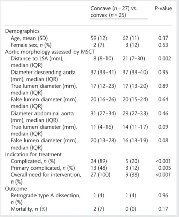
Table 2: Distribution of patients by different chronic
health conditions and in-hospital risk assessment stratified to the origin of the primary dissection entry
Concave (n = 27) vs. convex (n = 25) P-value Demographics Age, mean (SD) 59 (12) 62 (11) 0.37 Female sex,n (%) 2 (7) 3 (12) 0.53 Aortic morphology assessed by MSCT
Distance to LSA (mm), median (IQR)
8 (8–10) 21 (7–30) 0.002 Diameter descending aorta
(mm), median (IQR)
37 (33–41) 37 (33–40) 0.95 True lumen diameter (mm),
median (IQR)
17 (12–23) 17 (13–20) 0.89 False lumen diameter (mm),
median (IQR)
20 (16–26) 20 (15–24) 0.64 Diameter abdominal aorta
(mm), median (IQR)
31 (27–34) 29 (27–33) 0.46 True lumen diameter (mm),
median (IQR)
11 (4–16) 14 (11–17) 0.09 False lumen diameter (mm),
median (IQR)
20 (13–28) 16 (13–19) 0.08 Indication for treatment
Complicated,n (%) 24 (89) 5 (20) <0.001 Primary complicated,n (%) 13 (48) 3 (12) 0.005 Overall need for intervention,
n (%)
27 (100) 9 (38) <0.001 Outcome
Retrograde type A dissection, n (%)
1 (4) 1 (4) 0.96 Mortality,n (%) 2 (7) 0 (0) 0.17 SD: standard deviation; IQR: inter-quartile range.
Unless otherwise indicated, data are numbers (percentages). Primary complicated defined by occurrence of retrograde dissection, malperfusion or impending rupture.
A
O
RTIC
SURGER
Thesefindings warrant further attention. The incidence of com-plicated acute type B aortic dissection is reported to be 10–18% [14]. In our series, the incidence of primary complicated acute type B aortic dissections is 56%. We feel that the incidence of complicated type B aortic dissection might be underreported due to several reasons. Patients are classified as uncomplicated due to the absence of clinical signs of complications, such as malperfusion, haemodynamic compromise and pain and may therefore be discharged from hospital early. In our series, the time to development of complications was different with respect to the location of the primary entry tear. Interestingly, complica-tions, such as malperfusion and recurring pain, occurred within
thefirst 14 days with diminishing frequency with regard to the
time of the initial event, but not afterwards [15–17]. Indications for intervention afterwards were due to diameter increase but not due to classical indications [18]. Therefore, hospitalization of patients with acute type B aortic dissection can be recom-mended for 14 days as the probability for development of
classical complications afterwards is low. The incidence of retro-grade type A aortic dissection was comparable with recently published series [19]. However, retrograde type A aortic dissec-tion is not limited to the peri-intervendissec-tional time period and may also happen years after treatment [20]. As such, the need of stent-grafts especially for the treatment of acute and chronic aortic dissections is not sufficiently met by the industry as com-pliance mismatch between the very elastic aortic wall and the rigid graft and the resulting shear stress might well be causative for this phenomenon. Furthermore, the morphologically normal ascending aorta as well as the aortic wall may well be inheritably diseased and thereby prone to dissection.
There was a significant difference with regard to the
occur-rence of complications with regard to the location of the primary entry tear. Patients with a primary entry tear at the
Figure 4:Fluoroscopy of a 58-year old male patient with a type B aortic dis-section and the primary entry tear on the convexity of the distal aortic arch.
Figure 5:Kaplan–Meier calculation of time to intervention for both groups showing significant differences in the time to intervention related to the site of the primary entry tear.
Figure 3:A 75-year old male patient with a malperfusion syndrome of the left kidney. (A) Parasagital CT angiographic image showing an acute type B aortic dissection with a primary entry tear at the concavity of the distal aortic arch. (B) Axial image of the same patient at the level of the renal arteries showing a massive compression of the true lumen with subsequent malperfu-sion of the left kidney.
concavity were by far more likely to already present with com-plicated acute type B aortic dissection or to develop complica-tions within thefirst 72 h after the initial event. Interestingly, the distance from the primary entry tear to the left subclavian artery was significantly shorter in patients with complicated acute type
B aortic dissection. This finding warrants discussion. We
hy-pothesize that haemodynamics and consequently pressure gradi-ents in both lumina are affected by this morphological detail, leading to a higher pressurization of the false lumen due to the steeper angulation of the aortic arch at the level of the left sub-clavian artery. However, this theory has to be verified by further experimental and clinical work.
The need for intervention in this study was high and evidently corresponded to the high percentage of complicated type B aortic dissection. The primary strategy of intervention is closure of the primary entry tear in order to decompress the true lumen, restore distal perfusion by expanding the true lumen and finally, to stabilize segments with impending rupture. In the ma-jority of cases, the domino effect of readaption of the dissection membrane to the adventitia can be accomplished with the
closure of the primary entry tear by TEVAR. However, in specific
situations, the domino effect might be impaired by additional rupture of the membrane below the stent-graft or by a large communications between the lumina, thereby prohibiting effect-ive decompression of the true. In these situations, surgical mem-brane fenestration might be an option in addition to TEVAR or even as a sole therapeutically approach.
Cox regression analysis revealed the location of the primary entry tear at the concavity of the distal aortic arch as the sole in-dependent predictor of the presence or the development of
acute type B aortic dissection. This finding represents the core
statement of this study as morphology regarding the location of the primary entry tear was not taken into consideration to date. As such, this detail might represent an adjunct in the armentar-ium of the treating physician to stratify patients being at risk for complications and thereby anticipating the need for intervention.
Limitations of the study
It is clear that distribution patterns of the exact location of the primary entry tear do not adhere to geometrical algorithms and that a certain variability is present. We are fully aware of
this problem and have therefore stratified used a clear
defin-ition of what we regarded as concavity and what we regarded as convexity namely on axial CT scans, primary entry tears at the upper circumference (180°) of the distal aortic arch were
defined as to be at the convexity, and the remaining as to be
at the concavity. Interestingly, there few cases where we had doubt how to stratify these patients due to the fact that the entry was located exactly on the lateral wall. So it seems that to be that the natural occurrence of primary entry tears is asso-ciated with a clear correlation to one or the other circumfer-ence. Regarding the distance to the left subclavian artery, some self-criticism has to be applied as the angulation of the aortic arch has to be taken into account to a certain extent. In order to keep comparability, we adhered to a strict perpendicular
measurement. We feel that simplification is a major component
of a better understanding of diseases whose underlying patho-mechanisms have not been fully understood. As such, this
sim-plified delimitation of a highly complex problem is vital. We do
hope that these investigations will stimulate others to go in the
same direction and consequently future work will confirm our
findings or will put them into question.
Clinical relevance of the study
The clinical relevance of this study lies in increasing the aware-ness for surrogates of complications which have not been addressed to date. It is clear that thesefindings have no additive value in a patient being referred with already sustained compli-cations, but it helps to understand why it has happened. Furthermore, as has been pointed out in this study, there are patients with a primary entry tear at the concavity who are asymptomatic at the time of referral and who will develop com-plications within thefirst week after the acute event. Therefore, the additive value of this report lies in the newly created aware-ness of this subgroup at risk. As a consequence, we suggest liberal endovascular treatment in patients with this specific risk constellation of a primary entry tear at the concavity and a short distance to the left subclavian artery in order to prevent a very high probability of complications, especially malperfusion or retrograde type A aortic dissection.
Summarizing, a primary entry tear at the concavity of the aortic arch as well as a short distance between the primary entry tear and the left subclavian artery are frequently associated with the presence or the development of complicated acute type B aortic dissection. Based on thesefindings, the localization of the
primary entry should be implemented in risk stratification of
acute type B aortic dissection in addition to the common
cat-egorization in complicated and uncomplicated. These findings
may therefore also have an impact on primary treatment. Conflict of interest: none declared
REFERENCES
[1] Eggebrecht H, Nienaber CA, Neuhäuser M, Baumgart D, Kische S, Schmermund Aet al. Endovascular stent-graft placement in aortic dis-section: a meta-analysis. Eur Heart J 2006;27:489–98.
[2] Grabenwoger M, Fleck T, Czerny M, Hutschala D, Ehrlich M, Schoder M et al. Endovascular stent graft placement in patients with acute thoracic aortic syndromes. Eur J Cardiothorac Surg 2003;23:788–93.
[3] Ehrlich MP, Dumfarth J, Schoder M, Gottardi R, Holfeld J, Juraszek A et al. Midterm results after endovascular treatment of acute, complicated type B aortic dissection. Ann Thorac Surg 2010;90:1444–8.
[4] Dake MD, Kato N, Mitchell RS, Semba CP, Rasavi MK, Shimono Tet al. Endovascular stent graft placement for the treatment of acute aortic dis-section. N Engl J Med 1999;340:1546–52.
Table 3: Predictors of complicated dissection—Cox
regression analysis
HR 95% CI P-value Risk factor
Concave 7.26 3.03–17.36 <0.001 Retrograde component 1.44 0.73–2.85 0.29 Distance to subclavian artery (in
quartiles) 0.96 0.70–1.32 0.81 A O RTIC SURGER Y
[5] Khoynezhad A, Donayre CE, Omari BO, Kopchok GE, Walot I, White RA. Mid-term results of endovascular treatment of complicated acute type B aortic dissection. J Thorac Cardiovasc Surg 2009;138:625–31.
[6] Grimm M, Loewe C, Gottardi R, Funovics M, Zimpfer D, Rodler Set al. Novel insights into the mechanisms and treatment of intramural hema-toma affecting the entire thoracic aorta. Ann Thorac Surg 2008;86:453–6. [7] Zimpfer D, Czerny M, Kettenbach J, Schoder M, Wolner E, Lammer J et al. Treatment of an acute type B dissection with an intramural haema-toma in the ascending aorta by percutaneous endovascular stent-graft placement. Thorac Cardiovasc Surg 2006;54:500–1.
[8] DeBakey ME, Beall AC Jr, Cooley DA, Crawford ES, Morris GC Jr, Garrett HEet al. Dissecting aneurysms of the aorta. Surg Clin North Am 1966; 46:1045–55.
[9] Erbel R, Manterola FA, Boileau C, Dirsch O, Eber O, Haverich A et al. Diagnosis and management of aortic dissection. Eur Heart J 2001;22: 1642–81.
[10] Estrera AL, Miller CC, Goodrick J, Porat E, Achouh PE, Dhareshwar Jet al. Update on outcomes of acute type B aortic dissection. Ann Thorac Surg 2007;83:842–5.
[11] Uchidaa N, Katayamaa A, Tamuraa K, Sutoha M, Kuraokaa M, Muraoa N et al. Long-term results of the frozen elephant trunk technique for extended aortic arch disease. Eur J Cardiothorac Surg 2010;37:1338–45. [12] Czerny M, Hoebartner M, Sodeck G, Funovics M, Juraszek A, Dziodzio T
et al. The influence of gender on mortality in patients after thoracic endovascular aortic repair. Eur J Cardiothorac Surg 2011;40:e1–5. [13] Suzuki T, Mehta RH, Ince H, Nagai R, Sakomura Y, Weber Fet al. Clinical
profiles and outcomes of acute type B aortic dissection in the current era: lessons from the International Registry of Aortic Dissection (IRAD). Circulation 2003;108(Suppl. 1):II312–7.
[14] Trimarchi S, Eagle AK, Nienaber CA, Pyeritz RE, Jonker FHW, Suzuki T et al. on behalf of the International Registry of Acute Aortic Dissection (IRAD) Investigators. Importance of refractory pain and hypertension in acute type B aortic dissection. Circulation 2010;122:1283–9.
[15] Garbade J, Jenniches M, Borger MA, Barten MJ, Scheinert D, Gutberlet Met al. Outcome of patients suffering from acute type B aortic dissec-tion: a retrospective single-centre analysis of 135 consecutive patients. Eur J Cardiothorac Surg 2010;38:285–92.
[16] Genoni M, Paul M, Tavakoli R, Künzli A, Lachat M, Graves K et al. Predictors of complications in acute type B aortic dissection. Eur J Cardiothorac Surg 2002;22:59–63.
[17] Herold U, Piotrowski J, Baumgart D, Eggebrecht H, Erbel R, Jakob H. Endoluminal stent graft repair for acute and chronic type B aortic dissec-tion and atherosclerotic aneurysm of the thoracic aorta: an interdiscip-linary task. Eur J Cardiothorac Surg 2002;22:891–7.
[18] Narayan P, Wong A, Davies I, Angelini GD, Bryan AJ, Wilde Pet al. Thoracic endovascular repair versus open surgical repair—which is the more cost-effective intervention for descending thoracic aortic patholo-gies? Eur J Cardiothorac Surg 2011;40:869–74.
[19] Kpodonua J, Preventzab O, Ramaiaha VG, Shenniba H, Wheatley GH, JRodriquez-Lopeza Jet al. Retrograde type A dissection after endovascu-lar stenting of the descending thoracic aorta. Is the risk real? Eur J Cardiothorac Surg 2008;33:1014–8.
[20] Dumfarth J, Michel M, Schmidli J, Sodeck G, Ehrlich M, Grimm Met al. Mechanisms of failure and outcome of secondary surgical interventions after thoracic endovascular aortic repair (TEVAR). Ann Thorac Surg 2011; 91:1141–6.
APPENDIX. CONFERENCE DISCUSSION
Dr M. Grimm (Vienna, Austria): The paper studies the hypothesis that an ini-tially uncomplicated type B dissection starting at the concavity of the arch is significantly at risk of developing early complications within the subsequent few days, and I think this is, in the clinical setting, an extremely important period. In contrast (and this has to be discussed I think), if the tear is located in the convexity, the risk of early complications is low.
If you follow this theory, this has potentially saving but also life-threatening implications for the patient. Therefore I have three important questions I want to ask you. Firstly, I personallyfind it extremely difficult in a certain number of cases to identify the exact location of the primary entry tear. And given the scenario that maybe a less experienced radiologist and less experienced surgeons are on call late at night, do you think that an exact identification of the entry tear at the primary CT scan is really that reprodu-cible for all of us?
Secondly, does identification of the location of the primary entry tear in your opinion have different consequences? So maybe at the convexity, inter-vention by stent graft placement? Whereas primary entry tear at the concav-ity, due to the risk of retrograde type A dissection, has the consequence of surgical intervention?
And thirdly, could you share your opinion with us? Which types of stents do you use in such cases, because early intervention in acute type B dissec-tion carries a high risk of retrograde type A dissecdissec-tion.
Dr Weiss: As you know, I am not an expert on imaging, which is one reason why it was a retrospective study. We asked a radiologist, expert in aortic pathologies, to help with the evaluation of the CT scans and, together with Martin Czerny, who has a great experience in aortic dissections, we were able to analyse the CT scans and precisely define the location of the primary entry tear.
I believe that not every radiologist is able to do this analysis precisely, and an expert in aortic pathologies is definitely needed. I think in a late night setting, it may be difficult to find an experienced radiologist who is able to exactly locate the primary entry. Maybe you will be able to get expert help to assist with this aspect in the morning. Coming to the second question, I believe that it is too early to say that if the primary entry is located at the concavity of the distal aortic arch you should implant a stent graft or not. I think more work needs to be done in this area, and we will need more patients to clarify this question. I would not recommend doing an intervention just because the primary entry tear is located at the concavity of the distal aortic arch.
And the third question, we mainly used a stent graft with uncovered bare springs, but I think that we should reconsider this treatment approach in the future due to our high incidence of retrograde type A dissections after stent graft placement for type B dissection.
Dr E. Mostafa (Cairo, Egypt): There is actually a classification, which is most probably unknown, but I guess Jean Bachet knows it; a classification or first classification for type A dissection taking into consideration the multiple entries. My question to your group is, do you consider the multiple entries, not one single primary entry, as risk factors for complications?
Dr Weiss: I am sorry. I did not really understand the question.
Dr Mostafa: Multiple entries, because actually we have been speaking about one single primary entry which I guess is not so common. The most common cause for complications is actually the missed, undiagnosed multiple entries, and probably Jean Bachet can support this, according to their classi fi-cation orfirst classification.
Dr Weiss: Well, we detected first the primary entry tear and did not really concentrate on the multiple re-entries in this study. We tried tofind the exact site of the primary entry, and if these patients needed an intervention for complicated.
Dr Mostafa: I mean primary entries, entries, not one.
Dr M. Grabenwöger (Vienna, Austria): One is always the first. You have mul-tiple entries, but one is thefirst one.
Dr Weiss: Yes. But I guess the most common cause of complications is the missed multiple entries after that one which is very evident to us.
Dr M. Czerny (Berne, Switzerland): I think it is clear that the number of communications between both lumina has an impact, but our impression is that it does not have an impact on the development of complications. It has an impact on the development of late aneurysmal formation, but we were not able tofind an association. And the other thing is that it is extremely diffi-cult to visualize, let’s say to count the number of communications in the proximal thoracic aorta on the CT scan. I think for this we need functional imaging, and this should be the next step.
Dr A. Rajaii-Khorasani (Mashhad, Iran): Did you analyse the length of the aorta, meaning aortas as in the CT scan that you show, which is a torturous, elongated aorta?
Dr Weiss: I know, but we did not include the length of the aorta in our analysis. We just measured the lumen diameters and not the length, or if the aorta was kinked or not.
Dr Rajaii-Khorasani: I believe there is no data on this subject in the literature.
Dr Weiss: I agree with you.
Dr Rajaii-Khorasani: But based on my own experience, which is, I believe, an anecdote, the length of aorta is also a pathological sign. Everybody talks about the diameter. Even in the ascending aorta, if you are doing a simple coronary bypass and you see an elongated aorta, these are the ones that may dissect with partial clamping. These are the ones that may bleed from the site of cannulation. This is my experience. So I think if you look at the length of this aorta, it may have some significance.
Dr Czerny: I think this was a very important comment. I am sorry, we have to cut the discussion because we are running late. Maybe you could be so kind to discuss it later on during the break.