Genomic Structure of the Amphioxus Calcium Vector Protein
1 Hajime J. Yuasa,* Jos A. Cox,f and Takashi Takagi'^'Biological Institute, Graduate School of Science, Tohoku University, Sendai 980-8578; and * Department of Biochemistry, University of Geneva, CH-1211 Geneva 4, Switzerland
Received May 12, 1999; accepted July 2, 1999
Calcium vector protein (CaVP) is an EF-hand Ca2+-binding protein, which is unique to the protochordate, amphioxus. CaVP is supposed to act as a Ca2+ signal transductor, but its exact function remains unknown. Not only its function but also its exact evolutionary relationship to other Ca2+-binding proteins is unclear. To investigate the evolution of CaVP, we have determined the complete sequences of CaVP cDNAs from two amphioxus species,
Branchiostoma lanceolatum and B. floridae, whose open reading frame cDNA and amino
acid sequences show 96.5 and 98.2% identity, respectively. We have also elucidated the structure of the gene of B. Aoridae Ca VP, which is made up of seven exons and six introns. The positions of four of the six introns (introns 1, 2, 3, and 5) are identical with those of calmodulin, troponin C, and the Spec protein of the sea urchin. These latter proteins belong to the so-called troponin C superfamily (TnC superfamily) and thus CaVP likely also belongs to this family. Intron 6 is positioned in the 3' noncoding region and is unique to
CaVP, so it may represent a landmark of the CaVP lineage only. The position of intron 4 is
not conserved in the genes of the TnC superfamily or CaVP, and seems to result from either intron sliding or the addition of an intron (randomly inserted into or close to domain HI) to the genes of the TnC superfamily during their evolution.
Key words: amphioxus, calcium vector protein, cDNA sequences, EF-hand.
The EF-hand is a Ca2+-binding motif normally composed of 29 amino acids with a helix-loop-helix structure (1). Present-day proteins possess two to eight EF-hands and are classified as belonging to one protein family, called collec-tively the EF-hand family. The family comprises 39 subfamilies (2), and four of them, calmodulin (CaM), troponin C (TnC), myosin essential light chain (ELC), and myosin regulatory light chain (RLC), called the CTER subfamily, are evolutionarily closely related according to the results of analyses at both the amino acid (3) and cDNA levels (4). In fact, such a close relationship has been suggested from the earliest reports (5, 6), and the CTER together with parvalbumin (7) and Spec (8-10), a Ca2+ -binding protein isolated from the sea urchin, have often been called the "TnC superfamily". Except for parvalbu-min, they possess four EF-hand domains per molecule and may have evolved from a common four-domain ancestor. It is thought that parvalbumin lost the first domain during its evolution.
Calcium vector protein (CaVP) is a unique protein found 1 This work was supported in part by a grant from the Japan Society for the Promotion of Science. The determined nucleotide sequences have been submitted to the DDBJ under the accession numbers, AB001688 (Branchiostoma lanceolatum CaVP cDNA), AB001689 (B. floridae CaVP cDNA), and AB023071-AB023073 (B. floridae CaVP genomic clones).
'To whom correspondence should be addressed. Tel: + 81-22-217-6677, Fax: +81-22-263-9206, E-mail: [email protected] Abbreviations: CaM, calmodulin; CaVP, calcium vector protein; ELC, myosin essential light chain; nt, nucleotide; RLC, myosin regulatory light chain; TnC, troponin C.
© 1999 by The Japanese Biochemical Society.
in various tissues of amphioxus and is abundantly express-ed in muscle (11). It forms a stable complex with a protein of 26 kDa, CaVP-target protein (CaVPT, 12-14), and it is supposed to act as a Ca2+ signal transductor, although its
exact function remains unknown. The evolution of CaVP is also uncertain. Similar to the members of the TnC super-family, CaVP possesses four EF-hand domains, but binds only two Ca2+ per molecule. EF-hands I and II do not bind
Ca2+, and in the native protein, a unique disulfide bridge is
formed between these two domains. Kobayashi et al. (11) stressed that CaVP shows about 35% sequence identity with CaM and TnC. At the same time, however, they also suggested the possibility that CaVP is a chimera with a C-terminal half homologous to that of CaM and TnC, and an N-terminal half resembling those of parvalbumins and sarcoplasmic Ca2+ binding proteins. On the other hand,
CaVP clusters with ELC on the dendrogram constructed by the maximum parsimony method (15), and on analysis of the individual EF-hand domains, those of the CaVP were found to cluster near those of Si00, the small EF-hand protein which possess two domains per molecule (3).
In this study, we determined the complete cDNA se-quences of CaVPs from two species, Branchiostoma
lan-ceolatum and B. floridae, and elucidated the exon/intron
organization of the latter and discussed the evolutionary relationship of CaVP with the TnC superfamily.
MATERIALS AND METHODS
Cloning of B. lanceolatum CaVP cDNA—Total RNA of
adult B. lanceolatum was prepared according to the acid guanidium thiocyanate method (16), and mRNA was
purified with Oligotex dT-30 Super (Japan Roche). The single-stranded cDNA was synthesized using a First-Strand cDNA Synthesis Kit (Pharmacia). The cDNA of B.
lanceolatum CaVP was amplified by PCR using Ex Taq
DNA polymerase (Takara). The redundant oligomer used for PCR was 5'-GA(AG)GA(AG)AA(AG)GA(CT)GA(AG)-TG(CT) ATGAA-3', which was designed based on the amino acid sequence, EEKDECMK (residues 12-19), of B.
lan-ceolatum CaVP {11). The oligo-dT adaptor,
5'-GGGATCC-GAATTCT17-3', was used as another primer. The 5'
upstream of cDNA was determined as follows. The .EcoRI-ended double-stranded cDNA was synthesized from mRNA using a TimeSaver cDNA Synthesis Kit (Pharmacia), and the EcoBI Cassette (Takara) was ligated at each end of cDNA. The 5' upstream region was amplified by PCR using the cassette-specific primer Cl, 5'-GTACATATTGTCGT-TAGAACGCG-3', and 5'-CGAAGACAACACAGACTTT-ATTAC-3' (complementary to the sequence from nt 1057 to 1080).
Cloning of B. floridae CaVP cDNA—The A.ZAP II cDNA
library of embryonic B. floridae was kindly provided by Dr. L.Z. Holland (Scripps Institute of Oceanography, Univer-sity of California, San Diego). The B. floridae CaVP cDNA was also amplified by PCR using the cDNA library as a template. The primers used for 3'-half amplification were 5'-TGAGGGCGTTCAAGGTCTTC-3' (corresponding to the sequence from nt 275 to 294 of B. lanceolatum CaVP cDNA) and the T7 primer, 5'-TAATACGACTCACTATA-GGG-3', and those for 5'-half amplification were5'-GAAT-CCTGTGGACTAACTTA-3' (complementary to the se-quence from nt 487 to 506 of B. lanceolatum CaVP cDNA) and the T3 primer, 5'-ATTAACCCTCACTAAAGGGA-3'.
Screening of a Genomic Library—The A. FIX II genomic
library of B. floridae was also provided by Dr. L.Z. Holland, and was screened using the open reading frame of B.
lanceolatum CaVP cDNA as a probe, because the identity
between these two species, CaVP cDNAs is very high (96.5%, see below). The probe was labeled with DIG-DNA Labeling Mixture (Boehringer Mannheim) by PCR using the following forward primer, 5'-CTCTACCAGTCTTTC-AGTCC-3' (corresponding to the sequence from nt — 29 to
- 1 0 ) and the reverse primer, 5'-GAATCCTGTGGACTA-ACTTA-3' (complementary to the sequence from nt 487 to 506). Hybridization and washing were carried out according to the manufacturer's instructions (Boehringer Mann-heim), and positive clones were detected with a DIG Luminescent Detection Kit (Boehringer Mannheim).
DNA Sequencing—All PCR-amplified products and
iso-lated genomic clones were subcloned to the pCR II plasmid vector (TA-cloning kit, Invitrogen) or pUCl8 for sequenc-ing. The nucleotide sequences were determined by the dideoxy chain termination method with a Dye Primer Cycle Sequencing Kit (Applied Biosystems) using an automated DNA sequencer (Applied Biosystems 373A).
RESULTS AND DISCUSSION
CaVP cDNAs from Two Amphioxus Species—The cDNA
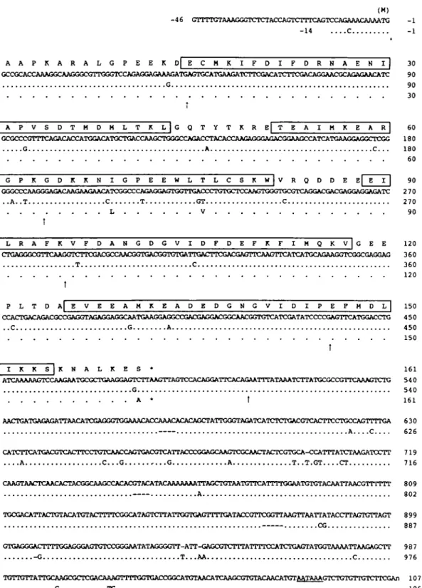
of B. lanceolatum CaVP was amplified by PCR and the complete sequence of 1,124 nucleotides was established from two overlapping fragments (Fig. 1). The open reading frame is composed of 489 nucleotides and encodes a protein of 162 amino acid residues including the initial Met.
Compared to the previously reported amino acid sequence of B. lanceolatum CaVP {11), the sequence of positions 152 to 155 is Lys-Lys-Ser-Lys instead of Lys-Ser-Lys-Lys. This region is Lys-rich and therefore the precise sequence is difficult to deduce from the results of peptide analysis. This and the fact that the same sequence is also observed in
B. floridae CaVP (Fig. 1) led us to conclude that the
previous report probably contained a peptide sequence error.
The cDNA of B. floridae CaVP is composed of 1,080 nucleotides and the open reading frame is 489 nucleotides long, encoding a protein of 162 amino acid residues includ-ing the initial Met, Le. it is of the same length as in B.
lanceolatum (Fig. 1). Within the coding regions, there are
17 nucleotide substitutions (96.5% identical) reflecting three amino acid differences (98.1% identical) between the CaVPs of the two species. Two of the three substitutions occur in domain II and the other at the C-terminus. Thus, there is likely no difference in Ca2+ binding between the two
CaVPs, i.e. both bind two CaJ+ per molecule at EF-hand
domains HI and TV.
The identity between the two species' CaVPs is as high as those of CaMs, TnCs and skeletal actins, which are known to be highly conserved proteins (see Table I). However, mitochondrial proteins, such as cytochrome b and NADH dehydrogenase, of the two amphioxus species show equally high identity, as compared to the lower level of identity between the human and mouse mitochondrial proteins. Therefore, the high similarity of the two species' CaVPs may not reflect the conservatism of CaVP, but may rather be due to relatively recent divergence of B. lanceolatum and
B. floridae.
Genomic Structure of the CaVP Gene—Using B. lan-ceolatum CaVP cDNA as a probe, we initially isolated three
positive clones (clones 1,2, and 3) from the genomic library of B. floridae. As clones 1 and 2 showed similar digestion patterns with several restriction enzymes, only clones 1 and 3 were used for further investigation. The nucleotide sequence of clone 1 was determined: it comprises 6,729 bp and spans the 5'-half of the CaVP gene up to Ile-90 (Fig. 2A). In this region, the CaVP gene consists of four exons divided by three introns (introns 1 to 3), and the nucleotide sequences of the exons exactly match those of the cDNA. All the introns start with GT and end with AG, and, according to the nomenclature3 of Kretsinger and
Naka-yama {17), the introns positions are —14/0, 1.01/1, and 2.13/1, respectively.
Clone 3 comprises 4,613 bp and spans the 3'-half of the
CaVP gene starting from Ile-105 (Fig. 2A). In this region,
the gene consists of three exons divided by two introns (introns 5 and 6); the position of the former intron is 4.21/ 1, and the last intron is inserted within the 3'-untranslated
' The positions of introns are indicated according to the nomenclature of Kretsinger and Nakayama (17). The first number corresponds to the EF-hand domain numbered sequentially from N to C. The second number (following the period) represents the residue number of the intron insertion in the EF-hand domain, which as a rule consists of 29 residues. The last number (following the slash) is the phase: 0 means that the intron lies between triplet codons, 1 between first and second nucleotides of the codon, and 2 between the second and third ones. For instance, 4.21/1 means insertion in domain IV, 21st residue and phase 1. —14/0 means phase 0, 14 residues before the beginning of domain I. 3 + 01/1 means phase 1, 1 residue beyond domain IE, within the region between domain HI and IV.
(H) - 4 6 GTTTTGTAAAGGGTCTCTACCAGTCTTTCAGTCCAGAAACAAAATG - 1 - 1 4 C - 1 A A P K A R A L G P E E K D E C M K I F D I F D R N A E y 30 GCCGCACCAAAGGCAAGGGCCTTGGGTCCAGAGGAGAAAGATGAGTGCATGAAGA 90 G 90 30 A P V S D T M D M L T K L | G Q T Y T K R E
L
T E A I M K E A_*]
| G P K G D K K N I G P E E W L T L C S K W | V R Q D D E 6 0 GCG<XCGTTTCAGACACCATGGACATGCTGACCAAGCTGGGCCAGAan'ACACCAAGAGGGAGAC^ 1 8 0 G A C. . . 1 8 0 60 270 90 | L R A F K V F D A H G D G V I D F D E F K F I H Q K V | G E E 1 2 0 CTCAGGGCGTTCAAGGTCTTCGACGCA\ACGGTGACGGTGTGATTGACTTCGAA^ 3 6 0 T C 360 120 t P L T D A E V E E A M K E A D E D G N G V I E P H D L 1 5 0 4 5 0 450 150 I K KJ J *
N A L K 1 6 1 ATCAAAAAGTCCAAGAATXXXXrTGAAGGAGTCTTAAGTTAGTCCACAGGATTCACAGAATT^ 540 G 540 A * t 1 6 1 AACTGATGAGAGATTAACATCGAGGGTGGAAACACCAAACACACAGCTATTGGGTAGATCATC 630 A C 626 CATCTTCATGACGTCACTTCCTGTCAACCAGTGACGTCArTACCCGGAGCAA 7 1 9 A C . . . G G A T . . T . G T CT 7 1 6 CAAGTAACTCAACACTACGGCAAGCCACACGTACATACAAAAAAAlTAGCItn^ATGTTC^TTTTGGA^ 8 0 9 A 8 0 2 TGCGACATTACTGTACATGTACITITCGGCATAGTCTTACTGGTGAGTTTTGATACCGTTCGGTrAAGTC 899 CG 887 GTGAGGGACTTTTGGAGGGAGTGTCCGGGAATATA<KGGTT-ATT-GAGCGTC^ 9 8 7 -G T...AA C 976 IWl'ltirmiTGCAAGCGCTCGACAAAGTITIGGTGACCGGCATGTAAaVTCAAGCGTGTACAACAT^ 1 0 7 8 C T G . - 1 0 6 6Fig. 1. Comparison of the cDNA and deduced amlno acid and shown by bars (-). The typical polyadenylation signals (AATAAA) sequences of CaVPs of two species of amphioxus. Upper lanes: B. are underlined, and the four EF-hand Ca>+ binding domains are boxed.
lanceolatum CaVP cDNA and deduced amino acid sequence; lower The initiator methionine (residue —1, parenthesized) likely is
re-lanes: B. floridae CaVP cDNA and deduced amino acid sequence, moved after translation. The upward arrows ( T ) indicate the positions Identical nucleotides and amino acids to those in B. lanceolatum CaVP of introns in the B. floridae CaVP genes,
are indicated by dots (.), and gaps are inserted for mniimpl similarity
region. Both introns are delineated in conformity to the GT/AG rule. Compared to the cDNA sequence, the geno-mic nucleotide sequence exhibits some differences: in the
open reading frame the GAT codon encoding Asp-143 changes to GAC in the genome and the 3'-noncoding region contains one substitution and three nucleotide insertions
(data not shown).
Since there is no overlapping region between clones 1 and 3, the region encoding from Leu-91 to Val-104 can not be adequately described. Therefore we performed further screening to obtain another clone containing the 91/104 region and succeeded in isolating clone 4. Clone 4 contains all the CaVP gene except exon 1 (see Fig. 2A). Of this clone only the exons and regions flunking the splice junctions were sequenced basically. Within the Leu-91/Val-104
TABLE I. The identity (%) of several molecules between two
species of amphioxus, and between human and mouse.
CaVP CaM TnC Skeletal actin Cytochrome b NADH 5° B. floridae vs. cDNA (ORF) 96.5 97.1 95.4 95.5 97.7 98.2 B. lanceolatum Amino acid 98.2 100.0 97.6 97.1 99.2 99.3 Human vs. Mouse cDNA (ORF) 91.8 90.3 90.8 73.5 65.2 Amino acid 100.0 98.8 100.0 76.7 63.0 'NADH 5, NADH dehydrogenase subunit 5. The accession numbers (EMBL/DDBJ/GenBank) of the sequences used are; Y09863 (B.
floridae CaM), Y09880 (B. lanceolatum CaM), M19311 (human CaM H), M27844 (mouse CaM 77), D88977 (B. floridae TnC),
D88976 (B. lanceolatum TnC), X07898 (human fast skeletal TnC), M57590 (mouse fast skeletal TnC), D87407 (B. floridae skeletal actin), Y13662 (B. lanceolatum skeletal actin), M20543 (human skeletal alpha-actin), M12347 (mouse skeletal alpha-actin), AF-035173 (B. floridae cytochrome 6), AF035172 (B. floridae NADH dehydrogenase subunit 5), Y16474 (B. lanceolatum mitochondria! DNA), D38112 (human mitochondria! DNA), and J01420 (mouse mitochondria! DNA).
region an intron (intron 4) was observed at position 3.08/0, whereas the positions of the other introns are identical to those in clones 1 and 3. The fourth intron was identified in clone 4 only, but in the B. lanceolatum CaVP gene an intron located at exactly the same position, as determined on sequencing of a partially amplified genomic PCR fragment (data not shown). To summarize, the conjectural organiza-tion of the B. floridae CaVP gene is shown in Fig. 2B, and it may consist of 7 exons and 6 introns (ca. 11.5 kbp).
When the intronic sequences of clones 1, 3, and 4 are compared, some substitutions and insertions/deletions are observed: the identity between each pair of splice junction flunking regions is ca. 90% on average. Moreover, in the exons four substitutions were observed, all of them being silent substitutions (data not shown). Thus, clone 4 may be derived from a gene that is different from in the case of clones 1 and 3. It is possible that clones 1 and 3 also come from distinct genes. There are two possible explanations for the observed discrepancies: (i) amphioxus CaVP may be multi-copy gene, or (ii) the different clones may be derived from distinct animal specimens, since the genomic library was not constructed from a single amphioxus specimen (18). Although the three clones are derived from different genes, it is supposed that the positions of the introns are identical in all genes.
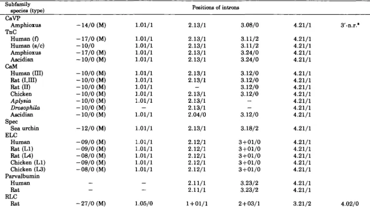
Comparison of Intron Positions among TnC Superfamily Genes—The distribution of introns in the genes of CaVP
and members of the TnC superfamily is shown in Table II. In all genes the first intron is basically inserted just after the initiation codon, ATG (shown as M in Table II). Furthermore, the positions of the second, third, and fifth
Clone 1 E EX I I I In I (3482bp) S E I I Clone 3 Ilc-90 lie-105 H H S X I I
-,—o^
1 In 5 " In 6 (l89Obp) (498bp) (l220bp) (H60bp)-CD-I kbp I 1
B
Clone 4 In 2 In 4 (358bp) • • ; . ; + • • • • ; . ' .(ca. I 9kbp) (ca. 0_5kbp) (ca. 1 2kbp) (ca. 1.2kbp)
Conjectured CaVP gene
In I (-1*0) ATG (initiation codon)
In 2 (1.01 1)
-0
In 4 (3.08 0)
Fig. 2. Structural organization of the B. floridae CaVP
geno-mic clones. A: Restriction and exon/intron maps of clones 1 and 3.
EcoHl (E), Pstl (P), Sail (S), and Xbal (X) restriction sites are
indicated. Exons are shown by boxes, the 5'- and 3'-flanking regions and introns (In) are shown by bars, and the length of each intron is indicated. As for clone 4, the regions whose sequences were not
In3 " (2.13 1)
In 5 (4.21 I)
In6
-CD-T.AA (termination codon)
determined are shown by dotted lines, and the length of each intron was estimated by PCR. B: The conjectural organization of the gene of B. floridae CaVP. It may be composed of 7 exons and 6 introns, and the position of each intron is indicated in parenthesis. The positions of the initiation codon (ATG) and the termination codon (TAA) are also indicated.
TABLE II. The intron positions of TnC snperfamily genes (based largely on Refs. 17 and 20).
Subfamily
species (type) Positions of introns
CaVP Amphioxus TnC Human (f) Human (s/c) Amphioxus Ascidian CaM Human (HI) Rat (I,m) Rat (II) Chicken Aplysia DrosophUa Ascidian Spec Sea urchin ELC Human Rat (LI) Rat(L4) Chicken (LI) Chicken (L3) Parvalbumin Human Rat RLC Rat -14/0 (M) -17/0 (M) -10/0 -17/0 (M) -10/0 (M) -10/0 (M) -10/0 (M) -10/0 (M) -10/0 (M) -10/0 (M) -10/0 (M) -10/0 (M) -12/0 (M) -09/0 (M) -09/0 (M) -08/0 (M) -09/0 (M) -08/0 (M) — — -27/0 (M) 1.01/1 1.01/1 1.01/1 1.01/1 1.01/1 1.01/1 1.01/1 1.01/1 1.01/1 1.01/1 — 1.01/1 1.01/1 1.01/1 1.01/1 1.01/1 1.01/1 1.01/1 — — 1.05/0 2.13/1 2.13/1 2.13/1 2.13/1 2.13/1 2.13/1 2.13/1 — 2.13/1 2.13/1 2.13/1 2.04/0 2.13/1 2.12/1 2.12/1 2.12/1 2.12/1 2.12/1 2.11/1 2.11/1 1 + 01/1 3.08/0 3.11/2 3.11/2 3.24/0 3.24/0 3.12/0 3.12/0 3.12/0 3.12/0 -— 3.12/0 3.18/2 3 + 01/0 3+01/0 3+01/0 3 + 01/0 3+01/0 3.23/2 3.23/2 2 + 03/1 4.21/1 4.21/1 4.21/1 4.21/1 4.21/1 4.21/1 4.21/1 4.21/1 4.21/1 4.21/1 4.21/1 4.21/1 4.21/1 4.21/1 4.21/1 4.21/1 4.21/1 4.21/1 4.21/1 4.21/1 3.21/2 3'-n.r." 4.02/0 (M) shows the intron is inserted just after the initiation codon, ATG. (—) shows the absence of intron. *The last intron of Ca VP is inserted within the 3'-noncoding region.
introns (1.01/1,2.13/1, and 4.21/1, respectively) of CaVP are identical with those of TnCs, CaMs, and Spec. Thus, the gene structure of amphioxus CaVP is typical of the TnC superfamily and CaVP is clearly a member of the TnC superfamily. Within the family CaVP seems more closely related to the following three members: TnCs, CaMs, and Spec (Table II). The sixth intron, which is observed in the 3'-noncoding region, is unique to CaVP, and may be a characteristic feature, Le. a landmark of the CaVP lineage. Except for in RLC,4 the exon/intron organizations are
well conserved among all members of the TnC superfamily (including CaVP). Basically they all possess five introns with the following characteristics: intron 1 is inserted just after the initiation codon (phase 0), and in a few exceptions, it is also inserted before domain I and the phase 0 mode is conserved; intron 2 is inserted at position 1.01/1 in all members without any exception; intron 3 is inserted at position 2.13/1 in CaM, TnC, Spec, and CaVP, whereas in the other members the insertion point is slightly different, but phase 1 is conserved (with the notable exception of intron 3 in the CaMs of the ascidian, Halocynthia roretzi with 2.04/0, Ref. 19); finally intron 5 is inserted at position 4.21/1 in all members without any exception.
Only the positions of intron 4 is not conserved at all in the TnC superfamily. Two explanations can account for this diversity: either intron 4 acquired different positions in the TnC superfamily genes through intron sliding, or the members of the TnC superfamily may have gained a fourth
4 The positions of introns in the RLC are different from those of the
other members of the TnC superfamily, but for conciseness thin particular diversity is not analyzed in this study.
intron independently during their evolution. According to the first hypothesis the fourth introns of the TnC super-family are derived from a common ancestral intron, but its sequence changed so rapidly that the homology quickly disappeared. Indeed these introns are different in length and sequence; there is no positive proof that they are homologous. In general the evidence of intron sliding itself is weak (20), and besides, except in one instance, between protochordate/vertebrate TnCs (21), no sliding has oc-curred within each lineage of subfamilies. The second hy-pothesis implies that the ancestor gene possessed only four introns and the present-day members gained a fourth intron independently during their evolution. Surprisingly, this intron 4 was gained within or near domain HI for all genes. Among the members of the TnC superfamily, the most conservative molecule is CaM, thus the common ancestor of the family is supposed to have been a CaM-like protein or, perhaps, CaM itself. Interestingly, in inverte-brate (DrosophUa and Aplysia) CaM genes (19, 22, 23) the fourth intron is absent, and this may reflect the absence of the fourth intron in the ancestor gene. The discovery of new members of the TnC superfamily may explain the evolu-tionary rules that govern the position of intron 4.
In this study, we elucidated the gene structure of a new Ca2+-binding protein and show that, although structurally
and functionally it is a unique protein, it belongs to the TnC superfamily. The gene structure may be used as a tool to assign a protein to a given superfamily. This gene structure sheds more light on the evolution of constant and variable elements in the genes of the TnC superfamily.
Oceanogra-phy, University of California San Diego, for providing us with the cDNA and genomic library of B. floridae larvae.
REFERENCES
1. Kretsinger, R.H. and Nockolds, C.E. (1973) Carp muscle cal-cium-binding protein. II. Structure determination and general description. J. Biol. Chenu 248, 3313-3326
2. Kawasaki, H. and Kretsinger, R.H. (1995) Calcium-binding proteins 1: EF-hands. Protein Profile 2, 305-490
3. Nakayama, S., Moncrief, N.F., and Kretsinger, R.H. (1992) Evolution of EF-hand calcium-modulated proteins. II. Domains of several subfamilies have diverse evolutionary histories. J. MoL EvoL 34, 416-448
4. Nakayama, S. and Kretsinger, R.H. (1993) Evolution of EF-hand calcium-modulated proteins, in. Exon sequences confirm most dendrograms based on protein sequences: calmodulin dendro-grams show significant lack of parallelism. J. MoL Evol. 36, 468-476
5. Collins, J.H. (1974) Homology of myosin light chains, troponin-C and parvalbumins deduced from comparison of their amino acid sequences. Biochem. Biophys. Res. Commun. 58, 301-308 6. Weeds, A.G. and McLachlan, A.D. (1974) Structural homology of
myosin alkali light chains, troponin C and carp calcium binding protein. Nature 252, 646-649
7. Dayhoff, M.O. (1978) Atlas of Protein Sequence and Structure, Vol. 5, supplement 3, pp. 273-283, National Biomedical Founda-tion, Washington, D.C.
8. Carpenter, CD., Bruskin, A.M., Hardin, P.E., Keast, M.J., Anstrom, J., Tyner, A.L., Brandhorst, B.P., and Klein, W.H. (1984) Novel proteins belonging to the troponin C superfamily are encoded by a set of mRNA in sea urchin embryos. Cell 36, 663-671
9. Hardin, S.H., Carpenter, CD., Hardin, P.E., Bruskin, A.M., and Klein, W.H. (1985) Structure of the gene encoding a major calcium-binding protein in the embryonic ectoderm of the sea urchin, Strongylocentrotus purpuratus. J. Mol. Biol 186, 243-255
10. Hardin, P.E., Angerer, L.M., Hardin S.H., Angerer, R.C, and Klein, W.H. (1988) Spec2 genes of Strongylocentrotus purpu-ratus. Structure and differential expression in embryonic aboral ectoderm cells. J. Mol. Biol 202, 417-431
11. Kobayashi, T.( Takagi, T., Konishi, K., and Cox, J.A. (1987) The
primary structure of a new M, 18000 calcium vector protein from amphioxus. J. Biol Chan, 262, 2613-2623
12. Takagi, T. and Cox, J.A. (1990) Primary structure of the target of calcium vector protein of amphioxus. J. Biol. Chem. 266, 19721-19727
13. Petrova, T.V., Comte, M., Takagi, T., and Cox, J.A. (1995) Thennodynamic and molecular properties of the interaction between amphioxus calcium vector protein and its 26 kDa target. Biochemistry 34, 312-318
14. Petrova, T.V., Takagi, T., and Cox, J.A. (1996) Phosphorylation of the IQ domain regulates the interaction between CaJ+-vector protein and its target in amphioxus. J. Biol Chem. 271, 26646-26652
15. Moncrief, N.D., Kretsinger, R.H., and Goodman, M. (1990) Evolution of EF- hand calcium-binding proteins. I. Relationships based on amino acid sequences. J. Mol. Evol 30, 522-562 16. Chomczynski, P. and Sacchi, N. (1987) Single-step method of
RNA isolation by acid guanidium thiocyanate-phenol-chloroform extraction. AnaL Biochem, 162, 156-159
17. Kreteinger, R.H. and Nakayama, S. (1993) Evolution of EF-hand calcium-modulated proteins. IV. Exon shuffling did not deter-mine the domain compositions of EF-hand proteins. J. Mol Evol 36, 477-488
18. Holland, P.W.H., Holland, L.Z., Williams, N.A., and Holland, N.D. (1992) An amphioxus homeobox gene: sequence conserva-tion, spatial expression during development and insights into vertebrate evolution. Development 116, 653- 661
19. Yuasa, H.J., Yamamoto, H., and Takagi, T. (1999) The struc-tural organization of the ascidian, Halocynthia roretzi, calmodu-lin genes. The vicissitude of introns during the evolution of calmodulin genes. Gene 229, 163-169
20. Stoltzfus, A., Logsdon, J.M., Jr., Palmer, J.D., and Doolittle, W.F. (1997) Intron "sliding" and the diversity of intron posi-tions. Proc. Natl. Acad. Sci. USA 94, 10739-10744
21. Yuasa, H.J., Sato, S., Yamamoto, H., and Takagi, T. (1997) Structure of the ascidian, Halocynthia roretzi, troponin C gene. J. Biochem. 121, 671-676
22. Smith, V.L., Doyle, K.E., Maune, J.F., Munjaal, R.P., and Beckingham, K. (1987) Structure and sequence of the Drosophila melanogaster calmodulin gene. J. Mol Biol 196, 471-485 23. Swanson, M.E., Stumer, S.F., and Schwartz, J.H. (1990)
Structure and expression of the Aplysia calfornica calmodulin gene. J. Mol Biol 216, 545-553