HAL Id: hal-00709677
https://hal.archives-ouvertes.fr/hal-00709677
Submitted on 15 Feb 2018
HAL is a multi-disciplinary open access
archive for the deposit and dissemination of
sci-entific research documents, whether they are
pub-lished or not. The documents may come from
teaching and research institutions in France or
abroad, or from public or private research centers.
L’archive ouverte pluridisciplinaire HAL, est
destinée au dépôt et à la diffusion de documents
scientifiques de niveau recherche, publiés ou non,
émanant des établissements d’enseignement et de
recherche français ou étrangers, des laboratoires
publics ou privés.
Distributed under a Creative Commons Attribution| 4.0 International License
Androctonus mauretanicus mauretanicus
Marie-France Martin-Eauclaire, Pierre E Bougis
To cite this version:
Marie-France Martin-Eauclaire, Pierre E Bougis. Potassium Channels Blockers from the Venom of
Androctonus mauretanicus mauretanicus. Journal of toxicology, Hindawi, 2012, 2012, pp.103608.
�10.1155/2012/103608�. �hal-00709677�
Hindawi Publishing Corporation Journal of Toxicology
Volume 2012, Article ID 103608,9pages doi:10.1155/2012/103608
Review Article
Potassium Channels Blockers from the Venom of
Androctonus mauretanicus mauretanicus
Marie-France Martin-Eauclaire and Pierre E. Bougis
Aix-Marseille University, CNRS, UMR 7286, CRN2M, Facult´e de M´edecine secteur Nord, CS80011, Boulevard Pierre Dramard, 13344 Marseille Cedex 15, France
Correspondence should be addressed to Marie-France Martin-Eauclaire, marie-france.eauclaire@univmed.fr and Pierre E. Bougis,pierre-edouard.bougis@univmed.fr
Received 2 February 2012; Accepted 16 March 2012 Academic Editor: Maria Elena de Lima
Copyright © 2012 M.-F. Martin-Eauclaire and P. E. Bougis. This is an open access article distributed under the Creative Commons Attribution License, which permits unrestricted use, distribution, and reproduction in any medium, provided the original work is properly cited.
K+channels selectively transport K+ions across cell membranes and play a key role in regulating the physiology of excitable and
nonexcitable cells. Their activation allows the cell to repolarize after action potential firing and reduces excitability, whereas channel inhibition increases excitability. In eukaryotes, the pharmacology and pore topology of several structural classes of K+channels
have been well characterized in the past two decades. This information has come about through the extensive use of scorpion toxins. We have participated in the isolation and in the characterization of several structurally distinct families of scorpion toxin peptides exhibiting different K+channel blocking functions. In particular, the venom from the Moroccan scorpion Androctonus
mauretanicus mauretanicus provided several high-affinity blockers selective for diverse K+channels (SK
Ca, Kv4.x, and Kv1.x K+
channel families). In this paper, we summarize our work on these toxin/channel interactions.
1. The Scorpion Venom Content
Scorpion venoms are very complex mixtures of molecules, constituting a diverse, naturally occurring peptide library, with most peptides displaying different kinds of biological activity [1,2]. These peptides can specifically bind to a vari-ety of pharmacological targets, in particular ion channels, resulting in neurotoxic effects. Toxins modulating Na+, K+,
Ca++, and Cl− currents have been described in scorpion
venoms [2].
Toxins that are highly lethal for mammals modify vol-tage-gated Na+ (Na
v) currents in excitable cells and are
referred to as “Navchannel long-chain toxin.” These toxins
are single-chain, small, basic peptides (60- to 75-amino-acid residue chain generally folded by four disulfide bridges). They have been described as α- or β-toxins due to their binding site on Nav channels as well as to their
pharmaco-logical effects [1,3]. α-Toxins bind in a voltage-dependent manner on the voltage sensor of the Nav channel domain
IV and inhibit the inactivation phase of the action potential.
β-Toxins act on the channel activation phase by binding
to extracellular loops located preferentially on the voltage sensor of the Navchannel domain II (but also occasionally
of domain III) [1,3–6].
Another class of scorpion toxins has also been widely studied, even if these toxins are devoid of serious lethal effect. They block different K+ channel subtypes (some of them in
the picomolar range) and are so-called “K+ channel toxins”
[1,2,7]. They are usually shorter than Nav channel toxins
but are structurally closely related to them. These Nav and
K+channels toxins share a common dense scaffold typically
formed by an α-helix and a β-sheet stabilized by disulfide bridges [8].
Several components of scorpion venom that act on Cl− and Ca++ channels have also been described [2]. However,
they have little or no influence on the venom toxicity for mammals.
This overview will focus, in particular, on our work done on the K+ channel blockers purified from the
Androc-tonus mauretanicus mauretanicus venom, which were among
the first K+ channel blockers characterized from scorpion
2. K
+Channel Blockers from Scorpion Venoms
K+ channels constitute a ubiquitous family of
transmem-brane proteins which play a key role in the regulation of a wide variety of physiological processes involved in cell excitability, including regulation of heart beat, muscle contraction, neurotransmitter release, hormonal secretion, signal transduction, and cell proliferation [9]. Multiple combinations of K+ channels result from the ability of
their subunits to coassemble as tetramers, thus considerably increasing the total number of functionally distinct K+
chan-nels. According to their functional and gating properties, K+
channels have been first divided into four groups: voltage-activated, Ca2+-activated, inward rectifier, and two-pore K+
channels [10]. Their 3D architecture has now been depicted by X-ray crystallography [11].
K+ channel blocker’s toxins (KTxs) from scorpion
ven-oms are short peptides, which are made usually of about 28–40-amino-acid residues reticulated by three or four disulfide bridges, forming compact and resistant molecules [7]. They have been invaluable tools for understanding the physiological role of K+ channels and have been exploited
to gain insights into the structure of the channel pore that they occlude via electrostatic and hydrophobic interactions [12, 13]. They block K+ channels from the extracellular
side and bind to their outer vestibules. In most cases, they possess at least two functionally crucial residues: examples include a lysine residue that plugs the channel pore with its side chain and a hydrophobic residue that strengthens the interaction between the toxin and its target. These residues are found in very low concentrations in the venoms (from 0.01 to 1% by weight of crude venom) and have almost no toxic effects in mice when injected by subcutaneous route. However, some of them could be very toxic following direct intracerebroventricular injection.
Based on primary amino acid sequences and cysteine pairing, KTxs have been classified into four families, theα-,
β-, γ-, and κ-KTx [2,7]. So far more than 120 KTxs, ranging
from 23 to 64 amino acids, have been isolated and sequenced. Most of their structures exhibit a common minimal motif, named the “Cystein-Stabilized-Helix” (CSH). As found in Buthidae scorpions, long and short toxins consist of one
α-helix and two or three β-strands, in which two disulfide
bridges covalently link a segment of the α-helix with one strand of theβ-sheet structure [8]. Even if their amino acid sequences are different, the conserved peptide fold allows insertions, deletions, and mutations conferring to the toxins diverse selectivity and affinity for their target. Only the recently characterizedκ-KTxs are formed by two parallel α-helices linked by two disulfide bridges [14].
Theα-KTx family is the largest one and is divided into
at least 22 subfamilies, defined according to the primary sequence alignments of the toxins. Each member has diverse, specific blocking activities against voltage-gated (Kv) and
calcium-activated (KCa) channels. Most of theα-KTxs bind
to the ion channel vestibule through theβ-sheet side of their structure.
Other, longer peptides, with 45- to 68-amino acid residues and cross-linked by three disulfide bridges, have
been characterized and classified as theβ-KTx family. They possess two structural and functional domains: an N-terminal α-helix (with cytolytic or antimicrobial activity like the insect defensins) and a tightly folded C-terminal region with the CSH motif (displaying K+channel-blocking
activities [15]). Finally, theγ-KTx family was described as specifically targeting hERG channels [16].
3. The
Androctonus mauretanicus
mauretanicus
Venom
Scorpion stings in Morocco are the primary cause of envenomation and constitute a largely underestimated health problem. An epidemiologic study of four regions of the Moroccan Kingdom, where scorpion stings are prevalent, showed that the stings are mainly due to the black scor-pion Androctonus mauretanicus mauretanicus (83% of the reported cases). Children, in desert areas far from medical centers, were the primary victims, with casualty rates up to 8% in those under ten years old. The Androctonus
mauretanicus mauretanicus venom is one of the most toxic
Buthidae venoms ever described (its median lethal dose ranges from 0.05 to 0.2 mg/kg by subcutaneous injection in mice) and immunotherapy remains the treatment of choice [17].
Previous fractionation studies of the venom allowed identification of several toxins that are active on different Navor Kvchannels [18–22]. At least several major proteins,
considered highly toxic to mice, have already been purified and chemically and pharmacologically characterized as clas-sical α-toxins [18,19]. All together, these toxins represent about 28% of the absorbance (wavelength 280 nm) of the crude venom and 73% of the total lethality for mice. The most represented and the most lethal classicalα-toxin Amm V alone constitutes 11% of the absorbance and 47% of the total lethality.
However, other toxins isolated from the Androctonus
mauretanicus mauretanicus venom gained popularity as
powerful tools because they have displayed some of the highest binding affinity and specificity for K+channels. They
have been extensively used to investigate the mechanisms of ion conduction and channel selectivity, as well as the architecture of the pore region. Finally, significant advances have been made by using solid-state NMR data to construct 3D structures from Kaliotoxin (KTX) in complex with a chimeric K+ channel KcsA-K
v1.3 [13]. These studies
allowed direct investigation of the molecular rearrangements associated with KTX binding on both the channel selectivity filter and the KTX itself.
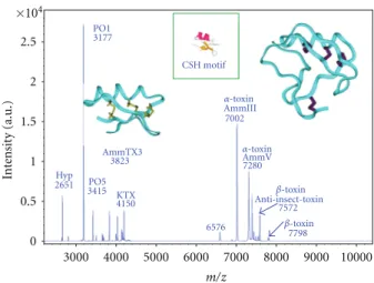
A recent MALDI-TOF mass spectrometry (MS) study has provided new information about the molecular composition of scorpion venom [23]. Through the developments in proteomics, MS is now widely used for accurate and sensitive determinations of molecular masses and identification of heterogenous complex mixtures such as crude venoms. Using an offline MALDI-TOF/MS analysis, we were able to determine the molecular masses of about 70 to 80 different compounds ranging between 3000 and 8000 Da in an Androctonus mauretanicus mauretanicus venom obtained
Journal of Toxicology 3
under manual stimulation from a pool of animals kept alive at the “Pasteur Institute” of Casablanca, Morocco. Figure 1
presents the snapshot of this crude venom.
4. The Smallest Toxins Identified so Far in the
Androctonus mauretanicus mauretanicus
Venom: P01 (
α-KTx8) and P05 (α-KTx5),
Ligands of SK
CaChannels
P01 (α-KTx 8.1 subfamily, 3177 Da) is devoid of significant toxicity in mouse [26]. This toxin of 28-amino-acid residues, which is the smallest K+channels ligand in the Androctonus
mauretanicus mauretanicus venom, is slightly negatively
charged with acidic amino acids localized at the beginning of theβ-turn and extending along the α-helix (Figure 2(a)). Surprisingly, P01 presents exactly the same sequence in
Androctonus mauretanicus mauretanicus, Androctonus aus-tralis, Androctonus amoreuxi, and Buthus occitanus venoms.
Usually, the polymorphism among scorpion toxins is so high that their sequences vary from a scorpion subspecies to another, even in the same structural, pharmacological, and immunological family. P01 is a poor-affinity ligand (300 nM) for the apamin-binding site on rat brain synaptosomes. In comparison, apamin purified from the bee venom displays a very high affinity (Kd= 8 pM) for its target, that is,
the small-conductance calcium-activated K+channel (SK Ca)
channel so-called apamin-sensitive channel. Since the first P01 characterization, other closely related analogs have been purified from other Buthidae venoms. Among them, one is particularly interesting because it shares 89% of identity with P01. This analog, called OdK1, was purified from the Iranian scorpion Odontobuthus doriae (α-KTx 8.5 subfamily). OdK1 is able to block Kv1.2 channels expressed in oocytes with
a median inhibition concentration (IC50) value of 183 nM
and has no effect on Kv1.1, Kv1.3, Kv1.4, Kv1.5, and the
Shaker channels [27]. It is important to mention that P01 has
not been tested yet on Kv1.2 channels in electrophysiological
experiments. P01 was only shown unable to compete with
125I-KTX (a K
v1.1 and Kv1.3 and not Kv1.2 blocker) bound
to its receptor on rat brain synaptosomes. Thus, we cannot totally exclude that P01 could exhibit a poor Kv1.2 blocking
activity. In the same line, OdK1 activity on SKCachannels has
not been tested yet either.
In contrast to P01, P05, another toxin from
Androc-tonus mauretanicus mauretanicus, is a high-affinity selective
ligand (Kd= 100 pM) for the apamin-binding site on rat
brain synaptosomes, and it has no effects on BKCa or Kv
channels. P05 is a 31-amino-acid long peptide (3415 Da), which belongs to the α-KTx5.x’s family (as Leiurotoxin1 or Scyllatoxin from the venom of the scorpion Leiurus
quinquestriatus Hebraeus) (Figure 2(b)) [29,30]. The toxins
from this family have only two-stranded β-sheet because of their short N-terminal side [28]. P05 is highly toxic (14 ng, i.e., 4 pmols for a 20 g mouse) and leads to an epileptic behavior when injected in mouse by the intracere-broventricular route. P05 possesses a short stretch of four amino acids similar to the one in the apamin sequence, which constitutes a highly positively charged region in the
3000 4000 5000 6000 7000 8000 9000 10000 ×104 2.5 2 1.5 1 0.5 0 In te nsit y (a.u.) m/z PO1 3177 CSH motif Hyp 2651 AmmTX3 3823 PO5 3415 KTX 4150 AmmV 7280 AmmIII 7002 Anti-insect-toxin 7572 7798 6576 α-toxin α-toxin β-toxin β-toxin
Figure 1: MALDI-TOF mass spectra of pool venom manually extracted from Androctonus mauretanicus mauretanicus. K+channel
blockers and voltage-gated Na+channels modulators are indicated.
P01 and P05, blockers of SKCachannels; AmmTX3, blockers of Kv4
and hERG channels; KTX, Kv1.1, and Kv1.3 blockers. Amm III and
Amm V are major lethalα-toxins; β-toxins specific for insects are also mentioned. The 3D structure of KTX (left) and of theα-toxin of reference AaH II from Androctonus australis (right) are shown [13,24,25]. The Cys-Stabilized-Helix (CSH) motif is shown in inset [8].
α-helix (containing in particular two Arg residues, Arg7
and Arg9) exposed to solvent. Extensive structure-function
studies using chemically synthesized analogs revealed that these two Arg were critical for P05’s interaction with its target as described for apamin [31]. When Arg were replaced by Lys, the activity dropped by a factor of one hundred, and when they were replaced by Ile, the affinity decreased even more to a Kd value in the micromolar range. Moreover,α-amidation of His in P05 C-terminal conferred a large gain of function leading to an almost irreversible binding to its receptor [21]. When the apamin C-terminal residue is in the free carboxylic form, only 0.06% of the pharmacological activity of the native C-amidated apamin is retained [32]. Both Arg and C-terminal His create a strong positive electrostatic potential, which drives the toxin to negative residues (Asp) of the channel pore. Using homology-modeling models of SK channels (rsk1, rsk2, and rsk3) and Brownian dynamics methods, the recognition between P05 and its targets was investigated [33]. It was found that the rsk2 channel, presenting the highest frequencies and lowest electrostatic interaction energies, was the most favorable target for P05 binding, while rsk3 was intermediate, and rsk1 was the least favorable. From the P05-rsk2 complex model, it was shown that P05 probably locates around the extracellular pore of SK channels and assures the contact with rsk2 channel using critical basic amino acid residues in itsα-helix: Arg6
(P05)-Asp364(SK), Arg7(P05)-Asn368(SK), and Arg13(P05)-Asp341
and Asp364 (SK). Further refinements of P05-rsk2 complex
model using molecular mechanics showed that six hydrogen bonding interactions exist between P05 and the rsk2 channel. These simulation results were in good agreement with our previous in vivo binding experiments and could explain
VSCEDCPEHCSTQAKCDNDKCVCEP PO1 SK2, Kv1.2, ??? (a) PO5 SK2, SK3 (b)
Figure 2: Amino acid sequences and 3D structures of P01 and P05. (a) Amino acid sequence (single letter notation) and 3D structure of P01 [26,28]. (b) Amino acid sequence (single letter notation) of P05 compared to that of the bee venom apamin (18 mers peptide). Amino acids common to P05 and apamin and crucial for the SKCa
blockade are in blue. These amino acids are materialized on the 3D structure of P05, view by theα-helix axe. The two positive Arg are in blue and the neutral Gln is in brown.
the interaction between P05 and SK channels at the level of the molecular structure.
5. The Most Noted K
+Channel Blocker
Characterized in the
Androctonus
mauretanicus mauretanicus
Venom:
Kaliotoxin (
α-KTx3 Subfamily)
The Kaliotoxin (KTX, 4150 Da) purified from the venom of the Androctonus mauretanicus mauretanicus was the first identified member of the α-KTx3.1 subfamily, which is currently composed of 13 highly homolog members sharing more than 75% sequence identity. KTX was first described as a BKCa low-affinity blocker [20], but further analysis
showed that KTX blocked more specifically Kv1.3 channels
with a very high affinity (Kd= 10 pM) and Kv1.1 with
a much weaker affinity (in the 10 nM range). KTX also exhibited a functional blockade of Ca2+-activated K+Gardos
channel from rabbit or human red blood cells with a median inhibition concentration value of 5 nM. Interaction between this channel and other peptide toxins indicated that the G´ardos channel was pharmacologically different in several interesting ways from the other Ca2+-activated K+
channels [34]. Interestingly, KTX was not toxic in mice by subcutaneous injection up to 500 micrograms (100 nmols),
but intracerebroventricularly injected mice showed tremor, paralysis, and death with a median lethal dose of only 24 ng of KTX (about 6 pmol for a 20 g mouse).
KTX was then widely used by different international groups to probe the vestibule topology from the lymphocyte Kv1.3 channel. The homotetrameric Kv1.3 channel controls
the resting potential membrane in T cells and plays a crucial role in human T-lymphocyte activation [35]. Its inhibition causes depolarization and an attenuation of the rise in the intracellular Ca2+ concentration that is required for T-cell
activation. Accordingly, Kv1.3 channel is a good therapeutic
target for the development of immunosuppressant drugs. It is important to note that Kv1.3 channel is a key mediator of
multiple sclerosis, type 1 diabetes mellitus, and rheumatoid arthritis pathologies [36]. In experimental autoimmune encephalomyelitis (EAE) rat, animal model for multiple sclerosis, KTX was capable of immunosuppressant activity
in vivo. Addition of KTX during Antigen/T-cell activation
led to a large reduction of the T-cell proliferative response, a decreased encephalitogenicity of T cells, and finally improved the symptoms of EAE [37].
A new therapeutic approach for inflammatory bone resorption by targeting Kv1.3 was also investigated and the
potential effect of KTX was tested on inflammatory lesions of periodontal disease. These lesions contain abundant activated/memory T and B cells, which control immuno-logical interactive networks and accelerate bone resorption. Systemic KTX administration finally resulted in an 84% decrease of the bone resorption. These results suggested that KTX could also constitute a potential therapy to prevent alveolar bone loss in periodontal disease [38].
Monoiodinated derivative of KTX (125I-KTX) binds
specifically to rat brain total membranes with a maximal binding capacity of about 14 fmol/mg of protein and with a high affinity (Kd= 80 pM). The distribution of125I-KTX
binding sites in rat brain was first studied using quantitative autoradiography on adult brain tissue sections. A compar-ison with the distribution of Kv1.1 and Kv1.3 α-subunits
by immunohistochemistry or in situ hybridization suggested that KTX recognizes channels containing these subunits [39]. Further, we used KTX injected by intracerebroventricular route to rat brain in order to investigate the involvement of Kv channels containing Kv1.1 and Kv1.3 α-subunits in
olfactory associative learning and memory. KTX facilitated cognitive processes as learning, in particular in a reference representation [40]. Therefore, it is likely that KTX-sensitive Kvchannels contribute to the repolarization of action
poten-tials at presynaptic terminals of hippocampal inhibitory neurons and induce facilitation of the transmission.
Concerning the KTX structure-function relationship studies, they were first performed using synthetic analogs such as KTX(1−37), KTX(1−37)-amideand short peptides
includ-ing KTX(27−37), KTX(25−32), and KTX(1−11). Concerning the
short peptides, which corresponded to secondary structural elements, only KTX(27−37)and KTX(25−32)were able to
com-pete with125I-KTX for its receptor on rat brain synaptosomes
and act as antagonists of KTX. These results demonstrated for the first time that the C-terminal region, particularly the toxinβ-sheet, was involved in the interaction with the
Journal of Toxicology 5
GVEINVKCSGSPQCLKPCKDAGMRFGKCMNRKCHCTPK
KTX
Kv1.1, Kv1.3
Figure 3: Amino acid sequence and 3D structure of KTX. The crucial amino acid residues involved in the receptor recognition are in red and are materialized on the 3D structure according to Lange et al., 2006 [13]. Theα-helix is in green, the β-sheet in red. Arrows in red indicate the three peptides able to compete (Kd=100 nM) with
the125I-KTX bound to its binding site on rat brain synaptosomes
[41].
receptor and the channel blockade [41].Figure 3summarizes these results.
Several international groups also produced KTX and numerous mutants using chemical synthesis or heterologous expression, in order to identify KTX residues implicated in the specific interaction with Kv1.3 channels. It was found
that the side chain of Lys27 of the toxin enters deeply into
the channel pore and interacts with the Asp402 residue of
each subunit [24]. Briefly, the KTX residues involved in the interaction with the channel were Arg24, Phe25, Lys27,
Met39, Asn30, Arg31. All these residues were present in the
short peptides that we first used to demonstrate theβ-sheet importance in the channel blockade. A model also predicted that Ser11 of the KTX made steric contacts with His404 and
Pro405of theα-subunit IV of the Kv1.3 and Thr36with His404
of the opposing Kv1.3α-subunit [42].
KTX binding to a chimeric K+ channel (KcsA-K v1.3) in
proteoliposomes was investigated using solid-state nuclear magnetic resonance (ssNMR) [13]. Upon complex for-mation, significant chemical shift changes of the residues implicated in the specific interaction were observed for both KTX and chimeric channel. For KTX, the conformational changes involved mainly β-sheet contacts between the first and thirdβ-strand and for the chimeric channel it affected the conformation of both the pore helix and the selectivity filter. The backbone conformation of Kcsa-Kv1.3 selectivity
filter adopted a novel structure with features of both the conducting and collapsed conformation of the KcsA. The ssNMR data directly showed that Asp64 in the KcsA-Kv1.3
vestibule represented an important interaction site for KTX. Large chemical shift changes were seen for Gly77, Tyr78,and
Gly79 in the selectivity filter and also for the side chains of
Glu71and Asp80that form carboxyl-carboxylate pairs on the
backside of the filter [13]. Finally, an enhanced backbone mobility was detected for two glycine residues within the selectivity filter that are highly conserved amongst potassium channels and are of core relevance to the filter structure and ion selectivity [43,44].
Combination of additional ssNMR studies, dynamic simulations, and electrophysiological measurements finally revealed the complete mechanism of KTX binding to its receptor site and showed a structural link between inactiva-tion and block of the channel. A mechanism of cooperative toxin-induced conformational changes that are structurally and functionally related to recovery from C-type inactiva-tion, would be the consequence of the very tight interac-tion between KTX and Kcsa-Kv1.3. The KTX affinity was
lowered by about 20-fold when the Kv1.3 channels entered in
C-type inactivation probably due to changes of the interac-tion surface between the toxin and the channel [43–45].
All these recent ssNMR data obtained between the chimeric KcsA-Kv1.3 channel and KTX have largely
con-tributed to decrypt the intimate interaction between a K+
channel blocker and its target, as well as to better understand the blockade of the K+conduction.
6. The
α-KTx15
Subfamily: Janus K
v4.x
and hERG Blockers
The first member of the α-KTx15 subfamily characterized
was Aa1 from the Androctonus australis venom (3869 Da) [46]. At the primary sequence level, the toxin has an unusual N-terminal pyroglutamic acid, like Charybdotoxin and Iberiotoxin, but the rest of its sequence was totally original. Aa1 completely blocked a fastIA-type K+ current
from cerebellum granular cells. Using whole-cell patch clamp recording on striatal neurons in culture, we selected another novel toxin, BmTX3 from Buthus martensii, which was also able to block a fastIA-type K+current. The sustained current
was unaffected with a micromolar dose of toxin whereas the
IA-type K+current completely disappeared independently of
the membrane potential. Autoradiograms of adult rat brain sections demonstrated a highly heterogeneous distribution of125I-BmTX3 binding sites throughout the adult rat brain.
High density of receptors was found in the striatum, the CA1 and CA3 field of the hippocampus, the superior colliculus, and in the granular layer of the cerebellum [47]. We then purified AmmTX3 (3827 Da), an analog of Aa1 and BmTX3, from the venom of Androctonus mauretanicus
mauretanicus [22]. Latter, several cDNAs encoding two
Aa1 isoforms, AaTX1 (3867 Da) and AaTX2 (3853 Da), were identified by PCR amplification from a venom gland cDNA library of Androctonus australis [48]. Also, another oligonucleotide sequence, AamTX (3751 Da), was amplified from a venom gland cDNA library of Androctonus amoreuxi [49]. Altogether these toxins constitute the first members of
theα-KTx15 subfamily (Figure 4). From a pharmacological
point of view, theses toxins were unable to compete with any other already described toxins purified from animal venom. However, they were toxic in mouse at high doses by intracerebral injections and induced epileptic status, which
Aa1 BmTX3 AmmTX3 AamTX∗ AaTX1∗ AaTX2∗ Discr.
Figure 4: Amino acid sequences of the toxins from theα-KTx15
subfamily. Aa1 [46], AaTX1, and AaTX2 [48] are from Androctonus
australis; BmTX3 [47] is from Buthus martensii; AmmTX3 [22] is from Androctonus mauretanicus; AamTX [49] is from Androctonus
amoreuxi. Discrepin [54] is from the Venezuelan scorpion Tityus
discrepans. Single letter notation is used for amino acid sequences.
Z is pyroglutamic acid.∗Indicates putative amino acid sequence deduced from cloned cDNA oligonucleotide sequence. Theβ-sheet consensus sequence is in grey.
could last 24 to 48 hours. Also, they all shared the same target on rat brain synaptosomes because they competed with each other for the same binding site. The nature of the K+
channels blocked by AmmTX3 was assessed by performing whole-cell patch recording of the K+ currents of striatal
neurons and of cerebellum granular cells in culture. In all cases, AmmTX3 inactivated the transient A-current without affecting the sustained K+current, as observed for Aa1 and
BmTX3. As well described, A-type K+-currents result mainly
from the expression of voltage-dependent Kv α-subunits
(Kv1.4, 3.4, 4.1, 4.2, 4.3) or from the association of Kv
β-subunit with Kv1 α-subunits [50, 51]. In the cerebellum
granular cells, the voltage-gated K+ channels K
v4 of the
Shal subfamily elicits A-type currents. These channels are fast transient K+ channels that regulate the kinetics of the
action potential [51,52]. The localizations of Kv4 subunits
and their auxiliary subunits were similar to the distribution of BmTX3 binding sites in numerous regions of the brain. For example, the high density of BmTX3 binding sites in the CA1 field and CA3 field followed Kv4.2 and Kv4.3
immunostaining patterns. In the molecular layer of the dendate gyrus, the BmTX3 binding pattern was also similar to the immunoreactivity pattern of Kv4 and KChIP subunits,
with a higher density in distal dendrites of the granular layer than in the more proximal dendrite areas [53].
Finally, electrophysiological analysis of mammalian cells expressing different A-type channels showed that BmTX3 completely inhibited the rapidly activating and inactivat-ing Kv4.1 current in a voltage-independent manner. This
inhibition was less effective on Kv4.2 and Kv4.3 channels
and the toxin did not show any effects on other tran-sient currents elicited by Kv1.4 and Kv3.4 [53]. Recent
electrophysiological studies using acute coronal midbrain slices and AmmTX3 confirmed that the targets of the α-KTx15 subfamily are Kv4 channels [55, Rudy B, personal
communication]. Interestingly, the radioiodinatedα-KTx15
toxins bound to their receptor on rat brain neurons were not displaced by any spider toxin, which block Kv4 channels.
This result suggests that these scorpion and spider Kv4
blockers have different binding sites. The α-KTx15 toxins
most probably block the Kv4 channel pore because tarantula
toxins inhibit Kv4 channels by binding to the voltage-sensor
paddles (crucial helix-turn-helix motifs within the voltage-sensing domains composed of the S3b and S4 helices) [56]. Moreover, deletions of the last two BmTx3 C-terminal residues (α-KTx15subfamily) were made to assess the role of
the penultimate Tyr residue in the receptor recognition. This Tyr resembled to the canonical dyad previously proposed as necessary to block the K+ channel conduction with high
efficacy. Our results showed that the truncated toxin bound to its receptor less efficiently than the wild-type toxin (by a factor of about 105) and had no more channel blocking
activity [57]. All these results suggest that the Kv4 channels
blocked by theα-KTx15subfamily might have a canonical K+
channel pore structure.
Moreover, we also described a significant hERG-blocking activity from the α-KTx15 toxins, as previously shown for
the γ-KTx peptides. AmmTx3 induced a hERG channel
block with no alteration of the gating kinetics. According to a model of theγ-KTx toxin BeKm-1 from Buthus eupeus docked on hERG channels, the toxin BeKm-1 is above the pore entrance and none of its side chains penetrate deeply into the pore. Whileα-KTx members usually interact with channels through theirβ-sheets, γ-KTxs modulate hERG in a different way than the one proposed for the interaction between Charybdotoxin and the Shaker channel or between KTX and the Kv1.3, in which the critical Lys27 protrudes
into the pore of the channel. BeKm-1 uses its α-helix and the following turn (possessing two basic residues Lys18
and Arg20), to interact with hERG channel [58, 59]. In
contrast to the other α-KTx15 members, a new toxin of
this subfamily isolated from the Venezuelan scorpion Tityus
discrepans, called Discrepin (α-KTx15−6), was also able to
block an A-type K+ current in cerebellum granular cells in
culture, but was ineffective to block the hERG channel. Its amino acid sequence displays only 50% identity with the other members purified mainly from Old-World scorpion venoms (Figure 4). Discrepin C-terminalβ-sheet, supposed to interfer with the Kv4 channel pore, presented a consensus
amino acid sequence similar to those found in the other members (in grey onFigure 4), but its “hot spot” for hERG channel blockade was altered. After several point mutations in AmmTX3 and introduction of positive charged residues in Discrepin, it was finally demonstrated that a common “hot spot” composed of two basic residues (Arg18and Lys19near
the end of theα-helix) conferred hERG blockade activity of
α-KTx15 peptides [54]. From a structural point of view, we
proposed that two separate functional surfaces (A and B) could coexist on the α-KTx15 toxins, and were responsible
for two different K+-current-blocking functions [60].
To extend our theory to other members from α-KTx subfamily, the effects of a “hot spot-” bearing toxin were tested on hERG channels and compared to the results obtained with a toxin without the “hot spot.” From these studies, it was concluded that only the α-KTxs possessing the “hot spot” were able to interact with the pore of hERG channels. This pharmacophore could either be -CKKX- or -CKXK- or -CXKKX-, with C being the third Cys, X being any nonpositively charged amino acid, and K being Lys or Arg [54].
Journal of Toxicology 7
7. Conclusion
Scorpion venom still remains a proven resource for the discovery of novel biologically active compounds, especially for the pharmacologists involved in research on ionic channels. A total of about 210 α-KTx oligonucleotide or amino acid sequences are now referenced in the UniProtKB data bank, but only some of these α-KTx peptides have really been shown able to block K+ currents. Unfortunately,
very often, there is no direct evidence described for the function of the reported peptides. Some of them represent new analogs of well-known families with described channel selectivity, but others exhibit novel structural features or activities. Majority of the α-KTx effects were determined on the Kv1.x subfamily or in a less extent on the Ca2+
-activated K+ channels (SK
Ca sensitive to the bee venom
apamin or BKCa). During the last two decades, we obtained
a lot of results that provided new insight into the targets and the mode of action of some α-KTxs isolated from different potent North African Androctonus venoms. In par-ticular, numerous studies on the Androctonus mauretanicus
mauretanicus venom “manually” extracted, which can be
considered as the physiological venom secretion, have greatly contributed to the chemical, immunological, structural, and pharmacological characterization of some highly specific K+
channel blockers. With AmmTX3, we have enlarged the α-KTx15 subfamily and defined more properly its target, the
Kv4 channels. KTX, from theα-KTx3subfamily, was finally
proven to be a powerful tool used by several international teams to depict the molecular mechanisms of interaction between K+ channels and peptide inhibitors, as well as to
demonstrate that the binding of K+channel specific scorpion
toxins does not take place only on the outer vestibule of the channel pore but also deeper into the selectivity filter. The binding involves a combination of hydrophobic, hydro-gen bonding and electrostatic interactions, which induces significant structural rearrangements in both interacting molecules. It was then proposed that structural flexibility of the K+ channel and the toxin represent an important
determinant of the high specificity of toxin/K+ channel
interactions [13].
Acknowledgments
The authors wish to thank Drs. N. Abbas, F. Bosmans, B. Card, C. Legros, J. Tytgat, and H. Vacher, H for their con-tribution at a moment or another in the purification, phar-macological or electrophysiological characterization and cloning and mutagenesis of the Androctonus mauretanicus toxins blocking K+channels.
References
[1] M. F. Martin-Eauclaire and F. Couraud, “Scorpion neurotox-ins: effects and mechanisms,” in Handbook Neurotoxicology, L. W. Chang and R. S. Dyer, Eds., pp. 683–716, Marcel Dekker, New York, NY, USA, 1995.
[2] L. D. Possani, E. Merino, M. Corona, F. Bolivar, and B. Becerril, “Peptides and genes coding for scorpion toxins that affect ion-channels,” Biochimie, vol. 82, no. 9-10, pp. 861–868, 2000.
[3] R. C. Rodr´ıguez De La Vega and L. D. Possani, “Overview of scorpion toxins specific for Na+channels and related peptides:
biodiversity, structure-function relationships and evolution,”
Toxicon, vol. 46, no. 8, pp. 831–844, 2005.
[4] S. Cest`ele and W. A. Catterall, “Molecular mechanisms of neu-rotoxin action on voltage-gated sodium channels,” Biochimie, vol. 82, no. 9-10, pp. 883–892, 2000.
[5] F. Bosmans, M. F. Martin-Eauclaire, and K. J. Swartz, “De-constructing voltage sensor function and pharmacology in sodium channels,” Nature, vol. 456, no. 7219, pp. 202–208, 2008.
[6] F. Bosmans, M. Puopolo, M. F. Martin-Eauclaire, B. P. Bean, and K. J. Swartz, “Functional properties and toxin pharmacol-ogy of a dorsal root ganglion sodium channel viewed through its voltage sensors,” Journal of General Physiology, vol. 138, no. 1, pp. 59–72, 2011.
[7] J. Tytgat, K. G. Chandy, M. L. Garcia et al., “A unified nomenclature for short-chain peptides isolated from scorpion venoms:α-KTx molecular subfamilies,” Trends in
Pharmaco-logical Sciences, vol. 20, no. 11, pp. 444–447, 1999.
[8] F. Bontems, C. Roumestand, B. Gilquin, A. M´enez, and F. Toma, “Refined structure of charybdotoxin: common motifs in scorpion toxins and insect defensins,” Science, vol. 254, no. 5037, pp. 1521–1523, 1991.
[9] O. Pongs, Regulation of Excitability by Potassium Channels.
Inhibitory Regulation of Excitatory Neurotransmission, vol.
44 of Results and Problems in Cell Differentiation, Springer, Heidelberg, Germany, 2007.
[10] G. A. Gutman, K. G. Chandy, S. Grissmer et al., “International Union of Pharmacology. LIII. Nomenclature and molecular relationships of voltage-gated potassium channels,”
Pharma-cological Reviews, vol. 57, no. 4, pp. 473–508, 2005.
[11] S. B. Long, E. B. Campbell, and R. MacKinnon, “Crystal structure of a mammalian voltage-dependent Shaker family K+channel,” Science, vol. 309, no. 5736, pp. 897–903, 2005.
[12] C. S. Park and C. Miller, “Interaction of charybdotoxin with permeant ions inside the pore of a k+channel,” Neuron, vol. 9,
no. 2, pp. 307–313, 1992.
[13] A. Lange, K. Giller, S. Hornig et al., “Toxin-induced conforma-tional changes in a potassium channel revealed by solid-state NMR,” Nature, vol. 440, no. 7086, pp. 959–962, 2006. [14] K. N. Srinivasan, V. Sivaraja, I. Huys et al., “kappa-Hefutoxin1,
a novel toxin from the scorpion Heterometrus fulvipes with unique structure and function. Importance of the functional diad in potassium channel selectivity,” Journal of Biological
Chemistry, vol. 277, no. 33, pp. 30040–30047, 2002.
[15] E. Diego-Garc´ıa, Y. Abdel-Mottaleb, E. F. Schwartz, R. C. R. De La Vega, J. Tytgat, and L. D. Possani, “Cytolytic and k+
channel blocking activities ofβ-KTx and scorpine-like pep-tides purified from scorpion venoms,” Cellular and Molecular
Life Sciences, vol. 65, no. 1, pp. 187–200, 2008.
[16] M. Corona, G. B. Gurrola, E. Merino et al., “A large number of novel Ergtoxin-like genes and ERG k+-channels blocking
peptides from scorpions of the genus Centruroides,” FEBS
Letters, vol. 532, no. 1-2, pp. 121–126, 2002.
[17] N. Oukkache, M. F. Martin-Eauclaire, F. Chgoury et al., “Identification des param`etres influenc¸ant la prise en charge de l’envenimation scorpionique au Maroc,” in Toxines et
Cancer, Rencontres en Toxinologie, pp. 295–300, LAVOISIER,
2006.
[18] J. P. Rosso and H. Rochat, “Characterization of ten proteins from the venom of the Moroccan scorpion Androctonus mauretanicus mauretanicus, six of which are toxic to the mouse,” Toxicon, vol. 23, no. 1, pp. 113–125, 1985.
[19] H. Zerrouk, P. E. Bougis, B. Ceard, A. Benslimane, and M. F. Martin-Eauclaire, “Analysis by high-performance liquid chromatography of Androctonus mauretanicus mauretanicus (black scorpion) venom,” Toxicon, vol. 29, no. 8, pp. 951–960, 1991.
[20] M. Crest, G. Jacquet, M. Gola et al., “Kaliotoxin, a novel pep-tidyl inhibitor of neuronal BK-type Ca2+-activated k+channels
characterized from Androctonus mauretanicus mauretanicus venom,” Journal of Biological Chemistry, vol. 267, no. 3, pp. 1640–1647, 1992.
[21] J. M. Sabatier, H. Zerrouk, H. Darbon et al., “P05, a new leiurotoxin I-like scorpion toxin: synthesis and structure-activity relationships of theα-amidated analog, a ligand of Ca2+-activated k+channels with increased affinity,”
Biochem-istry, vol. 32, no. 11, pp. 2763–2770, 1993.
[22] H. Vacher, M. Alami, M. Crest, L. D. Possani, P. E. Bougis, and M. F. Martin-Eauclaire, “Expanding the scorpion toxinα-KTX 15 family with AmmTX3 from Androctonus mauretanicus,”
European Journal of Biochemistry, vol. 269, no. 24, pp. 6037–
6041, 2002.
[23] N. Oukkache, J. P. Rosso, M. Alami et al., “New analysis of the toxic compounds from the Androctonus mauretanicus mauretanicus scorpion venom,” Toxicon, vol. 51, no. 5, pp. 835–852, 2008.
[24] J. Aiyar, J. M. Withka, J. P. Rizzi et al., “Topology of the pore-region of a k+channel revealed by the NMR-derived structures
of scorpion toxins,” Neuron, vol. 15, no. 5, pp. 1169–1181, 1995.
[25] J. C. Fontecilla-Camps, C. Habersetzer-Rochat, and H. Rochat, “Orthorhombic crystals and three-dimensional structure of the potent toxin II from the scorpion Androctonus australis Hector,” Proceedings of the National Academy of Sciences of the
United States of America, vol. 85, no. 20, pp. 7443–7447, 1988.
[26] H. Zerrouk, F. Laraba-Djebari, V. Fremont et al., “Character-ization of PO15 a new peptide ligand of the apamin-sensitive Ca2+ activated k+channel,” International Journal of Peptide
and Protein Research, vol. 48, no. 6, pp. 514–521, 1996.
[27] Y. Abdel-Mottaleb, E. Clynen, A. Jalali et al., “The first potassium channel toxin from the venom of the Iranian scorpion Odonthobuthus doriae,” FEBS Letters, vol. 580, no. 26, pp. 6254–6258, 2006.
[28] H. Darbon, E. Blanc, and J. M. Sabatier, “Three-dimensional structure of scorpion toxins: toward a new model of interac-tion with potassium channels,” Perspectives in Drug Discovery
and Design, vol. 16, pp. 41–60, 1999.
[29] P. Auguste, M. Hugues, B. Grave et al., “Leiurotoxin I (scyllatoxin), a peptide ligand for Ca2+-activated k+channels.
Chemical synthesis, radiolabeling, and receptor characteriza-tion,” Journal of Biological Chemistry, vol. 265, no. 8, pp. 4753– 4759, 1990.
[30] G. G. Chicchi, G. Gimenez-Callego, E. Ber, M. L. Garcia, R. Winquist, and M. A. Cascieri, “Purification and characteri-zation of a unique, potent inhibitor of apamin binding from Leiurus quinquestriatus hebraeus venom,” Journal of Biological
Chemistry, vol. 263, no. 21, pp. 10192–10197, 1988.
[31] C. Labbe-Jullie, C. Granier, F. Albericio et al., “Binding and toxicity of apamin. Characterization of the active site,”
European Journal of Biochemistry, vol. 196, no. 3, pp. 639–645,
1991.
[32] C. Devaux, M. Knibiehler, M. L. Defendini et al., “Recombi-nant and chemical derivatives of apamin. Implication of post-transcriptional C-terminal amidation of apamin in biological activity,” European Journal of Biochemistry, vol. 231, no. 3, pp. 544–550, 1995.
[33] M. Cui, J. Shen, J. M. Briggs et al., “Brownian dynamics simu-lations of the recognition of the scorpion toxin P05 with the small-conductance calcium-activated potassium channels,”
Journal of Molecular Biology, vol. 318, no. 2, pp. 417–428, 2002.
[34] C. Brugnara, C. C. Armsby, L. De Franceschi, M. Crest, M. F. Martin Euclaire, and S. L. Alper, “Ca2+-activated k+channels
of human and rabbit erythrocytes display distinctive patterns of inhibition by venom peptide toxins,” Journal of Membrane
Biology, vol. 147, no. 1, pp. 71–82, 1995.
[35] E. Honore, E. Guillemare, F. Lesage, J. Barhanin, and M. Lazdunski, “Injection of a k+ channel (K
v1.3) cRNA in
fertilized eggs leads to functional expression in cultured myotomal muscle cells from Xenopus embryos,” FEBS Letters, vol. 348, no. 3, pp. 259–262, 1994.
[36] M. D. Cahalan and K. G. Chandy, “The functional network of ion channels in T lymphocytes,” Immunological Reviews, vol. 231, no. 1, pp. 59–87, 2009.
[37] C. Beeton, J. Barbaria, P. Giraud et al., “Selective blocking of voltage-gated k+channels improves experimental
autoim-mune encephalomyelitis and inhibits T cell activation,” Journal
of Immunology, vol. 166, no. 2, pp. 936–944, 2001.
[38] P. Valverde, T. Kawai, and M. A. Taubman, “Selective blockade of voltage-gated potassium channels reduces inflammatory bone resorption in experimental periodontal disease,” Journal
of Bone and Mineral Research, vol. 19, no. 1, pp. 155–164, 2004.
[39] C. Mourre, M. N. Chernova, M. F. Martin-Eauclaire et al., “Distribution in rat brain of binding sites of kaliotoxin, a blocker of Kv1.1 and Kv1.3α-subunits,” Journal of
Pharma-cology and Experimental Therapeutics, vol. 291, no. 3, pp. 943–
952, 1999.
[40] S. Kourrich, C. Mourre, and B. Soumireu-Mourat, “Kalio-toxin, a Kv1.1 and Kv1.3 channel blocker, improves associative
learning in rats,” Behavioural Brain Research, vol. 120, no. 1, pp. 35–46, 2001.
[41] R. Romi, M. Crest, M. Gola et al., “Synthesis and charac-terization of kaliotoxin. Is the 26-32 sequence essential for potassium channel recognition?” Journal of Biological
Chem-istry, vol. 268, no. 35, pp. 26302–26309, 1993.
[42] G. M. Lipkind and H. A. Fozzard, “A model of scorpion toxin binding to voltage-gated k+channels,” Journal of Membrane
Biology, vol. 158, no. 3, pp. 187–196, 1997.
[43] C. Ader, R. Schneider, S. Hornig et al., “A structural link between inactivation and block of a k+channel,” Nature
Struc-tural and Molecular Biology, vol. 15, no. 6, pp. 605–612, 2008.
[44] C. Ader, O. Pongs, S. Becker, and M. Baldus, “Protein dynam-ics detected in a membrane-embedded potassium chan-nel using two-dimensional solid-state NMR spectroscopy,”
Biochimica et Biophysica Acta, vol. 1798, no. 2, pp. 286–290,
2010.
[45] U. Zachariae, R. Schneider, P. Velisetty et al., “The molecular mechanism of toxin-induced conformational changes in a potassium channel: relation to C-type inactivation,” Structure, vol. 16, no. 5, pp. 747–754, 2008.
[46] M. Pisciotta, F. I. Coronas, C. Bloch, G. Prestipino, and L. D. Possani, “Fast k+currents from cerebellum granular cells are
completely blocked by a peptide purified from Androctonus australis Garzoni scorpion venom,” Biochimica et Biophysica
Acta, vol. 1468, no. 1-2, pp. 203–212, 2000.
[47] H. Vacher, R. Romi-Lebrun, C. Mourre et al., “A new class of scorpion toxin binding sites related to an A-type k+
channel: pharmacological characterization and localization in rat brain,” FEBS Letters, vol. 501, no. 1-3, pp. 31–36, 2001. [48] C. Legros, P. E. Bougis, and M. F. Martin-Eauclaire,
Journal of Toxicology 9
the scorpion Androctonus australis,” Toxicon, vol. 41, no. 1, pp. 115–119, 2003.
[49] T. Chen, B. Walker, M. Zhou, and C. Shaw, “Molecular cloning of a novel putative potassium channel-blocking neurotoxin from the venom of the North African scorpion, Androctonus amoreuxi,” Peptides, vol. 26, no. 5, pp. 731–736, 2005. [50] M. C. Sanguinetti, J. H. Johnson, L. G. Hammerland et al.,
“Heteropodatoxins: peptides isolated from spider venom that block Kv4.2 potassium channels,” Molecular Pharmacology,
vol. 51, no. 3, pp. 491–498, 1997.
[51] W. A. Coetzee, Y. Amarillo, J. Chiu et al., “Molecular diversity of k+channels,” Annals of the New York Academy of Sciences,
vol. 868, pp. 233–285, 1999.
[52] H. Vacher and J. S. Trimmer, “Diverse roles for auxiliary subunits in phosphorylation-dependent regulation of mam-malian brain voltage-gated potassium channels,” Pflugers
Archiv European Journal of Physiology, vol. 642, no. 5, pp. 631–
643, 2011.
[53] H. Vacher, S. Diochot, P. E. Bougis, M. F. Martin-Eauclaire, and C. Mourre, “Kv4 channels sensitive to BmTX3 in rat
ner-vous system: autoradiographic analysis of their distribution during brain ontogenesis,” European Journal of Neuroscience, vol. 24, no. 5, pp. 1325–1340, 2006.
[54] Y. Abdel-Mottaleb, G. Corzo, M. F. Martin-Eauclaire et al., “A common ”hot spot” confers hERG blockade activity to α-scorpion toxins affecting k+channels,” Biochemical
Pharma-cology, vol. 76, no. 6, pp. 805–815, 2008.
[55] J. Amendola, A. Woodhouse, M. F. Martin-Eauclaire, and J. M. Goaillard, “Ca2+/cAMP-Sensitive co-variation of IA and
IH voltage dependences tune rebound firing in dopaminergic neurons,” The Journal of Neuroscience, vol. 32, no. 6, pp. 2166– 2181, 2012.
[56] L. R. Phillips, M. Milescu, Y. Li-Smerin, J. A. Mindell, J. I. Kim, and K. J. Swartz, “Voltage-sensor activation with a tarantula toxin as cargo,” Nature, vol. 436, no. 7052, pp. 857–860, 2005. [57] H. Vacher, R. Romi-Lebrun, M. Crest et al., “Functional con-sequences of deleting the two C-terminal residues of the scor-pion toxin BmTX3,” Biochimica et Biophysica Acta, vol. 1646, no. 1-2, pp. 152–156, 2003.
[58] G. N. Tseng, K. D. Sonawane, Y. V. Korolkova et al., “Probing the outer mouth structure of the hERG channel with peptide toxin footprinting and molecular modeling,” Biophysical
Jour-nal, vol. 92, no. 10, pp. 3524–3540, 2007.
[59] R. C. Rodr´ıguez De La Vega, E. Merino, B. Becerril, and L. D. Possani, “Novel interactions between k+channels and
scor-pion toxins,” Trends in Pharmacological Sciences, vol. 24, no. 5, pp. 222–227, 2003.
[60] I. Huys, C. Q. Xu, C. Z. Wang et al., “BmTx3, a scorpion toxin with two putative functional faces separately active on A-type k+and HERG currents,” Biochemical Journal, vol. 378, no. 3,
Submit your manuscripts at
http://www.hindawi.com
Pain
Research and TreatmentHindawi Publishing Corporation
http://www.hindawi.com Volume 2014
World Journal
Hindawi Publishing Corporationhttp://www.hindawi.com Volume 2014
Hindawi Publishing Corporation
http://www.hindawi.com Volume 2014
Toxins
Journal of
Vaccines
Journal of
Hindawi Publishing Corporation
http://www.hindawi.com Volume 2014
Hindawi Publishing Corporation
http://www.hindawi.com Volume 2014
Antibiotics
Toxicology
Journal of
Hindawi Publishing Corporation
http://www.hindawi.com Volume 2014
Stroke
Research and TreatmentHindawi Publishing Corporation
http://www.hindawi.com Volume 2014
Drug Delivery
Journal ofHindawi Publishing Corporation
http://www.hindawi.com Volume 2014
Hindawi Publishing Corporation
http://www.hindawi.com Volume 2014 Advances in Pharmacological Sciences
Tropical Medicine
Hindawi Publishing Corporation
http://www.hindawi.com Volume 2014
Medicinal ChemistryInternational Journal of
Hindawi Publishing Corporation
http://www.hindawi.com Volume 2014
Addiction
Journal of Hindawi Publishing Corporationhttp://www.hindawi.com Volume 2014
Hindawi Publishing Corporation
http://www.hindawi.com Volume 2014 BioMed
Research International Emergency Medicine International
Hindawi Publishing Corporation
http://www.hindawi.com Volume 2014
Hindawi Publishing Corporation
http://www.hindawi.com Volume 2014
Diseases
Hindawi Publishing Corporation
http://www.hindawi.com Volume 2014
Anesthesiology Research and Practice
Scientifica
Hindawi Publishing Corporation
http://www.hindawi.com Volume 2014
Journal of
Hindawi Publishing Corporation
http://www.hindawi.com Volume 2014
Pharmaceutics
Hindawi Publishing Corporation
http://www.hindawi.com Volume 2014
![Figure 3: Amino acid sequence and 3D structure of KTX. The crucial amino acid residues involved in the receptor recognition are in red and are materialized on the 3D structure according to Lange et al., 2006 [13]](https://thumb-eu.123doks.com/thumbv2/123doknet/14518607.531006/6.900.120.397.111.326/sequence-structure-residues-involved-recognition-materialized-structure-according.webp)

![[PDF] Cours sur la programmation impérative avec Python](data:image/gif;base64,R0lGODlhAQABAIAAAP///wAAACH5BAEAAAAALAAAAAABAAEAAAICRAEAOw==)