HAL Id: hal-01612697
https://hal.archives-ouvertes.fr/hal-01612697
Submitted on 5 Nov 2018
HAL is a multi-disciplinary open access
archive for the deposit and dissemination of sci-entific research documents, whether they are pub-lished or not. The documents may come from teaching and research institutions in France or abroad, or from public or private research centers.
L’archive ouverte pluridisciplinaire HAL, est destinée au dépôt et à la diffusion de documents scientifiques de niveau recherche, publiés ou non, émanant des établissements d’enseignement et de recherche français ou étrangers, des laboratoires publics ou privés.
Gillies
To cite this version:
Bo Fan, John Frederick Trant, Gauvin Hemery, Olivier Sandre, Elizabeth R. Gillies. Thermo-responsive self-immolative nanoassemblies: Direct and indirect triggering. Chemical Communications, Royal Society of Chemistry, 2017, 53 (89), pp.12068-12071. �10.1039/C7CC06410A�. �hal-01612697�
ChemComm
COMMUNICATION
Received 16th August 2017, Accepted 5th October 2017 DOI: 10.1039/c7cc06410a www.rsc.org/Thermo-responsive self-immolative nanoassemblies: Direct and in-direct triggering
Bo Fan,a John F. Trant,b Gauvin Hemeryc, Olivier Sandrec and Elizabeth R. Gillies*a,b
A thermo-responsive end-cap based on a retro-Diels-Alder and subsequent furan elimination reaction was developed. It was used to cap poly(ethyl glyoxylate), allowing end-to-end depolymerization upon thermal triggering. Using block copolymers, thermo-responsive micelles and vesicles were prepared and shown to disassemble upon heating. Thermal degradation could also be triggered indirectly by magnetic field hyperthermia after incorporation of iron oxide nanoparticles into the assemblies.
Nanoassemblies are of great interest due to their ability to mimic biological nanostructures and their capacity to perform diverse functions such as controlled release1, catalysis,2 and templating of inorganic structures.3 Stimuli-responsive macro-molecular assemblies have attracted significant attention,4 due to their ability to undergo morphological or functional changes in response to stimuli. Assemblies responsive to stimuli such as light,5 heat,6 and pH7 have been reported. Heat is particularly attractive as it can be easily applied either directly with good spatiotemporal control, or indirectly through photothermal8 or magnetothermal effects9. Many examples of thermo-respon-sive polymer assemblies employing polymers such as poly(N-isopropylacrylamide) that undergo solubility changes in re-sponse to temperature have been reported.10 There are very few examples involving thermally-initiated bond cleavage.11 Over the last decade, a new class of stimuli-responsive pol-ymers, often termed self-immolative polymers (SIPs), has been developed. These polymers can be triggered to degrade from end-to-end upon the cleavage of a stimulus-responsive end-cap from the terminus.12 The propagating depolymerization mech- anism amplifies the stimulus-mediated event. Various back- bones including polycarbamates,13 poly(benzyl ether)s,14 and
Scheme 1. Proposed end-cap cleavage and depolymerization mechanism of PEtG end-capped with a DA adduct (PEtG-DA).
polyaldehydes15-17 have been reported. A single SIP backbone can respond to different signals by simply changing the end-cap.18 SIPs responsive to stimuli including light,19 oxidizing or re-ducing agents,20 and enzymes13 have been reported. Assemblies such as vesicles19 and micelles21 have been prepared from SIPs and shown to undergo disintegration and payload release in re-sponse to stimuli.
Thus far, one thermo-responsive SIP end-cap was re-ported.22 This 1,2-oxazine end-cap underwent cycloreversion to an unstable carbamoylnitroso intermediate that hydrolyzed and decarboxylated to initiate depolymerization. However, the end- capping was challenging, as it required generation of the unsta-ble nitroso species to perform the cycloaddition. Furthermore, the end-cap cleavage and depolymerization were very slow, oc-curring over tens of days. Here we report that simple Diels-Alder (DA) adducts of furan and maleimides can serve as thermo-re-sponsive end-caps. Upon heating, a retro-Diels Alder reaction occurs, revealing a furan.23 Based on the known instability of similarly substituted furan derivatives,24 it is proposed that the released furfuryl carbonate undergoes an elimination reaction to release an uncapped poly(ethyl glyoxylate) (PEtG) SIP for de- polymerization (Scheme 1). Exploiting the easy synthetic modi-fication of this end-cap, amphiphilic block copolymers can be prepared, and self-assembled into thermo-responsive vesicles and micelles. Furthermore, indirect triggering of SIP
nanoas- semblies is demonstrated for the first time using the magneto-thermal effect with iron oxide nanoparticles (IONPs) under an alternating magnetic field. To test the proposed concept, we capped PEtG with the DA adduct and evaluated its thermo-responsiveness. First, N-ben-zylmaleimide (1a) was reacted with furfuryl alcohol (2) in a DA reaction to obtain 3a as a 70:30 mixture of endo:exo diastere-omers (Scheme 2). 3a was then activated as the chloroformate
4a. Polymerization of ethyl glyoxylate was conducted at
-20 C,15 and the resulting PEtG was end-capped in situ by reac- tion with 4a to afford PEtG-DA-Bn (Scheme 3). The number av-erage molecular weight (Mn) was 33 kg/mol and the dispersity (Đ) was 1.8 based on size exclusion chromatography (SEC). Suc- cessful end-capping was confirmed by thermogravimetric anal-ysis as the capped polymer was stable to > 165 °C in the solid state, whereas uncapped polymer degraded below 100 °C 15 (Fig. S28, Table S1).
To test the thermo-responsiveness of PEtG-DA-Bn, the pol-ymer was dissolved in 9:1 CD3CN:D2O and incubated at different temperatures. A non-responsive PEtG end-capped by benzyl chloroformate (PEtG-control, Scheme S1)25 was also examined. The depolymerization was monitored by 1H NMR spectroscopy. Initially, the spectrum of PEtG-DA-Bn consisted of broad peaks attributable to the polymer (Fig. 1). When the polymer was heated, a sharp peak at 5.1 ppm corresponding to the depoly-merization product ethyl glyoxylate hydrate (EtGH) emerged. The extent of depolymerization was quantified based on the in- tegrations of polymer and EtGH peaks. Over 24 h, 85% depoly-merization occurred at 75 C, 53 % at 60 C, and 8% at 40 C (Figs. 2a, S22). In contrast, PEtG-DA-Bn at 22
C and PEtG-con-trol at 75 C underwent less than 10% depolymerization even
after 4 days (Figs. 2a, S21, S23). Overall, the rate of end-cap
cleavage and depolymerization was much faster for PEtG-DA-Bn than that of the previously reported oxazine system,22 which required several days, resulting in substantial background deg-radation of the controls. Thus, DA adducts can provide rapid and selective thermo-responsive degradation.
To prepare thermally-responsive assemblies from am-phiphilic block copolymers, end-cap 4b with a propargyl group was synthesized (Scheme 2). 4b was installed on PEtG to afford PEtG-DA-alkyne with an Mn of 63 kg/mol and a Đ of 2.0 (Scheme 3). This polymer underwent depolymerization at rates very sim- ilar to those of PEtG-DA-Bn (Figs. 2a, S24-25). It was then cou- pled with two different lengths of azide-functionalized poly(eth-ylene glycol) (PEG-N3; 750 and 5000 g/mol) to give copolymers
PEtG-DA-PEG750 and PEtG-DA-PEG5000. The success of the
couplings was confirmed by SEC (Figs. S30-31) and 1H NMR and 13C NMR spectroscopy (Figs. S15-18).
Assemblies were prepared by nanoprecipitation involving the addition of H2O into THF for PEtG-DA-PEG750 and DMSO into H2 O for PEtG-DA-PEG5000. Based on dynamic light scatter-ing (DLS), the Z-average diameters of the assemblies were 480 ± 80 nm for PEG750 and 87 ± 3 nm for
PEtG-DA-PEG5000 (Figs. S33-34). Transmission electron microscopy
(TEM) showed that PEtG-DA-PEG750 formed vesicles, while
PEtG-DA-PEG5000 formed solid spherical nanoparticles (Fig. 3). Scheme 2. Synthesis of end-caps 4a and 4b. Scheme 3. Synthesis of end-capped PEtG and its block copolymers. Fig. 1. 1H NMR spectra of PEtG-DA-Bn incubated in 9:1 CD 3CN:D2O at 75 °C. Spectra are offset to allow the progression over time to be clearly observed. After incubation of the assemblies at 75 °C for 16 h, no re- maining assemblies were detected by TEM (Fig. S37). This sug-gests that depolymerization of the PEtG resulted in disassembly. This disassembly was further probed using DLS by fixing the de-tector attenuation factor and recording the mean count rate (CR), which is proportional to the number and molar mass of the scattering species. Both the thermo-responsive vesicles and mi-celles prepared from PEtG-DA-PEG750 and PEtG-DA-PEG5000 showed an 80% decrease in CR when incubated at 75 °C for 10 h (Fig. 2b). In contrast, when these systems were incubated at 22 °C less than a 20% change in CR was observed. To ensure O N O 1a R = CH2Ph 1b R = R O HO 3a (70 : 30 = endo : exo) 3b (80 : 20 = endo : exo) CH3CN 35 ˚C 14 h HO N O O O R + O N O O O R Cl O Phosgene /THF 22 oC,16 h CH2C CH 2 4a R = CH2Ph 4b R = CH2C CH O O O O nO O O O N O O O N O O O O O O O n O O O O N O O O N O O O PEtG-DA-alkyne OEt O O NEt3, trace H2O CH2Cl2, -20 oC 1)
2) one of end-capping reagents
4a,b NEt3, -20 oC to rt O O O O nO O O O N O O O N O O O PEG-N3 CuSO4 Sodium ascorbate DMF 750 or 5000 g/mol PEtG-DA-PEG750, m = 17 PEtG-DA-PEG5000, m = 115 PEtG-DA-Bn N NN O OCH3 m N NN O H3CO m
(from end-cap 4a)
J. Name., 2013, 00, 1-3 | 3 Fig. 2. a) Depolymerization of polymers in 9:1 CD3CN:D2 O monitored by NMR spectros-copy; Assembly degradation in pH 7.4 phosphate buffer monitored by b) DLS count rate changes, c) Nile red fluorescence changes, and d) NMR spectroscopy. Fig. 3. TEM images of a) PEtG-DA-PEG750 vesicles, b) PEtG-DA-PEG5000 micelles, c) un-loaded IONPs, d) IONP-loaded PEtG-DA-PEG5000 micelles. that the degradation at 75 °C did not arise from non-specific thermal bond cleavage, we prepared additional vesicle and mi- celle controls (Vesicle-control and Micelle-control) from previ- ously reported PEtG-PEG block copolymers containing a photo-cleavable nitrobenzyl end-cap/linker, that should not be thermo-responsive (Scheme S1).15 For both of these systems, the CR initially increased, which could be attributed to some ag-gregation, but it remained within 20% of its initial value. The release of cargo from the vesicles and micelles was also explored. Nile red is a hydrophobic dye with strong fluorescence emission in a hydrophobic environment, but greatly reduced fluorescence due to quenching in hydrophilic environments such as water. Nile red was incorporated into the responsive vesicles and micelles as well as their corresponding controls. When the thermo-responsive assemblies were incubated at 75
°C, the fluorescence intensity of Nile red decreased by 60-70% over 11 h (Fig. 2c), consistent with its release into the aqueous environment. However, for either the same assemblies bated at 22 °C or the non-thermo-responsive controls incu-bated at 75 °C, only ~10-20% intensity decrease was observed after 11 h. To confirm that breakdown of the assemblies was induced by the depolymerization of the PEtG blocks, the depolymerisa-tion of PEtG-DA-PEG5000 micelles was studied by 1 H NMR spec- troscopy. In this case, the assemblies were prepared by nano-precipitation of the polymer in DMSO-d6 into pH 7.4 phosphate buffered D2O (DMSO-d6: D2 O = 1:5). Only the peak correspond- ing to the PEG block was observed initially, consistent with self-assembly of PEtG at the particle cores (Fig. S26). However, after 1 h at 75 °C, peaks corresponding to EtGH appeared, confirming depolymerization. As shown in Fig. 2d, ~65% depolymerization had occurred over 10 h at 75 C, whereas less than 5% depoly-merisation occurred at 22 °C. For Micelle-control, at 75 C, there was only about 20% depolymerization, which could be in-duced by nonspecific hydrolysis of the carbonate group of the end-cap. Combined, these data support that the thermo-re-sponsive assemblies can degrade in response to external heat, and that this is due to the thermally responsive end-cap. For some applications, direct bulk heating to trigger depoly-merisation can be a viable process. However, in other cases, it would be necessary to apply a more selective and localized heating. Therefore, we also explored the incorporation of IONPs into the micelle core and the use of magnetic field hyperthermia (MFH) to obtain localized heating around the IONPs. This mag- netothermal effect has previously been shown to enable a sim-ilar cleavage of bonds at the IONP surface.26 Hydrophobic IONPs were synthesized via the “polyol” pro-cess (11.2 ± 1.9 nm diameter, Fig. 3c),8 and then coated with Beycostat NE surfactant to be incorporated into the micelle cores via nanoprecipitation of co-assembled IONPs and
PEtG-DA-PEG5000 from a THF solution into water.27 The pure micelle solution was transparent and colorless. When the IONPs were introduced, the colloidal suspension became brown and dark-ened as the concentration of iron increased (Fig. S39). However, even at 35 mass % of IONP relative to polymer, the suspension was still transparent, confirming the IONPs were well dispersed and not precipitating. TEM showed that the IONPs were aggre-gated in spherical shapes with dimensions of ~100 nm (Fig. 3d). Samples with the highest IONP content of 35 mass%, theo-retically able to produce the largest increases of temperature, were studied. Micelle-control was also loaded with IONPs. The samples were first heated to 72 °C and equilibrated for 1 h. The Z-average particle diameter and scattering CR were measured using an in situ MFH-DLS setup as previously reported (Fig. S40).9 No changes in CR or diameter were observed during the initial 1 h, suggesting that the composite structure may stabilize the assemblies. Then, magnetic field oscillations at maximum amplitude of 10.2 kA m-1 and 755 kHz were applied. Heat gen-erated by the IONP-loaded micelles led to only a slight increase in temperature of ~2 °C for the bulk suspension. Nevertheless, the MFH had a rapid effect on the magnetic micelles, leading to an increase in diameter and large decrease in the CR. Normally,
4 | J. Name., 2012, 00, 1-3 xx
Fig. 4. Bulk temperature, particle diameter, and count rate measured before, during, and
after magnetic hyperthermia using an in situ DLS for 35 mass% IONP-loaded a) PEtG-DA-PEG5000 micelles and b) Micelle-control.
an increase in diameter would be expected to lead to an in-crease in CR. However, based on the experiments carried out on non-magnetic micelles, we hypothesize that upon application of MFH, the polymer degraded, leading to an overall reduction of the concentration of scattering species and reduced CR. The re- sulting unstabilized hydrophobic IONPs then aggregated, result- ing in an increased diameter. In the case of the control IONP-loaded Micelle-control, a similar elevation of temperature was recorded, but diameters and intensities remained relatively constant. The experiment was also conducted at lower initial temperatures (53 °C) for the IONP-loaded thermo-responsive micelles but no significant changes were observed by DLS over the same time period (Fig. S41). Thus, while elevated initial tem-peratures were required for the MFH effect, these experiments demonstrate the ability to indirectly and selectively trigger the disassembly of the thermo-responsive micelles. A complemen-tary small angle neutron scattering (SANS) study confirmed the thermo-induced degradation of the assemblies, both for pure and IONP-loaded PEtG-DA-PEG5000 micelles heated at 80 °C for 30 min (Fig. S42).
In conclusion, we showed that a simple DA adduct of a furan and maleimide could serve as a new thermo-responsive end-cap for SIPs. It was readily functionalized to enable conjugation of PEG, forming block copolymers. Thermo-responsive PEtG- PEO copolymers with different PEG weight fractions were pre-pared and self-assembled to form micelles or vesicles. These nanoassemblies were triggered to disassemble upon heating as demonstrated by TEM, DLS, release of nile red, and NMR spec-troscopy. Furthermore, MFH was used as an indirect stimulus to trigger the degradation of IONP-loaded micelles. Future work will involve the tuning of the structures of the end-caps and the assemblies to enable them to respond to heat stimuli both di-rectly and indirectly at lower temperatures.
Acknowledgements
The authors thank the Natural Sciences and Engineering Re- search Council of Canada (DG and SPG) and the Agence Natio-nale de la Recherche (ANR-13-BS08-0017) for funding. Notes and references 1. S. Ganta, H. Devalapally, A. Shahiwala and M. Amiji, J. Controlled Release, 2008, 126, 187. 2. J. Ge, T. Huynh, Y. Hu and Y. Yin, Nano lett., 2008, 8, 931. 3. Y. Wang, A. S. Angelatos and F. Caruso, Chem. Mater., 2007, 20, 848. 4. J. Zhuang, M. R. Gordon, J. Ventura, L. Li and S. Thayumanavan, Chem. Soc. Rev., 2013, 42, 7421. 5. J. S. Katz and J. A. Burdick, Macromol. Biosci., 2010, 10, 339. 6. W. Agut, A. Brûlet, C. Schatz, D. Taton and S. Lecommandoux, Langmuir, 2010, 26, 10546. 7. J. Liu, Y. Huang, A. Kumar, A. Tan, S. Jin, A. Mozhi and X. J. Liang, Biotechnol. Adv., 2014, 32, 693. 8. X. Huang, I. H. El-Sayed, W. Qian and M. A. El-Sayed, J. Am. Chem. Soc., 2006, 128, 2115. 9. G. Hemery, E. Garanger, S. Lecommandoux, A. D. Wong, E. R. Gillies, B. Pedrono, T. Bayle, D. Jacob and O. Sandre, J. Phys. D: Appl. Phys., 2015, 48, 494001. 10. S. Qin, Y. Geng, D. E. Discher and S. Yang, Adv. Mater., 2006, 18, 2905. 11. A. W. Jackson, D. A. Fulton, Polym. Chem. 2013, 4, 31. 12. M. E. Roth, O. Green, S. Gnaim and D. Shabat, Chem. Rev., 2015, 116, 1309. 13. A. Sagi, R. Weinstain, N. Karton and D. Shabat, J. Am. Chem. Soc., 2008, 130, 5434. 14. M. G. Olah, J. S. Robbins, M. S. Baker and S. T. Phillips, Macromolecules, 2013, 46, 5924. 15. B. Fan, J. F. Trant, A. D. Wong and E. R. Gillies, J. Am. Chem. Soc., 2014, 136, 10116. 16. A. M. DiLauro, A. Abbaspourrad, D. A. Weitz and S. T. Phillips, Macromolecules, 2013, 46, 3309. 17. A. W. Knoll, D. Pires, O. Coulembier, P. Dubois and J. L. Hedrick, Adv. Mater., 2010, 22, 3361. 18. B. Fan, J. F. Trant and E. R. Gillies, Macromolecules, 2016, 49, 9309. 19. G. Liu, X. Wang, J. Hu, G. Zhang and S. Liu, J. Am. Chem. Soc., 2014, 136, 7492. 20. G. Liu, G. Zhang, J. Hu, X. Wang, M. Zhu and S. Liu, J. Am. Chem. Soc., 2015, 137, 11645. 21. B. Fan and E. R. Gillies, Mol. Pharmaceutics, 2017, 14, 2548. 22. G. I. Peterson, D. C. Church, N. A. Yakelis and A. J. Boydston, Polymer, 2014, 55, 5980. 23. A. Gandini, Prog. Polym. Sci., 2013, 38, 1. 24. J. E. Zanetti and J. T. Bashour, J. Am. Chem. Soc., 1939, 61, 2249. 25. B. Fan, J. F. Trant, R. E. Yardley, A. J. Pickering, F. Lagugné-Labarthet and E. R. Gillies, Macromolecules, 2016, 49, 7196. 26. T. T. N'Guyen, H. T. Duong, J. Basuki, V. Montembault, S. Pascual, C. Guibert, J. Fresnais, C. Boyer, M. R. Whittaker and T. P. Davis, Angew. Chem., Int. Ed., 2013, 52, 14152. 27. W. Agut, A. Brûlet, D. Taton, O. Sandre and S. Lecommandoux, Soft Matter, 2011, 7, 9744.
Thermo-responsive self-immolative nanoassemblies: Direct and
indirect triggering
Bo Fan1, John F. Trant2, Gauvin Hemery3, Olivier Sandre3, Elizabeth R. Gillies*1,2
1Department of Chemical and Biochemical Engineering, The University of Western Ontario, 1151 Richmond St.,
London, Canada, N6A 5B9; E-mail: [email protected].
2Department of Chemistry, The University of Western Ontario, 1151 Richmond Street, London, Canada, N6A 5B7. 3Laboratoire de Chimie des Polymères Organiques (LCPO), Université de Bordeaux, Bordeaux INP, ENSCBP, 16
avenue Pey Berland, Pessac Cedex, France, 33607.
Table of Contents
1. Chemical structures of control polymers ... 2
2. Experimental procedures ... 2
3. NMR Spectra of compounds and polymers ... 13
4. TGA curves of end-caps and polymers ... 27
5. SEC traces for polymers ... 28
6. DLS distributions of all assemblies ... 30
7. Additional TEM images ... 32
8. Study of the thermal degradation by small angle neutron scattering ... 34
9. References ... 37
1. Chemical structures of control polymers
Scheme S1. Chemical structure of a) PEtG-control, b) Micelle-control, and c) Vesicle control.
2. Experimental procedures
General materials and procedures. Micelle-control1a and PEtG-control1b were previously
reported and the same batches were used here. Ethyl glyoxylate in toluene solution (50% w/w),
2-(hydroxymethyl)furan, diisopropylethylamine were obtained from Alfa Aesar (Canada).Nile
red and phosgene solution (15 wt. % in toluene) were purchased from Sigma-Aldrich (USA).
Iron dichloride tetra-hydrate powder (FeCl2.4H2O), iron trichloride hexahydrate 45% solution
(FeCl3.6H2O), diethylene glycol, N-methyldiethanolamine, nitric acid were obtained from
Sigma-Aldrich (France). Beycostat NE (Ref NB09) was obtained from CECA chemicals (France). Triethylamine, pyridine, and dichloromethane were distilled from calcium hydride before use. Anhydrous tetrahydrofuran, acetonitrile were obtained from a solvent purification
system using aluminum oxide columns. All the other chemicals were of reagent grade and used
without further purification. 1H NMR spectra were obtained at 400 MHz or 600 MHz on Varian
Inova instruments. NMR chemical shifts (δ) are reported in ppm and were calibrated against
residual solvent signals of CDCl3 (δ 7.27), CD3CN (δ 1.94), or D2O (δ 4.75). Fourier transform
infrared (FTIR) spectra were obtained in attenuated total reflectance (ATR) mode using a
PerkinElmer UATR Spectrum Two with films drop cast from CH2Cl2 on diamond.
High-resolution mass spectrometry (HRMS) was performed with either a Thermo Scientific DFS (Double Focus Sector) mass spectrometer, using a reversed Nier Johnson geometry for electron impact (EI) ionization, or a Bruker microOTOF 11 for electrospray ionization (ESI). The SEC instrument was equipped with a Viscotek GPC Max VE2001 solvent module. Samples were analyzed using the Viscotek VE3580 RI detector operating at 30 C. The separation technique employed two Agilent Polypore (300 ´ 7.5mm) columns connected in series and to a Polypore guard column (50 ´ 7.5mm). Samples were dissolved in THF (glass distilled grade) at
approximately 5 mg/mL and filtered through 0.22 µm syringe filters. Samples were injected using a 100 µL loop. The THF eluent was filtered and eluted at 1 mL/min for a total of 30 min. A calibration curve was obtained from poly(methyl methacrylate) standards with molecular weight ranges of 1,540-1,126,000/mol. Thermogravimetric analyses (TGA) were performed on a TGA
Q50 from TA Instruments. The heating rate was 10 C /min between 30-500 C under N2.
Ultrapure water was obtained from a Barnstead EASYpure II system. Dialyses were performed using Spectra/Por regenerated cellulose membranes with 3500 g/mol molecular weight cut-off. The hydrodynamic diameters of the polymer assemblies were measured by dynamic light scattering (DLS) using a Zetasizer Nano Series ZS instrument from Malvern Instruments, at room temperature (25 ºC) in a 1 cm path length glass cuvette at a concentration of 0.8 mg/ml suspension of polymer assemblies. Fluorescence spectra were obtained using a QM-4 SE spectrometer from Photon Technology International (PTI) equipped with double excitation and emission monochromators. TEM imaging was performed using a Phillips CM10 microscope operating at an acceleration voltage of 80 kV. 3 µL of micelle suspension (0.08 mg / mL) was
placed onto a copper grid. The resulting sample was air-dried overnight before imaging. TEM for IONPs and IONPs loaded micelles was performed on a Hitachi H7650 microscope operated at 80 kV on samples deposited at ~1 mg/mL onto copper grids by a lab-made spraying tool.
Synthesis of Compound 3a. N-benzyl maleimide2 (2.00 g, 10.7 mmol) and 2-(hydroxymethyl) furan (931 µL, 1.05 g, 10.7 mmol) were dissolved in anhydrous acetonitrile under a nitrogen atmosphere in a flame-dried flask equipped with a magnetic stirring-bar. The reaction was stirred at 35 ºC for 14 h. When TLC indicated the reaction had reached equilibrium, the solvent was removed, and the reaction was concentrated under reduced pressure for 1 h. NMR spectroscopy of the unpurified reaction product indicated a ratio of (1:0.4:0.3) of endo-exo-unreacted
maleimide. The crude material was then purified by flash chromatography (6:4 to 4:6
hexanes-ethyl acetate) to provide 2.3 g of a mixture of the endo and exo in a 70:30 ratio and a 75 % isolated yield. A small amount of material was purified further by preparative TLC (8:2
hexanes-ethyl acetate, 7 elutions) to provide analytical samples of both the endo and exo products. Due to the inherent thermal instability, the material is stored at -20 ºC until needed.
3a-endo 1H NMR (600 MHz, CDCl 3): δppm 7.31-7.26 (m, 5H), 6.15 (dd, J = 5.8, 1.5 Hz, 1H), 6.06 (d, J = 5.8 Hz, 1H), 5.26 (dd, J = 5.5, 1.6 Hz, 1H), 4.47 (s, 2H), 4.25 (d, J = 12.2 Hz, 1H), 4.15 (d, J = 12.2 Hz, 1H), 3.63 (dd, J = 7.6, 5.5 Hz, 1H), 3.40 (d, J = 7.6 Hz, 1H), 2.11 (s, 1H); 13C NMR (100 MHz, CDCl 3): δppm 174.8, 174.4, 135.31, 135.28, 134.5, 129.0, 128.4, 128.0, 92.1,
79.5, 61.4, 47.9, 46.0, 42.3; HRMS (ESI): Calc’d for [M]+ (C16H15NO4): 285.1001; Found:
285.1012. 3a-exo 1H NMR (600 MHz, CDCl3): δppm 7.33-7.26 (m, 5H), 6.61 (d, J = 5.7 Hz, 1H),
6.54 (dd, J = 5.7, 1.5 Hz, 1H), 5.28 (d, J = 1.7 Hz, 1H), 4.66 (s, 2H), 4.09 (dd, J = 12.2, 8.8 Hz, 1H), 4.03 (dd, J = 12.2, 6.3 Hz, 1H), 3.02 (d, J = 6.5 Hz, 1H), 2.99 (d, J = 6.5 Hz, 1H), 2.76 (bt,
J = 7.4 Hz, 1H); 13C NMR (100 MHz, CDCl3): δppm 175.7, 175.6, 138.3, 136.9, 135.2, 128.6,
128.0, 127.8, 91.4, 80.8, 60.7, 50.0, 48.1, 42.5; HRMS (ESI): Calc’d for [M]+ (C
16H15NO4):
285.1001; Found: 285.0993.
THF (10 mL). The resulting solution was then added dropwise into a phosgene solution (15 wt% in toluene, 3.8 mL, 5.25 mmol, 3.0 equiv.) under an argon atmosphere at room temperature and was stirred for 24 h. The residual phosgene and solvent were then removed under high vacuum to yield chloroformate 4a (590 mg, 97%) as white gel. Phosgene collected in the liquid
nitrogen-cooled trap was then quenched with methanol (20 mL) and saturated sodium hydroxide
solution (20 mL). Caution! Phosgene is highly toxic. 1H NMR (400 MHz, CDCl
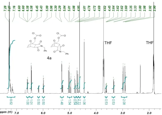
3): δppm 7.33-7.19 (m, 5H), 6.59 (dd, J = 5.5, 1.2 Hz, 2H), 6.44 (d, J = 5.5 Hz, 2H), 6.15 (dd, J = 5.9, 1.6 Hz, 1H), 5.98 (d, J = 5.9 Hz, 1H), 5.29 (d, J = 1.6 Hz, 2H), 5.26 (dd, J = 5.5, 1.6 Hz, 1H), 5.03-4.94 (m, 3H), 4.80-4.68 (m, 3H) 4.62 (s, 4H), 4.44 (s, 2H), 3.63 (dd, J = 7.8, 5.5 Hz, 1H), 3.34 (d, J = 7.8 Hz, 1H), 2.99-2.89 (m, 4H); 13C NMR (100 MHz, CDCl3): δppm 174.9, 173.6, 150.5, 138.0, 135.9, 135.0, 128.9, 128.6, 127.9, 127.8, 125.2, 88.8, 81.2, 49.7, 48.4, 46.2, 42.3;
HRMS (EI) calc’d. for [M]+ (C17H14ClNO5): 347.0561; Found: 347.0573.
Synthesis of compound 3b. N-propargyl maleimide3 (1.70 g, 12.6 mmol, 1.0 equiv.) and 2-(hydroxymethyl)furan (1.1 mL, 1.24 g, 12.6 mmol, 1.0 equiv.) were dissolved in anhydrous acetonitrile (10 mL) under a nitrogen atmosphere in a flame-dried flask equipped with a magnetic stirring-bar. The reaction was stirred at 35 ºC for 14 hours. When TLC indicated the reaction had reached equilibrium, the solvent was removed, and the reaction was concentrated under reduced pressure for 1 h. NMR spectroscopy of the unpurified product indicated a ratio of (3.7:0.7:1.0) of endo-exo-unreacted maleimide. The crude material was then purified by flash chromatography (1:1 to 4:6 hexanes-ethyl acetate) to provide 2.09 g of a mixture of the endo and exo in an 80:20 ratio and a 72 % isolated yield. A small amount of material was purified further by preparative TLC (7:3 hexanes-ethyl acetate, 4 elutions) to provide analytical samples of both the endo and exo products. Due to the inherent thermal instability, the material is stored at -20 ºC
until needed. 3b-endo 1H NMR (600 MHz, CDCl3): δppm 6.43 (dd, J = 5.8, 1.6 Hz, 1H), 6.31
(d, J = 5.8 Hz, 1H), 5.31 (dd, J = 5.5, 1.6 Hz, 1H), 4.26 (d, J = 12.8 Hz, 1H), 4.17 (d, J = 12.8 Hz, 1H), 4.06 (d, J = 2.5 Hz, 2H), 3.67 (dd, J = 7.6, 5.5 Hz, 1H), 3.48 (d, J = 7.6 Hz, 1H), 2.16
92.3, 79.7, 76.0, 71.4, 61.4, 50.1, 48.2, 27.6; HRMS (ESI): Calc’d for [M]+ (C12H11NO4): 233.0688. Found: 233.06824. 3b-exo 1H NMR (400 MHz, CDCl3): δppm 6.62 (d, J = 5.7 Hz, 1H), 6.55 (dd, J = 5.7, 1.6 Hz, 1H), 5.29 (d, J = 1.7 Hz, 1H), 4.25 (d, J = 2.5 Hz, 2H), 4.10 (d, J = 1.8 Hz, 2H), 3.68 (dd, J = 7.6, 5.5 Hz, 1H), 3.48 (d, J = 7.6 Hz, 1H), 2.21 (t, J = 2.5, 2.5 Hz, 1H), 1.64 (bs, 1H); 13C NMR (100 MHz, CDCl 3): δppm 174.5, 174.4, 138.3, 137.0, 91.5, 80.9, 76.2,
71.7, 60.5, 50.1, 48.1, 27.9; ESI (MS): Calc’d for [M]+ (C
12H11NO4): 233.0688; Found:
233.06892.
Synthesis of compound 4b. Compound 3b (500 mg, 2.15 mmol, 1.0 equiv.) was dissolved in
THF (10 mL). The resulting solution was then added dropwise into a phosgene solution (15 wt% in toluene, 4.6 mL, 6.44 mmol, 3.0 equiv.) under an argon atmosphere at room temperature and was stirred for 24 h. The residual phosgene and solvent were then removed by high vacuum to yield chloroformate 4b (610 mg, 97%) as a white gel. Phosgene collected in the liquid
nitrogen-cooled trap was then quenched with methanol (20 mL) and saturated sodium hydroxide
solution (20 mL). Caution! Phosgene is highly toxic. 1H NMR (600 MHz, CDCl3): δppm 6.66 (d,
J = 5.3 Hz, 2 H), 6.53 (d, J = 5.3 Hz, 1 H), 6.50 (d, J = 5.9 Hz, 2 H), 6.34 (d, J = 5.9 Hz, 1 H), 5.39 (d, J = 4.1 Hz, 1H), 5.35 (s, 2 H), 5.09-5.05 (m, 3 H), 4.88 (d, J = 12.3 Hz, 1H), 4.78 (d, J = 12.9 Hz, 2H), 4.25 (d, J = 2.9 Hz,4 H), 4.11 (d, J = 2.4 Hz, 3 H), 3.73 (dd, J = 5.3, 2.4 Hz, 1H), 3.46 (d, J = 7.6 Hz, 1H), 3.08 (d, J = 6.5 Hz, 2 H), 3.00 (d, J = 6.5 Hz, 2 H), 2.21 (t, J = 2.4 Hz, 1H), 2.19 (t, J = 2.4 Hz, 1H); 13C NMR (150 MHz, CDCl3): δppm 174.0, 172.8, 150.9, 138.4, 136.4, 133.7, 129.2, 128.4, 125.5, 89.3, 81.5, 76.2, 71.9, 68.1, 50.2, 48.8, 46.7, 28.3, 27.9;
HRMS (EI) calc’d. for [M]+ C13H10ClNO5: 295.0248; Found: 295.0257.
Synthesis of PEtG-DA-Bn and typical procedure for synthesis of end-capped PEtG. Ethyl
glyoxylate in toluene solution (20.0 mL) was distilled under vacuum (25 C, 0.3 mbar) over P2O5
to remove toluene and trace water in the first, discarded fraction. The residue was then distilled
twice successively over P2O5 at atmospheric pressure under argon at 130 C to obtain the highly
pure monomer. Purified ethyl glyoxylate (2.0 mL, 20 mmol, 1.0 equiv.) was dissolved in CH2Cl2
-20 C. Compound 4a (0.1 g, 280 µmol, 0.014 equiv.) and Et3N (38 µL, 280 µmol, 0.014 equiv.)
were added at -20 °C to end-cap the polymer. The solution was gradually warmed to room temperature and then stirred for 16 h. Purification was achieved by precipitation of the crude reaction mixture into methanol. After decanting the excess methanol, the residue was dried in
vacuo to provide 1.1 g of a white, sticky polymer in 55% yield. 1H NMR (600 MHz, CDCl 3): δ
7.28-7.32 (m, 10 H), 5.48-5.73 (m, 239 H), 4.15-4.31 (m, 449 H), 1.23-1.36 (m, 658 H). FT-IR:
2985, 1748, 1468, 1447, 1376, 1297, 1214, 1137, 1094, 1015, 989, 856, 675 cm-1. SEC: Mn = 33
kg/mol, Mw = 59 kg/mol, Đ = 1.8.
Synthesis of PEtG-DA-alkyne. The polymer is synthesized by the same procedure described for PEtG-DA-Bn except that compound 4b was used as the end-cap. The yield was 60%. 1H NMR
(600 MHz, CDCl3): δ 5.47-5.76 (m, 630 H), 4.14-4.29 (m, 1184 H), 1.24-1.40 (m, 1754 H). FTIR:
2985, 1748, 1468, 1447, 1376, 1297, 1214, 1138, 1094, 1016, 962, 857, 675 cm-1. SEC: Mn = 63
kg/mol, Mw = 130 kg/mol, Đ = 2.0.
Synthesis of Block Copolymer PEtG-DA-PEG750. 750 g/mol PEG-N3 (36.0 mg, 48 µmol, 6
equiv.) and PEtG-DA-alkyne (500.0 mg, 8 µmol, 1 equiv.) were dissolved in DMF (5.0 mL).
After removing the air and refilling with argon, CuSO4 (4.0 mg, 28 µmol, 3.5 equiv.) and sodium
ascorbate (5.0 mg, 28 µmol, 3.5 equiv.) were added into the solution, and the mixture was stirred at 40 C for 16 h. The reaction mixture was then transferred into a regenerated cellulose
membrane (50 kg/mol MWCO) and dialyzed against deionized water for 16 h (1 L, 2 solvent changes) to remove DMF and most free PEG. The dialyzed material was then lyophilized, washed 3 times with water to further remove free PEG, and then dried to afford 500 mg of the
product as a white, rubber-like, polymer in 98% yield. 1H NMR (600 MHz, CDCl3): δ 5.47-5.75
(m, 341 H), 4.07-4.33 (m, 704 H), 3.63 (s, 136 H), 3.37 (s, 6 H), 1.13-1.42 (m, 1075 H). 13C
NMR (150 MHz, CDCl3): δ 164.7-166.3, 90.3-94.7, 70.7, 62.3, 14. FTIR: 2985, 1750, 1468,
1447, 1376, 1298, 1216, 1138, 1017, 1095, 964, 857, 677 cm-1. SEC: Mn = 59 kg/mol, Mw = 111
Synthesis of PEtG-DA-PEG5000. The polymer was synthesized by the same procedure
described above for the synthesis of PEtG-DA-PEG750, except that 5000 g/mol PEG-N3 was
used. The yield was 98 %. 1H NMR (600 MHz, CDCl3): δ 5.45-5.70 (m, 670 H), 4.10-4.30 (m,
1399 H), 3.62 (s, 909 H), 3.36 (s, 3 H), 1.20-1.34 (m, 2110 H). 13C NMR (150 MHz, CDCl3): δ
164.6-166.7, 90.8-93.9, 70.5, 62.0, 13.8. FT-IR (thin film): 2985, 2874, 1750, 1468, 1448, 1376,
1298, 1216, 1136, 1095, 1018, 962, 856, 680 cm-1. SEC: M
n = 35 kg/mol, Mw = 71 kg/mol, Đ =
2.0.
Synthesis of Vesicle-control. The polymer was synthesized by the same procedure as
Micelle-control1a, except that 750 g/mol PEG-N3 was used. The yield was 86%. 1H NMR (600
MHz, CDCl3): δ 5.47-5.77 (m, 553 H), 4.07-4.36 (m, 1040 H), 3.65 (s, 136 H), 3.39 (s, 9 H),
1.13-1.47 (m, 1573 H). 13C NMR (150 MHz, CDCl3): δ 164.0-166.5, 90.0-94.2, 70.5, 62.0, 13.8.
FT-IR (thin film): 2985, 2874, 1752, 1467, 1448, 1376, 1297, 1217, 1140, 1096, 1018, 965, 855,
732 cm-1. SEC: M
n = 77 kg/mol, Mw = 177 kg/mol, Đ = 2.3.
Study of PEtG-DA-BnDepolymerization in Solution and Representative Procedure for Studying Depolymerization by NMR Spectroscopy. PEtG-DA-Bn (15.0 mg) was dissolved in
a 9:1 mixture of CD3CN: D2O (1.2 mL) at ambient temperature (22 C). The solution was then
divided between two NMR tubes. One tube was incubated at 75 C in an oven, while the other
one was stored at room temperature (22 C). 1H NMR spectra were recorded at defined intervals
to monitor the depolymerization of the materials. At the same time, benzyl chloroformate
end-capped PEtG (PEtG-control)1b was also subjected to the same procedure and its
depolymerization was monitored by NMR spectroscopy as non-triggerable control. The extent of depolymerization was calculated as % depolymerization = 100 – x, where x is the integration of the peak at 5.5 ppm, when the integration of the peak at 4.2 ppm was set to 200 (which remained
constant as it corresponds to the CH3CH2O- in both polymer and the depolymerization product).
Self-assembly of PEtG-PEG Block Copolymers. 8.0 mg of block copolymer was fully
Vesicle-control and DMSO for PEtG-DA-PEG5000 and Micelle-control). For vesicles, 0.9 mL
deionized water was added slowly into 0.1 mL of copolymer in THF with gentle stirring. For micelles, 0.1 mL of the copolymer in DMSO was injected quickly into 0.9 mL of rapidly stirring deionized water. After stirring for 10 minutes, the suspension was then dialyzed against
deionized water for 16 h (1 L, 2 solvent changes) to remove THF or DMSO, affording an aqueous suspension of assemblies.
Study of Assembly Decomposition by DLS. The assemblies were formed by the procedure
described above, except that the assemblies suspension was dialyzed against 100 mM pH 7.4 phosphate buffer solution. The assemblies were then transferred into plastic cuvettes and the CR was measured by DLS while fixing the attenuator. Samples were then incubated either at 75 C in an oven or at room temperature (22 C). CR changes were recorded at defined intervals to monitor the decomposition of assemblies.
Study of Nile Red Release.
A stock of copolymer assembly solution at a concentration of 0.8 mg/mL was prepared by the above standard self-assembly procedure in pH 7.4 phosphate buffer. 29 µL of a 0.1 mg/mL
solution of Nile red in CH2Cl2 was added to each of a series of vials and then the solvent was
evaporated to provide a thin film of Nile red. To each vial, 1.5 mL of assembly suspension was added, and the vials were gently shaken for 16 h to incorporate Nile red into the nanoparticles. After the initial fluorescence emission (600 nm) of the micelle suspension was measured using an excitation wavelength of 540 nm. Some samples were stored in an oven at 75 C, with others were stored at room temperature (22 C). The fluorescence emission was recorded at defined intervals to monitor the decomposition of assemblies.
Study of Assembly Depolymerization by 1H NMR Spectroscopy. 10.0 mg of block copolymer
was fully dissolved in 0.4 mL of DMSO-d6. 0.2 mL of the resulting solution was rapidly injected
into 1.0 mL of 100 mM, pH 7.4 phosphate buffered D2O. After stirring for 10 min, the micelle
while the other one was kept at room temperature. 1H NMR spectra were recorded at defined intervals to monitor the depolymerization of the materials. At the same time, Micelle-control was subjected to the same procedure. Percent depolymerization was determined using the sum of the integration of the methyl peaks corresponding to EtGH and ethanol (1.0-1.2 ppm), which plateaued at a very similar (1872) value to that of the methyl peak at 1.17-1.45 ppm in the block
copolymer taken in CDCl3 (integration 2111) when setting the PEG peak integral to 909. The %
polymer remaining was calculated as 100 - (sum of integration from 1.0-1.2 ppm/1872))´100.
“Polyol” Synthesis of Iron Oxide Nanoparticles4. 1.082 g (4.0 mmol) of FeCl3.6H2O and 398
mg (2.0 mmol) of FeCl2.4H2O were dissolved in a mixture of 40 g of di(ethylene glycol) (DEG)
and 40 g of N-methyldiethanolamine (NMDEA). Meanwhile, 640 mg (16 mmol) of NaOH was dissolved in a mixture of 20 g of DEG and 20 g of NMDEA. Both solutions were stirred
overnight under nitrogen flux to prevent the oxidation of Fe2+ species. The solutions were then
mixed and stirred for 3 h, before heating the mixture to 210 C with an oil bath, reflux set-up and mechanical agitation. The set-up was open and a flux of nitrogen helped to remove traces of water before temperature reaches 210 C. Once a temperature of 210 °C was reached, the set-up was closed, and 1 mL of water was injected in the system with a syringe through a septum, leading to a burst of nuclei. The formation of nanoparticles was carried on for 30 min, then the system was cooled to room temperature. The black sediment was separated magnetically and washed with a mixture of ethanol and ethyl acetate (1:1 v/v) 3 times. Possible iron hydroxides were removed by treatment with 10% nitric acid. The nanoparticles were then washed 2 times with acetone and 2 times with diethyl ether before being dispersed in water.
Coating of IONPs. 113 mg of Beycostat NE was deposited in a 50 mL beaker. 15.0 mL of water
was added and the solution was stirred with a mechanical agitator. 2.5 mL of a ferrofluid with an iron oxide concentration of 18.0 g/L (45 mg of iron oxide in solution) was added to the
surfactant. Then 2.6 mL of a 69 % w/w HNO3 solution was added to reach a final HNO3
concentration of 2.0 M. The solution was heated to 60 °C for 30 min with a water bath. The nanoparticles were then sedimented over a permanent magnet and washed 3 times with 50 mL of methanol, before being dispersed in 45 mL of dichloromethane or tetrahydrofuran (THF). The iron oxide content of this dispersion was estimated by dissolving 50.0 microliters of solution in 5.0 mL of 5.0 M HCl M with the help of a sonication bath. The absorption at 350 nm
correspondng to the peak of the [Fe(Cl)6]3- complex was compared to a calibration curve. The
final concentration was estimated at 6.0 g/L of Fe2O3.
Loading of IONPs into Micelles. 1.0 mg of PEtG-DA-PEG5000 or Micelle-control was
dissolved in 0.2 mL of THF, meanwhile IONPs (dispersed in THF with a concentration of 6.0 mg/mL) were mixed at different feed weight ratios (FWR) as needed with the polymer solution. This mixture was then added dropwise to 1.8 mL water via a micro-syringe while magnetically stirring. THF was allowed to evaporate by leaving the vials open for 24 h.
Dynamic Light Scattering Coupled with Magnetic Field Hyperthermia. The combined
DLS-MFH set-up based on a remote DLS setup VASCO Flex™ from Cordouan Technologies
(Pessac, France) has been previously reported5 and is shown in Fig. S40. Micelles loaded with 35
mass % IONPs were studied by DLS-MFH. The sample was heated up to the desired external temperature by a water bath. The temperature of the sample was measured with an optical fiber probe. After 1 h equilibration at the target temperature, the alternating magnetic field was applied at the maximum available amplitude of 10.2 kA/m and frequency of 755 kHz. Meanwhile, DLS was operating continuously at a backscattering angle of 165° to measure the sample diameter (Z-average), polydispersity index (PDI) and signal intensity changes during this period.
facility of LLB-CEA (Saclay, France) on the PAXY spectrometer equipped with a 2D
(anisotropic SANS) detector. Micelles were suspended at a concentration of 0.6 mg×mL-1 in pure
D2O, of neutron scattering length density SLD(D2O)=6.40´10-6 Å-2. The calculated SLD of iron
oxide and ethyl glyoxylate monomer are SLD(γ-Fe2O3)=6.98´10-6 Å-2 and SLD(EtG)=1.31´10-6
Å-2, respectively. The neutron scattering contrast of the micelles in heavy water thus arises
almost exclusively from the hydrophobic Poly(EtG) block of the polymer, the hydrophilic PEG block being highly hydrated, thus having negligible contribution to the neutron scattering contrast. Three beamline configurations were used to cover overlapping scattering vector (q) ranges of 1.92×10⁻³ – 2.84×10⁻², 1.05×10⁻² – 0.154, and 3.19×10⁻² – 0.427 Å-1, with the
following values of sample-to-detector distance D and neutron wavelength λ: D=7 m and λ=15 Å, D=3 m and λ=6 Å, D=1 m and λ=6 Å. The scattering intensity curves were divided by the
transmission factor and subtracted from the incoherent background, before normalizing by the flat signal of a cuvette filled with light water to correct the detector efficiency, yielding the absolute intensity in cm-1.
3. NMR Spectra of compounds and polymers
Figure S1. 1H NMR spectrum of Compound 3a-endo (CDCl
3, 600Hz).
Figure S3. 1H NMR spectrum of Compound 3a-exo (CDCl3, 600Hz)
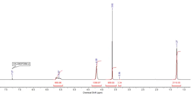
Figure S5. 1H NMR spectrum of Chloroformate 4a (CDCl3, 400Hz) (residual THF present).
Figure S6. 13C NMR spectrum of Chloroformate 4a (CDCl3, 100Hz).
Figure S7. 1H NMR spectrum of Compound 3b-endo (CDCl3, 600Hz).
Figure S9. 1H NMR spectrum of Compound 3b-exo (CDCl3, 400Hz).
Figure S11. 1H NMR spectrum of Chloroformate 4b (CDCl3, 400Hz).
Figure S13. 1H NMR spectrum of PEtG-DA-Bn (CDCl3, 600Hz).
Figure S15. 1H NMR spectrum of PEtG-DA-PEO750 (CDCl3, 600Hz). The success of the PEG
coupling is evidenced by the presence of the PEG peak at 3.6 ppm and its corresponding integration.
Figure S16. 13C NMR spectrum of PEtG-DA-PEO750 (CDCl3, 150Hz). The success of the PEG
coupling is evidenced by the presence of the PEG peak at 70 ppm.
Figure S17. 1H NMR spectrum of PEtG-DA-PEO5000 (CDCl3, 600Hz). The success of the
PEG coupling is evidenced by the presence of the PEG peak at 3.6 ppm and its corresponding integration.
Figure S18. 13C NMR spectrum of PEtG-DA-PEO5000 (CDCl3, 150Hz). The success of the
Figure S19. 1H NMR spectrum of Vesicle-control (CDCl3, 600Hz). The success of the PEG
coupling is evidenced by the presence of the PEG peak at 3.6 ppm and its corresponding integration.
Figure S20. 13C NMR spectrum of Vesicle-control (CDCl3, 150Hz). The success of the PEG
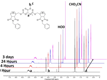
Figure S21. 1H NMR spectra of PEtG-DA-Bn incubated in 9:1 CD3CN:D2O at 22 C over time.
Spectra are offset to allow the progression over time to be clearly observed.
Figure S22. 1H NMR spectra of PEtG-DA-Bn incubated in 9:1 CD3CN:D2O at a) 40 C and b)
60 C. Spectra are offset to allow the progression over time to be clearly observed.
Figure S23. 1H NMR spectra of PEtG-control incubated in 9:1 CD3CN:D2O at 75 C. Spectra
are offset to allow the progression over time to be clearly observed.
Figure S24. 1H NMR spectra of PEtG-DA-alkyne incubated in 9:1 CD3CN:D2O at a) 75 C and
Figure S25. 1H NMR spectra of PEtG-DA-alkyne incubated in 9:1 CD3CN:D2O at a) 40 C and
b) 60 C. Spectra are offset to allow the progression over time to be clearly observed.
Figure S26. 1H NMR spectra of PEtG-DA-PEO5000 micelles incubated in 5:1 DMSO-d6:D2O
4. TGA curves of end-caps and polymers
Figure S28. TGA curves of thermo-responsive a) end-caps and polymers; b) homopolymer PEtG compared with the PEtG-PEG block copolymers. It can be noted that the polymers were more thermally stable than the DA adduct itself (compound 3a or 3b), which degraded at ~100 °C. There are two possible explanations for this phenomenon: 1) the polymer may serve as a matrix to protect the end-cap decomposition; 2) the elimination reaction following the retro-DA reaction is the rate-limiting step for thermal depolymerization. In the block copolymers, the presence of a second decomposition step at 300 °C provides can be attributed to the PEG block.
Table S1. Molecular weights, measured by SEC in THF relative to PMMA standards, for the
polymers, and thermal properties as measured by TGA. TO = onset degradation temperature
measured by TGA.
Polymer Mn (SEC) (kg/mol) Dispersity (Đ) TO (°C )
PEtG-DA-Bn 33 1.8 169 PEtG-control1b 42 1.4 N/A PEtG-DA-alkyne 63 2.0 154 PEtG-DA-PEG750 59 1.9 177 PEtG-DA-PEG5000 35 2.0 171 Micelle-control1a 40 2.1 N/A Vesicle-control 77 2.3 N/A
5. SEC traces for polymers
Figure S29. SEC curve of PEtG-DA-Bn.
Figure S30. Comparison of SEC curves of PEtG-DA-alkyne, PEG 750 g/mol, and
PEtG-DA-PEG750. No increase in molar mass was observed upon coupling of PEG, which is
consistent with previous work and can be attributed to the small mass fraction of the PEG as well as possible conformational changes that would cancel the effects of the addition of mass. This is
consistent with our previous reports.1a, 6
Figure S31. Comparison of SEC curves of PEtG-DA-alkyne, PEG 750 g/mol, and
PEtG-DA-PEG5000. As THF is not an ideal solvent for PEG, after coupling with PEG5000, the
hydrodynamic radius of PEG-DA-PEG5000 decreased rather than increased likely due to some polymer chain collapse. However the absence of a free PEG peak combined its presence in the NMR spectra clearly confirmed the presence of PEG in the block copolymer.
6. DLS distributions of all assemblies
Figure S33. DLS intensity (top) and volume (bottom) distributions for PEtG-DA-PEG750 vesicles.
Figure S34. DLS intensity (top) and volume (bottom) distributions for PEtG-DA-PEG5000 micelles.
Figure S35. DLS intensity (top) and volume (bottom) distributions for Vesicle-control.
Figure S36. DLS intensity (top) and volume (bottom) distributions for Micelle-control.
7. Additional TEM images
Figure S37. TEM images of a) PEtG-DA-PEG750 vesicles and b) PEtG-DA-PEG5000 micelles after incubation at 75 C for 16 h. No assemblies were observed.
Figure S38. TEM images of a) Vesicle-control and b) Micelle-control.
Figure S39. Photos of IONP-loaded PEtG-DA-PEG5000 micelles with increasing mass % of IONP.
Figure S41. Bulk temperature, particle diameter, and count rate measured before, during, and after magnetic hyperthermia using an in situ DLS for IONP-loaded PEtG-DA-PEG5000 with a bulk temperature of 53 °C.
8. Study of the thermal degradation by small angle neutron scattering
The thermal degradation of the thermosensitive micelles was also investigated by small-angle neutron scattering (SANS). Micelles loaded with IONPs were prepared as previously described,with few changes to adapt to this other characterization technique. The IONPs (dTEM=10.5 nm)
and the polymer were mixed in deuterated THF, and nano-precipitated in deuterated water to improve the contrast between the micelles and their solvent. Pure PEtG-DA-PEG5000 micelles
(BO14) or magnetically loaded thermosensitive micelles (BO15,35 mass% iron oxide relative to
polymer) were prepared this way, at a concentration of 0.6 mg×mL-1. The effect of long heating
and after heating at 80 °C in an oven for 30 min. The curves of micelles before heating were well fitted by a polydisperse sphere form factor multiplied by a “sticky hard sphere” structure factor to take into account short-range attractions. The fitted radius and volume fraction of the micelles
were respectively R0=10.9 nm and f=0.00028 (R0=11.7 nm and f=0.00030) for the unloaded
micelles BO14 (respectively magnetically loaded micelles BO15) with a high dispersity s=0.4
(Log-normal distribution) in both cases. The weight-average radius of the micelles that take into
account this Log-normal dispersity can be calculated using 𝑅w = 𝑅$ 𝑅% = 𝑅&∙ exp 7𝜎- 2 ,
leading to Rw=19.1 nm for pure micelles and Rw=20.5 nm for magnetic ones. These SANS sizes
are still much lower than the hydrodynamic radius of the micelles found around 50 nm. However, this can be explained by the relatively high hydration level of PEtG blocks compared to standard hydrophobic polymers, leading to a dominant contribution to the neutron scattering contrast from the dense dehydrated cores of the micelles compared to their hydrated shells. This is also why the fitted volume fractions around 0.03 vol% are about twice lower than expected from the 0.6 mg/mL total concentration in polymer.
The curves of the heat-treated micelles were not fitted by a model shape. Nonetheless, they can be interpreted by a drastic reduction of the volume fraction of suspended copolymer micelles, together with an increase of the size of the remaining objects (as ascribed to aggregation of the remaining IONPs). This SANS experiment thus brings another evidence of the thermosensitivity of PEtG-DA-PEG5000 copolymer micelles that did not disappear when embedding IONPs in their core.
Figure S42. SANS curves of pure micelles BO14 and magnetically loaded micelles BO15 before and after treatment at 80°C for 30 min (the SANS curves being acquired afterwards, at 20°C). The curves of the untreated micelles were fitted by a polydisperse sphere form factor multiplied by a “sticky hard sphere” structure factor. Solid lines represent simulated curves using the
SasView 3.1.2 software (http://sasview.org) with a polydisperse sphere form factor P(q)
multiplied by a “sticky hard sphere” structure factor S(q) to take into account short-range attractive interactions between the micelles.7 In addition to the SLDs that were fixed to their theoretical values, the parameters of the polydisperse sphere form factor were the volume
fraction f, the incoherent background level, the median radius R0 of the micelles and the width s
of the distribution of radii as described by a Log-normal law. Other fitting parameters of the
sticky hard sphere structure factor S(q) were the “stickiness” t=0.047±0.006 (respectively
t=0.036±0.0015) and the “perturbation distance” e=0.637±0.019 (respectively e=0.605±0.012) for the pure micelles BO14 (respectively magnetically loaded micelles BO15).