HAL Id: hal-02530966
https://hal-amu.archives-ouvertes.fr/hal-02530966
Submitted on 3 Apr 2020
HAL is a multi-disciplinary open access
archive for the deposit and dissemination of
sci-entific research documents, whether they are
pub-lished or not. The documents may come from
teaching and research institutions in France or
abroad, or from public or private research centers.
L’archive ouverte pluridisciplinaire HAL, est
destinée au dépôt et à la diffusion de documents
scientifiques de niveau recherche, publiés ou non,
émanant des établissements d’enseignement et de
recherche français ou étrangers, des laboratoires
publics ou privés.
Distributed under a Creative Commons Attribution| 4.0 International License
antidepressant treatment
Raoul Belzeaux, Victor Gorgievski, Laura Fiori, Juan Pablo Lopez, Julien
Grenier, Rixing Lin, Corina Nagy, El Chérif Ibrahim, Eduardo Gascon,
Philippe Courtet, et al.
To cite this version:
Raoul Belzeaux, Victor Gorgievski, Laura Fiori, Juan Pablo Lopez, Julien Grenier, et al..
GPR56/ADGRG1 is associated with response to antidepressant treatment. Nature Communications,
Nature Publishing Group, 2020, 11, �10.1038/s41467-020-15423-5�. �hal-02530966�
ARTICLE
GPR56/ADGRG1 is associated with response to
antidepressant treatment
Raoul Belzeaux
1,2,3
, Victor Gorgievski
4,5
, Laura M. Fiori
1
, Juan Pablo Lopez
1
, Julien Grenier
6
, Rixing Lin
1
,
Corina Nagy
1
, El Chérif Ibrahim
2,3
, Eduardo Gascon
2
, Philippe Courtet
3,7
, Stéphane Richard-Devantoy
1
,
Marcelo Berlim
1
, Eduardo Chachamovich
1
, Jean-François Théroux
1
, Sylvie Dumas
8
, Bruno Giros
1
,
Susan Rotzinger
9
, Claudio N. Soares
10,11
, Jane A. Foster
9
, Naguib Mechawar
1
, Gregory G. Tall
12
,
Eleni T. Tzavara
3,4,5,13
, Sidney H. Kennedy
9,10
& Gustavo Turecki
1,13
✉
It remains unclear why many patients with depression do not respond to antidepressant
treatment. In three cohorts of individuals with depression and treated with
serotonin-norepinephrine reuptake inhibitor (
N = 424) we show that responders, but not
non-responders, display an increase of GPR56 mRNA in the blood. In a small group of subjects we
also show that GPR56 is downregulated in the PFC of individuals with depression that died by
suicide. In mice, we show that chronic stress-induced Gpr56 downregulation in the blood and
prefrontal cortex (PFC), which is accompanied by depression-like behavior, and can be
reversed by antidepressant treatment. Gpr56 knockdown in mouse PFC is associated with
depressive-like behaviors, executive dysfunction and poor response to antidepressant
treatment. GPR56 peptide agonists have antidepressant-like effects and upregulated AKT/
GSK3/EIF4 pathways. Our
findings uncover a potential role of GPR56 in antidepressant
response.
https://doi.org/10.1038/s41467-020-15423-5
OPEN
1Douglas Mental Health University Institute, Department of Psychiatry, McGill University, Montreal, QC, Canada.2Aix-Marseille Univ, AP-HM, CNRS, INT,
Inst Neurosci Timone, Hôpital Sainte Marguerite, Pôle de psychiatrie, Marseille, France.3Fondation FondaMental, Créteil, France.4CNRS (Integrative
Neuroscience and Cognition Center, UMR 8002), Paris, France.5Université Paris Descartes, Sorbonne Paris Cité, Paris, France.6INSERM UMR-S 1124 ERL
3649, Université Paris Descartes, Paris, France.7Department of Emergency Psychiatry and Acute Care, Lapeyronie Hospital, CHU Montpellier,
Montpellier, France.8Oramacell, 75006 Paris, France.9Centre for Mental Health, Department of Psychiatry, University Health Network, Krembil Research
Institute, University of Toronto, Toronto, ON, Canada.10St Michael’s Hospital, Li Ka Shing Knowledge Institute, Centre for Depression and Suicide Studies,
Toronto, ON, Canada.11Department of Psychiatry, Queen’s University, Kingston, Ontario, Canada.12Department of Pharmacology, University of Michigan,
Ann Arbor, MI, USA.13These authors jointly supervised this work: Eleni T. Tzavara, Gustavo Turecki. ✉email:gustavo.turecki@mcgill.ca
123456789
M
ajor depressive disorder (MDD) is a common
psy-chiatric disorder
1and one of the leading causes of
disability worldwide
2. Antidepressants are the
first-line treatment for moderate to severe major depressive episodes
(MDE)
3, and although they are effective, not every patient
responds to antidepressant treatment. Approximately 40%
of patients respond to their
first antidepressant trial, and
fol-lowing multiple trials, response rates increase up to 70%
4.
Antidepressants are thought to act through modulation of
mono-amines, but the precise mechanisms whereby they affect
therapeutic response, as well as the underlying causes of
treatment-response variability, remain poorly understood.
Therefore, there is an important need to better understand
molecular pathways and mechanisms involved in antidepressant
response. In this study, we examined peripheral gene expression
in three cohorts of individuals with MDD undergoing
anti-depressant treatment and identified one gene, G-protein coupled
receptor 56 (GPR56) whose expression was consistently
asso-ciated with antidepressant response. We further characterized
the function and signaling properties of this gene in vivo and
in vitro, and found it to be related to depressive-like behaviors
and executive functioning, and to upregulate classical
anti-depressant signaling pathways upon activation.
Results
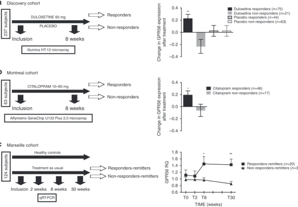
Gene expression analysis in the discovery cohort.
Anti-depressant response involves a complex interplay between genetic
and environmental factors. Using a double-blind, randomized
clinical trial design (Fig.
1
a), we
first set out to investigate mRNA
changes associated with antidepressant response in patients
undergoing a MDE (N
= 237) treated with either the
anti-depressant duloxetine (N
= 112), a serotonin-norepinephrine
reuptake inhibitor (SNRI), or with placebo (N
= 125), over eight
weeks. After treatment, 89 (79.5%) and 51 (40.8%) patients were
responders in the duloxetine and placebo arms, respectively
(Supplementary Table 1). Using the Human HT-12 v4 Expression
Bead Chip (Illumina), we found 42 probes, corresponding to 41
different annotated genes, that were overexpressed following
duloxetine treatment in responder patients (FDR < 1%). Two of
these probes were also upregulated in the placebo group, but
among non-responders, while the remaining 40 were specifically
overexpressed in duloxetine responders (Supplementary Table 2).
No downregulated probes were found by our initial analysis, while
1752 and 1670 probes were found to be downregulated and
overexpressed, respectively, using a t-test without correction (p <
0.05). G protein-coupled receptor 56 (GPR56), also known as
ADGRG1, a G protein-coupled receptor of the adhesion class, was
the most significantly upregulated mRNA based on fold change
(FC) and q-value during duloxetine treatment (FC
= 1.19, q-value
< 0.01; Fig.
1
a). Using a general linear model (GLM) for repeated
measures, we confirmed that GPR56 was specifically increased
only in patients who responded to duloxetine (F(1,199)
=
8.47, p
= 0.004; Fig.
1
a). These results were technically validated
using qRT-PCR (Supplementary Fig. 1) and were not explained by
potential clinical or biological confounders including sex, age, BMI,
or sample cellular composition (Supplementary Fig. 2).
Replication of overexpression of GPR56 during antidepressant
response. We next investigated whether the results observed in
our discovery cohort could be replicated in two independent
cohorts with similar clinical characteristics, but treated with
dif-ferent antidepressants. The Montréal cohort consisted of patients
treated with the selective serotonin reuptake inhibitor (SSRI)
citalopram over 8 weeks (N
= 63, Fig.
1
b). Similar to the
dis-covery cohort, we found that peripheral levels of GPR56 mRNA
significantly increased after treatment only in patients who
responded to antidepressant treatment (F(1,61)
= 4.273, p =
0.043; Fig.
1
b). Our third cohort (Marseille cohort) was designed
as a naturalistic 30-week-follow-up study, which investigated
patients with depression (N
= 64) and healthy subjects (N = 87)
(Fig.
1
c). Patients received antidepressant treatment as prescribed
by their physician/psychiatrist (Supplementary Table 3). As
observed in the other two cohorts, we found that GPR56 mRNA
levels significantly increased as a function of response after
8 weeks (N
= 30, FC = 1.26; paired t-test t = −2.52, p = 0.018)
while no change were observed in non-responders (N
= 34, t =
0.35, p
= 0.73). Interestingly, we also saw no change in untreated
healthy control subjects (t
= 0.50, p = 0.62). A GLM for repeated
measures confirmed a significant time × group interaction (F
(2,148)
= 4.98, p = 0.008; Supplementary Fig. 3). Moreover, we
found that GPR56 mRNA remained stably overexpressed over a
30-week-follow-up among those who initially responded and then
achieved remission after 30 weeks of treatment
(responders-remitters, N
= 20) in comparison to others (N = 44) (linear
mixed model including 0 week, 2 week, 8 week and
30-week-follow-up, F(1,230.199)=14.79, p < 0.001; Fig.
1
c).
Effects of unpredictable chronic mild stress (UCMS) on Gpr56.
Our results above suggest that GPR56 may be involved in
mechanisms associated with antidepressant response that are
common to different classes of antidepressants, but interestingly,
not involved in mechanisms of placebo response. To further
examine the potential function and regulation of Gpr56 in
depression and antidepressant response, we conducted studies in
animals, using the unpredictable chronic mild stress (UCMS)
paradigm, a well validated murine model of depression
5, followed
by treatment with
fluoxetine, a standard SSRI, to model
anti-depressant effects (Fig.
2
a). In this model, stress-exposure leads to
depressive-like behaviors (displayed as increased anhedonia and/
or resignation) that can be alleviated by subsequent
administra-tion of an antidepressant. We have previously adapted this model
to distinguish between responder and non-responder mice
5.
Here, chronic stress-induced depressive-like behaviors were
effectively reversed in 60% (responders) of the
fluoxetine-treated
mice. Thus, we investigated peripheral Gpr56 mRNA levels in
mice subjected to UCMS and found a significant decrease in mice
that manifested depressive-like symptoms as compared to
non-stressed mice (FC
= 0.81; Fig.
2
b). Interestingly, reversal of the
depressive-like behaviors with antidepressant treatment was
paralleled by normalization of blood Gpr56 mRNA expression in
responder mice, i.e., demonstrating improvement in
depressive-like phenotype. In contrast, blood Gpr56 mRNA levels remained
low in non-responder mice, in close analogy to the Gpr56
expression biosignature seen in the human studies detailed above
(F
= 6.15, p = 0.001; Fig.
2
b).
Using the same model, we then sought to investigate the effects
of stress-induced depression and antidepressant treatment on
Gpr56 expression in the central nervous system (CNS). We
focused on four regions of interest: the dorsal and ventral
hippocampal areas (HD and HV, respectively), the prefrontal
cortex (PFC) and the Nucleus Accumbens (NAcc), all previously
implicated in stress and depression, albeit in a different manner
6.
A repeated measures two-way ANOVA analysis between groups
and brain regions showed a significant interaction between brain
region and phenotypes (F(9,151)
= 3.112; p = 0.0018) (Fig.
2
c).
Subsequent post hoc analyses showed that stress-induced a
significant reduction in Gpr56 expression in the PFC (FC = 0.65,
p
= 0.0006) and no effect in the NAcc (p = 0.08). In the
hippocampus we observed a difference in the HD (FC
= 0.62,
p
= 0.0001), in accordance with a previous study
7, but not in the
HV (p
= 0.88). In the PFC, we observed a bimodal regulation of
Gpr56 mRNA by UCMS and antidepressant treatment. UCMS
exposure led to reduced Gpr56 mRNA expression (Fig.
2
c), which
was normalized by antidepressant administration in responder
mice, but not in non-responder mice (Fig.
2
b), a pattern
remarkably similar to that seen in the mouse and human blood
samples. In contrast, in the HD, while UCMS induced a
downregulation of Gpr56 mRNA, antidepressant treatment had
no effect in responder mice (p
= 0.38). Although brain-blood
correlation of gene expression remains a matter of debate, it is
worth noting that Gpr56 mRNA levels were significantly
correlated between the PFC and peripheral blood in stressed
mice (r
= 0.51; p = 0.02; Supplementary Fig. 4) while in control
mice, we found no correlation between blood and PFC (p
= 0.21).
Taken together, our results suggest that an increase in Gpr56
expression levels may be an integral part of effective
antidepres-sant action. Our results also suggest a specific role for the PFC in
relation to Gpr56 mRNA variation in depressive-like behaviors
and antidepressant action, as we found no significant effect of
antidepressant-related regulation of Gpr56 in several other brain
regions, including the HD.
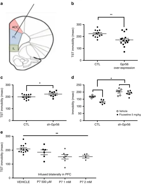
Effects of Gpr56 over-expression and knockdown on mouse
behavior. To investigate a possible causal relationship between
Gpr56 mRNA variation in the PFC and behavioral responses to
stress, we used a viral vector strategy to locally manipulate Gpr56
expression levels selectively in the PFC, and thus determine the
influence of increased or decreased expression of Gpr56 on
depressive-like behaviors and/or antidepressant action in the
mouse (Fig.
3
and Supplementary Fig. 5). In naive mice, bilateral
PFC infusions of a lentivirus-Gpr56 construct resulted in PFC
Gpr56 overexpression (FC
= 2.02, t = 4.09, p = 0.003,
Supple-mentary Fig. 5B), while bilateral PFC infusions of a
lentivirus-sh-Gpr56 construct resulted in PFC lentivirus-sh-Gpr56 downregulation (FC
=
0.49, t
= 3.37, p = 0.007, Supplementary Fig. 5C). Behavioral
analysis showed that PFC Gpr56 downregulation was sufficient to
produce depressive-like behaviors in unstressed mice, as seen by
increased immobility in the TST (Fig.
3
b, t
= 2.2, p = 0.048). This
test is among the most commonly used procedures to detect
clinically effective antidepressant agents because of its high degree
of predictive validity, and has been previously used to identify
mouse strains that are resistant or hyporesponsive to treatment
8.
In the same test, PFC Gpr56 overexpression in naive mice
induced the opposite effect, namely decreased immobility, the
hallmark effect of antidepressant action (Fig.
3
c, t
= 3.07, p =
0.005). In both cases there was no effect on locomotor activity,
indicating no change in general ambulatory behavior
(Supple-mentary Figs. 6A and 7A), but rather a targeted effect of Gpr56
PFC manipulations on stress-triggered behaviors. This
observa-tion was further strengthened by similar results in the forced
Discovery cohort Montreal cohort Marseille cohort DULOXETINE 60 mg CITALOPRAM 10–60 mg Healthy controls Treatment as usual PLACEBO Inclusion 8 weeks InclusionInclusion 2 weeks 8 weeks 30 weeks qRT-PCR 8 weeks Responders 0.4 Duloxetine responders (n=75) Citalopram responders (n=46) Citalopram non-responders (n=17) Duloxetine non-responders (n=21) Placebo responders (n=44) Placebo non-responders (n=63) 0.2 0.0 –0.2 –0.4 0.4 0.2 0.0 –0.2 –0.4 1.8 GPR56 RQ 1.6 1.4 1.2 1.0 0.8 0.6 T0 T2 T8 TIME (weeks) T30 Change in GPR56 e xpression after treatment Change in GPR56 e xpression after treatment Non-responders Responders Responders-remitters Non-responders-remitters Responders-remitters (n=20) Non-responders-remitters (n=34) Non-responders Illumina HT-12 microarray
Affymetrix GeneChip U133 Plus 2.0 microarray
237 subjects 63 subjects 124 subjects
a
b
c
Fig. 1GPR56 mRNA is related to antidepressant response. a In the discovery cohort, 237 patients in a major depressive episode were randomized to
double-blind treatment with either duloxetine (n = 112) or placebo (n = 125), for up to 8 weeks. Using two-class paired significant analysis of microarray
(SAM) with correction for multiple testing (FDR < 1%) in patients who responded to duloxetine,GPR56 mRNA is the most significantly upregulated mRNA
in whole blood after duloxetine treatment, based on fold change andq-value (FC = 1.19, q-value < 0.01). General linear model (GLM) demonstrated a time x
treatment interaction,F(1,199)=8.468, p = 0.004, that confirms the specificity of GPR56 mRNA increase in responders to duloxetine. b In the first
replication cohort (Montréal), patients were treated with citalopram in an open-label trial.GPR56 mRNA in whole blood demonstrated an increase only in
responders. GLM demonstrated a time x group interaction,F(1,61)=4.27, p = 0.043 (not adjusted for multiple testing). c In the second replication cohort
(Marseille), psychiatrically healthy subjects and patients with depression were included in a naturalistic design. In patients who responded and achieved
remission after 30 weeks of treatment (responders-remitters,n = 20), GPR56 mRNA is not different at inclusion and 2 weeks, however was then
overexpressed at 8 weeks in comparison to others (n = 44) (two-sided t-test t = 2.085, p = 0.049) and remained stably overexpressed over a 30-week
follow-up (n = 18 responders-remitters in comparison to others n = 31) two-sided t-test t = 3.076, p = 0.005); Linear Mixed model (F(1,230.199) = 14.79,
p = 0.0001). Bars represent mean. Error bars represent standard error of the mean. **p < 0.01, *p < 0.05. Source data are provided as a Source Data file.
UCMS + Vehicle 4 weeks
Test and sacrifice
a
b
c
Responders or non-responders UCMS + Fluoxetine 4 weeks
UCMS alone 3 weeks
1.5 1.0 GPR56 RQ GPR56 RQ 0.5 0.0 2.5 Non-stressed (n=16) UCMS vehicle (n=16) UCMS fluoxetine non-responders (n=6) UCMS fluoxetine responders (n=8) 2.0 1.5 1.0 0.5 0.0 PFC HV NAcc HD
Non-stressed (PFC, HV and HC n=17, NAcc n=12) UCMS vehicle (PFC, HV and HC n=12, NAcc n=11)
UCMS fluoxetine responders (PFC n=8, HV and HC n=9 and NAcc n=7) UCMS fluoxetine non-responders (n=6 for all regions)
Fig. 2 Unpredictable chronic mild stress (UCMS) and antidepressant response dysregulateGpr56 mRNA in blood and CNS in mice. a Gpr56 expression
was analysed by qRT-PCR in blood and several brain regions, including the dorsal and ventral hippocampal areas (HD and HV, respectively), the prefrontal
cortex (PFC) and the Nucleus Accumbens (NAcc) in non-stressed or stressed mice (exposed to UCMS) and receiving vehicle orfluoxetine. Mice treated
byfluoxetine were classified as “responders” or “non-responders” according to behavioral tests. b In whole blood, a one-way ANOVA showed between
group differences forGpr56 expression (F = 6,150, p = 0.001). Blood Gpr56 mRNA expression was decreased in mice subjected to UCMS, while reversal of
depressive-like behaviors withfluoxetine was paralleled by normalization of blood Gpr56 mRNA expression in responder mice (post hoc analysis p < 0.01).
c In brain, a two-way ANOVA between group and brain regions showed a significant interaction between brain region and mice group (F(9,151) = 3.112;
p = 0.0018). Post hoc analysis demonstrated a specific PFC effect, a decrease of Gpr56 in PFC between stressed and non-stressed mice, with a reversal effect of antidepressant only in responder mice. Sample numbers vary between tissues due to removal of poor quality RNA samples from the analyses. Bars
represent mean. Error bars represent standard error of the mean. **p < 0.01. Graph represents Box and Whiskers Min to Max. Source data are provided as
swim test (FST) for both up- and downregulation of Gpr56
(Supplementary Figs. 6B and Fig. 7B). We also found behavioral
effects for Gpr56 downregulation in the sucrose preference and
O-maze tests (Supplementary Fig. 7C–D). Cognitive symptoms
are often associated with MDE and may have prognostic and
therapeutic implications, in particular related to executive
func-tioning and PFC funcfunc-tioning
9. As a consequence, we also
con-ducted a set shifting test (SST), a well validated cognitive test in
mice related to PFC functioning, in sh-Gpr56-PFC mice
10.
Interestingly, in these mice PFC specific Gpr56 downregulation is
accompanied by impairments in the SST (Supplementary Fig. 8).
Overall, these results indicate that Gpr56 downregulation in the
PFC of non-stressed mice produces depressive-like responses
similar to those induced by UCMS. On the contrary, Gpr56
overexpression in the PFC of naive mice produces behaviors
similar to those elicited by classical antidepressants in the TST or
FST.
In order to directly probe the link between increased PFC
Gpr56 mRNA and antidepressant response, mice
under-expressing Gpr56 were tested for their response to
fluoxetine in
the TST. Mice infused in the PFC with control virus or with
sh-Gpr56-virus were injected acutely with saline or
fluoxetine (5 mg/
kg) 30 min before the TST trial. Fluoxetine decreased immobility
in control animals, while this effect was strongly attenuated in
300
a
b
c
e
d
200 TST immobility (msec) TST immobility (msec) TST immobility (msec) Immobility (msec) 100 ACC PL IL 250 200 150 100 50 0 0 300 200 100 0 300 200 100 0 CTL CTL VEHICLE Vehicle Fluoxetine 5 mg/kg P7 500 μM Infused bilaterally in PFC P7 1 mM P7 2 mM CTL sh-Gpr56 sh-Gpr56 Gpr56 over-expressionFig. 3Gpr56 regulates depressive-like behaviors. a Mice were injected bilaterally in the pre-frontal cortex with control (CTL) or lentivirus-Gpr56
constructs, as well as with CTL or lentivirus-sh-Gpr56 (inhibitor) constructs. b Overexpression of Gpr56 (n = 15) was associated with lower immobility time
in the TST in comparison to control animals (n = 12), two-sided t = 3.07, p = 0.005. c Downregulation of Gpr56 (n = 8) produced increased immobility
time, i.e. depressive-like behavior in the tail suspension test (TST) in comparison to control animals (n = 11), two-sided t = 2.203, p = 0.048. d Fluoxetine
decreased immobility in control animals (n = 5), while this effect was strongly attenuated in sh-Gpr56-virus infused animals (n = 6); two-way ANOVA for
repeated measures; treatment × group interactionF(1,9) = 6.80, p = 0.028; main effects for group F (1,9) = 25.4, p = 0.001 (as compared to control) and
for treatmentF(1,9) = 6.80 p = 0.001 (as compared to vehicle). e GPR56 agonist P7 has antidepressant-like effects. GPR56 agonist P7 peptide infused
bilaterally with escalating doses in PFC decreases immobility time and demonstrates antidepressant-like effects, ANOVAF(1,4) = 4.88 p = 0.008. In
comparison to vehicle (n = 13), both 1 mM (n = 7) and 2 mM (n = 4) doses demonstrate a significant decrease of immobility time (p < 0.01 and p = 0.01
respectively, two-sided post hoc test). Bars represent mean. Error bars represent standard error of the mean. **p < 0.01, *p < 0.05. Source data are
provided as a Source Datafile.
sh-Gpr56-virus infused animals (treatment*group interaction F
(1,9)
= 6.80, p = 0.028; Fig.
3
d).
Overall, we showed opposite and bidirectional associations
between Gpr56 expression in the PFC, and depressive- and
antidepressant- like responses. Indeed, chronic stress decreased
Gpr56 expression in the PFC whereas antidepressant response
normalized this downregulation. Gpr56 overexpression in the
PFC produced antidepressant-like effects, whereas downregulated
Gpr56 in the PFC produced depressive-like behavior, executive
function alterations and impaired antidepressant response. Thus,
our animal-model results suggest that Gpr56 may have an
important role in the adaptations to stress or in depressive-like
behaviors and antidepressant response, and that the PFC is a key
region involved in these effects.
Effects of Gpr56 agonist treatment on mouse behavior.
Fol-lowing activation of the GPR56 receptor by its ligands, the
extracellular and transmembrane domains of GPR56 dissociate to
reveal a tethered-peptide-agonist
11. Based on this mechanism,
synthetic
peptides
(i.e.,
P7
“TYFAVLM-NH2” and P19
“TYFAVLMQLSPALVPAELL-NH2”), comprising the specific
portion of the tethered-peptide-agonist, have been generated and
demonstrate GPR56 agonist properties
11,12. We bilaterally
infused these peptides and their inactive controls in the mouse
PFC to explore the behavioral effects of GPR56 activation.
Par-allel to our results with Gpr56 overexpression, behavioral analyses
showed that GPR56 agonists produced antidepressant-like effects
in unstressed mice, as seen by decreased immobility in the tail
suspension test (TST) for P7 with a dose-response profile (Fig.
3
e,
ANOVA F(1,4)
= 4.88 p = 0.008) and for P19 (Supplementary
Fig.
9).
Interestingly,
we
confirmed the specificity of
antidepressant-like effects of GPR56 in the PFC by using the same
peptides infused in the NAcc, which produced no behavioral
effects (Supplementary Fig. 10). Finally, the antidepressant-like
effect of the GPR56 agonists was not explained by basic
loco-motion differences, as we did not
find any differences in
ambu-lations across time between active peptides and their controls
(Supplementary Fig. 11). These data provide evidence that
acti-vation of GPR56 through pharmacological manipulation by
GPR56-specific ligands has antidepressant-like-effects, specifically
in the PFC. These experiments further support a role of GPR56 in
depressive-like behaviors and antidepressant response.
Further-more, they indicate that GPR56 may represent a molecular target
for treatment of MDD.
GPR56 expression in depressed human brains and relation to
executive function. Similar to our results in mice (Fig.
2
), GPR56
is expressed in all brain regions in humans (Supplementary
Fig. 12). Therefore, we next investigated the expression of GPR56
in the PFC (BA44) from individuals who died during an episode
of MDD and compared them with psychiatrically healthy
indi-viduals. We found that GPR56 expression was significantly lower
in cases in comparison to controls (FC
= 0.56, U = 385, Z =
−2.81, p = 0.005, Fig.
4
a). Controlling for covariates and possible
confounders, such as age, sex, PMI and tissue pH, did not have an
impact on our results (F(1,69)
= 4.91, p = 0.030).
A central role of the PFC is executive function. Thus, we
hypothesized that variation of GPR56 levels associated with
antidepressant response may result in cognitive changes in
patients, a hypothesis consistent with previous data suggesting
that improvement in executive functioning is associated with
antidepressant response
9. Neuropsychological testing was
con-ducted in a subset of individuals who participated in the
duloxetine trial. We investigated executive functioning using the
Stroop interference test, and analyzed the data as a function of
GPR56 mRNA variation. Variation in the Stroop interference
score was negatively correlated with variation of GPR56 mRNA
levels (r
= −0.71, p = 0.009, Fig.
4
b), associating increased GPR56
mRNA with improved executive functions, a
finding that mirrors
results in the mouse. A partial correlation analysis confirmed this
result after correction by response status, age, and gender
(correlation
= −0.796, df = 7, p = 0.01).
GPR56 agonists upregulate AKT/GSK3/EIF4 pathways in
neuroblastoma cells. In order to gain a more complete
under-standing of downstream signaling processes initiated by GPR56
activation, we investigated the transcriptional consequences of
treatment with the two GPR56 agonists described above. These
experiments were performed in vitro, using a human
neuro-blastoma cell line, which was treated with the agonist peptides for
24 h then examined using RNA sequencing. We used these cells
because they are derived from neural cells, express GPR56
receptors, and express genes from several important pathways
that have been associated with antidepressant response, including
the serotonin signaling pathway
13,14.
To functionally characterize the gene expression variation
associated with agonist-induced activation of GPR56, we used
Gene Set Enrichment Analysis
15. 6,568 gene sets with sizes
between 15 and 500 genes were included in the analysis after gene
set size
filtering. Among them, we identified significant
enrich-ment of nine gene sets (FWER < 0.20, Suppleenrich-mentary Table 4).
Interestingly, AKT, GSK3 and EIF4 pathways demonstrated the
highest normalized enrichment scores and lowest FWER p-values
for upregulated gene sets. These pathways were upregulated in
cells treated with the agonists, in comparison to control
conditions. These pathways are highly related and have been
described as downstream biological mechanisms involved in
depression and antidepressant action of several different drugs,
including SSRIs and ketamine
16–19. As a consequence, our results
suggest that GPR56 agonists may have antidepressant effects
through pathways that are similar to those activated by
established antidepressants.
Discussion
Taken together, our preclinical and clinical results identified
GPR56 as a player in depressive symptomatology and a key
mediator of antidepressant response in blood and in the brain. In
blood, GPR56 mRNA increased in parallel to antidepressant
response and could be used to monitor antidepressant response.
In the brain, namely in the PFC, decreased GPR56 expression
associated with depression in humans or depressive-like
beha-viors in mice, whereas in mice increased PFC GPR56 expression
was necessary and sufficient for antidepressant action, an effect
that might involve cognitive modulation. Using two agonist
peptides, we confirmed in mice that activation of GPR56 in the
PFC is associated with behavioral responses that are commonly
associated with antidepressant treatment. Moreover, based on
cell experiments and RNA sequencing, we found that GPR56
agonists upregulated AKT-GSK3-EIF4 pathways, downstream
biological mechanisms previously associated with depression
and antidepressants action
16–19. Although we did not examine
these pathways in vivo, it is possible that the upregulation of
these pathways explains the antidepressant effects that were
observed following agonist treatment. Overall, our results suggest
that
GPR56
is
a
potential
target
for
development
of
antidepressant drugs.
GPR56 is involved in a number of biological functions relevant to
the pathophysiology of depression, including neurogenesis,
oligo-dendrocyte development and progenitor cell migration in brain, as
well as myelin repair
20–23, in parallel to its important role in
immune cell functioning
24–26. GPR56 ligands comprise two general
subtypes: (1) proteins from the surface of neighboring cells, and (2)
extracellular matrix proteins. Known extracellular ligands of GPR56
include collagen III, transglumatinase 2, and heparin
23,27,28.
Fol-lowing activation of the GPR56 receptor by its ligands, the
extra-cellular and transmembrane domains dissociate to reveal a
tethered-peptide-agonist
11. To date, it remains unclear which ligand could be
related to depressive behavior and antidepressant effects of GPR56
in the PFC. GPCRs are particularly appealing drug targets. While
further efforts are needed to develop compounds with optimised
pharmacokinetic properties for in vivo administration, our
findings
identify a previously unsuspected GPCR as a possible target for
antidepressants.
Our study does have several limitations. Firstly, the human
cohorts have relatively modest sample sizes that may limit the
generalisation of our results. However, we replicated our
findings in
three relatively different treatment cohorts. Secondly, as we only
conducted these studies using SSRI and SNRI antidepressants, we
cannot conclude if GPR56 is involved in antidepressant response in
general or limited to specific antidepressant classes. Thirdly, GPR56
is expressed in numerous cell types and tissues, and is involved in
different processes that may be related to antidepressant
response
21–25. In the brain, single-cell sequencing data from the
frontal cortex indicates that GPR56 is expressed in all cell types,
including glutamatergic neurons, and astrocytes, where relatively
higher levels are observed
29. These data are consistent, although not
fully concordant, with results we observed in the mouse using
fluorescence in situ hybridization (Supplementary Fig. 13). In the
future, cell-type specific studies should be conducted in order to
elucidate the mechanisms through which GPR56 is involved in
depression and antidepressant response. It remains unclear how
GPR56 may be regulated by antidepressants in both the brain and
blood. Moreover, it is unclear if variation in the blood only mirrors
a process in the brain, or reflects an active biological process that
influences antidepressant response. Fourthly, we conducted our
animal experiments to test the causal relationship between Gpr56
under- and over-expression, as well as pharmacological testing, only
in acute stress paradigms of depressive-like symptoms (i.e.,
TST, FST).
Despite these limitations, by integrating several clinical
cohorts, animal studies and post-mortem brain analyses, our
results provide a greater understanding of the pathophysiology of
depression, and suggest a drug target for the treatment of MDD.
Methods
Study design. Participants: Our study involved three cohorts of living subjects and one cohort of post-mortem brain samples. Informed written consent was obtained from all participants, or next-of-kin. Each study was approved by the appropriate ethics committee.
The discovery cohort consisted of 237 patients in a MDE (69.6% female) who were randomized to double-blind treatment with either duloxetine (60 mg die,
N= 112), a SNRI, or placebo (N = 125), for up to 8 weeks (www.ClinicalTrials.
gov11984ANCT00635219; 11918ANCT00599911; 13267ANCT01140906).
These studies were sponsored by Lundbeck and samples were provided as a donation to the Canadian Biomarker Integration Network in Depression program. Patients were excluded from the study if they suffered from bipolar disorder, schizophrenia and/or comorbid substance use disorder. Depressive symptoms were assessed using the Montgomery Åsberg Depression Rating Scale (MADRS) at baseline and at the end of the trial. The mean age was 46.8 years ± 12.8 SD. Before treatment, mean depression severity level using MADRS total score was 31.2 ± 3.7 SD, ranging from 26 to 46. Patients were classified as
responders or non-responders according to a MADRS reduction of≥50% or
<50%, respectively. Haematological data (blood cell counts) were collected at inclusion and at the end of treatment.
Thefirst replication cohort (Montréal cohort) was an independent group of 63
patients treated with citalopram, a SSRI, in an open-label trial previously
published30. This study was conducted in Douglas Mental Health Institute with the
necessary approval from the hospital ethics and internal review board. In this study, patients experiencing a MDE received citalopram for 8 weeks. The same exclusion criteria as above were applied. At the end of the follow-up, patients were classified as responders (N = 46) or non-responders (N = 17), according to a
Hamilton Depression Rating Scale 21 items (HDRS-21) reduction of≥50% or
<50%, respectively.
Our second replication cohort (Marseille cohort) was a naturalistic prospective cohort that included 64 patients and 87 healthy controls
(ClinicalTrials.govNCT02209142)31. Patients were included during a MDE with
HDRS-17 > 19 at the inclusion. Patients were excluded from this study if they suffered from bipolar disorder, schizophrenia and/or comorbid substance use disorder. All patients were treated at the inclusion with treatment as usual upon discretion of the treating psychiatrist (Supplementary Table 3). Healthy controls were free of any psychiatric disorder according to a semi-structured interview. All subjects included in the analysis were followed for 30 weeks with four points of evaluation (i.e., inclusion, 2 weeks later, 8 weeks later and 30 weeks later). After an 8-week-follow-up, 30 patients were classified as responders according to
a HDRS-17 reduction of≥50%. At the end of the study (30 weeks), 20 patients
were classified as responders at 8 weeks that achieved remission at 30 weeks (i.e.,
HDRS-17≤ 7) and 44 patients were classified as never responders or responders
without remission.
The post-mortem cohort was comprised of 75 post-mortem PFC samples (Brodmann Area 44) obtained from the Douglas-Bell Canada Brain Bank (Douglas Mental Health University Institute, Montreal, Quebec, Canada). Ethics approval was obtained from the institutional review board of the Douglas Mental Health University Institute. Brain pH and post-mortem interval (PMI) were used as tissue integrity measures. Subjects were either individuals who were
suffering from a MDE at time of death by suicide (N= 49), or psychiatrically
normal controls (N= 26), as assessed by psychological autopsies using DSM-IV
critepost-mortem intervalria. 8
a
b
1.5 1.0 0.5 –0.5 –1.0 –0.5 0.0 0.5 1.0 0.0 GPR56 RQ Δ Stroop interf erence before and after dulo
x
etine
Δ GPR56 expression before and after duloxetine 6
4
2
0
C MDD
Fig. 4GPR56 expression is altered in the prefrontal cortex from post-mortem brain tissue of individuals with depression and is related to pre-frontal
cortex functioning in patients. aGPR56 expression was measured by qRT-PCR in post-mortem brain tissue (BA44). Expression was lower in individuals
with depression (MDD,n = 49) in comparison to psychiatrically healthy controls (n = 26). FC = 0.56, two-sided U = 385, Z = −2.81, p = 0.005. Graph
represents Box and Whiskers Min to Max.b Changes in Stroop interference score, a neuropsychological test that involves pre-frontal cortex function, were
correlated with changes inGPR56 expression in whole blood following antidepressant treatment (n = 12 from discovery cohort, Pearson coefficient of
correlation= −0.71, two-sided p = 0.009). Reduction of interference score was associated with an improvement of Stroop interference test, i.e., an
improvement in pre-frontal cortex functioning. Source data are provided as a Source Datafile.
Sample collection and processing. Whole blood samples were collected using PAXgene Blood RNA Tubes (PreAnalytix®) for the discovery cohort and Montréal cohort, while Marseille samples were collected in EDTA tubes and later processed
using Leukolockfilters. Brain tissues (post-mortem cohort) were processed and
dissected at 4 °C, then snap-frozen in liquid nitrogen before storage at−80 °C.
Total RNA was extracted using the Qiagen miRNeasy Micro Kit (discovery, Montreal, post-mortem cohorts) or Ambion spin columns (Marseille cohort), with DNase treatment. RNA integrity was evaluated using an Agilent Bioanalyzer. All samples had a RNA integrity number (RIN) > 6.
Microarray quantification and data processing. RNA from the discovery cohort
was hybridized to the Illumina Human-HT-12 v4 microarray. Samples were ran-domized to avoid batch effects. All array probes and samples were subjected to
quality control using Flexarray® package implemented in R (version 1.6.3). Data
were normalized using background adjustment and log2 transformation, variance stabilization transformation (VST) correction, and quantile normalization. A
principal component analysis was used to identify outliers resulting in identi
fica-tion and exclusion of 13 samples. After exclusion of outliers, 443 remaining samples were re-used in the same normalization procedure, comprising 237 dif-ferent individual subjects. In total, 47,323 probes were present in the microarray.
All probes werefiltered using a detection P value < 0.01 in at least 10% of the
samples, resulting in available expression data for 16,674 remaining probes. Gene expression from the Montréal cohort was measured in whole blood using the Affymetrix GeneChip Human Genome U133 Plus 2.0 array as previously
described30. We used normalized andfiltered data to assess the expression of
GPR56 in this data set.
Quantitative real-time polymerase chain reaction (qRT-PCR). For the dis-covery and post-mortem cohorts, total RNA was reverse-transcribed using M-MLV Reverse Transcriptase (200 U/uL) (ThermoFisher®) and oligo (dT) 16 primers (Invitrogen). Real-time PCR (qRT-PCR) were run in triplicate using the
Quant-StudioTM6 Flex System and data collected using QuantStudioTMReal-Time PCR
Software v1.1. Expression levels were calculated using the absolute (standard curve
method) or relative (2−ΔΔCt) quantification method, depending on experimental
design. GAPDH was used as the endogenous control. The following primers were used in the study: GPR56 (FW: CCCATCTTTCTGGTGACGCT; REV: GATCC AGCACATGGAAGGGT) and GAPDH (FW: TTGTCAAGCTCATTTCCTGG; REV: TGTGAGGAGGGGAGATTCAG).
For the Marseille cohort, total RNA was reverse transcribed using the High-Capacity cDNA Reverse Transcription kit (Life Technologies, Applied Biosystems, Foster City, CA). Real-time PCR reactions were performed in duplicate using the TaqMan Universal PCR Master Mix II with no UNG (Life Technologies, Applied Biosystems, Foster City, CA), with an ABI PRISM 7900HT thermocycler under the following conditions: 10 min at 95 °C, 50 cycles of 15 s at 95 °C and 1 min at 60 °C. Primers/TaqMan probe assays (Hs00173754_m1) purchased from Applied Biosystems were used to determine the level of expression of GPR56 transcripts. We
used CRYL1 (Hs00211084_m1) as a reference gene as previously described32.
Expression levels were calculated using the relative (2−ΔΔCt) quantification method.
Evaluation of executive functioning. For a sub-sample of the discovery cohort, executive functioning was evaluated using a standard color-word Stroop task at
inclusion and after treatment (N= 12). In this task, the participant is asked to
name the colors of a series of words“red,” “green,” and “blue” as quickly as
possible without making mistakes. During a congruent task, the actual observed colors of the words match the colors that the words denote, while during an incongruent task, the series of color words does not match with the actual color. The interference Stroop score was calculated as the difference between time of reading during incongruent and congruent tasks. As such, the higher the inter-ference score is, the greater the impairment of executive functioning.
Mouse studies. Animals: All mice used were male adults (3–6-months old). The
following strains were used: C57Bl6, and BALB/cJico. All UCMS experiments were
performed using BALB/c mice, as previously published33. Targeted manipulations
of Gpr56 expression in the PFC through lentiviral particle infusions were per-formed using C57Bl6. The mice were kept under standard conditions at 22 ± 1 °C, and a 12-h light-dark cycle with food and water available ad libitum except when food/water deprivation was part of the experimental protocol. Humidity levels were between 45 and 55%. Behavioral assessments were performed during the second half of the light phase. All animal protocols and welfare complied with French and European Ethical regulations. The experimental protocols were approved by the local Ethical Committee (Comité d'éthique en expérimentation animale Charles Darwin N°5).
Unpredictable chronic mild stress (UCMS): After a two week acclimation period BALB/c male mice (8-week old) were individually housed and subjected to
UCMS as described33. Stressors, typically wet bedding, tilted cages, lights on at
night, crowding, difficult access to food, paired housing with intruder restraint and forced swim were applied twice a day for a two hour period and overnight in a randomized order. No food deprivation was used. Control (non-stressed) mice were standard housed in a room distinct to that of the stressed mice. Throughout
the UCMS protocol the animal’s weight was measured every five days. At the end of the chronic stress protocol the emotional state of the animals was evaluated in
the TST and the sucrose preference tests as described33. The stress procedure was
maintained with items compatible with behavioral testing. Control (non-stressed) mice were left undisturbed throughout the protocol.
Pharmacological effects and response to antidepressants in UCMS-subjected mice were assessed with a reversal protocol (45 days of UCMS; treatment during
the last 3 weeks) as previously described5. Namely, mice were subjected to the
UCMS-protocol for 45 days starting Day 0. During thefirst 3 weeks there was no
treatment for any of the groups. From week 4 until the end of the protocol, mice
were treated daily with saline or the reference antidepressantfluoxetine at 15
mg/kg, i.p., daily. In the literature it has been reported thatfluoxetine
administered in a reversal mode exerts a bimodal effect; it elicits a response in a sub-group of responder mice and has no effect on a distinct sub-group of
non-responders34. We therefore sought to distinguish betweenfluoxetine responders
and non-responders for subsequent qPCR determinations of Gpr56 mRNA expression.
Lentiviral particle-infusions in the PFC: Lentiviral particles: The following commercially available lentiviral particle solutions were used: for Gpr56
overexpression, we used the Adgrg1 (NM_018882) Mouse Tagged ORF Clone
Lentiviral Particle, >107TU/mL (Origene CAT#: MR210044L2V), and for Gpr56
downregulation, we used Gpr56 shRNA (m) Lentiviral Particles (Santa Cruz: sc-60750-V).
Infusions: For stereotaxic delivery, mice were anesthetized with a ketamine/ xylazine mixture (100/10 mg/kg, i.p.) and then given bilateral microinjections of
0.8μl/side of lentivirus solution at a rate of 0.1 μl/min. The following stereotaxic
coordinates were used for viral delivery:+1.9 mm (anterior/posterior), +0.75
(lateral),−2.75 (dorsal/ventral) at an angle of 15° from the midline (relative to
bregma). Animals were left to recover for 4–5 weeks before behavioral testing. The
correct placement of the injection site was verified histologically at the end of the
experiments (Fig. S5A).
Gpr56 agonist: To test the antidepressant-like effect of Gpr56 agonists, we bilaterally infused synthetic peptides (P7 and P19) as well as control or an inactive modified peptide (P19 Y ->N: “TNFAVLMQLSPALVPAELL-NH2”) previously
described12, both in the PFC and in the NAcc of mice. Mice were anesthetized with
a ketamine/xylazine mixture (100/10 mg/kg, i.p.) and stereotaxically implanted
with 12 mm long canulae in the left and right PrL Area (anterior (AP)+ 1.9 from
the bregma; lateral (ML)+/− 0.5; ventral (DV) −1.3) or in the left and right
nucleus accumbens (NAcc) (AP+ 1,6; ML +/− 0.7; DV −3.3). Animals were left
to recover for at least 7 days. On the test day, infusion needles (30 Gauge) were inserted into the canulae (needles were 13 mm long i.e. ending 1 mm deeper than the guide canulae) and mice were locally infused with a pump (UNIVENTOR), at a
rate of 0.5μl/min, with P7 (0.5 mM, 1 mM or 2 mM) or vehicle (vehicle: 80%
saline+ 10% DMSO + 10% Cremophor), or with P19 (1 mM) or its inactive
control peptide P19YN (1 mM). The needles were left in place for another 2 min to ensure compound diffusion. Mice were subsequently placed in their cage until the TST session (30 min after infusion).
Behavioral studies: Behavioral testing was performed using 7–22 animals per group.
Tail suspension test (TST): Immobility was measured in the TST as previously
described35. Mice were individually tail-suspended by using a paper adhesive tape
that was placed 1 cm from the tip of the tail, in such manner as to down rate the probability of the mice reaching their tail. Immobility time (seconds) was measured during a 6 min test period. In case of the mouse catching its tale the measure was discarded. We tested 8–15 mice/group.
Forced swimming test (FST): The forced swimming test was conducted in
clear plastic cylinders (diameter 20 cm; height 25 cm)filled with 6 cm of water
(22–25 °C) for 6 min. The duration of immobility was measured manually during
the last 4 min of the 6 min trial. A mouse was regarded as immobile whenfloating
motionless or making only those movements necessary to keep its head above the water. We tested 11–22 mice/group.
Locomotor activity: Horizontal activity (ambulations) was assessed in transparent activity cages (20 × 15 × 25 cm), with automatic monitoring of photocell beam breaks (Imetronic, France). Locomotor activity (ambulations defined as breaking two consecutive beams) was recorded for a 1-h period and we
conducted analyses between groups, both for thefirst 6 min and the total duration
of the test. We tested 7–21 mice/group.
O-maze: The O-maze consisted of a white circular path (runway width 5.5 cm,
Ø= 46 cm) with two opposing compartments protected by walls (height = 10 cm)
and two open sectors of equal size. The maze was elevated 40 cm above the ground and illuminated from the top with white light (50 Lux). At the start of the testing session, mice were placed at the end of one of the two closed compartments. The test was recorded with a camera for 5 min. We tested 10–18 mice/group.
Sucrose consumption: For the sucrose preference test mice werefirst habituated
to drink from two graduated pipettes: onefilled with water, and the other with
sucrose solution for 3 days. The side of the sucrose pipette was alternated each day. On day 4 and after an overnight (15 h) deprivation of water, the two pipettes were
presented again; however, one wasfilled with water and the other with 4% sucrose.
The water and sucrose solution consumed over a 3-h period were measured. The
sucrose preference index is defined as (sucrose consumed)/(sucrose consumed +
Cognitive testing: To test the impact of mouse PFC Gpr56 inhibition on cognition, we used an attentional set shifting test (ASST) for mice as previously
described10,36. The extra-dimensional shift task of the ASST has been associated
with medial-frontal lesion and is considered as a measure of executive functioning associated with PFC in primate and rodent animal models.
Measurement of Gpr56 expression: RNA extractions: Brain punches were made with a Rodent Brain Matrice, ASI Instruments RBM-2000C, and a Harris
Uni-Core, Hole 1.0 mm biopsy tool. Regions were punched according to37. Total RNA
from tissue punches was obtained using Trizol® reagent (Invitrogen, France) and 1 µg was reverse transcribed with random primers from Biolabs (Beverly, MA) and Reverse Transcriptase MLV-RT from Fisher scientific (France), according to manufacturers’ instructions.
Real-time quantitative polymerase chain reaction (qRT-PCR): qRT-PCR was performed to assess Gpr56 expression using SYBR green (ABgene, France) on an ABI PRISM 7000. Each reaction was performed in triplicate and the mean of at least three independent experiments was calculated. All results were normalized to
26S or Gapdh and calculated using the relative (2−ΔΔCt) quantification method.
The primer sequences used in real time PCR are: 26S (FW: AGGAGAAACAACGG TCGTGCCAAAA, REV: GCGCAAGCAGGTCTGAATCGTG), and GPR56 (FW: TCCAGGCATACTCGCTGTTGCT, REV: CTTCTCACCCAGGACTTGGCTA). Cell experiments. Cell Culture: Human neuroblastoma cells (SK-N-AS, ATCC CRL-2137) were cultured in Dulbecco’s Modified Eagle Medium (DMEM) sup-plemented with 10% FBS, 1% non-essential amino acids, 100 U/ml penicillin and
100μg/ml streptomycin (Invitrogen) in a 5% CO2humidified incubator at 37 °C.
Cells were treated with 25 µM of peptide (P7, P19, P19Y N) or vehicle (DMSO) for 24 h then collected in TRI reagent. RNA was extracted using the DirectZol kit with DNase treatment (Zymo). Three experiments were performed in triplicate. For sequencing, we pooled the triplicates from each experiment.
RNA sequencing: All libraries were prepared using the NEB mRNA stranded
protocol following the manufacturer’s instructions. Samples were sequenced at the
McGill University and Genome Quebec Innovation Centre (Montreal, Canada) using the Illumina HiSeq4000 with 100nt paired-end reads. Based on the number of reads, their length and the estimated human exome size being around 3 Mb, the average sequencing depth across all samples is 115×.
FASTX Toolkit (v0.0.14) and Trimmomatic (v0.36) were respectively used for quality and adapter trimming. TopHat (v2.1.1), using Bowtie2 (v2.2.9) was used
to align the cleaned reads to the reference genome (GRCh38,https://www.ncbi.
nlm.nih.gov/assembly/GCF_000001405.39). Reads that lost their mates through the cleaning process were aligned independently from the reads that still had
pairs. Quantification on each gene’s expression was estimated using
HTSeq-count and a reference transcript annotation from ENSEMBL (v77). Counts for the paired and orphaned reads for each sample were added to each other. Genes
counts for each sequenced library were normalized using DESeq2’s median ratio
normalization method38.
To facilitate downstream analyses, we chose to correct our normalized counts
for the effect of potential covariates using limma’s removeBatchEffect function39.
We specifically regressed out the effects of a possible batch effect associated with
the cell culture as well the expected heterogeneity associated with the use of the two different peptides and their respective controls. Our analysis demonstrated that GPR56 was expressed in each cell line.
Gene set enrichment analysis: To functionally characterize the gene expression variation associated with GPR56 agonist treatment, we used gene set enrichment
analysis15. Based on the largest differences in expression between cells receiving
agonist or not, GSEA allowed us to calculate enrichment for predefined gene sets related to functional pathways based on enrichment scores and p-values, as well as
Familywise-error rate FWER p < 0.2040, adjusted for gene set size and multiple
hypotheses testing. We used gene sets previously described41.
Statistical analysis. Data collection was conducted using Excel 2013 or Epidata V3.1.
For the discovery cohort, data were expressed as proportions and frequency for categorical variables or means and standard deviations (SD) for continuous variables. Differences between groups were compared using Chi-square tests for categorical data or two-sided t-tests for continuous variables. Repeated measures were analyzed using paired t-tests. Moreover, group by time interactions were evaluated using GLM for repeated measures. If necessary, potential confounding factors according to univariate analyses and/or current knowledge were also added in GLM. Correlations between continuous variables were conducted using Pearson correlation coefficient calculation and partial correlation analysis to include potential confounding factors.
For microarray expression data analysis in the discovery cohort, wefirst used
two-class paired significant analysis of microarray (SAM)42to determine
differential gene expression before and after treatment in the duloxetine responders group with MultiExperiment Viewer 4 (MeV4, TM4 software suite). False discovery rate (FDR) threshold was set at 1% and q-values were computed. To identify specific probes which were differentially expressed between the two time points in the duloxetine responders group, we also performed two-class paired t-test comparisons between both times in the duloxetine non-responders group, the placebo responders group, and the placebo non-responders group. To confirm
specificity of gene expression variation across time in the duloxetine responders group, we built a GLM for repeated measures including all available samples to identify the effect of time, group (placebo or duloxetine) and response, as well as the interaction between them. To control for potential confounding factors, we also included age, gender and BMI.
For the replication cohorts (Montréal cohort and Marseille cohort), we performed a two-paired t-test to identify gene expression variation across time in responders, non-responders and healthy controls. To confirm specificity in the responders, we built a GLM for repeated measures as described above. For the Marseille cohort, we also conducted a Linear Mixed Model to compare GPR56 mRNA between groups across the four visits.
For the post-mortem cohort, we used a Mann–Whitney test, according to the skewness of the data, to compare the level of expression of GPR56 between healthy controls and individuals with depression who died by suicide. We log2-transformed raw data to achieve a normal distribution, in order to conduct a GLM to control for potential confounding factors.
For animal experiments, to compare behavior measures and gene expression levels, we used t-tests, one-way or two-way ANOVAs and adapted post hoc test. Correlation analyses were conducted using Pearson’s coefficient calculation.
All statistical analyses were conducted using SPSS V21 and GraphPad Prism 5 and p-value < 0.05 was considered as significant, except where noted.
Reporting summary. Further information on experimental design is available in the Nature Research Reporting Summary linked to this paper.
Data availability
The source data underlying Figs.1a–c,2b, c,3b–e,4a, b, Supplementary Figs. 1–12 and
Supplementary Tables 1 and 2 are provided as a Source Datafile. Microarray data
obtained in this study are available under accession codeGSE146446.
Received: 5 February 2019; Accepted: 10 March 2020;
References
1. Waraich, P., Goldner, E. M., Somers, J. M. & Hsu, L. Prevalence and incidence
studies of mood disorders: a systematic review of the literature. Can. J.
Psychiatry 49, 124–138 (2004).
2. WHO. Depression and Other Common Mental Disorders: Global Health
Estimates (2017).
3. Kennedy, S. H. et al. Canadian Network for Mood and Anxiety Treatments
(CANMAT) 2016 Clinical Guidelines for the Management of Adults with Major Depressive Disorder: Section 3. Pharmacological Treatments. Can. J. Psychiatry 61, 540–560 (2016).
4. Rush, A. J. et al. Acute and longer-term outcomes in depressed outpatients
requiring one or several treatment steps: a STAR*D report. Am. J. Psychiatry 163, 1905–1917 (2006).
5. Apazoglou, K. et al. Antidepressive effects of targeting ELK-1 signal
transduction. Nat. Med. 24, 591–597 (2018).
6. McIntosh, A. L., Gormley, S., Tozzi, L., Frodl, T. & Harkin, A. Recent advances
in translational magnetic resonance imaging in animal models of stress and depression. Front. Cell Neurosci. 11, 150 (2017).
7. Suzuki, G. et al. Stress and electroconvulsive seizure differentially alter GPR56
expression in the adult rat brain. Brain Res. 1183, 21–31 (2007).
8. Svenningsson, P. et al. Involvement of striatal and extrastriatal DARPP-32 in
biochemical and behavioral effects offluoxetine (Prozac). Proc. Natl Acad. Sci.
USA 99, 3182–3187 (2002).
9. Wagner, S. et al. Early improvement of executive test performance during
antidepressant treatment predicts treatment outcome in patients with major depressive disorder. PLoS ONE 13, e0194574 (2018).
10. El Khoury, M. A., Gorgievski, V., Moutsimilli, L., Giros, B. & Tzavara, E. T. Interactions between the cannabinoid and dopaminergic systems: evidence from
animal studies. Prog. Neuropsychopharmacol. Biol. Psychiatry 38, 36–50 (2012).
11. Stoveken, H. M., Hajduczok, A. G., Xu, L. & Tall, G. G. Adhesion G protein-coupled receptors are activated by exposure of a cryptic tethered agonist. Proc.
Natl Acad. Sci. USA 112, 6194–6199 (2015).
12. Stoveken, H. M., Larsen, S. D., Smrcka, A. V. & Tall, G. G. Gedunin- and Khivorin-derivatives are small-molecule partial agonists for adhesion G protein-coupled receptors GPR56/ADGRG1 and GPR114/ADGRG5. Mol. Pharm. 93, 477–488 (2018).
13. Gupta, M. et al. TSPAN5, ERICH3 and selective serotonin reuptake inhibitors in major depressive disorder: pharmacometabolomics-informed
pharmacogenomics. Mol. Psychiatry 21, 1717–1725 (2016).
14. Qi, X. R. et al. Abnormal retinoid and TrkB signaling in the prefrontal cortex in mood disorders. Cereb. Cortex 25, 75–83 (2015).
15. Subramanian, A. et al. Gene set enrichment analysis: a knowledge-based approach for interpreting genome-wide expression profiles. Proc. Natl Acad. Sci. USA 102, 15545–15550 (2005).
16. Zanos, P. & Gould, T. D. Mechanisms of ketamine action as an antidepressant.
Mol. Psychiatry 23, 801–811 (2018).
17. Beaulieu, J. M. A role for Akt and glycogen synthase kinase-3 as integrators of dopamine and serotonin neurotransmission in mental health. J. Psychiatry
Neurosci. 37, 7–16 (2012).
18. Gould, T. D. & Manji, H. K. Glycogen synthase kinase-3: a putative molecular target for lithium mimetic drugs. Neuropsychopharmacology 30, 1223–1237 (2005).
19. Aguilar-Valles, A. et al. Translational control of depression-like behavior via phosphorylation of eukaryotic translation initiation factor 4E. Nat. Commun. 9, 2459 (2018).
20. Bae, B. I. et al. Evolutionarily dynamic alternative splicing of GPR56 regulates regional cerebral cortical patterning. Science 343, 764–768 (2014). 21. Bai, Y., Du, L., Shen, L., Zhang, Y. & Zhang, L. GPR56 is highly expressed in
neural stem cells but downregulated during differentiation. Neuroreport 20,
918–922 (2009).
22. Giera, S. et al. The adhesion G protein-coupled receptor GPR56 is a cell-autonomous regulator of oligodendrocyte development. Nat. Commun. 6, 6121 (2015).
23. Giera, S. et al. Microglial transglutaminase-2 drives myelination and myelin repair via GPR56/ADGRG1 in oligodendrocyte precursor cells. Elife 7, e33385 (2018).
24. Peng, Y. M. et al. Specific expression of GPR56 by human cytotoxic lymphocytes. J. Leukoc. Biol. 90, 735–740 (2011).
25. Hamann, J. et al. International Union of Basic and Clinical Pharmacology. XCIV. Adhesion G protein-coupled receptors. Pharm. Rev. 67, 338–367 (2015).
26. Della Chiesa, M. et al. GPR56 as a novel marker identifying the CD56dull
CD16+ NK cell subset both in blood stream and in inflamed peripheral
tissues. Int. Immunol. 22, 91–100 (2010).
27. Luo, R. et al. G protein-coupled receptor 56 and collagen III, a receptor-ligand pair, regulates cortical development and lamination. Proc. Natl Acad. Sci. USA 108, 12925–12930 (2011).
28. Chiang, N. Y. et al. Heparin interacts with the adhesion GPCR GPR56, reduces receptor shedding, and promotes cell adhesion and motility. J. Cell Sci. 129, 2156–2169 (2016).
29. Habib, N. et al. Massively parallel single-nucleus RNA-seq with DroNc-seq. Nat. Methods 14, 955–958 (2017).
30. Mamdani, F. et al. Gene expression biomarkers of response to citalopram treatment in major depressive disorder. Transl. Psychiatry 1, e13 (2011). 31. Consoloni, J. L. et al. Serotonin transporter gene expression predicts the
worsening of suicidal ideation and suicide attempts along a long-term
follow-up of a major depressive episode. Eur. Neuropsychopharmacol. 28, 401–414
(2018).
32. Belzeaux, R. et al. Responder and nonresponder patients exhibit different peripheral transcriptional signatures during major depressive episode. Transl. Psychiatry 2, e185 (2012).
33. Farley, S., Apazoglou, K., Witkin, J. M., Giros, B. & Tzavara, E. T. Antidepressant-like effects of an AMPA receptor potentiator under a chronic mild stress paradigm. Int J. Neuropsychopharmacol. 13, 1207–1218 (2010). 34. Dournes, C., Beeske, S., Belzung, C. & Griebel, G. Deep brain stimulation in
treatment-resistant depression in mice: comparison with the CRF1 antagonist,
SSR125543. Prog. Neuropsychopharmacol. Biol. Psychiatry 40, 213–220 (2013).
35. Crozatier, C. et al. Calcineurin (protein phosphatase 2B) is involved in the
mechanisms of action of antidepressants. Neuroscience 144, 1470–1476
(2007).
36. Birrell, J. M. & Brown, V. J. Medial frontal cortex mediates perceptual
attentional set shifting in the rat. J. Neurosci. 20, 4320–4324 (2000).
37. Paxinos, G. & Franklin, K. B. J. The Mouse Brain in Stereotaxic Coordinates, 2nd edn. (Academic Press, 2001).
38. Love, M. I., Huber, W. & Anders, S. Moderated estimation of fold change and dispersion for RNA-seq data with DESeq2. Genome Biol. 15, 550 (2014). 39. Ritchie, M. E. et al. limma powers differential expression analyses for
RNA-sequencing and microarray studies. Nucleic Acids Res. 43, e47 (2015). 40. Benjamini, Y. & Hochberg, Y. Controlling the false discovery rate: a practical
and powerful approach to multiple testing. J. R. Stat. Soc. Ser. B (Methodol.)
57, 289–300 (1995).
41. Merico, D., Isserlin, R., Stueker, O., Emili, A. & Bader, G. D. Enrichment map: a network-based method for gene-set enrichment visualization and interpretation. PLoS ONE 5, e13984 (2010).
42. Tusher, V. G., Tibshirani, R. & Chu, G. Significance analysis of microarrays applied to the ionizing radiation response. Proc. Natl Acad. Sci. USA 98, 5116–5121 (2001).
Acknowledgements
We are grateful to all patients for their involvement in this research. This manuscript
contains data from four registered clinical trials:www.ClinicalTrials.gov NCT00635219,
NCT00599911,NCT01140906, andNCT02209142. Clinical trialsNCT00635219,
NCT00599911, andNCT01140906were sponsored by Lundbeck, and samples were provided as a donation to the Canadian Biomarker Integration Network in Depression (CAN-BIND) program. This research was conducted with the support of CAN-BIND, an Integrated Discovery Program, with funding from the Ontario Brain Institute, an
independent non-profit corporation, funded partially by the Ontario government. RB
received a grant from Fondamental Foundation (Post-doctoral fellowship). RB and EI were supported by the Agence Nationale de la Recherche (ANR-13-SAMA-0002). VG holds a Labex-Biopsy Fellowship. ETT is supported by Fondation de Recherche sur le Cerveau and Fondation de France. ETT is a past recipient of the Bodossakis Foundation Young Scientist Award. RB, ECI, ETT and GT were supported by ERA-NET NEURON (Grant ANTARES). GT holds a Canada Research Chair (Tier 1) and a NARSAD Dis-tinguished Investigator Award. He is supported by grants from the Canadian Institute of Health Research (CIHR) (FDN148374 and EGM141899), and by the Fonds de recherche
du Québec– Santé (FRQS) through the Quebec Network on Suicide, Mood Disorders
and Related Disorders. We would like to thank ML Niepon (Institute of Vision, Paris) for slide scanning for the FISH experiments.
Author contributions
R.B., E.T.T., S.D.H., G.G.T., and G.T. designed the study. R.B., V.G., L.M.F., E.C.I., E.G., E.T.T., and G.T. wrote the manuscript. R.B., E.C.I., P.C., S.R.D., M.B., E.C., S.D.H., and G.T. conducted the clinical studies (inclusion, evaluation of subjects and analyses of clinical data). R.B., E.C.I., and J.P.L. performed the sample processing from clinical studies. R.B., J.P.L., R.L., and E.C.I. performed the gene expression studies in the clinical cohorts. R.B., J.P.L., C.N., and N.M. performed the experiments on post-mortem brain samples. V.C. and J.G. performed and analysed the animal experiments. L.M.F. and R.L. performed the cell experiments. G.G.T. produced agonist peptides. S.D. performed FISH analyses. R.B., V.G., L.M.F., E.T.T., and J.F.T. performed the statistical analyses. C.N.S., S.R., J.A.F., and B.G. contributed to study design and writing the manuscript.
Competing interests
The authors declare no competing interests.
Additional information
Supplementary informationis available for this paper at
https://doi.org/10.1038/s41467-020-15423-5.
Correspondenceand requests for materials should be addressed to G.T.
Peer review informationNature Communications thanks Ronald Duman and the other,
anonymous, reviewer(s) for their contribution to the peer review of this work. Peer reviewer reports are available.
Reprints and permission informationis available athttp://www.nature.com/reprints
Publisher’s note Springer Nature remains neutral with regard to jurisdictional claims in
published maps and institutional affiliations.
Open Access This article is licensed under a Creative Commons
Attribution 4.0 International License, which permits use, sharing, adaptation, distribution and reproduction in any medium or format, as long as you give appropriate credit to the original author(s) and the source, provide a link to the Creative Commons license, and indicate if changes were made. The images or other third party
material in this article are included in the article’s Creative Commons license, unless
indicated otherwise in a credit line to the material. If material is not included in the
article’s Creative Commons license and your intended use is not permitted by statutory
regulation or exceeds the permitted use, you will need to obtain permission directly from
the copyright holder. To view a copy of this license, visithttp://creativecommons.org/
licenses/by/4.0/. © The Author(s) 2020