HAL Id: inserm-02180285
https://www.hal.inserm.fr/inserm-02180285
Submitted on 11 Jul 2019
HAL is a multi-disciplinary open access archive for the deposit and dissemination of sci-entific research documents, whether they are pub-lished or not. The documents may come from teaching and research institutions in France or abroad, or from public or private research centers.
L’archive ouverte pluridisciplinaire HAL, est destinée au dépôt et à la diffusion de documents scientifiques de niveau recherche, publiés ou non, émanant des établissements d’enseignement et de recherche français ou étrangers, des laboratoires publics ou privés.
Slimane Allali, Céline Dietrich, François Machavoine, Rachel
Rignault-Bricard, Valentine Brousse, Mariane de Montalembert, Olivier
Hermine, Thiago Trovati Maciel, Maria Leite-De-Moraes
To cite this version:
Slimane Allali, Céline Dietrich, François Machavoine, Rachel Rignault-Bricard, Valentine Brousse, et al.. Innate-like T cells in children with sickle cell disease. PLoS ONE, Public Library of Science, 2019, 14 (6), pp.e0219047. �10.1371/journal.pone.0219047�. �inserm-02180285�
Innate-like T cells in children with sickle cell
disease
Slimane AllaliID1,2,3*, Ce´line Dietrich4, Franc¸ois Machavoine4, Rachel Rignault-Bricard2,4,
Valentine Brousse1,3, Mariane de Montalembert1,3, Olivier Hermine2,3,5, Thiago Trovati Maciel2,3☯, Maria Leite-de-MoraesID4☯*
1 Department of General Pediatrics and Pediatric Infectious Diseases, Hoˆ pital Necker-Enfants malades, Paris Descartes – Sorbonne Paris Cite´ University, Assistance Publique-Hoˆpitaux de Paris, Paris, France,
2 Laboratory of Cellular and Molecular Mechanisms of Hematological Disorders and Therapeutical
Implications, Paris Descartes – Sorbonne Paris Cite´ University, Imagine Institute, Inserm U1163, Paris, France, 3 Laboratory of Excellence GR-Ex, Paris, France, 4 Laboratory of Immunoregulation and Immunopathology, Institut Necker-Enfants malades, Centre National de la Recherche Scientifique (CNRS) UMR 8253, Inserm UMR 1151, Paris Descartes – Sorbonne Paris Cite´ University, Paris, France,
5 Department of Hematology, Hoˆpital Necker-Enfants malades, Paris Descartes – Sorbonne Paris Cite´
University, Assistance Publique-Hoˆ pitaux de Paris, Paris, France ☯These authors contributed equally to this work.
*slimaneallali@hotmail.fr(SA);maria.leite-de-moraes@parisdescartes.fr(MLM)
Abstract
Background
The implication of lymphocytes in sickle cell disease pathogenesis is supported by a number of recent reports. These studies provided evidence for the activation of invariant natural killer T (iNKT) cells in adult patients, but did not investigate the involvement of other innate-like T cell subsets so far.
Methods
Here we present a monocentric prospective observational study evaluating the number and functional properties of both circulating conventional and innate-like T cells, namely iNKT, Mucosal-Associated Invariant T (MAIT) and gammadelta (γδ) T cells in a cohort of 39 chil-dren with sickle cell disease.
Results
Relative to age-matched healthy controls, we found that patients had a higher frequency of IL-13- and IL-17-producing CD4+T cells, as well as higher MAIT cell counts with an increased frequency of IL-17-producing MAIT cells. Patients also presented increased Vδ2 γδT cell counts, especially during vaso-occlusive crisis, and a lower frequency of IFNγ -pro-ducing Vδ2γδT cells, except during crisis. iNKT cell counts and the frequency of IFNγ -pro-ducing iNKT cells were unchanged compared to controls. Our study revealed positive correlations between 1) the frequency of IFNγ-producing CD4+, CD8+and Vδ2γδT cells and the number of hospitalizations for vaso-occlusive crisis in the previous year; 2) the fre-quency of IFNγ-producing iNKT cells and patients’ age and 3) the frequency of IL-17-pro-ducing Vδ2γδT cells and hemoglobin S level.
a1111111111 a1111111111 a1111111111 a1111111111 a1111111111 OPEN ACCESS
Citation: Allali S, Dietrich C, Machavoine F, Rignault-Bricard R, Brousse V, de Montalembert M, et al. (2019) Innate-like T cells in children with sickle cell disease. PLoS ONE 14(6): e0219047.
https://doi.org/10.1371/journal.pone.0219047 Editor: Philippe Connes, Universite´ Claude Bernard Lyon 1, FRANCE
Received: May 17, 2019 Accepted: June 15, 2019 Published: June 28, 2019
Copyright:© 2019 Allali et al. This is an open access article distributed under the terms of the
Creative Commons Attribution License, which permits unrestricted use, distribution, and reproduction in any medium, provided the original author and source are credited.
Data Availability Statement: All relevant data are within the manuscript and its Supporting Information files.
Funding: This work was supported by funds from INSERM, CNRS and Paris Descartes University, Paris, France (MLM). The funders had no role in study design, data collection and analysis, decision to publish, or preparation of the manuscript. Competing interests: The authors have declared that no competing interests exist.
Conclusion
These results strongly suggest a role of innate-like T cells in sickle cell disease pathophysi-ology, especially that of IL-17-producing MAIT andγδT cells.
Introduction
Sickle cell disease (SCD) is a common life-threatening genetic hemoglobin disorder affecting millions of people worldwide and characterized by chronic hemolysis, recurrent painful vaso-occlusive events and progressive organ damage [1]. It originates from a single nucleotide mutation of theβ-globin gene, leading to polymerization of the abnormal deoxygenated hemo-globin S (HbS), and resulting in small vessel obstruction by sickle-shaped erythrocytes. The understanding of SCD pathophysiology has greatly progressed over the last years, revealing multicellular cascades driven by inflammatory stimuli [2,3]. SCD can now be considered a chronic inflammatory disease associated with increased levels of multiple cytokines during both vaso-occlusive crisis (VOC) and steady state [4–7]. The list of these pro-inflammatory cytokines, such as TNF-α, IL-1β and IL-6, has recently been extended to IFNγ and IL-17A (hereafter referred to as IL-17), which are classically produced not only by conventional Th1 and Th17 CD4+T cells, but also by innate-like T cells [8–10].
Innate-like T (ILT) cells are unique unconventional lymphocytes sharing features of both innate and adaptive immune systems. They include invariant natural killer T (iNKT), mucosal-associated invariant T (MAIT) and gammadelta (γδ) T cells, which are characterized by a restricted T cell receptor (TCR) usage [11]. iNKT and MAIT cells express an antigen-specific semi-invariant TCR, TRAV10-TRAJ18 and TRAV1-2-TRAJ33, respectively [12].γδ T cells are not a homogeneous population but the Vδ2+
subset is predominant in human peripheral blood [12,13]. By contrast with conventional T cells, which recognize peptides, iNKT cells are acti-vated by glycolipids presented by CD1d, MAIT cells are targeted by vitamin B metabolites pre-sented by the MHC-related protein 1 (MR1) molecules, andγδ T cells are activated by a wide range of antigens without requiring MHC or MHC-related molecules [12–14]. These cells are able to produce large amounts of cytokines shortly after stimulation and play an important role in first-line defense against microbial infections [15–18]. However, ILT cells are also involved in a growing number of inflammatory diseases, as they can shift toward a pro-inflammatory state, with increased production of pathogenic cytokines, including IL-17 [19–24].
Recent studies have highlighted the possible implication of ILT cells in the inflammatory condition associated with SCD. Increased numbers of circulating iNKT cells with upregulated activation markers and increased IFNγ production during VOC have been reported in adult SCD patients versus healthy controls [25–27]. However, these results have not been confirmed in children and, to our knowledge, the possible implication of MAIT andγδ T cells in SCD pathophysiology has not been investigated so far. In the present study we address this issue by assessing the number and the cytokine-producing capacities of conventional as well as ILT lymphocytes, namely iNKT, MAIT and Vδ2 T cells, in the peripheral blood of children with SCD compared to age-matched controls of Afro-Caribbean origin.
Methods
Study design
Our monocentric prospective observational study was performed between 2017 and 2018 in a pediatric French university hospital SCD reference center. Patients of all SCD types, including
HbSS, HbSC, HbS/β0and HbS/β+, were eligible at an age of � 1 year. Exclusion criteria were other diseases potentially affecting ILT cells (e.g., asthma, inflammatory or infectious diseases), treatments that could modify ILT cells (e.g., immunosuppressive therapies) and recent VOC (<1 month) for steady state patients or patients in a monthly exchange transfusion (MET) pro-gram. All patients were recruited: 1) during a routine visit, in steady state; 2) during a MET; or 3) during hospitalization for VOC. Controls were recruited among unaffected siblings (HbAA) of SCD patients. Blood samples were collected in ethylenediamine tetraacetic acid, at the end of the routine visit, immediately before transfusion for patients on a MET program and during hospitalization for patients undergoing VOC.
The medical files of all patients were analyzed, and several clinical and biological data were recorded (Table 1).
Informed consent was obtained from the parents or legal guardians and assent was obtained from the child when appropriate. The study was approved by a Medical Ethics Com-mittee (GR-Ex/CPP-DC2016-2618/CNIL-MR01).
Flow cytometry
Freshly isolated peripheral blood mononuclear cells (PBMC) were isolated by using Ficoll-Paque density centrifugation (1.077 g/mL; PAA Laboratories GmbH). Cells were then washed in fluorescence activated cell sorting (FACS) buffer (2% (w/v) Bovine Serum Albumin, 0.1% NaN3 sodium azide in Phosphate Buffered Saline) then stained with fluorochrome-conjugated antibodies for 30 minutes at 4˚C. Doublets were excluded using pulse-height and pulse-area and side scatter properties. Fixable viability dye was used to exclude dead cells (ThermoFischer Scientific). MAIT cells (CD3+TCRVα7.2+
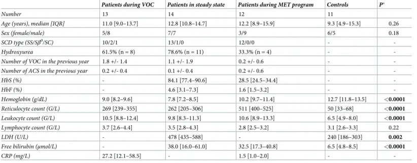
CD161+) were identified by the following monoclo-nal antibodies: anti-CD3-AlexaFluor700 (BD Biosciences), anti-Vα7.2-FITC (Biolegend) and Table 1. Main clinical and biological characteristics of patients and controls.
Patients during VOC Patients in steady state Patients during MET program Controls P�
Number 13 14 12 11
Age (years), median [IQR] 11.0 [9.0–13.7] 12.8 [10.8–14.7] 12.2 [8.9–15.9] 9.3 [4.9–15.3] 0.26
Sex (female/male) 5/8 7/7 3/9 6/5 0.18
SCD type (SS/Sβ0/SC) 10/2/1 13/1/0 12/0/0 -
-Hydroxyurea 61.5% (n = 8) 78.6% (n = 11) 33.3% (n = 4) -
-Number of VOC in the previous year 1.8 +/- 1.4 1.1 +/- 1.9 0.2 +/- 0.6 -
-Number of ACS in the previous year 0.2 +/- 0.4 0.1 +/- 0.4 0.2 +/- 0.6 -
-HbS (%) - 84.1 [77.4–90.6] 28.5 [24.5–34.4] - -HbF (%) - 4.6 [3.1–7.3] 1.6 [1.5–3.2] - -Hemoglobin (g/dL) 9.0 [8.2–9.6] 7.8 [7.2–8.5] 10.2 [9.7–11.4] 12.7 [11.8–13.5] <0.0001 Reticulocyte count (G/L) 269 [239–355] 262 [205–306] 511 [400–525] 50 [33–68] <0.0001 Leukocyte count (G/L) 10.5 [8.8–12.4] 9.8 [8.3–11.3] 10.6 [8.9–13.3] 6.5 [4.9–8.0] <0.0001 Lymphocyte count (G/L) 3.7 [2.6–4.4] 3.5 [2.8–4.3] 2.8 [2.5–3.2] 3.1 [2.6–3.3] 0.22 LDH (U/L) - 478 [435–588] - 240 [186–303] 0.002
Free bilirubin (μmol/L) - 38.0 [16.0–61.0] 32.5 [17.3–40.8] 6.5 [4.8–8.5] <0.0001
CRP (mg/L) 27.2 [12.1–58.5] - 1.5 [1.0–2.0] -
-Data are expressed as median [interquartile range], mean± SD or percentage.
�Comparison between all SCD patients and controls. Results with a P-value <0.05 are indicated in bold.
ACS: acute chest syndrome. CRP: C-reactive protein. HbF: fetal hemoglobin. HbS: hemoglobin S. LDH: lactate dehydrogenase. MET: monthly exchange transfusion. SCD: sickle cell disease. VOC: vaso-occlusive crisis.
anti-CD161-BV785 (eBiosciences). iNKT cells were identified by using CD1-PBS57-APC tet-ramers (NIH Tetramer Core Facility) associated with anti-CD3-AlexaFluor700 mAb, while Vδ2+γδ T cell subset was identified by anti-Vδ2-PE and anti-CD3-AlexaFluor700 mAbs (BD Biosciences).
For intracellular cytokine analysis allowing the identification of 17, IFNγ, 4 and IL-13-producing T cells, freshly isolated PBMC were cultured during 5 hours with 25 ng/mL phorbol-12-myristate 13-acetate (PMA) and 10−6M ionomycin, in the presence of 10 ng/mL Brefeldin A (Sigma-Aldrich), as previously described [28,29]. Following surface staining, cells were washed and fixed with 4% paraformaldehyde and permeabilized with 0.5% saponin (Sigma-Aldrich) before further incubation for 45 minutes with anti-IL-17-BV605, anti-IFNγ-BV711, anti-IL-4-PE-Cy7 (Sony Biotechnology) and anti-IL-13-V450 (BD-Biosciences) [22].
Cells were identified by flow cytometry using a FACS Fortessa™ (BD-Biosciences) and ana-lyzed with FlowJo software.S1 Figshows a representative FACS analysis illustrating how the distinct T cell populations were gated for cytokine assays.
Statistical analysis
Data are expressed as median [interquartile range (IQR)] or percentage. Differences between groups were assessed with the Mann-Whitney test. Correlation analyses between cytokine pro-duction by conventional T cells and innate-like T cells and several clinical and biological fac-tors (Table 1) were made using Spearman’s rank correlation analysis. Statistical significance threshold was set at a P-value of 0.05. Data were analyzed using GraphPad Prism (GraphPad Software).
Results
Patients
Our study comprised a cohort of 39 children with SCD (14 in steady state, 12 on a MET pro-gram and 13 undergoing VOC) and 11 age-matched healthy controls (Table 1). Patients were homozygous HbSS (n = 35, 89.7%), HbS/ß0(n = 3, 7.7%) and HbSC (n = 1, 2.6%). Their median [IQR] age was 12.4 [9.3–15.0] years. Median basal hemoglobin level was 7.5 [7.2–8.3] g/dL and 8.5 [7.7–9.0] g/dL for patients receiving hydroxyurea. Twenty-three patients (59.0%) were currently receiving hydroxyurea with a median dose of 20 [19.5–22.5] mg/kg/day and median treatment duration of 3.5 (1.2–7.3) years. Twelve patients (30.8%) were on a MET pro-gram (median duration of 4.6 [1.2–6.4] years) for a history of stroke (33.3%), abnormal tran-scranial Doppler velocities (50.0%) or frequent VOC and acute chest syndromes (ACS) despite treatment with hydroxyurea (16.7%). For patients in VOC, flow cytometry analyses were per-formed after a median of 2.0 [2.0–3.0] days of pain.
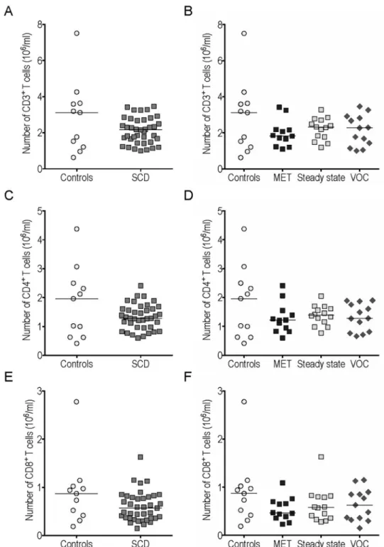
Conventional T cells. The number of CD3+T cells among circulating lymphocytes was similar in SCD patients and controls (Fig 1A), as were conventional CD4+and CD8+T cell counts (Fig 1C and 1E). We found no significant difference between CD3+, CD4+and CD8+T cell counts in the 3 SCD subgroups (MET, steady state and VOC) (Fig 1B, 1D and 1F).
The frequency of IL-4+cells among gated conventional CD4+T cells was unchanged in SCD patients (whatever the subgroup), compared to controls (Fig 2A and 2B). By contrast, the proportion of CD4+T cells expressing IL-13, another typical Th2 cytokine, was higher in SCD patients and remained significantly higher, when comparing MET and VOC groups with con-trols (Fig 2C and 2D).
A highly significant increase in the proportion of IL-17-producing CD4+Th17 cells was observed in SCD patients (whatever the subgroup) versus controls (Fig 2E and 2F).
Fig 1. Conventional T cell counts in peripheral blood were similar in sickle cell disease (SCD) patients and controls. The CD3+(A-B), CD4+(C-D) and CD8+(E-F) cell counts were compared between SCD patients and
controls (A, C, E) and between different subgroups (B, D, F). MET: monthly exchange transfusion; VOC: vaso-occlusive crisis.
Fig 2. IL-13 and IL-17 production by conventional CD4+T cells was increased in peripheral blood of sickle cell
disease (SCD) patients compared to controls. The frequency of IL-4+(A-B), IL-13+(C-D), IL-17+(E-F) cells among gated CD4+cells and of IFNγ+
(G-J) cells among gated CD4+(G-H) and CD8+(I-J) cells were compared between SCD patients and controls.�p<0.05;��p<0.01.
The frequency of IFNγ+cells among gated CD4+T cells was unchanged in SCD patients rel-ative to controls (Fig 2G). However, the frequency of these IFNγ+CD4+Th1 cells was signifi-cantly enhanced in the VOC versus the MET group (Fig 2H). It is noteworthy that similar results were obtained regarding the frequency of IFNγ+
cells among gated CD8+T cells (Fig 2I and 2J). Medians [IQR] and P-values <0.05 of the results presented in Figs1and2are summa-rized inS1andS2Tables.
Innate-like T cells. We found no significant difference regarding iNKT cell counts and
the frequency of IFNγ-producing iNKT cells between SCD patients, or any of the 3 SCD sub-groups, and controls (Fig 3A–3D). However, a slight statistically non-significant trend toward increased IFNγ production by iNKT cells was observed during VOC compared with steady state and MET (p = 0.28 and p = 0.12).
MAIT cell counts were significantly higher in SCD patients, whatever the subgroup consid-ered (Fig 4A and 4B). The frequency of IL-17- but not of IFNγ-producing cells among this population was higher in SCD patients than in controls (Fig 4C and 4E). Similar results were obtained when comparing MET and steady state patients with controls, while no significant increase occurred during VOC (Fig 4D). The frequency of IFNγ+cells among gated MAIT cells was similar in the three patient subgroups (Fig 4F).
The number of Vδ2 T cells, the major peripheral blood γδ T subset, was increased in SCD patients compared with controls, and further enhanced during VOC (Fig 5A and 5B). The fre-quency of IFNγ+
cells among this population was lower in SCD patients than in controls (Fig 5C), which was also the case for MET and steady state groups (Fig 5D). By contrast, a statisti-cally non-significant trend toward increased IFNγ-producing Vδ2 T cells was observed during VOC compared to steady state (p = 0.07) (Fig 5D). IL-17-producing Vδ2 T cells also showed a tendency toward enhancement in SCD patients, without reaching statistical significance (p = 0.09) (Fig 5E). We found no significant difference between the patient subgroups in terms of IL-17 production by Vδ2 T cells (Fig 5F). Medians [IQR] and P-values <0.05 of the results presented in Figs3–5are summarized inS1andS2Tables.
Clinical and biological factors correlated with IFNγ and IL-17 production by conven-tional T cells and Innate-like T cells. The frequency of IFNγ+
cells among gated CD4+, CD8+and Vδ2 T cells was positively correlated with the number of hospitalizations for VOC in the previous year (r = 0.45, p = 0.004; r = 0.51, p = 0.001; and r = 0.37, p = 0.021, respec-tively) and negatively with the time to last hospitalization for VOC (r = -0.38, p = 0.028; r = -0.45, p = 0.008; and r = -0.37, p = 0.029, respectively) (S2 Fig). The frequency of IFNγ+
cells among gated iNKT cells was positively correlated with patients’ age (r = 0.32, p = 0.048). The frequency of IL-17+cells among gated Vδ2 T cells was positively correlated with HbS level (r = 0.47, p = 0.028). No other statistically significant correlation was found with any of the parameters listed inTable 1, including treatment with hydroxyurea.
Discussion
In this study, we provide the first evidence for numerically and functionally distinct MAIT and Vδ2 ILT cells in children suffering from SCD relative to age-matched controls of Afro-Carib-bean origin. We observed higher MAIT cell counts with an increased proportion of IL-17+ MAIT cells, and higher Vδ2 T cell counts, especially during VOC, with a decreased proportion of IFNγ+Vδ2 T cells in off-crisis patients. These results are consistent with an involvement of innate-like T cells in SCD pathophysiology.
Higher levels of circulating iNKT cells have been previously reported in adult patients with SCD, together with increased production of IFNγ during VOC [25–27]. We could not repro-duce these results in our pediatric population, besides a slight trend toward increased IFNγ
production by iNKT cells during crisis. The small number of patients in our cohort, which reduces the statistical power, might explain this discrepancy. However, the positive correlation between the frequency of IFNγ-producing iNKT cells and patients’ age may also account for this difference.
Apart from iNKT cells, modifications of ILT populations had not yet been examined in SCD patients. We show here that both MAIT andγδ T cells may be involved in SCD patho-physiology, thus widening the spectrum of potential actors of innate immunity in SCD. Fur-thermore the increased Vδ2 T cell counts during VOC suggest a specific role for this ILT population during painful crises.
SCD can be viewed as an inflammatory disease leading to chronic inflammation, exacer-bated during VOC [3,30]. In addition to classical pro-inflammatory cytokines, such as TNF-α, IL-1β, IL-6, IL-8 and IFNγ, raised levels of IL-17 have been previously observed in the plasma of steady state SCD patients but the cells responsible for this activity were not clearly character-ized [8–10]. We provide evidence for increased IL-17 production not only by CD4+(Th17) but also by MAIT and possibly byγδ T cells in steady state SCD patients, as compared with Fig 3. The number of iNKT cells and their intracellular cytokine production in peripheral blood were similar in sickle cell disease (SCD) patients and controls. iNKT cell counts (A-B) and frequency of IFNγ+cells among gated
iNKT cells (C-D) from SCD patients and controls.
Fig 4. MAIT cell counts were increased with enhanced IL-17 production in peripheral blood of sickle cell disease (SCD) patients compared to controls. MAIT cell counts (A-B) and frequency of IL-17+(C-D) and IFNγ+
(E-F) cells among gated MAIT cells from SCD patients and controls.�p<0.05;��p<0.01;���p<0.001.
Fig 5. Vδ2 γδ T cell counts were increased with decreased IFNγ production in peripheral blood of sickle cell disease (SCD) patients compared to controls. Vδ2+cell counts (A-B) and frequency of IFNγ+
(C-D) and IL-17+(E-F) cells among gated Vδ2+cells from SCD patients and controls.�p<0.05;��p<0.01.
controls. In these different T cell populations, IL-17 production did apparently not increase further during VOC, which is concordant with the data reported for IL-17 plasma levels [9,
10]. IL-17 is an essential pro-inflammatory T cell-derived cytokine which has been found to play a pivotal role in the defense against extracellular and fungal infections [31,32]. However, this cytokine can also become deleterious, inasmuch as overwhelming production may lead to chronic inflammation and severe immunological conditions [32,33]. IL-17 has pleiotropic effects on multiple target cells and promotes inflammation mainly by inducing several cyto-kines and chemocyto-kines, and enhancing neutrophil recruitment [34,35]. During the last years, the crucial role of neutrophils in SCD has been largely described [3]. Moreover, IL-17 is a potent activator of the endothelium and can induce the expression of endothelial adhesion markers such as E-selectin, VCAM-1, and ICAM-1, which are known as major contributors to SCD pathophysiology [3,5,36]. Finally, IL-17 is involved in the pathogenesis of several auto-immune diseases, whose frequency seems to be increased in SCD [37,38]. It has been previ-ously suggested that IL-17 could represent a marker of SCD severity, as decreased IL-17 plasma levels have been observed in hydroxyurea-treated patients [9]. In addition, a trend toward decreased IL-17 levels has been documented in SCD patients with Benin/Benin haplo-type, which gives rise to a less severe phenohaplo-type, compared to Bantu/Bantu haplotype [39]. In a similar line of evidence, we found a positive correlation between the frequency of IL-17-pro-ducing Vδ2 T cells and HbS level. Together with the fact that exchange transfusions reduce SCD severity by decreasing HbS level, these results support an association between IL-17 and SCD severity. Furthermore, it has been shown that IL-17 is a potent stimulant of lung micro-vascular endothelial cells to produce chemoattractants such as CXCL8, selectively driving neu-trophil chemotaxis [36]. IL-17 may therefore play a role in the pathophysiology of acute chest syndrome (ACS). None of the patients included in our study suffered from ACS, but it might be relevant for future studies to analyze IL-17 production by Th17, MAIT andγδ T cells in SCD patients undergoing ACS.
Enhanced IL-4 secretion during VOC has been previously reported, suggesting a possible shift of the CD4+T cell response towards a Th2 phenotype [40]. We found no significant dif-ference regarding IL-4 production by CD4+T cells, neither between children with SCD and controls, nor between VOC and other SCD subgroups. However, IL-13+cells among gated CD4+T cells were more frequent in patients than in controls, suggesting that the CD4+T cell response is skewed toward Th2 and Th17 phenotypes with increased IL-13 and IL-17 production.
Higher IFNγ plasma levels have been observed in steady state SCD patients compared to controls, with a slight rise during VOC [8,41]. Here, we report increased IFNγ production by CD4+, CD8+and possibly by iNKT and Vδ2 T cells in patients undergoing VOC compared to off-crisis patients. Furthermore, we found that IFNγ production by CD4+
, CD8+and Vδ2 T cells was positively correlated with the frequency of VOC and negatively with the time to last hospitalization for VOC. These results suggest that IFNγ may be involved in VOC pathophysi-ology, whereas IL-17 essentially contributes to the chronic pro-inflammatory state of SCD. On the other hand, the reduced production of IFNγ by Vδ2 T cells in off-crisis SCD patients versus controls suggests chronic down-modulation of these cells, similarly to what has been described in other inflammatory disorders, such as Behcet’s disease [42].
It has been reported that hydroxyurea could have a potential modulatory impact on several immune cells but in our study, no correlation was found between hydroxyurea and any of the analyzed cell subsets [43].
Further studies are required for deciphering the mechanisms leading to the observed alter-ations of MAIT andγδ T cells in SCD patients. Microbial infections are well-known activators of ILT cells but all SCD patients included in our study were receiving antibiotic prophylaxis
and no blood sample was collected during an infection. By analogy with other innate immune cells, a role for chronic hemolysis and inflammation may be suspected [3].
There are some limitations in our study, such as the small number of patients in each group, which may reduce statistical power. Nevertheless, several statistically significant differ-ences between the patient groups could be identified. We are aware that the use of antigens presented by antigen-presenting cells (APC) would be more physiologically relevant than stim-ulation with PMA and ionomycin to detect the ability of peripheral blood T cells to secrete IL-17, even though these mitogens are currently used by other research groups [44,45]. Unfortu-nately, antigen and APC stimulation requires a larger volume of blood than what we were authorized to collect. In addition, antigen stimulation induces T cell receptor (TCR) down-regulation, which may compromise the identification of MAIT cells.
In conclusion, this is the first demonstration in patients with SCD of increased MAIT cell counts with enhanced IL-17 production, increased Vδ2 T cell counts with decreased IFNγ pro-duction, and enhanced IL-13 and IL-17 production by CD4+T cells, as compared to controls. It opens new perspectives for the study of innate-like T cells and Th17 cells, as likely important actors of SCD pathophysiology. Further studies are required to assess if these parameters could help predicting the severity of disease outcome. Since IL-17 inhibitors have shown promising results in treating other inflammatory diseases, a similar approach may be relevant for patients affected with SCD.
Supporting information
S1 Fig. Flow cytometric gating strategy. Flow cytometric gating strategy used to identify
iNKT (CD3+CD1d-PBS57 tetramer+), Vδ2 γδ T cells (CD3+
CD1d-PBS57 tetramer-Vδ2+ ), MAIT (CD3+CD1d-PBS57 tetramer-Vδ2-CD161+TCRVα7.2+), CD4+(CD3+CD1d-PBS57 tetramer-Vδ2 -CD161-TCRVα7.2 -CD8-CD4+) and CD8+(CD3+CD1d-PBS57 tetramer -Vδ2
-CD161-TCRVα7.2-CD8+CD4-) cells from peripheral blood and their ability to produce IL-4, IL-13, IL-17 or IFNγ.
(TIFF)
S2 Fig. Correlation between the frequency of IFNγ+
cells among gated CD4+(A), CD8+ (B) and Vδ2+
(C) cells and the time to last hospitalization for vaso-occlusive crisis (VOC).
(TIF)
S1 Table. Number and cytokine production of conventional and innate-like T cells.
(DOCX)
S2 Table. P-values for statistically significant comparisons between patient groups.
(DOCX)
Acknowledgments
The authors would like to thank the National Institutes of Health Tetramer Core Facility for providing CD1d tetramer reagents.
Author Contributions
Conceptualization: Slimane Allali, Olivier Hermine, Thiago Trovati Maciel, Maria
Leite-de-Moraes.
Formal analysis: Slimane Allali, Rachel Rignault-Bricard, Thiago Trovati Maciel, Maria
Funding acquisition: Maria Leite-de-Moraes.
Investigation: Slimane Allali, Ce´line Dietrich, Franc¸ois Machavoine, Valentine Brousse,
Mar-iane de Montalembert.
Methodology: Slimane Allali, Thiago Trovati Maciel, Maria Leite-de-Moraes. Supervision: Thiago Trovati Maciel, Maria Leite-de-Moraes.
Validation: Thiago Trovati Maciel, Maria Leite-de-Moraes. Writing – original draft: Slimane Allali.
Writing – review & editing: Valentine Brousse, Mariane de Montalembert, Olivier Hermine,
Thiago Trovati Maciel, Maria Leite-de-Moraes.
References
1. Ware RE, de Montalembert M, Tshilolo L, Abboud MR. Sickle cell disease. Lancet. 2017; 390 (10091):311–23.https://doi.org/10.1016/S0140-6736(17)30193-9PMID:28159390.
2. Manwani D, Frenette PS. Vaso-occlusion in sickle cell disease: pathophysiology and novel targeted therapies. Blood. 2013; 122(24):3892–8.https://doi.org/10.1182/blood-2013-05-498311PMID: 24052549.
3. Zhang D, Xu C, Manwani D, Frenette PS. Neutrophils, platelets, and inflammatory pathways at the nexus of sickle cell disease pathophysiology. Blood. 2016; 127(7):801–9. https://doi.org/10.1182/blood-2015-09-618538PMID:26758915.
4. Francis RB Jr., Haywood LJ. Elevated immunoreactive tumor necrosis factor and interleukin-1 in sickle cell disease. J Natl Med Assoc. 1992; 84(7):611–5. PMID:1629925.
5. Graido-Gonzalez E, Doherty JC, Bergreen EW, Organ G, Telfer M, McMillen MA. Plasma endothelin-1, cytokine, and prostaglandin E2 levels in sickle cell disease and acute vaso-occlusive sickle crisis. Blood. 1998; 92(7):2551–5. PMID:9746797.
6. Croizat H. Circulating cytokines in sickle cell patients during steady state. Br J Haematol. 1994; 87 (3):592–7. PMID:7527647.
7. Pathare A, Kindi SA, Daar S, Dennison D. Cytokines in sickle cell disease. Hematology. 2003; 8 (5):329–37.https://doi.org/10.1080/10245330310001604719PMID:14530175.
8. Pathare A, Al Kindi S, Alnaqdy AA, Daar S, Knox-Macaulay H, Dennison D. Cytokine profile of sickle cell disease in Oman. Am J Hematol. 2004; 77(4):323–8.https://doi.org/10.1002/ajh.20196PMID: 15551290.
9. Keikhaei B, Mohseni AR, Norouzirad R, Alinejadi M, Ghanbari S, Shiravi F, et al. Altered levels of pro-inflammatory cytokines in sickle cell disease patients during vaso-occlusive crises and the steady state condition. Eur Cytokine Netw. 2013; 24(1):45–52.https://doi.org/10.1684/ecn.2013.0328PMID: 23608554.
10. Vilas-Boas W, Veloso Cerqueira BA, Figueiredo CV, Santiago RP, da Guarda CC, Pitanga TN, et al. Association of homocysteine and inflammatory-related molecules in sickle cell anemia. Hematology. 2016; 21(2):126–31.https://doi.org/10.1179/1607845415Y.0000000048PMID:26334689.
11. Lanier LL. Shades of grey—the blurring view of innate and adaptive immunity. Nat Rev Immunol. 2013; 13(2):73–4.https://doi.org/10.1038/nri3389PMID:23469373.
12. Godfrey DI, Le Nours J, Andrews DM, Uldrich AP, Rossjohn J. Unconventional T Cell Targets for Can-cer Immunotherapy. Immunity. 2018; 48(3):453–73.https://doi.org/10.1016/j.immuni.2018.03.009 PMID:29562195.
13. Vantourout P, Hayday A. Six-of-the-best: unique contributions of gammadelta T cells to immunology. Nat Rev Immunol. 2013; 13(2):88–100.https://doi.org/10.1038/nri3384PMID:23348415.
14. Crosby CM, Kronenberg M. Tissue-specific functions of invariant natural killer T cells. Nat Rev Immunol. 2018; 18(9):559–74.https://doi.org/10.1038/s41577-018-0034-2PMID:29967365.
15. Chandra S, Kronenberg M. Activation and Function of iNKT and MAIT Cells. Adv Immunol. 2015; 127:145–201.https://doi.org/10.1016/bs.ai.2015.03.003PMID:26073984.
16. Salou M, Franciszkiewicz K, Lantz O. MAIT cells in infectious diseases. Curr Opin Immunol. 2017; 48:7–14.https://doi.org/10.1016/j.coi.2017.07.009PMID:28750261.
17. Martin B, Hirota K, Cua DJ, Stockinger B, Veldhoen M. Interleukin-17-producing gammadelta T cells selectively expand in response to pathogen products and environmental signals. Immunity. 2009; 31 (2):321–30.https://doi.org/10.1016/j.immuni.2009.06.020PMID:19682928.
18. Trottein F, Paget C. Natural Killer T Cells and Mucosal-Associated Invariant T Cells in Lung Infections. Front Immunol. 2018; 9:1750.https://doi.org/10.3389/fimmu.2018.01750PMID:30116242.
19. Sherlock JP, Joyce-Shaikh B, Turner SP, Chao CC, Sathe M, Grein J, et al. IL-23 induces spondyloar-thropathy by acting on ROR-gammat+ CD3+CD4-CD8- entheseal resident T cells. Nat Med. 2012; 18 (7):1069–76.https://doi.org/10.1038/nm.2817PMID:22772566.
20. Venken K, Elewaut D. IL-23 responsive innate-like T cells in spondyloarthritis: the less frequent they are, the more vital they appear. Curr Rheumatol Rep. 2015; 17(5):30. https://doi.org/10.1007/s11926-015-0507-2PMID:25874346.
21. Mortier C, Govindarajan S, Venken K, Elewaut D. It Takes "Guts" to Cause Joint Inflammation: Role of Innate-Like T Cells. Front Immunol. 2018; 9:1489.https://doi.org/10.3389/fimmu.2018.01489PMID: 30008717.
22. Lezmi G, Abou Taam R, Dietrich C, Chatenoud L, de Blic J, Leite-de-Moraes M. Circulating IL-17-pro-ducing mucosal-associated invariant T cells (MAIT) are associated with symptoms in children with asthma. Clin Immunol. 2018; 188:7–11.https://doi.org/10.1016/j.clim.2017.11.009PMID:29196148.
23. Michel ML, Keller AC, Paget C, Fujio M, Trottein F, Savage PB, et al. Identification of an IL-17-producing NK1.1(neg) iNKT cell population involved in airway neutrophilia. J Exp Med. 2007; 204(5):995–1001. https://doi.org/10.1084/jem.20061551PMID:17470641.
24. Papotto PH, Ribot JC, Silva-Santos B. IL-17(+) gammadelta T cells as kick-starters of inflammation. Nat Immunol. 2017; 18(6):604–11.https://doi.org/10.1038/ni.3726PMID:28518154.
25. Wallace KL, Marshall MA, Ramos SI, Lannigan JA, Field JJ, Strieter RM, et al. NKT cells mediate pul-monary inflammation and dysfunction in murine sickle cell disease through production of IFN-gamma and CXCR3 chemokines. Blood. 2009; 114(3):667–76.https://doi.org/10.1182/blood-2009-02-205492 PMID:19433855.
26. Lin G, Field JJ, Yu JC, Ken R, Neuberg D, Nathan DG, et al. NF-kappaB is activated in CD4+ iNKT cells by sickle cell disease and mediates rapid induction of adenosine A2A receptors. PLoS One. 2013; 8 (10):e74664.https://doi.org/10.1371/journal.pone.0074664PMID:24124453.
27. Field JJ, Lin G, Okam MM, Majerus E, Keefer J, Onyekwere O, et al. Sickle cell vaso-occlusion causes activation of iNKT cells that is decreased by the adenosine A2A receptor agonist regadenoson. Blood. 2013; 121(17):3329–34.https://doi.org/10.1182/blood-2012-11-465963PMID:23377438.
28. Moreira-Teixeira L, Resende M, Coffre M, Devergne O, Herbeuval JP, Hermine O, et al. Proinflamma-tory environment dictates the IL-17-producing capacity of human invariant NKT cells. J Immunol. 2011; 186(10):5758–65.https://doi.org/10.4049/jimmunol.1003043PMID:21478400.
29. Baron M, Belo R, Cathelin D, Moreira-Teixeira L, Cartery C, Rondeau E, et al. Innate-like and conven-tional T cell populations from hemodialyzed and kidney transplanted patients are equally compromised. PLoS One. 2014; 9(8):e105422.https://doi.org/10.1371/journal.pone.0105422PMID:25144742.
30. Platt OS. Sickle cell anemia as an inflammatory disease. J Clin Invest. 2000; 106(3):337–8.https://doi. org/10.1172/JCI10726PMID:10930436.
31. Milner JD, Brenchley JM, Laurence A, Freeman AF, Hill BJ, Elias KM, et al. Impaired T(H)17 cell differ-entiation in subjects with autosomal dominant hyper-IgE syndrome. Nature. 2008; 452(7188):773–6. https://doi.org/10.1038/nature06764PMID:18337720.
32. Miossec P, Korn T, Kuchroo VK. Interleukin-17 and type 17 helper T cells. N Engl J Med. 2009; 361 (9):888–98.https://doi.org/10.1056/NEJMra0707449PMID:19710487.
33. Kirkham BW, Kavanaugh A, Reich K. Interleukin-17A: a unique pathway in immune-mediated diseases: psoriasis, psoriatic arthritis and rheumatoid arthritis. Immunology. 2014; 141(2):133–42.https://doi.org/ 10.1111/imm.12142PMID:23819583.
34. Iwakura Y, Nakae S, Saijo S, Ishigame H. The roles of IL-17A in inflammatory immune responses and host defense against pathogens. Immunol Rev. 2008; 226:57–79.https://doi.org/10.1111/j.1600-065X. 2008.00699.xPMID:19161416.
35. Gaffen SL. An overview of IL-17 function and signaling. Cytokine. 2008; 43(3):402–7.https://doi.org/10. 1016/j.cyto.2008.07.017PMID:18701318.
36. Roussel L, Houle F, Chan C, Yao Y, Berube J, Olivenstein R, et al. IL-17 promotes p38 MAPK-depen-dent endothelial activation enhancing neutrophil recruitment to sites of inflammation. J Immunol. 2010; 184(8):4531–7.https://doi.org/10.4049/jimmunol.0903162PMID:20228195.
37. Konya C, Paz Z, Apostolidis SA, Tsokos GC. Update on the role of Interleukin 17 in rheumatologic auto-immune diseases. Cytokine. 2015; 75(2):207–15.https://doi.org/10.1016/j.cyto.2015.01.003PMID: 26028353.
38. Li-Thiao-Te V, Uettwiller F, Quartier P, Lacaille F, Bader-Meunier B, Brousse V, et al. Coexistent sickle-cell anemia and autoimmune disease in eight children: pitfalls and challenges. Pediatr Rheumatol Online J. 2018; 16(1):5.https://doi.org/10.1186/s12969-017-0221-xPMID:29343274.
39. Bandeira IC, Rocha LB, Barbosa MC, Elias DB, Querioz JA, Freitas MV, et al. Chronic inflammatory state in sickle cell anemia patients is associated with HBB(*)S haplotype. Cytokine. 2014; 65(2):217– 21.https://doi.org/10.1016/j.cyto.2013.10.009PMID:24290434.
40. Musa BO, Onyemelukwe GC, Hambolu JO, Mamman AI, Isa AH. Pattern of serum cytokine expression and T-cell subsets in sickle cell disease patients in vaso-occlusive crisis. Clin Vaccine Immunol. 2010; 17(4):602–8.https://doi.org/10.1128/CVI.00145-09PMID:20130127.
41. Qari MH, Dier U, Mousa SA. Biomarkers of inflammation, growth factor, and coagulation activation in patients with sickle cell disease. Clin Appl Thromb Hemost. 2012; 18(2):195–200.https://doi.org/10. 1177/1076029611420992PMID:21949038.
42. Parlakgul G, Guney E, Erer B, Kilicaslan Z, Direskeneli H, Gul A, et al. Expression of regulatory recep-tors on gammadelta T cells and their cytokine production in Behcet’s disease. Arthritis Res Ther. 2013; 15(1):R15.https://doi.org/10.1186/ar4147PMID:23336215.
43. Nickel RS, Osunkwo I, Garrett A, Robertson J, Archer DR, Promislow DE, et al. Immune parameter analysis of children with sickle cell disease on hydroxycarbamide or chronic transfusion therapy. Br J Haematol. 2015; 169(4):574–83.https://doi.org/10.1111/bjh.13326PMID:25753210.
44. Hinks TS, Zhou X, Staples KJ, Dimitrov BD, Manta A, Petrossian T, et al. Innate and adaptive T cells in asthmatic patients: Relationship to severity and disease mechanisms. J Allergy Clin Immunol. 2015; 136(2):323–33.https://doi.org/10.1016/j.jaci.2015.01.014PMID:25746968.
45. Mamessier E, Nieves A, Lorec AM, Dupuy P, Pinot D, Pinet C, et al. T-cell activation during exacerba-tions: a longitudinal study in refractory asthma. Allergy. 2008; 63(9):1202–10.https://doi.org/10.1111/j. 1398-9995.2008.01687.xPMID:18699937.