`
Role of two UDP-Glycosyltransferases from the L group
of arabidopsis in resistance against pseudomonas syringae
Benoît Boachon&Jordi Gamir&Victoria Pastor&Matthias Erb&John V. Dean&Víctor Flors&
Brigitte Mauch-Mani
Accepted: 2 April 2014 / Published online: 13 April 2014 # Koninklijke Nederlandse Planteziektenkundige Vereniging 2014
Abstract The role of the salicylic acid (SA) glyco-sides SA 2-O-β-D-glucose (SAG), SA glucose ester (SGE) and the glycosyl transferases UGT74F1 and UGT74F2 in the establishment of basal resistance of Arabidopsis against Pseudomonas syringae pv toma-to DC3000 (Pst) was investigated. Both mutants altered in the corresponding glycosyl transferases
(ugt74f1 and ugt74f2) were affected in their basal resistance against Pst. The mutant ugt74f1 showed enhanced susceptibility, while ugt74f2 showed en-hanced resistance against the same pathogen. Both mutants have to some extent, altered levels of SAG and SGE compared to wild type plants, however, in response to the infection, ugt74f2 accumulated higher levels of free SA until 24 hpi compared to wild type plants while ugt74f1 accumulated lower SA levels. These SA levels correlated well with reduced expres-sion in PR1 and EDS1 in ugt74f1. In contrast, ugt74f2 has enhanced expression of Enhanced Disease Susceptibility 1 (EDS1) but a strong reduc-tion in the expression of several jasmonate (JA)-dependent genes. Bacterial infection interfered with the expression of Fatty Acid Desaturase (FAD), Lipoxygenase2 (LOX2), carboxyl methyltransferase1 (BSMT1) and 9-cis-epoxycarotenoid dioxygenase (NCED3) genes in ugt74f1, thus promoting an an-tagonistic effect with SA-signalling and leading to enhanced bacterial growth. UGT74F2 might be a target for bacterial effectors since bacterial mutants affected in effector synthesis were impaired in induc-ing UGT74F2 expression. These results suggest that UGT74F2 negatively influences the accumulation of free SA, hence leading to an increased susceptibility due to reduced SA levels and increased expression of the JA and ABA markers LOX-2, FAD and NCED-3.
Keywords Salicylic acid . SA 2-O-β-D-glucose . SA glucose ester . Pseudomonas syringae
Electronic supplementary material The online version of this article (doi:10.1007/s10658-014-0424-7) contains supplementary material, which is available to authorized users.
B. Boachon
:
V. Pastor:
B. Mauch-Mani (*)Laboratory of Molecular and Cellular Biology, Institute of Botany, University of Neuchâtel,
Rue Emile-Argand 11, Case Postale 158, 2009 Neuchâtel, Switzerland
e-mail: Brigitte.mauch@unine.ch
J. Gamir
:
V. FlorsMetabolic Integration and Cell Signaling Group, Plant Physiology Section, Department of CAMN, Universitat Jaume I,
Castellón 12071, Spain M. Erb
Laboratory for Fundamental and Applied Research in Chemical Ecology (FARCE), University of Neuchâtel, Neuchâtel, Switzerland
J. V. Dean
Department of Biological Sciences, DePaul University, Chicago, IL, USA
M. Erb
Root-Herbivore Interactions Group, Max Planck Institute for Chemical Ecology,
Introduction
The immune response of a plant following attack by pathogens is regulated by plant hormones. Among the many changes occurring during this expression of basal resistance, is a prominent rise in salicylic acid (SA) levels, especially during interactions with biotrophic pathogens (de Vos et al.,2005).
SA is synthesized from shikimate/chorismate-derived products (about 90 %) essentially through the activity of ISOCHORISMATE SYNTHASE 1 (ICS1) (Garcion et al., 2008; Wildermuth et al., 2001; Wildermuth,2006). Signaling downstream of SA bio-synthesis requires the interaction between ENHANCED DISEASED RESISTANCE 1 (EDS1) and its partner PHYTOALEXIN DEFICIENT 4 (PAD4) to activate SA accumulation (Feys et al.,2001; Wiermer et al.,2005). Subsequent signaling of SA is mostly controlled by NON-EXPRESSOR of PR PROTEIN 1 (NPR1) whose function is regulated by SA-mediated modulation of the cellular redox state (Mou et al.,2003). Cytosolic local-ization of NPR1 monomers regulates the SA-mediated repression of jasmonic acid (JA)-responsive genes (Spoel et al., 2003). NPR1 also interacts with TGA transcription factors in the nucleus to regulate the ex-pression of SA-inducible genes such as PR-1 (Kesarwani et al.,2007; Mou et al.,2003).
The role of SA in plant immune responses depends on a poorly understood interplay between its free and conjugated forms. SA can be metabolized into several conjugates through glycosylation, methylation and con-jugation with amino acids (Loake and Grant,2007; Vlot et al., 2009). The methylated form methylsalicylate (MeSA) is catalyzed from SA by BSMT1 (Chen et al.,
2003). MeSA has been proposed to be the long-distance signal for transduction of systemic acquired resistance (SAR) since it can release free SA in systemic tissue by the activity of SA BINDING PROTEIN 2 (SABP2) (Liu et al., 2010; Park et al., 2007; Vlot et al., 2008). However, MeSA production and BSMT1 expres-sion are not required for SAR establishment in Arabidopsis (Attaran et al., 2009). Instead, several studies support that MeSA production is involved in the JA-mediated inhibition of the SA pathway (Attaran et al., 2009; Koo et al., 2007; Song et al.,
2008; Song et al., 2009). In Arabidopsis, most MeSA produced during compatible and incompat-ible interactions with P. syringae is released to the atmosphere (Attaran et al., 2009).
In regards to glycosylated forms, in tobacco, the bulk of SA produced during pathogen infections or exogenous SA application is primarily metabolized into SA 2-O-β-D-glucose (SAG) by a SA glyco-syltransferase (SAGT) (Edwards, 1994; Lee and Raskin, 1999; Dean et al., 2005; Song, 2006). SAG accumulates in the vacuole presumably as a hydrolysable source of free SA available for plant defense (Dean et al., 2005). In tobacco, SAG can be converted into SA by a β-glucosidase (Seo e t a l . , 1 9 9 5) . F u r t h e r m o r e , t o b a c c o o v e r-expressing a β-glucosidase accumulates less SAG and elevated SA levels resulting in enhanced re-sistance against Tobacco Mosaic Virus (TMV) (Yao et al., 2007). In Arabidopsis, in vitro catalyt-ic analysis determined that the glycosyltransferase UGT74F1 can form SAG while UGT74F2 can form both SAG and SGE but with a 10-fold higher specific activity for SGE formation (Lim et al., 2002). The specific activity of UGT74F1 was 58-fold higher than of UGT74F2 for SAG formation. The mutant ugt74f1 only accumulates SGE in untreated plants and accumulates both SAG and SGE in SA-treated plants due to UGT74F2 activity (Dean and Delaney, 2008). The mutant ugt74f2 does not accumulate SGE in both water- and SA-treated plants but still pro-duces SAG in SA-treated plants due to UGT74F1 activity (Dean and Delaney, 2008).
The biological function of SAG and SGE is not well understood. Some glycosyltransferases from phyloge-netic group L have been shown to be relevant for resis-tance against Pst (Langlois-Meurinne et al., 2005). Additionally, overexpression of UGT74F2 leads to en-hanced susceptibility to Pst (Song et al.,2008). Previous reports suggest the relevance of glycosylation in resis-tance against Pst. A member of the uridine 5 ’-disphosphate (UDP)-glycosyltransferase (UGT) (At1g05680) confers enhanced resistance against the bacterium. The mutant ugt74e2 shows augmented sys-temic immunity during SAR, and increased levels of PR1, PR2 and PR5 transcripts (Park et al., 2011). UGT74E2 is a member of the L subclass of UGTs (Ross et al.,2001) and together with UGT74F2 has been shown to transfer glucose to SA and anthranilate al-though their biological substrates have not been clearly identified.
Recent findings also describe the SA-related priming activity of several novel chemicals called imprimatins
(Noutoshi et al.,2012). Their priming activity against Pst seems to be linked to the inhibition of glycosyltrans-ferase activity. Thus by treating Arabidopsis plants with these compounds free SA increases and the SA-O-β-glucoside is reduced. Interestingly, mutants in the UGT74F1 display enhanced resistance against virulent Pst DC3000 and avirulent Pst avrRmp1. However, only increased levels of SA upon inoculation with the avirulent strain Pst avrRmp1 have been determined whereas SA and SAG levels upon inoculation with virulent Pst were not determined. In addition, the mutant ugt74f1 used in these experiments showed a surprising up-regulation of UGT74F1 mRNA (Noutoshi et al., 2012). These investigations rein-force the role of UGT74F1 in SA glycosylation, however, the role of this transferase in resistance still remains controversial. Interestingly, glycosyl-transferases affecting the SA pathway are not re-stricted to those using SA as a substrate. In fact, a novel role for the UGT76B1 gene has been dis-covered (von Saint Paul et al., 2011). By using recombinant systems, it has been demonstrated that this gene encodes an enzyme that uses isoleucic acid (ILA) as a substrate. The absence of this transferase up-regulates the SA pathway and down-regulates JA signalling. In addition, the ugt76b1 mutants are more resistant to P. syringae but more susceptible to Alternaria brassicicola (von Saint Paul et al., 2011). Although the rela-tionship between ILA and SA is not clear, it is obvious that glycosylation of both bioactive com-pounds inactivates them and this has a negative impact on Arabidopsis resistance against Pst.
These recent advances provide the impetus for addi-tional studies the role of SA metabolites in plant disease r e s i s t a n c e . I n t h i s s t u d y, t h e r o l e o f b o t h glycosyltranferases UGT74F1 and UGT74F2 in Arabidopsis basal resistance against Pst was investigat-ed. The transcriptional profile of genes involved up-stream and downup-stream of the SA signaling pathway, SA biosynthesis and SA metabolism upon bacterial infection was examined. Accumulation of SA, SAG and SGE and the emission of MeSA were also moni-tored. Surprisingly, the mutants showed resistant pheno-types opposite from one another following bacterial infection. They also differed in their response to the bacterial effector Coronatine, suggesting that each has a contrasting function in Arabidopsis basal resistance against Pst.
Material and methods Biological material
Wild-type Arabidopsis thaliana accession Col-0 and Ws-0 was obtained from Lehle Seeds (Round Rock, TX). The Col-0 mutants npr1, sid1, sid2-1 were kindly provided by C. Nawrath (University of Lausanne, Switzerland). The Ws-0 mutant ugt74f1 was kindly provided by John V. Dean (DePaul University, USA). The Col-0 mutant ugt74f2 was a gift from Judith Bender (Brown University, USA). Plants were grown singly in 30 mL Jiffy® peat tablets (Ryomgaard, Denmark), maintained at 20 °C day/18 °C night temperatures with 9 h of light (150μmol m−2s−1) per 24 h and 70 % RH. The virulent strain of Pseudomonas syringae pv. tomato DC3000 (Pst) (Whalen et al., 1991) was grown over-night at 28 °C in liquid King’s medium B containing 50μg/mL of rifampicine. Pst strains DC3000 hrpA A9 (HrpA) and DC3118 coronatine− (COR−) were kindly provided by Sheng Yang He (Michigan State University, USA) and grown in King’s medium B containing 50 μg/ mL of each rifampicine and kanamycin.
Plant inoculation and sampling
For the infection, five-week-old plants were inoculated by dipping the leaves in MgSO4mock suspension or
Pst, HrpA and COR−containing 5×10⁷colony-forming units ml−1in 10 mM MgSO40.01 % v/v Silwet L-77
(Lehle Seeds, Round Rock, TX). After inoculation, plants were grown under 100 % relative humidity until sampling. For the P. syringae bioassays, 3 days after inoculation, leaves showing necrotic or water-soaked lesions surrounded by chlorosis were scored and the percentage of leaves presenting disease symptoms was determined. At the indicated time-points leaves were sampled for specific genetic and hormonal analyses, frozen in liquid N2and stored at -80ºC.
Quantitative real-time RT-PCR analysis of transcripts and PCR-based quantification of P. syringae infection For quantification of plant gene expression powdered samples from frozen leaf material was homogenized. Total RNA was isolated using the Rneasy plant minikit (Qiagen). cDNA was obtained from 2μg of RNA using oligo(dT)18 and Superscript III reverse transcriptase
manufacturer and then diluted to a final volume of 200μL in sterile water.
The PCR was performed in a rotor Gene 6000 (Corbett Life Science) and plate samples were prepared with a ROBOT CAS1200 (Corbett Robotics). Two μL of diluted cDNA were ampli-fied using 3 μl of SensiMixPLus SYBR Kit (Quantace) with 250 mM of primers and adjusted to a final volume of 8 μl. Primers are described in the Suplementary Table 1. For the quantification of each transcript, standard curves (number of copies/log Ct) were constructed using serial dilu-tions of known copy numbers of plasmids (108 to 101 copies.μl−1). Plasmids for each transcript were transformed with the corresponding PCR product obtained with the same primers used for the real-time PCR and cloned in the pGEM-T easy Vector (Promega). Results of gene aqRT-PCR expression were expressed as copy number of the target gene per total ng of cDNA.
The quantification of P. syringae growth and tran-scripts accumulation was performed by real-time PCR in the same real-time equipment. Diluted cDNA was amplified using the SensiMix plus SYBR Kit (Quantance) in the same conditions de-scribed for the plant gene expression. The cycling conditions for Psoprf were 95ºC for 10 min follow-ed by 40 cycles at 95ºC for 10 s, 58ºC for 15 s and 72ºC for 20 s, followed by a melting point analysis. Standard curves (Copy number/log Ct) were con-structed using plasmids transformed with the respec-tive cDNA or DNA product. Results of the quanti-fication of P. syringae growth were expressed as copy numbers of the Psoprf gene per copy numbers of the AtTUB4 gene. For the absolute quantification of transcript accumulation (aqRT-PCR), the quantifi-cation of total cDNA of each sample was used for the normalization and performed using the Quant-iTTM PicoGreen® dsDNA reagent (Invitrogen). Quantification of SA, SAG and SGE
Frozen material was lyophilized and SA, SAG and SGE were analyzed as is described by Pastor et al.
2012. Briefly, 50 mg of dried material was mixed with internal standard and homogenized with a polytron, using an extraction solution containing MeOH/H2O (10/90, v/v), containing 0.01 % of
HCOOH. After centrifugation the supernatant was
filtrated through a 0.22 μm cellulose acetate filter. A 20 μl aliquot of this solution was injected into a liquid chromatography coupled to tandem mass spectrometry. Masslynx 4.1 (Waters, Manchester, UK) software was used to process the quantitative data obtained from calibration standards and samples.
Quantification of MeSA emission
Three Arabidopsis plans per condition were placed in glass bottles 24 h post Pst infection. A constant airflow of charcoal-filtered, humidified air entered the bottle at a rate of 0.6 l/min. Filters containing 25 mg of the absorbent SuperQ (ARS) were at-tached to the outlet of the bottle. The system was hermetically closed, thereby forcing all the head space volatiles across the filter. At different time points, the filters were detached and the trapped volatile compounds were eluted with 150 μL MeCl2. Then, 10 μl of a mixture of internal
stan-dards (n-octane and nonyl-acetate, 20 ng/mL, Sigma) was added to each sample. All extracts were stored at -80 ºC until analyses. Volatiles were identified and quantified using a gas chromato-graph (Agilent 6,890 Series GC system G1530A) coupled to a mass spectrometer (Agilent 5,973 Network Mass Selective Detector) that operated in electron impact mode (transfer line 230 ºC, source 230 ºC, ionization potential 70 eV, scan range 33–280 m/z). A 2 μL aliquot of each sam-ple was injected in the pulsed splitless mode onto an apolar capillary column (HP-1, 30 m, 0.25 mm ID, 0.25 μm film thickness; Alltech Associates). Helium at constant flow (0.9 ml/min) was used as carrier gas. After injection, the column temperature was maintained at 40 ºC for 3 min and then in-creased to 100 ºC at 8 ºC/min and subsequently to 125 ºC at 5 ºC/min followed by a post run of 5 min at 250 ºC. The detected volatiles were identified by comparison of their mass spectra with those of the NIST 05 library and authentic standards of MeSA (Sigma-Aldrich, Switzerland). Quantification of MeSA was carried out by comparing the integrated total ion peaks with that of the internal standard nonyl-acetate. After the sampling period, plants were weighted and the emission of volatiles was determined as the mass of MeSA emitted per mass of fresh weight per hour.
Results
UGT74F1 and UGT74F2 play a role in basal resistance against Pseudomonas
To get a more precise insight into the role of glycosyl-transferases involved in the synthesis of SAG and SGE, the mutants ugt74f1 and ugt74f2 were inoculated with a virulent strain of Pst. Compared to wild type plants, bacterial titers in ugt74f1 were higher starting at 48 hpi (Fig.1a). Accordingly, the disease levels expressed as a
percentage of infected leaves per plant were higher and the visible symptoms were more pronounced in the mutant (Fig. 1a). On the contrary, ugt74f2 was more resistant to the infection by Pst supporting reduced bacterial growth, showing a lower percentage of dis-eased leaves and displaying reduced symptoms com-pared to the wild type (Fig.1b).
In addition to these observations and in agreement with previous results, UGT74F2 but not UGT74F1 was induced in wild type plants challenged with Pst in both backgrounds tested (Fig. 2). Interestingly, UGT74F1
Fig. 1 Basal resistance of Arabidopsis wild type and ugt74f1 and ugt74f2 mutants against Pseudomonas syringae pv. tomato DC3000. A) Five-week old Col-0, Ws-0, ugt74f1 and ugt74f2
plants were challenged with 5.107colony-forming units mL−1of
P. syringae DC3000. Bacterial growth was measured by real-time PCR at the indicated time-points. Data presented are the means of three technical replicates (±SD) of the ratio of Psorf gene copy number/AtTUB4 gene copy number. Asterisk means statistically
significant differences (T-test; p<0.05). B) Disease rate was mea-sured by calculating the percentage of diseased leaves per plant (n=20,±SD). Data are from a representative experiment that was repeated four times with similar results. Asterisk means statistical-ly significant differences (T-test; p<0.05). Photograph shows a representative picture of disease symptoms in both genetic backgrounds
and UGT74F2 displayed a cooperative function upon infection. This cooperative influence was clearly evi-denced when UGT74F2 was disrupted since expression of UGT74F1 was severely affected in the mutant ugt74f2 (Fig.2). Hence, UGT74F2 seems to contribute negatively to Arabidopsis resistance towards Pst. The hypersusceptible phenotype of ugt74f1 is due to reduced SA levels and a concomitant enhancement of antagonistic pathways
In order to understand the reasons for the observed phenotype of ugt74f1, gene expression of the main disease resistance pathways was assessed in this mutant. As expected, UGT74F1 expression was absent in the knockout mutant (Fig. 2). Gene expression of SA-related genes Enhanced Disease Susceptibility1 (EDS1) and Pathogenesis Related-1 (PR-1) was clearly
reduced while Isochorismate Syntase1 (ICS1) was hard-ly altered in ugt74f1 compared to the wild type plants (Fig.3a). Interestingly, the levels of free SA in ugt74f1 were reduced at later time points in the infection by Pst (Fig.3b).
According to previous studies, most SAG produced in wild type Arabidopsis is due to the activity of UGT74F1 while SGE is only formed by UGT74F2 (Dean and Delaney,2008; Lim et al.,2002). In infected ugt74f1 plants, the accumulation of SAG is delayed and lower than in wild type plants whereas SGE is hardly affected (Fig.3b). In ugt74f1 compared to wild type, the accumulation of Fatty Acid Desaturase (FAD), Lipoxygenase (LOX2) and 9-cis-epoxycarotenoid dioxygenase (NCED3) transcript levels are enhanced (Fig.3a), pointing to an up-regulation of the oxylipin and the ABA signalling pathway. In contrast, the JA and JA/Et marker genes VSP2 and PDF1.2 were less
Fig. 2 Profile of glycosyltransferase gene expression upon P. syringae DC3000 infection. UGT74F1 and UGT74F2 were measured at the given time-points by real-time PCR. Data show the mean of three technical replicates (±SD). The experiment was
repeated three times with similar results. For inoculation and
further experimental details see Fig.1. Asterisk means statistically
expressed in the mutant compared with wild type plants (Fig. 3a). This switch between JA up-stream and JA down-stream genes suggests a diversion in the pathway
that may be clearly re-organized towards the accumula-tion of other oxylipins that differ from JA or its down-stream products. On the other hand, the increase of
Fig. 3 Profile of plant defence pathways upon Pst infection in Ws-0 wild type and ugt74f1 mutant. A) Expression of genes representing key components of the SA, JA and ABA pathways was measured at the given time-points by real-time PCR. Data show the mean of three technical replicates (±SD). B) The levels of SA, SAG and SGE upon infection were determined by LC-MS. C) The accumulation of MeSA released along the time periods
indicated was determined by GC-MS. Gene expression data and MeSA are from representative experiments that were repeated twice with similar results. SA, SAG and SGE data are from a representative experiment that was repeated four times. In the figures A, B and C the asterisk means statistically significant differences within each time-point (T-test; p<0.05)
BSMT1 gene expression in ugt74f1 correlates with a strong increase in the MeSA emission (Fig.3c) that may contribute to the observed decrease in free SA levels.
ugt74f2 displays enhanced resistance due to a transient accumulation of free SA and a concomitant negative crosstalk with antagonistic pathways
Although no major changes in the expression of PR1 and ICS1 were observed, the expression of EDS1 was clearly up-regulated in ugt74f2 (Fig.4a). Mutant plants displayed a transient increase in free SA until 24 hpi (Fig. 4b). Despite the fact that UGT74F2 has been reported to be involved in the synthesis of SAG and SGE (Dean and Delaney,2008; Lim et al.,2002), sim-ilar levels of SAG were found in mutant and wild type plants upon infection, while SGE in ugt74f2 was hardly detected either in the presence or in the absence of infection (Fig. 4b). The expression of UGT74F2 was almost absent in ugt74f2 (Fig.2).
In contrast to the observed enhancement of the JA pathway in ugt74f1, expression levels of LOX2, VSP2 and FAD were strongly down-regulated in ugt74f2 after infection (Fig.4a). PDF1.2 was expressed higher in the mutant. NCED3 expression was also down-regulated albeit to a lesser extent. Therefore, the mutation in UGT74F2 contributes to a shift in the negative crosstalk between SA and JA-ABA signalling pathways towards SA-dependent responses. A likely explanation for the fast SA accumulation in ugt74f2 is the down-regulation of BSMT1 and the resulting reduction in MeSA emis-sion (Fig.4c). Noteworthy the JA levels are higher in ugt74f1 at 24, 72 and 96 upon infection while they are only increased after 96hpi in the ugt74f2 mutant (Fig.S1).
To determine whether an alteration in the SA path-way could also affect glycosyltransferase gene expres-sion and glucoside formation. SA mutants and NahG were as expected more susceptible to Pst (Fig. S2a). Neither UGT74F1 nor UGT74F2 gene expression was significantly altered following infection with Pst in the above-mentioned mutants except in NahG plants (Fig.S2b). As expected, sid1 and sid2 mutants affected in SA synthesis and NahG in which SA is immediately converted to catechol accumulates neither conjugated nor free forms of SA, although these compounds were clearly not affected in the signaling mutant npr1 (Fig.S2c).
Functional effector secretion is required for the induction of UGT74F2
In order to check whether effector secretion was necessary for the up-regulation of UGT74F2 upon infection by Pst, wild type plants were inoculated with COR− and hrpA mutants of Pst. Bacterial growth was impaired for both mutant strains com-pared to WT Pst (Fig. 5a). This was also reflected in the number of affected leaves (Fig. 5b). The expression of UGT74F1 was the same following inoculation with wild type and mutant Pst strains, respectively (Fig. 5c). It is noteworthy, that the induction of UGT74F2 expression was strongly induced by the virulent strain of Pst while it was reduced in the absence of both the Coronatine and HrA effectors (Fig. 5c). An additional support for the importance of functional effector secretion was found when Col-0 plants where treated with the Pathogen Associated Molecular Pattern (PAMP) Flg22 (Fig. S3). A transient up-regulation of UGT74F2 was observed at 24 hpt and had reverted to mock levels at 48 hpt. This might suggest that its activation upon infection is depen-dent on the effector machinery of Pst at the be-ginning of the infection process but not by the elicitor Flg22.
Discussion
In Arabidopsis, about 120 genes encoding UDP-glycosyltranferases have been described. These genes have been classified into 14 groups (Ross et al.,2001). Although the exact function of all these genes has not been yet determined, many of them share similar cata-lytic properties and appear to be involved in the glyco-sylation of plant hormones and other substrates (Langlois-Meurinne et al., 2005). The first identified function of a UGT was flavonol glycosylation (Dooner and Nelson,1977). The down-regulation of this gene in tobacco resulted in an enhanced susceptibility towards TMV. In Arabidopsis, UGT73B3 and UGT73B5 have been shown to participate in resistance towards Pst and this resistance seems to be dependent on SA signalling (Langlois-Meurinne et al., 2005). The AtSAG1 (UGT74F2, At2g43820) gene is directly induced upon SA treatments and therefore it could be involved in SA signalling dependent defence (Song,2006). All three,
free and conjugated, forms of SA (SA, SAG and SGE) rise upon infection by Pst (Song et al., 2009; Pastor
et al., 2012) and also after wounding in a time-dependent manner (Ogawa et al.,2010), clearly pointing
Fig. 4 Profile of plant defence pathways upon Pst infection in Col-0 wild type and ugt74f2 mutant. A) Expression of genes representing key components of the SA, JA and ABA pathways was measured at the given time-points by real-time PCR. Data show the mean of three technical replicates (±SD). B) The levels of SA, SAG and SGE upon infection were determined by LC-MS. C) The accumulation of MeSA released along the time periods
indicated was determined by GC-MS. Gene expression data and MeSA are from representative experiments that were repeated twice with similar results. SA, SAG and SGE data are from a representative experiment that was repeated four times. In the figures A, B and C the asterisk means statistically significant differences within each time-point (T-test; p<0.05)
to an interplay between these molecules following biotic challenges. In the results presented here, mutants in two SA-glycosyltransferases display opposite resistance phenotypes against Pst.
A mutant in UGT74F1, previously reported to be responsible for SAG formation, is hypersusceptible to
Pst. On the other hand, a mutant in UGT74F2, that is supposed to be involved in both SAG and SGE forma-tion, is more resistant to Pst (Fig. 1). In contrast, Noutoshi et al. (2012) have reported that ugt74f1 is more resistant 3 days after inoculation. There may be several reasons for such an apparent contradiction. First, the
method of inoculation is different. In this study, plants were dip-inoculated while Noutoshi and colleagues syringe-infiltrated the bacteria, thus bypassing the con-stitutive barriers and stomatal responses to infection. The second and most relevant difference is that the mutant used by Noutoshi et al. (2012) was not impaired in UGT74F1 expression. In fact, their mutant showed enhanced levels of UGT74F1 mRNA and therefore was seemingly a gain of function mutant rather than a trans-lational UGT74F1 impaired mutant. The T-DNA inser-tional mutant used in the present research is a full knockout with hardly any detectable gene expression.
The evidence presented highlights the relevance of a fine regulation of both genes during defence reactions. In accordance with our results, a line overexpressing AtSGT1 (UGT74F2) was more susceptible to Pst and accumulated less free SA, SAG and SGE (Song et al.,
2008). Unexpectedly, this line also accumulated more MeSA and MeSA-2-O-β-D-glycoside. As shown in Fig. 3, the mutant ugt74f1 shows less induction of ICS1 after Pst inoculation. Accordingly, after 24 hpi it accumulated less SA in response to the infection com-pared with Ws-0 and this correlates with a lower PR1 activation. The mutant ugt74f2 shows the opposite re-sponse since it accumulates free SA until 24hpi. In contrast, Song et al. (2009) showed that a RNAi line of AtSGT1 (UGT74F2) displayed no altered phenotype following infection by Pst. These differences in results are probably due to the fact that the mutant used in the
present research is a knockout T-DNA insertional mutant that completely blocks UGT74F2 expression as shown in Fig.2.
A further observation is the altered accumulation of both glucosides in the mutants in response to the bacte-rial infection. While SAG is mainly affected in ugt74f1, SGE is strongly affected in ugt74f2 (Figs. 3 and 4). Noutoshi et al. (2012) showed that ugt74f1 accumulated higher levels of SA, but in response to the avirulent strain Pst-avrRpm1 that was spray-inoculated while the resistant phenotype was obtained following infiltra-tion of the bacterium into the leaves. In addiinfiltra-tion to glycosyl conjugates, other SA conjugates such as MeSA are also induced after Pst and Pseudomonas maculicola (Psm) infection (Attaran et al.,2009; Song et al., 2008). UGT74F2 overexpressing plants release much higher levels of MeSA after infection and are more susceptible to the bacteria (Song et al., 2008). Accordingly, as shown here, ugt74f1 releases more MeSA and displays enhanced activity of BSMT1 that may contribute to its enhanced susceptibility. On the other hand, the resistant mutant ugt74f2 shows a strong-ly attenuated expression of BSMT1 and very low levels of MeSA.
An interesting observation in the ugt74f1 mutant upon infection is a strong up-regulation of the oxylipin-dependent genes LOX2, and FAD, proba-bly as a consequence of the host manipulation by bacterial effectors. Both genes are upstream in the LOX-9 regulated oxylipin pathway (Vellosillo et al., 2007). In contrast, VSP2 and PDF1.2, both markers downstream of COI1 and JA signalling (Fig. 3), are down-regulated. This suggests that the reduction in SA and altered UGT74F1 function affects the oxylipin pathway and intermediates may be diverted into a branch pathway leading to metabolites that differ from JA and its deriva-tives. In contrast, the enhanced SA signalling in ugt74f2 leads to a very low transcription of these JA-dependent genes with the exception of PDF1.2 (Fig. 4). Since this marker gene is also responsible for ethylene (ET) signalling (de Vos et al., 2005), it is very likely that ugt74f2 contributes to an enhancement of other ET-dependent responses, otherwise, VSP2 would be induced in this mutant upon infection and indeed, this is not the case (Fig. 4).
Similar resistance is found when another glycosyl-transferase UGT76B1 is absent. The knockout mutant
Fig. 5 Basal resistance of Arabidopsis Col-0 against P. syringae DC3000 wild type (Pst), the coronatine deficient strain (COR-) or with the TTSS deficient strain (HrpA). A) Bacterial growth was measured by real-time PCR at the indicated time-points. Data presented are the means of three technical replicates (±SD) of the ratio of Psorf gene copy number/AtTUB4 gene copy number. Asterisks mean statistical significant differences within time-point (T-test, p<0.05). B) Disease rate was measured by calculating the percentage of diseased leaves per plant (n=20,±SD). Data are from a representative experiment that was repeated four times with similar results. Different letters mean statistical significant differ-ences within treatments (ANOVA; LSD, p<0.05). C) Profile of UGT74F1 and UGT74F2 gene expression upon Pst infection with strains compromised in coronatine production and TTSS. Five week-old plants were challenged with the P. syrinage DC3000
wild type (DC3000), the coronatine deficient strain (COR−) or
with the TTSS deficient strain (HrpA) at 5.107c.f.u.ml−1. Gene
expression was measured at indicated time-points by real-time PCR. Data represent the mean (±SD) of technical triplicates. Data are from a representative experiment that was repeated twice with similar results. Different letters mean statistical significant differ-ences within treatments at each time-point (ANOVA; LSD, p<0.05)
ugt76b1 presents higher basal levels of SA as well as gene induction in those genes sensibles to SA, while the JA-responsive genes are down-regulated (von Saint Paul et al.,2011). In this case, again, the overexpression of UGT76B1 leads to an opposite result. Attaran et al. (2009) showed that upon bacterial infection MeSA for-mation is regulated by the JA pathway. Following in-fection by virulent Pst, the MeSA synthesis depends on the presence of coronatine contributing to the repression of the SA-dependent defences. In the present research it is shown that the induction of UGT74F2 by the bacteria requires the presence of coronatine and HrpA that con-tributes to the up-regulation of the JA-pathway in re-sponse to the additional UGT74F2. Surprisingly, the loss of function of the UGT74F2 gene correlates with a reduction in UFT74F1 expression. On the other hand, although to a lower extent, the mutant ugt74f1 is also affected in UGT74F2 expression upon infection, sug-gesting a cross-regulation between these two glycosyl-transferases. The expression levels of both transferases seem not to be significantly affected by mutations in genes in the SA pathway such as ICS1 or NPR1. On the contrary, the scavenging of SA in NahG triggers a down-regulation of both UGTs (Fig. S1). Obviously, SA is necessary as a substrate for the generation of SAG and SGE and mutants with disrupted levels of
the free hormone are impaired in the accumulation of the glycosides.
In conclusion, UGT74F1 down-regulates the JA-pathway, therefore, when it is absent in the corre-sponding mutant, JA signalling is enhanced contrib-uting to increase BSMT1 expression. This creates an alternative sink for the free form of SA that is convered into MeSA, hence, SA signalling in ugt74f1 is repressed and it becomes more suscepti-ble. Noteworthy, only COI1 upstream signals are up-regulated in ugt74f1, which suggests that the positive crosstalk between MeSA and oxylipins does not directly involve JA or JA-metabolites but earlier signalling within this pathway. On the other hand, UGT74F2 is needed for the up-regulation of the JA-pathway in the presence of coronatine and HrpA. Consequently, in the mutant ugt74f2 the JA-pathway is down-regulated, free SA accumulates and SA-dependent signalling is up-regulated resulting in a resistant phenotype (Fig. 6). As a final remark, it is noteworthy that two genes with 76 % of se-quence similarity have opposite effects on the regu-lation of Arabidopsis resistance against Pst. It may be possible that the bacterial effectors are targeting these small differences between the two genes, al-though this is matter of future research.
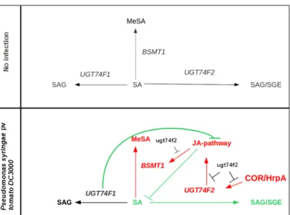
Fig. 6 Model showing the effects of UGT74F1 and UGT74F2 during bacterial infection in Arabidopsis. In the absence of infec-tion UGT74F1 and UGT74F2 show low levels of basal expression which leads to low amounts of SGE and SAG. Once infection occurs, UGT74F2 is targeted by bacterial effectors that enhance its expression. Its activation stimulates an up-regulation of a
PDF1.2-independent branch of JA-signaling and BSMT1 contributing to increase Pst susceptibility. Although UGT74F1 is not responding to the infection, it plays a key role down-regulating JA-signaling, that is strongly activated in the ugt74f1 mutant. (Ws) Events found in the Ws-0 background; (Col) events found in the Col-0 background
Acknowledgments We thank F.J. Uribe-Romeo and Sheng Yang He (Michigan State University, USA) for providing Pst
strains DC3000 hrpA A9 and DC3118 coronatine−. We are
grate-ful to P. Saindrenan (IBP, Paris, France) and D. Heintz (IBMP, Strasbourg, France) for providing SGE standards. We thank Pro. Ted C. J. Turlings for acces to the volatile trapping and GS/MS systems. We also thank the SCIC of the Universitat Jaume I for its technical support and the Plan de promoción de la Investigación Universitat Jaume I P1.1B2010-06. Financial support by the Swiss National Science Fundation Grant 31003A_140593 is gratefully acknowledged.
References
Attaran, E., Zeier, T. E., Griebel, T., & Zeier, J. (2009). Methyl salicylate production and jasmonate signaling are not essen-tial for systemic acquired resistance in Arabidopsis. Plant
Cell, 21, 954–971.
Chen, F., D’Auria, J. C., Tholl, D., Ross, J. R., Gershenzon, J.,
Noel, J. P., & Pichersky, E. (2003). An Arabidopsis thaliana gene for methylsalicylate biosynthesis, identified by a bio-chemical genomics approach, has a role in defense. Plant J.,
36, 577–588.
de Vos, M., van Oosten, V. R., van Poecke, R. M. P., van Pelt, J. A., Pozo, M. J., Mueller, M. J., Buchala, A. J., Métraux, J. P., van Loon, J. C., Dicke, M., & Pieterse, C. J. (2005). Signal signature and transcriptome changes of Arabidopsis during pathogen and insect attack. Molecular Plant Microbe
Interactions, 18, 923–937.
Dean, J. V., & Delaney, S. P. (2008). Metabolism of salicylic acid in wild-type, ugt74f1 and ugt74f2 glucosyltransferase
mu-tants of Arabidopsis thaliana. Physiol. Plant., 132, 417–425.
Dean, J. V., Mohammed, L. A., & Fitzpatrick, T. (2005). The formation, vacuolar localization, and tonoplast transport of salicylic acid glucose conjugates in tobacco cell suspension
cultures. Planta, 221, 287–296.
Dooner, H. K., & Nelson, O. E. (1977). Genetic control of UDPglucose:flavonol 3-O-glucosyltransferase in the
endo-sperm of maize. Biochem. Genet., 15, 509–519.
Edwards, R. (1994). Conjugation and metabolism of salicylic acid
in tobacco. J. Plant Physiol., 143, 609–614.
Feys, B. J., Moisan, L. J., Newman, M. A., & Parker, J. E. (2001). Direct interaction between the Arabidopsis disease resistance
signaling proteins, EDS1 and PAD4. EMBO J., 20, 5400–
5411.
Garcion, C., Lohmann, A., Lamodiere, E., Catinot, J., Buchala, A., Doermann, P., & Metraux, J. P. (2008). Characterization and biological function of the ISOCHORISMATE SYNTHASE
2 gene of Arabidopsis. Plant Physiol., 147, 1279–1287.
Kesarwani, M., Yoo, J. M., & Dong, X. N. (2007). Genetic interactions of TGA transcription factors in the regulation of pathogenesis-related genes and disease resistance in
Arabidopsis. Plant Physiol., 144, 336–346.
Koo, Y. J., Kim, M. A., Kim, E. H., Song, J. T., Jung, C., Moon, J. K., Kim, J. H., Seo, H. S., Song, S. I., Kim, J. K., Lee, J. S., Cheong, J. J., & Do Choi, Y. (2007). Overexpression of salicylic acid carboxyl methyltransferase reduces salicylic acid-mediated pathogen resistance in Arabidopsis thaliana.
Plant Mol. Biol., 64, 1–15.
Langlois-Meurinne, M., Gachon, C. M. M., & Saindrenan, P. (2005). Pathogen responsive expression of glycosyltransfer-ase genes UGT73B3 and UGT73B5 is necessary for resis-tance to pseudomonas syringae pv tomato in Arabidopsis.
Plant Physiol., 139, 1890–1901.
Lee, H., & Raskin, I. (1999). Purification, cloning and expression of pathogen-inducible UDP-glucose: salicylic acid glucosyltransferase from tobacco. J. Biol. Chem., 274,
36637–36642.
Lim, E. K., Doucet, C. J., Li, Y., Elias, L., Worrall, D., Spencer, S. P., Ross, J., & Bowles, D. J. (2002). The activity of Arabidopsis glycosyltransferases toward salicylic acid, 4-hydroxybenzoic acid, and other benzoates. J. Biol. Chem.,
277, 586–592.
Liu, P. P., Yang, Y., Pichersky, E., & Klessig, D. F. (2010). Altering expression of benzoic acid/salicylic acid carboxyl methyl-transferase 1 compromises systemic acquired resistance and PAMP-triggered immunity in Arabidopsis. Mol. Plant
Microbe Interact., 23, 82–90.
Loake, G., & Grant, M. (2007). Salicylic acid in plant defence-the
players and protagonists. Curr. Opin. Plant Biol., 10, 466–
472.
Mou, Z., Fan, W. H., & Dong, X. N. (2003). Inducers of plant systemic acquired resistance regulate NPR1 function through
redox changes. Cell, 113, 935–944.
Noutoshi, Y., Okazaki, M., Kida, T., Nishina, Y., Morishita, Y., Ogawa, T., Suzuki, H., Shibata, D., Jikumaru, Y., Hanada, A., Kamiya, Y., & Shirasu, K. (2012). Novel plant immune-priming compounds identified via high-throughput chemical screening target salicylic acid glucosyltransferases in
Arabidopsis. Plant Cell, 24, 3795–804.
Ogawa, T., Ara, T., Aoki, K., Suzuki, H., & Shibata, D. (2010). Transient increase in salicylic acid and its conjugates after wounding in Arabidopsis leaves. Plant Biotechnology, 27,
205–209.
Park, S. W., Kaimoyo, E., Kumar, D., Mosher, S., & Klessig, D. F. (2007). Methyl salicylate is a critical mobile signal for plant
systemic acquired resistance. Science, 318, 113–116.
Park, H. J., Kwon, C. S., Woo, J. Y., Lee, G. J., Kim, Y. J., & Paek, K. H. (2011). Suppression of UDP-glycosyltransferase-coding Arabidopsis thaliana UGT74E2 gene expression leads to increased resistance to pseudomonas syringae pv.
Tomato DC3000 infection. Plant Pathol. J., 27, 170–182.
Pastor, V., Vicent, C., Cerezo, M., Mauch-Mani, B., Dean, J., & Flors, V. (2012). Detection, characterization and quantifica-tion of salicylic acid conjugates in plant extracts by ESI tandem mass spectrometric techniques. Plant Physiol.
Biochem., 53, 19–26.
Ross, J., Li, Y., Lim, E., & Bowles, D. J. (2001). Higher plant
glycosyltransferases. Genome Biol., 2, 30041–30046.
Seo, S., Ishizuka, K., & Ohashi, Y. (1995). Induction of salicylic-acid beta-glucosidase in tobacco-leaves by exogenous
salicylic-acid. Plant Cell Physiol., 36, 447–453.
S o n g , J . T. ( 2 0 0 6 ) . I n d u c t i o n o f a s a l i c y l i c a c i d glucosyltransferase, AtSGT1, is an early disease response in
Arabidopsis thaliana. Molecules and Cells, 22, 233–238.
Song, J. T., Koo, Y. J., Seo, H. S., Kim, M. C., Do Choi, Y., & Kim, J. H. (2008). Overexpression of AtSGT1, an Arabidopsis salicylic acid glucosyltransferase, leads to in-creased susceptibility to Pseudomonas syringae.
Song, J. T., Koo, Y. J., Park, J. B., Seo, Y. J., Cho, Y. J., Seo, H. S., & Choi, Y. D. (2009). The expression patterns of AtBSMT1 and AtSAGT1 encoding a salicylic acid (SA) methyltrans-ferase and a SA glucosyltransmethyltrans-ferase, respectively, in Arabidopsis plants with altered defense responses.
Molecules and Cells, 28, 105–109.
Spoel, S. H., Koornneef, A., Claessens, S. M. C., Korzelius, J. P., Van Pelt, J. A., Mueller, M. J., Buchala, A. J., Metraux, J. P., Brown, R., Kazan, K., Van Loon, L. C., Dong, X. N., & Pieterse, C. M. J. (2003). NPR1 modulates cross-talk be-tween salicylate- and jasmonate-dependent defense pathways
through a novel function in the cytosol. Plant Cell, 15, 760–
770.
Vellosillo, T., Martínez, M., López, M. A., Vicente, J., Cascón, T., Dolan, L., Hamberg, M., & Castresana, C. (2007). Oxylipins produced by the 9-lipoxygenase pathway in Arabidopsis regulate lateral root development and defense responses
through a specific signaling cascade. Plant Cell, 19, 831–
846.
Vlot, A. C., Liu, P. P., Cameron, R. K., Park, S. W., Yang, Y., Kumar, D., Zhou, F. S., Padukkavidana, T., Gustafsson, C., Pichersky, E., & Klessig, D. F. (2008). Identification of likely orthologs of tobacco salicylic acid-binding protein 2 and their
role in systemic acquired resistance in Arabidopsis thaliana. Plant J., 56, 445–456.
Vlot, A. C., Dempsey, D. A., & Klessig, D. F. (2009). Salicylic acid, a multifaceted hormone to combat disease. Annual
Revue of Phytopathology, 47, 177–206.
Von Saint Paul, V., Zhang, W., Kanawati, B., Geist, B., Faus-Kessler, T., Schmitt-Kopplin, P., & Schäfner, A. R. (2011). The Arabidopsis glucosyltransferase UGT76B1 conjugates isoleucic acid and modulates plant defense and senescence.
Plant Cell, 23, 4124–4145.
Wiermer, M., Feys, B. J., & Parker, J. E. (2005). Plant immunity: the
EDS1 regulatory node. Curr. Opin. Plant Biol., 8, 383–389.
Wildermuth, M. C. (2006). Variations on a theme: synthesis and modification of plant benzoic acids. Curr. Opin. Plant Biol.,
9, 288–296.
Wildermuth, M. C., Dewdney, J., Wu, G., & Ausubel, F. M. (2001). Isochorismate synthase is required to synthesize
salicylic acid for plant defence. Nature, 414, 562–565.
Yao, J., Huot, B., Foune, C., Doddapaneni, H., & Enyedi, A. (2007). Expression of a beta-glucosidase gene results in increased accumulation of salicylic acid in transgenic Nicotiana tabacum cv. Xanthi-nc NN genotype. Plant Cell