The Rockefeller University Press $30.00
Omenn syndrome (OS) is an inherited disorder
characterized by the paradoxical coexistence of
immunodeficiency and autoimmunity. OS is a
genetically heterogeneous condition caused by
a variety of genetic defects impairing
lympho-cyte development (Villa et al., 2008). Affected
patients manifest with symptoms of severe
combined immunodeficiency (SCID),
includ-ing an increased occurrence of life-threateninclud-ing
infections, failure to thrive, and, in particular,
autoimmune-like clinical features including
early-onset severe erythrodermia, alopecia,
hepato-splenomegaly, and lymphadenopathy
(Omenn, 1965; Ochs et al., 1974). The
best-characterized defects leading to OS are
hypo-morphic mutations in RAG genes, the first
CORRESPONDENCEAnna Villa: anna.villa@itb.cnr.it Abbreviations used: CRC, clonally related cell; CSR, class switch recombination; FO, follicular; GC, germinal center; IFA, immunofluorescence; ISC, Ig-secreting cell; MZ, marginal zone; OS, Omenn syndrome; PD-1, programmed death-1; SCID, severe combined
immunodeficiency. E. Traggiai and A. Villa contributed equally to this paper.
Homeostatic expansion of autoreactive
immunoglobulin-secreting cells in the Rag2
mouse model of Omenn syndrome
Barbara Cassani,
1Pietro Luigi Poliani,
2Veronica Marrella,
3,4Francesca Schena,
5Aisha V. Sauer,
6Maria Ravanini,
2Dario Strina,
3,4Christian E. Busse,
7Stephan Regenass,
8Hedda Wardemann,
7Alberto Martini,
5Fabio Facchetti,
2Mirjam van der Burg,
9Antonius G. Rolink,
10Paolo Vezzoni,
3,4Fabio Grassi,
11,12Elisabetta Traggiai,
5and Anna Villa
3,61Fondazione Humanitas per la Ricerca, 20089 Rozzano, Italy 2Department of Pathology, University of Brescia, 25100 Brescia, Italy
3Italian National Research Council (CNR)-Istituto Tecnologie Biomediche, 20090 Milan, Italy 4Istituto Clinico Humanitas, 20089 Rozzano, Italy
5Laboratory of Immunology and Rheumatic Disease, IGG, 16147 Genoa, Italy 6San Raffaele Telethon Institute for Gene Therapy (HSR-TIGET), 20132 Milan, Italy
7Max Planck Research Group Molecular Immunology, Max Planck Institute for Infection Biology, D-10117 Berlin, Germany 8Division of Clinical Immunology, University Hospital Zürich, Zürich CH 8091, Switzerland
9Department of Immunology, Erasmus MC, University Medical Center Rotterdam, 3015 GE Rotterdam, Netherlands 10Department of Biomedicine, University of Basel, 4031 Basel, Switzerland
11Institute for Research in Biomedicine, 6500 Bellinzona, Switzerland
12Dipartimento di Biologia e Genetica per le Scienze Mediche, Universita’ degli Studi di Milano, 20133 Milan, Italy
Hypomorphic RAG mutations, leading to limited V(D)J rearrangements, cause Omenn
syn-drome (OS), a peculiar severe combined immunodeficiency associated with autoimmune-like
manifestations. Whether B cells play a role in OS pathogenesis is so far unexplored. Here we
report the detection of plasma cells in lymphoid organs of OS patients, in which circulating
B cells are undetectable. Hypomorphic Rag2
R229Qknock-in mice, which recapitulate OS,
revealed, beyond severe B cell developmental arrest, a normal or even enlarged compartment
of immunoglobulin-secreting cells (ISC). The size of this ISC compartment correlated with
increased expression of Blimp1 and Xbp1, and these ISC were sustained by elevated levels
of T cell derived homeostatic and effector cytokines. The detection of high affinity
patho-genic autoantibodies toward target organs indicated defaults in B cell selection and tolerance
induction. We hypothesize that impaired B cell receptor (BCR) editing and a serum B cell
activating factor (BAFF) abundance might contribute toward the development of a
patho-genic B cell repertoire in hypomorphic Rag2
R229Qknock-in mice. BAFF-R blockade reduced
serum levels of nucleic acid-specific autoantibodies and significantly ameliorated
inflamma-tory tissue damage. These findings highlight a role for B cells in OS pathogenesis.
© 2010 Cassani et al. This article is distributed under the terms of an Attribution– Noncommercial–Share Alike–No Mirror Sites license for the first six months– after the publication date (see http://www.rupress.org/terms). After six months it is available under a Creative Commons License (Attribution–Noncommercial– Share Alike 3.0 Unported license, as described at http://creativecommons.org/ licenses/by-nc-sa/3.0/).
inflammatory response, ultimately leading to tissue damage and
autoimmunity (Münz et al., 2009). Such a process may be
favored in OS patients, in part because of defaults in the
cellu-lar and molecucellu-lar components responsible for keeping
inflam-mation in check (Villa et al., 2008).
Here, we have investigated the central and peripheral
development of B cells in Rag2
R229Qmice, their function, as
well as their contribution to the OS immunopathology. Our
results show that in the presence of a severe BM B cell
devel-opmental defect, homeostatic and effector cytokines sustain
the peripheral expansion of a few “nonconventional”
ma-ture, activated B cells, which engender a relatively increased
compartment of Ig-secreting cells (ISCs). The resulting
pe-ripheral B cells are responsive to TLR agonists and T cell–
independent antigens. We demonstrate that sera from
Rag2
R229Qmice contain high-affinity anti-dsDNA and
tissue-specific autoantibodies. Rag2
R229QB cells display impaired
receptor editing, and mutant mice have increased serum
BAFF levels. Notably, BAFF-mediated rescue of
autoreac-tive B cell clones was prevented by selecautoreac-tive BAFF-R
block-ade, resulting in significant amelioration of tissue damage.
Collectively, these findings point to an as yet unrecognized
role of B cells in the OS immunopathology.
RESULTS
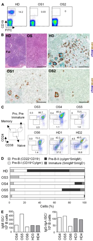
Detection of plasma cells in lymphoid organs of OS patients
OS patients are characterized by oligoclonal T cells, although
they are virtually devoid of circulating B cells (Fig. 1 A and
Table S1
). However, high IgE and residual IgG and/or IgM
serum levels indicate that Ig-producing plasma cells are
ac-tively present. Histological analysis of LN biopsies from two
OS patients revealed a severe alteration of the microscopic
architecture of the organ characterized by expansion of the
paracortical area, along with severe depletion of the lymphoid
cell population and lack of B cell follicles (Fig. 1 B, top left).
Late B cell differentiation stages, including plasma cells and a
subset of GC B cells, which are committed to plasma cell
dif-ferentiation (Angelin-Duclos et al., 2000), are characterized
by the expression of both Blimp1 and CD138, or Blimp1
alone, respectively. Accordingly, LN biopsies from healthy
donors revealed the presence of a relatively small number of
Blimp1
+CD138
–late GC B cells, as well as Blimp1
+CD138
+terminally differentiated plasma cells (Fig. 1 B, top right and
inset). We detected Blimp1
+CD138
+double-positive cells in
both LN biopsies from OS patients, albeit at different levels
(Fig. 1 B, bottom left and right). However, in contrast to
LNs from healthy donors, Blimp1
+CD138
–cells were only
occasionally detected (unpublished data). Thus, in LNs from
OS patients with undetectable circulating B cells, the large
majority of Blimp1
+cells represent an advanced stage of
plasma cell differentiation.
We analyzed BM samples from four OS patients bearing
RAG mutations for B cell subset composition. As expected
from the recombinase defect, a severe block in B cell
differen-tiation was evident, and mature B cells were barely detectable
(Fig. 1 C). To better characterize this developmental arrest, we
players in V(D)J recombination (Villa et al., 1998, 1999). The
hallmark of OS, as a consequence of residual recombinase
ac-tivity, is a peculiar immune phenotype made up of normal or
elevated numbers of activated yet poorly functional T cells,
with a highly restricted oligoclonal TCR repertoire. Such
T cells infiltrate various organs, including skin, gut, spleen,
and liver, resulting in profound tissue damage (Harville et al.,
1997; Rieux-Laucat et al., 1998; Signorini et al., 1999). More
recently, we and others have reported hypomorphic Rag
mouse mutants that mimic many features of human OS (Khiong
et al., 2007; Marrella et al., 2007); the study of these mice has
led to a better understanding of the complexity of OS
patho-genesis. Together, these models have clearly demonstrated
that, in lymphopenic conditions, abnormal compensatory
pe-ripheral T cell proliferation and reduced thymic output could
favor the expansion of T cell clones with inappropriate
self-reactivity, and predispose to the development of
immuno-pathology. Moreover, the lack of thymic Aire expression and
the markedly reduced number of Foxp3
+regulatory T cells
suggested that impairment in both central and peripheral
mechanisms of tolerance may contribute toward the
devel-opment of autoimmunity both in mice (Marrella et al., 2007)
and in humans (Poliani et al., 2009; Cassani et al., 2010).
In contrast, the B cell defect still remains one puzzling
as-pect of OS. Most of the OS patients have high IgE and
resid-ual IgG and/or IgM serum levels, though are virtresid-ually devoid
of circulating B cells. On the other hand, later studies have
shown that hypomorphic RAG mutations can, indeed, be
as-sociated with milder B cell phenotypes and, in such cases, Ig
may be variably present (Villa et al., 2001; Sobacchi et al.,
2006). The basis for this broad clinical spectrum is largely
un-known, but epigenetic and environmental factors may play a
causative role. Consistent with these observations,
spontane-ous hypomorphic Rag1 mutant mice showed high serum
lev-els not only of IgE but also of IgG and IgM isotypes. In the
periphery, partial B cell maturation occurred, displaying a
stricted BCR repertoire. Moreover, B cells in these mice
re-sponded to antigen challenge and T cell help, in agreement
with the presence of functional germinal centers (GCs; Khiong
et al., 2007). In contrast to the leaky B cell defect in this
mu-rine model, B cell differentiation seemed more heavily
af-fected in Rag2
R229Qmice, similar to patients with typical OS
(Noordzij et al., 2002). Indeed, a severe arrest at the pro–B
stage was evident in the BM and was associated to a dearth of
mature functional B lymphocytes in the peripheral lymphoid
organs, which are depleted of B cell follicles (Marrella et al.,
2007). Analogous to archetypal OS, the origin of elevated
serum IgE levels in these mice remains to be elucidated.
Several lines of evidence led us to hypothesize that defects
in RAG-mediated Ig gene editing/revision, either in the BM
or in peripheral lymphoid tissues, might contribute to the
de-velopment of the autoimmune phenotype (Hillion et al., 2005;
Wang and Diamond, 2008). In addition to genetic
susceptibil-ity, autoreactive B cells can arise from the inability of a
defec-tive immune response to eradicate environmental pathogens.
This results in a compensatory, often exaggerated chronic
used the differential expression of marker molecules to define
various stages of B cell differentiation (van Zelm et al., 2005).
In the BM from age-matched healthy children, 18% of
the precursor B cell compartment consisted of cytoplasmic
(Cy) Ig
cells, whereas in BM from all analyzed OS
pa-tients, 92% (82–98%) of the precursor B cell compartment
had this phenotype (Fig. 1 D). Interestingly, some CyIg
+pre–
B-II cells and surface membrane (Sm) IgM
+immature B cells
could be detected in patients OS4 and OS5 (Fig. 1 D). The
mature B cell population (CD19
+SmIgM
+SmIgD
+) was
dra-matically reduced in patients (<0.6% vs. 24% in healthy
con-trols; unpublished data). However, ELISpot analysis revealed
the presence of ISCs in the BM of all OS patients, producing
either IgM or IgG/A (Fig. 1 E).
Characterization of BM and splenic B cell compartment
in Rag2
R229Qmice
Given the obvious limitation in performing experiments with
tissue samples from OS patients, we addressed the role of
B cells in the pathogenesis of Rag2
R229Qmice, which
recapitu-late the main immune system alterations observed in OS
pa-tients (Marrella et al., 2007). Indeed, similarly to the majority
of patients, Rag2
R229Qmice showed absent or dramatically
re-duced numbers of circulating B cells (Fig. 2 A), which is
in-dicative of arrested B cell development. In the BM, Rag2
R229Qmice had a significantly reduced absolute number and
per-centage of B220
+B cells (P < 0.0001; Fig. 2 B and
Fig. S1 A
).
Analysis of early B cell subsets showed a severe block in B cell
differentiation at the pro–B cell stage, as indicated by the
selective increase of early pro–B cells (B220
+CD43
+IgM
–HSA
lowBP-1
–; Hardy et al., 1991; P = 0.03; Fig. S1 A and
not depicted) and the severely reduced pre–B cells (B220
+CD43
–IgM
–), as well as IgM
+IgD
–immature B cells (P < 0.0001;
Fig. 2 B and Fig. S1 A). As a consequence, the absolute number
and relative proportion of splenic B220
+B cells were
pro-foundly reduced in Rag2
R229Qmice (P < 0.0001; Fig. 2, C and D,
and Fig. S1, B and C), with the complete absence of
conven-tional transiconven-tional (T1 and T2) and mature B cells (Fig. 2 C and
Fig. S1 B). The peripheral B cell pool was mainly composed
of IgM
+IgD
–B cells, half of which expressed B220 (43.8 ±
12.3%; Fig. 2 C, bottom), and were negative for the AA4.1
and CD138 markers (not depicted). Notably, the expression
of several cell activation markers (CD40, CD69, CD86, and
MHC II) was augmented in B220
+IgM
+cells from Rag2
R229Qmice when compared with the same subset from WT littermates
Figure 1. Analysis of the B cell compartment in OS patients.
(A) Peripheral blood mononuclear cells were stained with anti-CD19 mAb and analyzed by flow cytometry in two independent experiments. FACS plots of one representative age-matched healthy donor (HD) and two OS patients with RAG mutations are shown. Numbers indicate percentage of
cells. (B) Representative LN biopsies from HD and two OS patients have been stained by H&E and double immunostained with anti-CD138 and Blimp1. Bars: (top left) 200 µM; (top right and bottom left and right) 100 µM.
(C) BM-derived mononuclear cells from patients and healthy donors were stained with anti-CD19, -CD24, and -CD38 mAbs. FACS plots shown are gated on CD19+ cells. Numbers indicate percentage of cells for each
sub-set. (D) Composition of the precursor B cell compartment in OS patients (n = 4) compared with age-matched healthy children (n = 9). The BM
precursor B cell compartment was set at 100% after exclusion of CD10/
SmIgM+/SmIgD+ mature B cells. (E) Frequency of ISCs in cultures of IgM
and IgG-IgA isotypes. The number of spots/105 CD19+ B cells is reported.
(Fig. 2 C). Among IgM
+B cells, the mature naive splenic
B cell compartment is made up of two populations designed
as follicular (FO) B cells (B220
+CD23
highCD21
low) and
mar-ginal zone (MZ) B cells (B220
+CD23
lowCD21
high). Rag2
R229Qmice had barely detectable IgM
+MZ B cells and lacked
ma-ture IgD
+FO B cells (Fig. 2 D and Fig. S1 C), consistent with
the almost complete disruption of both MZ and FO
organiza-tion (not depicted). In contrast, a CD21
–CD23
–B220
+lym-phocyte population was overrepresented in the mutant mice
in comparison with WT littermates (P = 0.0037; Fig. S1 C).
The CD21
–CD23
–cells were CD43
–/lowCD5
–Mac-1
–and
negative for sIg (not depicted), thus excluding the
conven-tional B-1 lineage. These findings collectively suggest that in
Rag2
R229Qmice some B cells may overcome the
developmen-tal block and colonize the periphery.
The presence of peripheral B cells in Rag2
R229Qmice
prompted us to analyze the IgH repertoire. To this end, we
cloned and sequenced the cDNAs encoding the Ig heavy chain
(IgH) variable domain of IgM from bulk sorted B220
+spleno-cytes (
Table S2
). The analysis of these sequences showed one
dominant set of clonally related cells (CRCs) in each of the
mutant mice, whereas we found only four small CRC clusters
in the WT animal (
Fig. S2 A
). We additionally performed
single-cell PCRs on individually sorted B220
+CD19
+spleno-cytes of WT and Rag2
R229Qmice. Again, although we could
not find CRCs in the normal mice, two out of three Rag2
R229Qanimals showed CRC clusters (Fig. S2 A). The absence
of CRCs from Rag2
R229QB220
+CD19
+B cells in the third
sorting experiment is likely caused by the small number of
sequences analyzed. Although we could not definitely
demon-strate that Rag2
R229Qmice display a restricted Igh-V repertoire
(Fig. S2 B), the results from single-cell PCR show expansion
of a few CRCs in Rag2
R229Qmice.
ISC expansion in secondary lymphoid organs
of Rag2
R229Qmice
With high variability, the production of serum IgG
1, IgG
2,
and IgM was preserved in Rag2
R229Qmice (aged 1–9 mo),
whereas IgG
3and IgA were reduced (P < 0.0001; Fig. 3 A).
Consistent with the OS phenotype, IgE levels were
signifi-cantly higher in mutant mice (P < 0.0001). Thus, Rag2
R229Qmice are able to produce serum Ig of all isotypes, in spite of
Figure 2. Characterization of the B cell compartment in Rag2R229Q
mice. (A) Analysis of circulating B cells from WT and Rag2R229Q mice.
(B) Total BM cells were labeled with anti-B220, CD43, IgM, and IgD to determine B cell developmental stages. Representative FACS plots are shown for mice (WT = 35 and Rag2R229Q = 42) analyzed in six independent
experiments. Numbers indicate percentage of cells for each gate. (C) Dot plots show analysis of splenic mature and transitional B cell populations from WT and Rag2R229Q mice, using IgD and IgM markers. Histograms
show expression of CD40, CD86, CD69, and MHCII activation markers in gated splenic B220+ IgM+ cells. Numbers indicate percentage of cells
for each gate. (D) Splenocytes were labeled with anti-B220, -CD21, and -CD23 and analyzed by flow cytometry. Dot plots are gated on B220+
cells. Numbers indicate percentage of cells for each gate. The results shown are representative of mice (WT = 43 and Rag2R229Q = 57) analyzed in
sorted B220
+cell pool, these findings suggest that splenic B cells
from Rag2
R229Qmice might have progressed further through a
plasmacytic differentiation pathway than those of WT mice.
Accordingly, in mutant mice, Xbp1 and Blimp1 expression
were further increased in B220
IgM
+B cells with respect to
B220
+IgM
+B cells, indicative of an advanced stage of plasma
cell differentiation (unpublished data). Furthermore, Rag2
R229QB220
+cells retained increased expression of Aid (P = 0.029;
Fig. 3 C), which is essential for somatic hypermutation and
iso-type switching/class switch recombination (CSR).
Activated effector/memory T lymphocytes sustain ISC
expansion, but do not help an antigen-specific response
in Rag2
R229Qmice
B cell activation and plasmacytic differentiation might be
co-operatively sustained by T cell activation. In Rag2
R229Qmice,
CD4
+T cells were skewed toward T
H
-1 polarization, as
in-dicated by increased production of IFN-, but not of IL-4 and
IL-10 cytokines upon PMA/ionomycin stimulation (Fig. 4 A).
Interestingly, CD4
+T cells from Rag2
R229Qmice had a
substan-tially increased percentage of IL-17–producing cells (Fig. 4 A).
Accordingly, Rag2
R229QCD4
+T cells displayed
signifi-cantly increased mRNA levels for the Th1-specific
T-bet transcription factor and reduced mRNA levels for
GATA-3, which directs Th2 polarization (P = 0.03; Fig. 4 D;
Szabo et al., 2002). Significantly higher mRNA levels for the
Th17-specific RORt transcript were also detected. Thus,
Th1 and Th17 cells could be implicated in the inflammatory
condition characteristic of Rag2
R229Qmice.
Th17 cells
se-crete large amounts
of IL-21, which then
acts to amplify the
the apparent lack of mature B cells. To clarify this apparent
discrepancy, we enumerated ISCs in cell suspensions derived
from BM, spleen, and LNs by ELISpot. Using this analysis,
we found a greater proportion of ISCs in secondary
lym-phoid organs of all Rag2
R229Qmice assayed compared with
WT mice (
Fig. S3 A
). More pronounced differences were
scored when ISC counts were normalized to B cell numbers.
Indeed, mutant mice displayed 6–165-fold increases in IgM-,
IgG-, IgA-, and IgE-secreting cells within the B cell
com-partment of spleen and LNs, compared with WT littermates
(IgM, IgG, IgA, P = 0.008; IgE, P = 0.028; Fig. 3 B). In
contrast, in the Rag2
R229QBM, ISCs were retrieved to a lesser
extent and with higher variability (Fig. 3 B). This observation
correlated with the reduced expression of CXCL12 in
Rag2
R229QBM, the chemokine required for plasma cells
homing and survival in BM niches (Tokoyoda et al., 2004;
Fig. S3 B). These data revealed a larger expansion of the ISC
population in Rag2
R229Qmice, mainly within the secondary
lymphoid organs.
Plasmacytic differentiation is sustained by a program of gene
expression, of which Blimp1 is considered to be the primary
trigger (Martins and Calame, 2008). By quantitative PCR, we
found that expression of Blimp1 is increased by threefold in
splenic B220
+B cells isolated from mutant mice as compared
with controls (P = 0.029; Fig. 3 C). Increased Blimp1
expres-sion also resulted in the induction of Xbp1, which is required
for antibody production and secretion (Reimold et al., 2001;
P = 0.029; Fig. 3 C). Differentiation to ISC with Blimp1
up-regulation is associated with transcriptional repression of Bcl6
and c-Myc, characteristic of GC B cells (Lin et al., 1997; Shaffer
et al., 2000). Accordingly, these transcripts were significantly
reduced in Rag2
R229QB220
+cells (P = 0.029; Fig. 3 C).
There-fore, although partially limited by the heterogeneity of the
Figure 3. Serum Ig levels and ISC in lym-phoid organs. (A) Sera from naive WT (n = 49)
and Rag2R229Q (n = 67) mice (5–37 wk-old)
were collected and their IgG1, IgG2a, IgG2b, IgG3, IgM, and IgA concentrations were deter-mined by luminex technique using a Bioplex reader. Each serum sample was run in dupli-cate, and five independent assays were per-formed. Serum IgE concentrations were measured by ELISA in five independent experi-ments. (B) The frequency of IgM-, IgG-, IgE-, and IgA-secreting cells in the BM, spleen, and LNs of naive WT and Rag2R229Q mice
(10–12 wk old) was determined by ELISpot. The number of spots/105 B220+ cells is indicated.
Results are mean ± SD of five independent experiments testing a total of 17 mice/group. (C) Transcriptional programming sustains ISC differentiation in Rag2R229Q mice. B220+
lym-phocytes were purified by cell sorter from total splenocytes of WT (n = 6) and Rag2R229Q
mice (n = 12). Expression levels of different
transcription factors were normalized to 18S rRNA. Quantitative RT-PCR was run in tripli-cates. Relative measures are indicated as arbitrary units (A.U.). Data represent one of the three independent experiments with consistent results. *, P < 0.05; **, P ≤ 0.01; ***, P ≤ 0.001.
serum level in Rag2
R229Qmice (Fig. 5 C) and a substantial
decrease in the concentrations of CSR-driving cytokines,
IFN- and IL-21 (Fig. 5 D). Thus, these findings highlight
the contribution of CD4
+T cells in sustaining B cell
activa-tion and plasma cell differentiaactiva-tion in Rag2
R229Qmice.
We next investigated whether Rag2
R229Qmice could
mount a T cell–dependent immune response in vivo. Mutant
and WT mice (10 wk of age) were immunized into the
foot-pad with OVA in CFA and boosted with recall antigen in
immunofluorescence (IFA) on day 21. At regular intervals,
sera were analyzed simultaneously for total and OVA-specific
antibodies. As expected, in normal mice immunization
resulted in a rapid and significant increase of total IgG serum
levels (P = 0.03; Fig. 6 A and not depicted), whereas no IgG
elevation was observed in Rag2
R229Qmice (Fig. 6 A).
More-over, in WT animals OVA-specific IgG levels were
detect-able immediately after the boost and sustained after 48 d
(Fig. 6 B). In contrast, we could not detect a secondary
anti-OVA IgG response in the sera from Rag2
R229Qmice (Fig. 6 B).
These results were matched by the analysis of the frequency
of antigen-specific ISC as measured by ELISpot. Unimmunized
Rag2
R229Qmice made significantly more IgG-secreting cells
than WT mice (P = 0.008; Fig. 6 C). However, after
boost-ing, an increased number of IgG plasma cells were detectable
exclusively in the spleens of WT mice (P = 0.029) from days
26 to 33, and returned to nearly baseline levels by day 48,
which is consistent with the IgG serum levels (Fig. 6 C). A
simi-lar trend was observed in the response of WT mice using the
ELISpot assay (Fig. 6 D). No OVA-specific IgG-secreting cells
could be detected
in the Rag2
R229Qmice at any of the
time points assayed
(Fig. 6 D), indicating
Th17 response in an autocrine fashion (Korn et al., 2007;
Zhou et al., 2007). Analysis of serum cytokines by ELISA
revealed that mutant mice had elevated levels of IL-21
(P = 0.0113) and IFN- (P = 0.0007; Fig. 4 B). Because
pro-duction of both IL-17 and IL-21 cytokines has been
associ-ated to FO helper T cells (T
FHcells; Bauquet et al., 2009;
Chtanova et al., 2004), in addition to T
H-17 cells, we
ana-lyzed the CD4
+T cell subset for the expression of CXCR5
and ICOS, crucially involved in T
FHcells homing and
dif-ferentiation (Schaerli et al., 2000; Mak et al., 2003). An
in-creased proportion of ICOS
+cells was observed in Rag2
R229QCD4
+splenocytes (41.6% vs. 7.7%; n = 6; P = 0.008), and
more CD4
+ICOS
hiT cells expressed CXCR5 in Rag2
R229Qmice (7.7% vs. 1.5%; n = 6; P = 0.008; Fig. 4 C). Rag2
R229QCD4
+CXCR5
+ICOS
hiT cells expressed WT levels of
CD40L, but increased levels of programmed death-1 (PD-1;
Fig. S4
), both of which are co-stimulatory molecules
nec-essary for T
FHcell generation and function (Vinuesa et al.,
2005). The mRNA encoding the transcription factor Bcl6,
a determinant of the T
FHcell fate (Nurieva et al., 2009),
was also significantly increased in Rag2
R229QCD4
+spleno-cytes (P = 0.03; Fig. 4 D). Overall, these data define subsets
of T helper cells with the potential to activate B cells
and promote their differentiation into ISC in Rag2
R229Qmice. To better clarify the T cell contribution to B cell
phenotype in Rag2
R229Qmice, we treated mutant mice with
the anti-CD4 mAb (GK1.5) for 7 wk. This treatment
resulted in a complete depletion of circulating, splenic,
and BM CD4
+T cells (Fig. 5 A). Concomitant to the
de-pletion of CD4
+T cells, we observed a significant
reduc-tion of the frequency of IgG and IgA but not IgM-secreting
cells in the spleen and BM of treated mice (Fig. 5 B),
corre-lating with the serum Ig levels (Fig. 5 C). Anti-CD4
treat-ment determined a complete normalization of the IgE
Figure 4. Characterization of effector T cell subsets in Rag2R229Q mice. (A)
Cyto-kine production profile of splenic CD4+ T cells.
Total splenocytes were stimulated for a total of 5 h in the presence of PMA and ionomycin. Cytokine production was assessed by flow cytometry with intracellular staining. Plots are gated on CD4+ T cells. Numbers indicate the
percentage of cells in the respective quad-rants. Data represent one of the three inde-pendent experiments with consistent results. (B) Serum levels of IL-21 (WT, n = 30; Rag2R229Q, n = 50) and IFN- (WT, n = 30; Rag2R229Q, n = 50) cytokines in WT and Rag2R229Q mice (8–24 wk old) as analyzed by
ELISA in three independent experiments. (C) Expression of CXCR5 and ICOS by CD4+
splenic T cells from WT and Rag2R229Q mice.
Numbers in quadrants indicate percentage of cells in each. (D) Expression of indicated mol-ecules in sorted splenic CD4+ T cell population
assessed by qRT-PCR. Samples were run in duplicate, and the target mRNA was normal-ized to Ppia mRNA. The RNA contents are
shown as arbitrary units (A.U.). Means ± SD of six mice/group. Data represent one of the three independent experiments with con-sistent results. *, P < 0.05; **, P ≤ 0.01; ***, P ≤ 0.001.
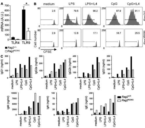
Ig production. Analysis of Ig in the supernatants at day 4 of
culture indicated that mutant B cells were able to switch and
secrete Ig at a rate comparable to control B cells, and
partic-ularly upon CpG stimulation (Fig. 7 C). To confirm in vivo
the T cell–independent humoral responsiveness of Rag2
R229QB cells, we immunized mutant mice i.p. with a mixture of
pneumococcal antigens (Pneumovax23-P23) and monitored
total and specific IgM serum antibodies at different time
points after immunization (from day 10 to 60). The increase
in total and specific serum IgM was lower in Rag2
R229Qwith
respect to WT littermates (Fig. 8, A and B). However, in the
Rag2
R229QB220
+population we detected a higher frequency
of ISC secreting P23-specific IgM which decayed rapidly
(
Fig. S5, C and D
). The possible homing of specific ISC in
the BM was excluded by the failure to detect P23-specific
ISC in the BM of mutant mice (unpublished data). The
ap-parent discrepancy between ELISpot results and serum titers
could be explained by considering the markedly reduced
size of the splenic B cell compartment in mutant mice. Low
absolute numbers of P23-specific IgM ISC would be unable
to determine WT antibody titers. Moreover, Rag2
R229Qdis-played a more rapid decrease in specific serum antibodies
reaching the pre-boost level at day 20, indicating a selective
impairment in the generation of long-term humoral
re-sponses (Fig. 8, A and B). Collectively, our results show that
Rag2
R229Qmice are able to mount short-term specific
hu-moral immune responses, which do not require T cell help
for their generation.
that the observed absence in IgG production resulted from an
impaired generation of specific antibody-secreting cells.
Humoral response to TLR agonists and T cell–independent
antigen in Rag2
R229Qmice
It is well established that Toll-like receptor (TLR)–mediated
activation of mature B cells is required for eliciting humoral
immune responses (Pasare and Medzhitov, 2005). Mature
murine B cells can be stimulated in vitro by LPS or CpG
(the TLR4 and TLR9 ligand, respectively) to proliferate and
secrete antibodies (Watson, 1979; Krieg et al., 1995). We
evaluated the expression of TLR4 and TLR9 by real-time
PCR in purified Rag2
R229QB220
+cells, and found a
reduc-tion in comparison to WT cells (Fig. 7 A). Moreover,
Rag2
R229QB220
+cells did not cycle in response to LPS or
CpG and IL-4 co-stimulation (Fig. 7 B). Because proper
in-terpretation of these results could be hindered by the
hetero-geneity of the B220
+cell subset, we also evaluated the
Figure 5. CD4 T cells sustain ISC differentiation in Rag2R229Q mice.
Rag2R229Q mice (5 wk-old) were injected i.v. with two doses of anti-CD4
mAb (0.25 mg/mouse) 3 wk apart and were analyzed after 7 wk. (A) Rep-resentative FACS plots of CD4 and CD8 stainings in treated mice and con-trols. (B) ISC by ELISpot assay. Data represent the mean ± SD number of spots obtained from n = 5–6 mice/group analyzed and normalized to
105 B220+ cells. Serum Ig (C) and cytokine (D) levels in treated and control
mice. Data represent the mean ± SD of concentrations observed in
n = 5–6 mice/group analyzed. *, P < 0.05; **, P ≤ 0.01.
Figure 6. Rag2R229Q mice fail to induce specific humoral response
to OVA. WT and Rag2R229Q mice were immunized into the footpad with
OVA emulsified in CFA and boosted at day 21 in IFA. PBS was injected in control mice. Total IgG1 (A) and anti-OVA–specific IgG (B) antibody titers were determined at days 26, 33, and 48 after challenge by ELISA. Each point represents mean values + SD from n = 3–4 mice and were corrected
for background binding. Frequency analysis of total (C) and specific (D) ISCs by ELISpot assay on splenocytes from WT and Rag2R229Q mice at the
indicated times after OVA challenge. Data represent the mean ± SD num-ber of spots obtained from n = 3–4 mice/group analyzed in two
indepen-dent experiments and normalized to 105 B220+ cells. n.d., not detectable.
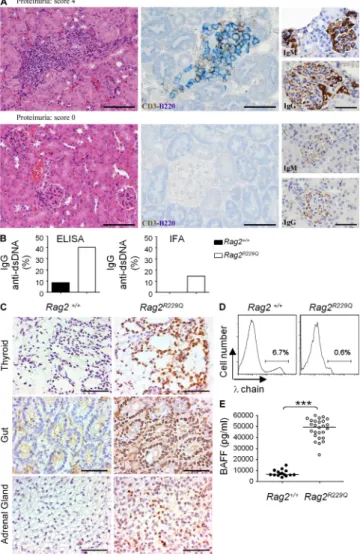
Rag2
R229Qmice. Nonetheless, a direct
role of B cells in the disease
pathogen-esis was suggested by the severe
glom-erulus infiltration of IgM
+and IgG
+cells (Fig. 9 A, top) in mice with high proteinuria index,
whereas no tissue infiltration was observed in nonproteinuric
mice (Fig. 9 A, bottom). To further investigate whether
autoantibody production might contribute to the
autoim-mune phenotype, we screened a large cohort of Rag2
R229Qand control animals for the presence of anti–double-
stranded DNA (anti-dsDNA) measured by ELISA and IFA
with Chionodoxa luciliae, two complementary assays for
detec-tion of DNA autoantibodies characterized by low and high
specificity, respectively. We observed a significant increase in
the frequency of sera positive for both IgG and IgM specific
for dsDNA in the group of mutant mice as compared with
WT (P < 0.01; Fig. 9 B and not depicted). Notably, high
ti-ters of IgG anti-dsDNA were detected in animals with severe
kidney infiltrations and urine protein loss (unpublished data).
Staining of frozen sections of multiple organs from Rag2/Il2rc
double knock-out mice (devoid of endogenous Ig) with sera
derived from mutant mice confirmed the
presence of autoreactive Ig targeting
differ-ent tissues (Fig. 9 C).
B cells from Rag2
R229Qmice actively participate to
tissue damage
A proportion of Rag2
R229Qmice spontaneously develop
se-vere alopecia, skin erythrodermia, and wasting syndrome
caused by colitis (Marrella et al., 2007). In addition, a
minor-ity of them presented with differing degrees of renal damage,
resulting in proteinuria. A marked infiltration of oligoclonal
activated T lymphocytes was consistently evident in different
tissues of mutant mice and correlated with abnormal CXCL10
serum level (P = 0.002;
Fig. S6 A
). Further characterization
of these infiltrates revealed that they were also composed of
B220
+Ig
+B cells (Fig. 9 A; Fig. S6, B and C). The
abun-dance of B220
+and IgM
+cell infiltrates was lower with
re-spect to CD3
+T cells (P = 0.048 and P = 0.0098, respectively;
Fig. S6 C), which may reflect the absence of circulating
B cells, as well as the significantly lower representation of
B lymphocytes in primary and secondary lymphoid organs of
Figure 7. Rag2R229Q mice have impaired
proliferative responses to TLR agonists but intact Ig responses. (A) Splenic B220+
lym-phocytes were purified by cell sorting, and the relative mRNA levels of TLR4 and TLR9 expres-sion were determined by RT-PCR. The samples were run in duplicates. The obtained values were normalized to 18S rRNA and indicated as arbitrary units (A.U.). Mean values ± SD of three independent experiments testing eight mice/group. (B) Total splenocytes were CFSE labeled, and then stimulated in vitro with LPS (10 µg/ml) and CpG (2.5 µg/ml) in the absence or presence of IL-4 (10 ng/ml). CFSE dilution was analyzed by FACS at day 4 of culture. Representative FACS histograms of CFSE pro-file in B220+ cell gate are shown (percentage
is reported above the gate). The histogram plot is representative of three independent experiments (C) In parallel, Ig levels were measured in the culture supernatants. Ig con-centrations were normalized for the number of B220+ cells in the culture. *, P < 0.05.
Figure 8. Rag2R229Q mice show short-term
humoral response to Pneumovax23. WT and
Rag2R229Q mice were immunized i.p. with P23. PBS
was injected in control mice. Total (A) and anti-P23– specific IgM (B) antibody titers were determined at different days after challenge by ELISA. Each group represents mean values ± SD from n = 5 mice and
were corrected for background binding. The data are representative of two independent experiments.
antibody gene rearrangements, usually involving the and
light chain gene loci. Repeated variable joining
rearrange-ments eventually lead to exhaustion of the recombination
po-tential at the Igk-V gene locus and expression of a light chain.
A markedly reduced frequency of chain
+B220
+cells was
observed in mutant mice (1.5 ± 1% vs. 4.7 ± 1.6%; P = 0.0002;
Fig. 9 D), thus suggesting that Rag2
R229Qmutation impairs the
process of secondary recombination. In normal conditions,
pe-ripheral self-reactive B cells compete with non–self-reactive
B cells for survival factors. BAFF is the key regulator of B cell
homeostasis beyond the late transitional B cell stage
(Meyer-Bahlburg et al., 2008), when expression of BAFF-R is acquired
on B cells. Because increased BAFF levels are expected in mice
with reduced B cell numbers (Lesley et al., 2004; Lavie et al.,
2007), we measured BAFF serum levels in our model.
Signifi-cantly, higher levels of BAFF were already detected in
Rag2
R229Qmice (P < 0.0001; Fig. 9 E) early in life (5-wk-old
animals), whereas in WT mice the increase in BAFF serum
levels was age-dependent (Fig. S6 D).
Effect of BAFF-BAFF-R signaling blockade
in Rag2
R229Qmice
As BAFF promotes B cell survival, its overexpression could
potentially break B cell tolerance by rescuing self-reactive
B cells from deletion. Indeed, mice transgenic for Tnfsf13b (the
gene encoding BAFF) develop severe autoimmune symptoms
(Mackay et al., 1999; Lesley et al., 2004; Thien et al., 2004).
Based on this evidence, we speculated that increased BAFF
serum levels in Rag2
R229Qmice could play a crucial role in
B cell–mediated autoimmunity by favoring the survival and
differentiation of autoreactive ISCs. To directly test this
hy-pothesis, we examined the in vivo effect of selective
BAFF-BAFF-R signaling blockade. Mutant mice displaying an overt
pathological phenotype (i.e., presence of serum anti-dsDNA
IgG and proteinuria) were i.v. injected with three 0.5-mg
doses of anti–BAFF-R mAb (9B9) known to prevent BAFF
binding (Rauch et al., 2009), and followed for 2 mo. Rag2
R229Qmice treated with anti–BAFF-R mAb showed a significant
de-crease in the number of splenic B220
+B cells compared with
PBS-treated (control) animals (P = 0.008; Fig. 10 A). In WT
mice, administration of anti–BAFF-R resulted in 90%
deple-tion of FO B cells and 68% depledeple-tion of MZ B cells
(unpub-lished data). Interestingly, a marked reduction of both CD4
and CD8 T cells was observed in mutant, but not WT mice,
consistent with the role of BAFF signaling on activated T cells
(Mackay and Leung, 2006; unpublished data).
The frequency of IgM, IgG, and IgA-ISC in the spleen
was not significantly different in treated versus control mutant
mice, but because of the smaller splenic B cell counts, the total
number of ISC was significantly lower in anti–BAFF-R–
treated Rag2
R229Qmice than controls (P < 0.001; Fig. 10 B and
not depicted). After euthanization, histological examinations
were performed and the degree of cell infiltration was scored
in different organs (liver, gut, lung, kidney, and skin).
Re-markably, a significantly decreased infiltration index was
gen-erally observed in Rag2
R229Qanti–BAFF-R–treated mice
These findings are indicative of defaults in B cell selection
and tolerance. Receptor editing is the mechanism by which
B lymphocytes alter their antigen receptors through secondary
Figure 9. Residual autoreactive B cells in Rag2R229Q mice
contrib-ute to tissue damage in autoimmune-like manifestations. (A)
Char-acterization of kidney infiltrates and correlation with clinical symptoms. Kidney sections from a mouse with proteinuria (score 4: ≥ 2000 mg/dl; top) and a mouse without renal disease (bottom). Kidneys from mice with proteinuria show infiltrates positive for staining with CD3, B220, IgM and IgG. Bars: (left) 100 µM; (middle) 50 µM; (right) 60 µM. (B) The presence of IgG anti–double-strand DNA autoantibodies was analyzed in the sera of WT (n = 70) and Rag2R229Q (n = 97) mice by ELISA and IFA in five
inde-pendent experiments. Bars indicate the frequency of positive sera in mice aged 8–24 wk. (C) Ig targeting tissues are detected in the sera of
Rag2R229Q mice. Frozen sections obtained from, gut, thyroid, and adrenal
gland of Rag2/Il2rc double-KO mice were incubated with sera obtained
from WT and Rag2R229Q mice. Staining with anti–mouse IgG Ab followed
by incubation with secondary, anti–mouse IgG peroxidase-conjugated antibody was performed. Representative images from 1 out of 10 mice tested/group. Bars, 100 µM. (D) FACS analysis of light chain expression in gated B220+ IgM+ cells. Results are representative of 11 mice/group
analyzed in three independent experiments. (E) Serum BAFF in Rag2R229Q
(n = 26) and WT littermates (n = 15), measured by ELISA in two
antibodies was detected in the majority of the treated mice
(Fig. 10 D). Overall, these results support a causative role of
B cells in the Rag2
R229Qimmunopathology, providing
prelimi-nary evidence for a potential therapeutic application of
BAFF-BAFF-R signaling blockade.
DISCUSSION
In this study, we have exploited Rag2
R229Qmice, which
repro-duce the immunopathology observed in OS patients, to
ad-dress whether B cells play any role in the pathogenesis of the
disease. The impairment of early B cell development in
Rag2
R229Qmice determines a dramatic reduction of the
periph-eral B cell compartment, which is characterized by the
pres-ence of Ig-negative precursors and only
a few IgM
+IgD
–B cells, which express
several activation markers. FO B cells
were almost undetectable, whereas
splenic MZ B cells were barely present.
In contrast, mutant animals contained
an expanded CD21
–CD23
–IgM
–lym-phocyte population. This phenotype
could be reminiscent of mice with
de-fective B cell development caused by
lack of the surrogate light chain. In
fact, these mice displayed an expanded
B220
+CD21
–CD23
–IgM
lowcell subset
and produced ANAs upon in vitro
polyclonal stimulation (Harfst et al.,
compared with controls, and specifically in the extent of
infil-trating B220
+and IgM
+B cells (Fig. 10 C). The potential
clin-ical effect of anti–BAFF-R administration was also evaluated.
The Rag2
R229Qmice studied manifested similar proteinuria
levels at the beginning of treatment, which significantly
wors-ened over time in mice treated with PBS (P = 0.04; Fig. 10 D).
In contrast, stabilization of kidney disease was observed in anti–
BAFF-R–treated mice, with 4/7 mice displaying a reduction in
the proteinuria index (Fig. 10 D). Consistently, the treated
mice had markedly reduced signs of glomerular damage
com-pared with the control mice that showed mesangial cell
prolif-eration and heavy deposition of IgG (Fig. 10 D). Furthermore,
2 mo after treatment, the disappearance of IgG anti-dsDNA
Figure 10. Effect of anti-BAFF-R mAb administration in Rag2R229Q mice. Mutant
mice were treated with three doses of 0.5 mg anti–BAFF-R mAb or PBS, and followed for a total period of 2 mo. (A) Splenic B220+ cell
counts. (B) Absolute numbers of splenic ISC. Mean values ± SD obtained from n = 6–7
mice/group. (C) The global and cell-specific (CD3+, B220+, IgM+) degree of infiltration was
scored in different organs (liver, kidney, lung, and gut) from anti–BAFF-R mAb and PBS-treated mice. Bars indicate the mean ± SD infiltration index calculated for all the organs from n = 6–7 mice/group. Images show
im-munostains of gut and liver from representa-tive PBS and anti–BAFF-R mAb-treated mice. Bars: (left) 200 µM; (right) 50 µM. (D) The presence of high-affinity IgG anti-dsDNA autoantibodies was analyzed in the sera of PBS and anti–BAFF-R Rag2R229Q mice by IFA,
before and after the treatment. Bars indicate the frequency of positive sera. Proteinuria was assessed before and after treatment of
Rag2R229Q mice with PBS and anti–BAFF-R,
respectively. Mean values ± SD of proteinuria index in n = 6–7 mice/group. (bottom)
Repre-sentative images of glomeruli from untreated proteinuric mice and anti-BAFFR–treated mice. Bars, 50 µM.
that T cell help for B cells was not dependent on cognate
interaction, as Rag2
R229Qmice were severely impaired in the
antibody response to TD antigens. The lack of OVA-specific
IgG production might be caused by impaired T-B cell
col-laboration in the absence of GC-like structures. On the other
hand, in this issue,
Walter et al.
show that adoptive transfer
of WT CD4
+T lymphocytes was unable to correct
defec-tive antibody response to TNP-KLH, indicating B cell
in-trinsic abnormalities.
In Rag2
R229Qmice, a skewed pattern of cytokines
expres-sion and T helper cell bias may favor B cell–mediated
auto-immunity. Dysregulated T
FHcells produce large amounts of
IL-21, which promotes B cell activation as well as secretion of
pathogenic anti-dsDNA autoantibodies and contributes to
chronic T cell–mediated autoimmune inflammation by the
generation of Th17 cells. Interestingly, it has been shown that
IL-17 acts in synergy with BAFF in supporting B cell
prolifer-ation and differentiprolifer-ation to plasma cells in SLE patients (Hutloff
et al., 2004; Iwai et al., 2003). In our model, homeostatically
proliferating T cells are critical for the generation of ISCs, as
well as the induction of the hyper-IgE state in mutant mice,
and most likely contribute toward the pathogenesis of
distinc-tive OS clinical features.
On the other hand, autoimmunity develops to the same
extent in T cell–sufficient and T cell–deficient BAFF-transgenic
mice (Groom et al., 2002), indicating that BAFF-mediated
rescue of self-reactive B cells may be T cell independent.
High-affinity anti-dsDNA serum IgG autoantibodies
to-gether with severe kidney damage with proteinuria, which
we observed in Rag2
R229Qmice, are pathognomonic signs of
B cell–mediated autoimmunity (Sobel et al., 1991; Manson
et al., 2009). In the hypomorphic Rag1 murine model
de-scribed by Walter et al. (2010), only low-affinity IgG
auto-antibodies were detected and the majority of the mice were
devoid of clinical symptoms. This variability in clinical
phe-notype may reflect the different impact of the genetic defect
on the recombination activity and the contribution of
envi-ronmental factors.
Fewer B cells expressing chain in the mutant mice is
suggestive of defects in the secondary rearrangements (Vela
et al., 2008). Because an impairment in this process would also
affect autoreactive Ab-expressing B cells, which could not
therefore be silenced by light chain editing, it can be
specu-lated that hypomorphic Rag2 mutation leading to impaired
RAGs activity limits the potential to edit autoimmune BCRs.
Under physiological conditions, self-reactive B cells that
es-cape deletion and receptor editing, compete with
nonautore-active B cells for peripheral survival signals. Among these
signals, BAFF has a crucial role in controlling peripheral B cell
homeostasis (Mackay et al., 2003). In Rag2
R229Qmice
in-creased BAFF serum level is likely a consequence of the B cell
lymphopenic environment (Lesley et al., 2004; Lavie et al.,
2007). It has been shown that increased BAFF levels favor the
persistence of self-reactive B cells normally deleted at
devel-opmental checkpoints (Lesley et al., 2004; Thien et al., 2004).
Indeed, high levels of BAFF have been detected in the serum
2005; Keenan et al., 2008). Based on this, it can be speculated
that in Rag2
R229Qmice most of the peripheral B cells carry
autoreactive potential.
The analysis of a large cohort of mutant mice revealed, in
some animals, a normal or even higher Ig production
com-pared with WT mice. This variability, resembling the clinical
phenotype reported in OS patients (Villa et al., 2001), is
ac-tually greater than the one previously reported by Marrella
et al. (Marrella et al., 2007). Such a discrepancy could likely
reflect the mixed genetic background of our model and the
effects of stochastic events occurring during B cell repertoire
generation. In mutant mice, the residual serum Ig production
is maintained by an enlarged ISC compartment. Excessive
commitment to plasma cell fate is sustained by a defined
tran-scription program. The observed increase in Blimp1 likely
leads to cessation of cell cycling activity in Rag2
R229QB cells
(Lin et al., 1997), repression of genes required to establish
GC reactions, and induction of the Ig secretory pathway
(Shaffer et al., 2002; Shaffer et al., 2004). Our findings
corre-late with previous observations in immunodeficient and/or
irradiated hosts, reconstituted with a limited number of
pe-ripheral B cells (Agenès and Freitas, 1999). In these animals,
B cells quickly expanded, displayed an activated phenotype,
and gave rise to an increased proportion of plasma cells.
Hence, a compensatory homeostatic regulation might account
for the skewed B cell phenotype in our model. During
inflam-mation Aid and Blimp1 expression can be induced in
imma-ture/T1 B cells by TLR engagement in a T cell–independent
manner (Ueda et al., 2007). Consistently, Aid
+immature/T1
B cells were preferentially found outside the conventional
GC, at sites exposed to exogenous antigens (Wang et al.,
2000; Ueda et al., 2007). The most severe inflammatory and
autoimmune-like manifestations in Rag2
R229Qmice affect
or-gans like the gut and the skin, where exposure to microbial
antigens could potentially select immature B cell clones for
expansion and antibody production. We showed that B cells
from Rag2
R229Qmice efficiently responded to TLR ligands
and polysaccharides Ag by undergoing rapid plasmacytic
dif-ferentiation and Ig class switching. The limited variability
consequent to impaired recombinase activity and the stochastic
nature of B cell repertoire generation might represent factors
determining the phenotypic heterogeneity of OS patients.
Several experiments in this study addressed the role of
T cells in B cell homeostasis in Rag2
R229Qmice. We found
that CD4 T cells were required for B cell activation and
dif-ferentiation to plasmablasts in Rag2
R229Qmice. We provided
evidence that the dominating T cell subsets in mutant mice,
namely Th1, Th17, and T
FHcells do have a crucial role in
B cell abnormalities. In fact, anti-CD4 treatment caused a
significant reduction of ISC compartment and a
normaliza-tion of serum IgE. These results correlated with the decreased
serum concentration of cytokines involved in CSR such as
IFN- and IL-21. Interestingly, IL-21 has recently been
re-ported to stimulate IgE synthesis in humans, thus highlighting its
important role in the occurrence of allergic and atopic
dis-orders (Kobayashi et al., 2009). Furthermore, we demonstrated
Flow cytometry and cell sorting. Mononuclear cells from the
periph-eral blood and BM of patients were labeled with the following antibodies: anti–human-CD19, CD24, CD38, CD22 (BD); SmIgM, SmIgD, CyIgM (Kallestad/Sanofi-Synthelabo). Cell suspensions from murine BM, spleens, and LNs were stained in PBS 2% FCS-containing antibodies. To reduce nonspecific binding, cells were pretreated with anti-CD16 (BD). The fol-lowing anti–mouse antibodies were purchased from eBioscience: B220, CD5, CD21, CD23, BP-1, CD40L, and PD-1. Reagents purchased from BD include: IgM, IgD, MHCII, CD40, ICOS, CD69, CD80, CD86, AA4.1, Mac-1, CD4 (RM4-5 and GK1.5), CD138, chain, HSA, strepta-vidin, and appropriate isotype controls. Anti–mouse-CD43 and CXCR5 were obtained from BioLegend. To detect cytokines production by T cells, splenocytes were cultured for 5 h with PMA (50 ng/ml), ionomycin (1 µg/ ml), and monensin (1 µg/ml; Sigma Aldrich), and then intracellular IFN-, IL-4, IL-10, and IL-17 were stained according to the manufacturer’s instructions (BD). Samples were acquired on a FACSCanto II system (BD) and analyzed with FLOWJO software (version 4.5.4; Tree Star Inc.). Splenic B lympho-cytes (B220+) used for mRNA analyses were sorted with a FACSAria (BD).
The purity was >95%.
ELISA and ELISpot. Levels of IgG isotypes, IgA, and IgM were measured
in culture supernatants and sera by multiplex assay kit (Beadlyte Mouse Immu-noglobulin Isotyping kit; Millipore) according to the manufacturer’s instruc-tions and run using a Bio-Plex reader (BioRad Laboratories). Levels of IgE were determined by ELISA assay (BD). Cytokine and chemokine levels in su-pernatants and sera were measured with specific ELISA kits for mouse BAFF, IL-21, IFN-, and CXCL10 (R&D Systems). ISCs were analyzed by ELISpot using the species-specific purified and biotinylated anti-IgG, IgM, IgA, and IgE mAbs (Southern Biotech) and the 3-amino-9-ethylcarbazole (Sigma-Aldrich) as a chromogenic substrate.
Determination of auto-antibody profile. For anti-dsDNA, 1:10 diluted
serum was applied to fixed C. luciliae slides (Antibodies Inc.), with biotin- conjugated goat anti–mouse IgG (Southern Biotech), and Alexa Fluor 555– conjugated streptavidin (Invitrogen) as detection reagent. A reader blinder to the genotype/treatment of the mice read slides at 1,000×. Anti-dsDNA anti-bodies were also evaluated by ELISA assay. In brief, polystyrene plates were coated with poly-L-lysine (Sigma-Aldrich) and DNA from calf thymus (Sigma-Aldrich); after post coating with 50 µg/ml of Polyglutamic Acid for 45 min and blocking with PBS 3% BSA, serial dilutions of serum from 1:20 to 1:1,280 were incubated overnight. Bound Abs were detected with alkaline phosphatase-conjugated goat anti–mouse IgG (Southern Biotech). The score of positivity was assigned to sera that were positive for dilution of 1:20 or higher. For anti-cardiolipin antibodies, polystyrene plates were coated with 50 µg/ml of cardiolipin diluted in ethyl alcohol.
Autoantibodies against murine tissues were detected by indirect immu-nohistochemistry on cryostat tissue sections from adult Rag2/Il2rc/ mice
to avoid interference from endogenous Ig, and stained with 1/20 dilutions of sera from Rag2R229Q mice and control littermates. Slides were examined
on a Zeiss Axioplan-2 microscope and images were acquired by Olympus DP70 camera using CellF imaging software (Soft Imaging System GmbH).
Functional in vitro assay. Total splenocytes were labeled with 0.5 µM
CFSE (Invitrogen) for 8 min at room temperature. After quenching the labeling reaction by adding FCS, cells were washed twice. Labeled cells (105 cells) were cultured in a 96-well U-bottomed plate with the following
stimuli: 2.5 µg/ml CpG-B oligodeoxynucleotide (5-tccatgacgttcctgacgtt-3; Alexis Co.), 10 µg/ml LPS (R&D Systems), and 10 ng/ml IL-4 (R&D Sys-tems). The proliferation profile of propidium iodide-negative viable B220+
cells was analyzed at day 4 of culture. On the same day, culture supernatants were collected for Ig quantification.
Histology. Formalin-fixed paraffin-embedded tissue sections from inguinal
LN biopsies of OS patients and patients with unrelated nonimmunological diseases were subjected to routine hematoxylin and eosin (H&E) and double
of patients with various autoimmune conditions (Zhang et al.,
2001; Groom et al., 2002; Lindh et al., 2008). In Rag2
R229Qmice high levels of serum BAFF might rescue autoreactive
clones and favor their differentiation to ISC. We showed that
blocking of BAFF-BAFF-R signaling in Rag2
R229Qmice with
an anti-BAFF-R monoclonal antibody led to the
disappear-ance of anti-dsDNA IgG antibodies, a significant amelioration
of inflammatory infiltrates and an arrest of renal disease
pro-gression. Thus, these data strongly suggest that B cell
abnor-malities and increased BAFF serum levels play a significant
role in autoimmune manifestations in Rag2
R229Qmice. Walter
et al. (2010), in the companion paper, also measured elevated
serum BAFF levels and autoantibody production in a large
proportion of patients with OS and leaky SCID caused by
hypomorphic rag mutations. Based on the results presented in
this study, we propose that defects in B cell differentiation and
function actively contribute to the OS pathogenesis. In
sup-port of this hypothesis, we resup-ported the presence of terminally
differentiated plasma cells in the lymphoid organs of OS
pa-tients devoid of circulating B cells. The similarities between
murine and human studies validate the Rag2
R229Qmodel for
further investigation on the contribution of B cell defects to
autoimmunity. In conclusion, we have shown that
hypo-morphic rag mutations do not only result in profound
impair-ment of B cell developimpair-ment but are also associated with the
generation of an autoimmune BCR repertoire. We showed
that the peripheral cytokines milieu crucially influences the
generation of ISC and B cell–mediated immunopathology.
The contribution of B cells to autoimmune pathology could
provide the basis for a potential use of B cell–directed
inter-vention in OS treatment.
MATERIALS AND METHODS
Patients and cells. Six unrelated patients with hypomorphic RAG defects
whose clinical, immunological, and molecular features were consistent with OS were studied (Table S1). Human samples were collected after signed in-formed consent in accordance with the protocols of Spedali Civili of Brescia and Erasmus MC Ethical Committees. Mononuclear cells from peripheral blood and BM were purified on Ficoll gradient (Nycomed Pharma A/S).
Mice treatment and immunization. 129Sv/C57BL/6 knock-in Rag2R229Q
mice were previously generated (Marrella et al., 2007), and housed in specific pathogen–free facilities. Immunizations were performed in mice aged 2–3 mo. T cell–dependent antigen response was induced by footpad injection of 100 µl of a 1:1 emulsion of CFA and 100 µg OVA (grade V; Sigma-Aldrich). Secondary response was elicited by boosting with 50 µg OVA in IFA (Sigma-Aldrich). T cell–independent antigen response was induced by i.p. injection of 115 µg Pneumovax-23 (Merck). For CD4+ T cell depletion, mice were
injected i.v. with two doses of GK1.5 (0.25 mg) 3 wk apart and followed for a total of 7 wk. Anti–BAFF-R (9B9) mAb treatment was performed in mice (3–5 mo age) previously tested for serum autoantibody and urine proteinuria. 3 doses of 0.5 mg were administered i.v. at 3 wk intervals. The mice were followed for 2 mo. In all experiments control mice were injected with PBS. All procedures were performed according to protocols approved by the Insti-tutional Animal Care and Use Committee of the San Raffaele Scientific In-stitute (IACUC318).
Proteinuria evaluation. Proteinuria was evaluated using Albustix stick
(Bayer). Proteinuria index was scored as follows: 0 < 30 mg/dl; 1 = 30 mg/dl; 2 = 100 mg/dl; 3 = 300 mg/dl; and 4 ≥ 2,000 mg/dl.