Should living related kidney transplantation be considered for patients with renal failure due to Fabry's disease?
3
0
0
Texte intégral
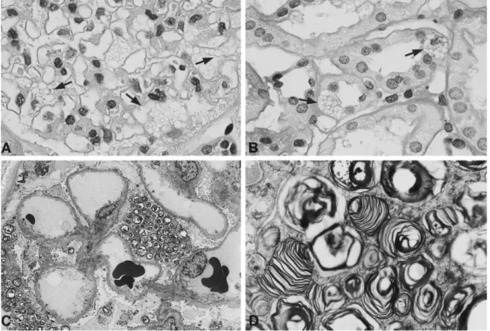
(2) Fabry’s disease and living related kidney donation. cion of cornea verticillata). Nephrological tests showed a normal urinary sediment without proteinuria, a normal serum creatinine, and a normal renal ultrasound. Inulin and PAH clearances were then performed, revealing a GFR of 120 ml/min/1.73 m2 and a RPF of 563 ml/min/m2 (both considered normal ). The patient then underwent percutaneous renal biopsy to test for evidence of GSL accumulation in the kidney. Figure 1 demonstrates that glomerular epithelial cells and some proximal tubular cells were filled with typical clear vacuoles in a mosaic pattern, representing the GSL deposits. The immunofluorescence staining showed no IgG, IgM, or C3 deposition (not shown). Electron microscopy examination of the biopsy also revealed typical vacuoles filled with lamellated electrondense myelin figures in visceral and parietal glomerular epithelial cells. Inclusions were also found in endothelial cells. Overall the GSL deposition was extensive and contrasted with the unremarkable clinical presentation. Because of the remarkable GSL deposition in the kidney of the heterozygous mother it was decided not to proceed with living related organ donation but to consider this 27-year-old patient for cadaveric allograft transplantation instead. Several months later he was successfully transplanted with a cadaveric kidney and so far has had an uneventful course.. 2935. Discussion The situation of an affected male hemizygous patient with Fabry’s disease presenting with his asymptomatic mother for living related kidney donation illustrates the problems transplant physicians are faced with when examining clinically normal heterozygous donors of hereditary diseases for renal allograft transplantation. Although the mother did not show clinical or laboratory signs of kidney disease on routine examination she was found to have abundant osmiophilic electrondense inclusions in glomerular epithelial and in some proximal tubular cells. In the literature a few reports have shown that female heterozygous carriers of Fabry’s disease can develop renal disease which is characterized by proteinuria and haematuria and later by the development of chronic, and very rarely of end-stage renal failure [3,5–9]. The majority of female carriers do not develop renal disease, however, and negative GSL deposition has been documented by Gubler et al. in a biopsy of a female carrier [5]. Since it is impossible to predict which carrier will develop renal involvement one needs to perform a renal biopsy to determine the extent of renal involvement.. Fig. 1A-D. Renal biopsy of the 48-year-old potential heterozygous female donor. (A) Glomerulus with visceral epithelial cells which are filled with clear vacuoles (arrows) (H&E ×400). (B) Individual cells in proximal tubules are also filled with clear vacuoles (arrows) (PAS ×400). (C ) Overview by electron microscopy, demonstrating ‘myelin figures’ in podocytes (×1850). (D) Electron micrograph demonstrating the lamellated structure of the ‘myelin figures’ (×8200)..
(3) 2936. A case has been reported where living related renal transplantation has been performed from an asymptomatic female heterozygotic carrier to her unaffected daughter who developed renal failure due to chronic glomerulonephritis with nephrotic syndrome [10]. The typical lesions of Fabry’s disease, which were seen in the graft at the time of transplantation, remained unchanged in successive biopsies, suggesting that hyperfiltration in a normal a-galactosidase environment did not affect renal morphology adversely. The donor’s subsequent course over 7 years was also favourable and the renal function stayed stable. Another case of living related donation has been reported where a normal unaffected sister donated a kidney to her hemizygous affected brother [4]. Most transplants reported for patients with Fabry’s disease have been performed from cadaveric donors. The outcome for these patients has been satisfactory with 1-year graft and patient survival times of over 80% and 90% respectively [11]. What is clear from the literature is that a recurrence of GSL deposition is detectable when a normal cadaveric kidney is transplanted into a patient with Fabry’s disease, suggesting that these storage products can accumulate in a transplanted organ despite possessing enzymatic activity for a-galactosidase A (reviewed in [12]). GSL accumulation is usually confined to the endothelium, although more widespread graft involvement can occur. It is therefore quite possible that a heterozygous kidney with signs of renal involvement could become severely affected after a short time with danger of subsequent allograft loss when transplanted into an agalactosidase-deficient environment. Some authors have suggested that living related donation should always be avoided in patients with Fabry’s disease [4]. Based on our experience with the reported case we would recommend the performance of a kidney biopsy to evaluate the extent of renal involvement in heterozygous females with absence of proteinuria, haematuria, or signs of renal failure. In. R. P. Wu¨thrich et al.. case of a positive biopsy, living related kidney donation should not be performed because of the danger for allograft loss due to ‘recurrent’ disease and because of a possible risk of the donor’s developing chronic renal failure.. References 1. Desnick RJ, Ioannou YA, Eng CM. a-Galactosidase A deficiency: Fabry disease. In: Scriver R, Beaudet AL, Sly WS, Valle D. The Metabolic and Molecular Basis of Inherited Disease. McGraw-Hill, New York, 7th edn, 1995; 2741–2784 2. Desnick RJ, Astrin KH, Bishop DF. Fabry disease: molecular genetics of the inherited nephropathy. Adv Nephrol 1989; 18: 113–127 3. Burda CD, Winder PR. Angiokeratoma corporis diffusum universale (Fabry’s disease) in female subjects. Am J Med 1967; 42: 293–301 4. Schweitzer EJ, Drachenberg CB, Bartlett ST. Living kidney donor and recipient evaluation in Fabry’s disease. Transplantation 1997; 54: 924–927 5. Gubler M-C, Lenoir G, Gru¨nfeld J-P, Ulmann A, Droz D, Habib R. Early renal changes in hemizygous and heterozygous patients with Fabry’s disease. Kidney Int 1978; 13: 223–235 6. Rodriguez FH, Hoffmann EO, Ordinario AT, Baliga M. Fabry’s disease in a heterozygous woman. Arch Pathol Lab Med 1985; 109: 89–91 7. Chen HC, Tsai JH, Lai YH, Guh JY. Renal changes in heterozygous Fabry’s disease—a family study. Am J Kidney Dis 1990; 15: 180–183 8. Fukushima M, Tsuchiyama Y, Nakato T et al. A female heterozygous patient with Fabry’s disease with renal accumulation of trihexosylceramide detected with a monoclonal antibody. Am J Kidney Dis 1995; 26: 952–955 9. Van Loo A, Vanholder R, Madsen K et al. Novel frameshift mutation in a heterozygous woman with Fabry disease and endstage renal failure. Am J Nephrol 1996; 16: 352–357 10. Gru¨nfeld J-P, Le Porrier M, Droz D, Bensaude I, Hinglais N, Crosnier J. La transplantation re´nale chez les sujets atteints de maladie de Fabry: transplantation du rein d’un sujet he´te´rozygote a` un sujet sain. Presse Med 1975; 4: 2081–2085 11. Tsakiris D, Simpson HKL, Jones EHP et al. Rare diseases in renal replacement therapy in the ERA-EDTA registry. Nephrol Dial Transplant 1996; 11 [Suppl 7]: 4–20 12. Gantenbein H, Bruder E, Burger HR, Briner J, Binswanger U. Recurrence of Fabry’s disease in a renal allograft 14 years after transplantation. Nephrol Dial Transplant 1995; 10: 287–289 Received for publication: 13.5.98 Accepted in revised form: 15.6.98.
(4)
Figure
Documents relatifs
Doux ballet mis en scène approuvé pas les anges, Ton âme ne sera que ferveur et louanges. Union de vos deux chairs, sentiment d’absolu, D’individu souffrant à un
In contrast, in Chlamydomonas we have previously shown the existence of three proteases, two of which, namely SPP-2 and SPP-1, cleaved pSS in vitro at specific sites and
Samples, such as woody debris, benthic algae, particulate organic material contained in terrestrial soil sediment (SPOM) – in the following denoted as particulate organic matter
Fourth, it would be helpful, in particular for medical stu- dents and residents, if the authors could rank the relevance of the different topics within a chapter (e.g.,
[r]
Plus encore, on peut penser que le film de Melville — comme Rashomon (1950) d’Akira Kurosawa — se concentre sur la relativité de la perception (le film de
Model outputs overlayed on an isometric projection of the study area topography: (a) simulated 137 Cs inventories, (b) simulated soil redistribution rates by tillage erosion, and (c)
a- Kapsiki 1- Tombe d’un homme avec une jarre entourée de stèles en carré 2- Tombe d’une femme avec une meule entourée de stèles en triangle 3- Tombe d’un enfant b- Podoko 1-
