REGULATION OF CORONARY BLOOD FLOW DURING ETHER AND HALOTHANE ANAESTHESIA
G. WOLFF, B. CLAUDI, M. R I S T , M. R. WARDAK, W. NIEDERER AND E. GRAEDEL
SUMMARY
The effects of ether (6-10%) and halothane (1-2%) were studied on coronary flow regulation in dogs. In one group of experiments the kft coronary artery was perfused mechanically, coronary perfusion pressure being either kept constant or adjusted to aortic pressure, and the heart itself had to pump the blood to all other arteries. The preload on the heart was changed by varying die intravascular volume. In another group, bodi coronary arteries were perfused mechanically under constant pressure; the other arteries were also perfused mechanically by a cardiopulmonary bypass. In this group measurements were carried out on the empty beating heart. Halothane had a direct effect on die heart, myocardial contractility was reduced, cardiac work and myocardial oxygen consumption were diminished and coronary vasoconstriction followed. Edier effects on the heart were principally die same as diose due to halodiane, but to a lesser degree. Coronary vasoconstriction caused by halodiane did not produce myocardial hypoxia. Coronary vasoconstriction occurred as an autorcgulatory mechanism preventing "unnecessary" hyperperfusion as long as cardiac work and oxygen consumption were diminished. Bodi edier and halodiane reduced systemic vascular resistance.
In 1968 we reported a decrease in mean coronary blood flow in the dog after changing from edier to halothane anaesdiesia (Wolff, Griidel and Niederer, 1968). This decrease was caused by a rise in coronary vasomotor tone. At the same time we observed a decrease in systemic blood pressure and cardiac output. But it was not dear whedier die change was caused by die starting of halodiane or die stopping of edier administration, or die com-bined result of these two. There remained die question of whedier die change in anaesdietic agents produced a change in catecholamine concentration, and if catecholamines were responsible for die observed effects. Finally, a primary influence of edier and halothane on heart work (direcdy or in-direcdy) could not be excluded. In diis case die observed change in coronary vasomotor tone could have been secondary to changed heart work or myo-cardial oxygen consumption.
Recendy it was found in die dog (Weaver, Bailey and Preston, 1970) diat halodiane reduced coronary artery flow to an extent equal to diat in cardiac output and proportional to the decrease in myo-cardial oxygen consumption and myomyo-cardial work. But variations of coronary artery pressure could have influenced coronary vascular resistance.
Furthermore, coronary vascular resistance was not distinguished from die non-vascular component of total coronary resistance. Since die non-vascular component of total resistance depends on intra-mural myocardial pressure, while die vascular com-ponent depends on coronary vasomotor tone, die mechanism of regulation of the coronary flow could not be determined from diese studies.
Our original experiments were continued with a modified technique in order to obtain more informa-tion on diese points.
METHOD
In one series of experiments basal anaesdiesia was used; halodiane or edier could be started or stopped independendy as additional anaesdietic agents. In diis way we were able to study separately the effects of diese two anaesdietics. By varying die intra-vascular blood volume, the load on die heart was changed in an attempt to balance variations in
G. WOLFF, M.D.; B. CLAUDI, MJX; M. RIST, MJ>.; M. R. WAHDAJC, MJ>.J W. NIEDERER, MJ).; E. GRAEDKL MJ>.; from the Division of Intensive Medicine of the Depart-ment of Surgery, the Clinic for Cardio-Thoracic Surgery and the Institute of Anaesthesiology, University of Basle, Switzerland.
systemic blood pressure caused by ether or halo-thane. Myocardial oxygen consumption was measured to assess its influence on coronary flow regulation. The coronary artery pressure was kept constant or adapted to the aortic pressure during repeated short periods of halothane or ether admini-stration in order to assess the role of intracoronary autoregulation of flow.
In a second series, blood from both atria and both ventricles was drained to an oxygenator; phasic blood flow of both coronary arteries was recorded and the animal was perfused by cardio-pulmonary bypass. In this way the heart performed no external work. A fixed-rate atrial pacemaker excluded any variation in heart rate which could have changed the duration of diastole.
Group A.
In 9 mongrel dogs, weighing 25-30 kg, a previously described technique was used (Wolff, Gradel and Niederer, 1968). As shown in figure 1 the femoral arterial blood was pumped to a small reservoir. From here blood was driven at an adjustable pressure through a cannula into the left coronary arterial tree; the cannula being fixed in the vessel by a ligature. The coronary artery pressure (CAP) was recorded in the left coronary artery itself, and the left coronary artery blood flow was measured by an electromagnetic flowmeter1 attached to the left coronary cannula. Zero point of flow registration could be controlled repeatedly by the same method as described for the experiments in group B. Mean and phasic blood flows were registered simultaneously. The right coronary artery was perfused by the animal's heart. Throughout the experiment the animal was kept anaesthetized by ventilating it with 50% nitrous oxide and an infusion of suxamethonium 0.2 mg/min/kg body weight; Pcoj was kept between 35 and 45 mm Hg. Metabolic acidosis was prevented by continuous administration of THAM. Base excess, Pco3 and Po, were measured every 30 min. Blood pressure was measured by a cannula which was placed in the aorta. Blood loss was compensated with a plasma expander (Hemaccel, Behring).
Halothane3 or ether' was added to the inspired gas mixture for variable periods. In this preparation 1 System of Khoury and Gregg; amplifier by Biotronics
Laboratory, Silver Springs, Maryland, U.S.A.
1 Oxford miniature vaporizer, calibrated with the aid of a Hook and Tucker halothane meter.
* Gardner universal vaporizer.
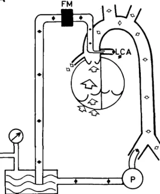
FIG. 1. Schematic diagram of the experimental procedure on the "working heart" (group A). After having cannu-lated a femoral artery the blood for the left coronary artery (black arrows) is transported by an ocdusive pump (P) to a smal reservoir. Driven at an adjustable pressure, the blood passes through an electromagnetic blood flowmeter and through a metal cannula into the left coronary artery. Coronary artery pressure is recorded by a cannula directly in the left coronary artery. The heart pumps the blood to all other arteries by itself (open arrows). For details
see text.
(3 dogs, figs. 3 and 4) mean coronary artery pressure was kept constant. Any variation of cardiac output or afterload (peripheral resistamce) must influence myocardial work, myocardial oxygen consumption and, therefore, coronary blood supply.
In an attempt to exclude these factors, the second femoral artery was connected with a blood reservoir in a modified preparation, used in three further animals in which mean coronary artery pressure was kept constant. By varying the height of this reservoir above the animal, the amount of extra-corporeal blood volume added to the intravascular space could be varied at will. The arterial blood pressure could thus be deliberately adjusted. Since such variations in intravascular volume influence right atrial pressure, coronary driving pressure ( = effective coronary artery pressure=CAP,,f) in
these three animals was calculated as coronary artery pressure minus right atrial pressure. Coronary driving pressure was used for calculating coronary resistance.
In a further three experiments coronary artery mean pressure was adjusted for a certain period of time to the level of mean aortic pressure and com-pared with other periods in the same experiment, coronary artery pressure being kept constant.
In six of these nine experiments samples of coronary sinus blood were taken for measurement of oxygen saturation.4
The effects of ether and halothane were con-sistently reproducible in all experiments. From each type of experiment an example is presented in detail.
Group B.
A different preparation was developed for this series (seven mongrel dogs weighing 22-28 kg) (diagram B, fig. 2).
General anaesthesia was maintained by con-tinuous intravenous administration of pentobarbi-tone 4 mg/kg/hr and suxamethonium 0.2 mg/min/ kg body weight. The heart was exposed through a sternal incision and, after administration of heparin 3 mg/kg body weight, a large angled Portex3 cannula with a series of side holes was inserted through the right atrial appendage into the right ventricle to drain the right atrium and ventricle into the venous reservoir of a heart-lung machine.' After cannulation of a femoral artery, cardiopulmonary bypass was established with an oxygenator/ A similar Portex cannula was introduced via the left atrial appendage into the left atrium, its tip passing the mitral valve so as to drain left atrial and ven-tricular blood into the venous reservoir. The mitral and tricuspid valves were kept incompetent. Fresh heparinized blood from donor dogs was used as the priming fluid of the cardiopulmonary bypass circuit. Normothermic perfusion was carried out at a haematocrit of about 30%. Two additional cannulae were placed in both ventricles to monitor mean pressure in the left and right ventricle. They were kept at zero by varying the suction force on the cannulae draining the heart. The pulmonary artery was ligated and mechanical ventilation discontinued.
* American Optical Co. Oxymeter. 5 Portex, Hythe, Kent.
• Rygg-Kyvsgaard, Mark IV, Polystan, Kopenhagen-Herlev, Denmark.
7 Temptrol-Oxygenator Q 110, Bentley Laboratories, Santa Anna, California, U.S.A.
B
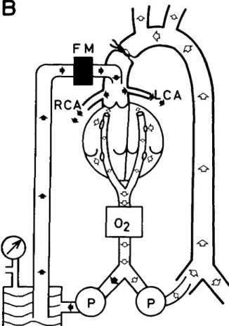
FIG. 2. Schematic diagram of the experimental procedure on the "empty beating heart" (group B). Both atria and both ventricles are drained into a venous reservoir. All blood passes through an oxygenator (O2). Having cannu-lated a femoral artery, the animal is kept alive by extra-corporeal perfusion (open arrows). By a second occlusive pump, oxygenated blood is driven to a small reservoir and at an adjustable pressure through an electromagnetic blood flowmeter, and through a metal cannula into the ascend-ing aorta. Because the aorta is clamped distal to the can-nula, all blood recorded by the flowmeter perfuses
the coronary arteries (black arrows).
Arterial blood from the oxygenator supplied a similar system, as described in figure 1. The arterial blood was pumped to a small reservoir and then through a cannulating flow-probe of the electro-magnetic flowmeter1 and through a wide steel cannula inserted by a purse-string suture in the ascending aorta. The perfusion pressure of the arterial line could be varied, at will. After occluding the aorta distal to the cannula, all blood passing through the flowprobe supplied the coronary arteries as long as the aortic valve was competent. The coronary artery pressure (CAP) was measured by a
cannula in this artificial aortic chamber. A Czakko stop-cock was used (Wolff, Gra'del and Niederer,
1968) to register an undamped phasic flow and to
control repeatedly the mechanical zero of the flow-meter without interrupting the coronary circulation. Blood was aspirated simultaneously and con-tinuously by a mechanically driven double syringe from a cannula in the coronary sinus and from the arterial coronary perfusion line. The syringes were filled after seven minutes and taken as samples for measurement of oxygen saturation.7 The systemic blood pressure was measured via a cannula in the descending aorta. Since the volume pumped into the animal was kept constant (100 ml/min/kg body weight) during the whole experiment, an increase in systemic blood pressure reflected a systemic vaso-constriction (and vice versa). During coronary per-fusion the mean coronary artery pressure was kept constant (80 mm Hg). The heart rate was kept constant by a fixed-rate pacemaker (pacing wires attached to the right atrial wall).
By changing Fioj and Fioo, in the oxygenating gas mixture, Paoj and Paooa were kept within narrow limits (150-200 mm Hg, 37-43 mm Hg respec-tively). The usual decrease in plasma potassium concentration and the gradual development of meta-bolic addosis during cardiopulmonary bypass were compensated by a continuous and adjustable intra-venous infusion of THAM (0.3 m) and KC1 (0.5 m.equiv/1.). Paoj,' Paooa,8 BE," and haematocrit were measured every 30 min.
Ether was added to the gas mixture in a concen-tration of 10% for a 30-min period. After a recovery period of 30 min 2% halothane was added for another 30 min. Again recovery was observed for 30 min. After the experiment the heart was excised to determine its weight.
The following were recorded continuously: mean systemic blood pressure; mean coronary artery pressure; diastolic coronary artery pressure10; mean coronary artery blood flow; diastolic coronary artery blood flow. Coronary arteriovenous oxy-gen difference and myocardial oxyoxy-gen uptake were calculated. Coronary arteriolar dilatation was thought to have occurred when the coronary • Combi-Analysator, Eschweiler and Co., Kiel, Ger-many.
8 Method of Astrup, Radiometer, Copenhagen, Den-mark.
11 Statham Transducers P 23 Db; amplifiers and 4-channel recorder of Hellige GmbH, Freiburg i.Br.,
Germany.
resistance at ± e end of diastole (=Rd) decreased (diastolic coronary pressure divided by diastolic coronary flow = vascular component of total resis-tance). Similarly, constriction was assumed to have occurred when the end-diastolic coronary resistance increased. The end-diastolic point of the heart cycle was chosen as a reference point, since at this moment extravascular compression is minimal and undergoes minimal changes with time. Thus the arterial inflow is little influenced by changes in the volume of blood contained in the arteries distal to the flowmeter (Dennison and Green, 1958).
Total coronary resistance (=Rm) was calculated as mean coronary pressure divided by mean coro-nary flow. We realize that the "nonvascular com-ponent of resistance" is not a value which corres-ponds to an exactly defined physiological function, but it is reasonable to assume that the "nonvascular component of resistance" rises with the myocardial intramural pressure during systole. Blood flow, resis-tances and oxygen uptake were calculated per 100 g heart weight.
The mean values for all experiments in group B were calculated. Their standard deviations were already marked during the control readings, because of differences between individual animals after the preliminary surgery. In order to eliminate these differences, or individual trends in a given animal during the experiment, each value obtained under new conditions was additionally related to that of die previous state, the difference being expressed as a percentage change from the previous (control) value. In both groups (A and B) it took between 90 and 120 min to prepare the dogs for study. When a stable state had been achieved for at least 30 min, the experiments were started. Obvious deterioration did not take place during the experimental period.
RESULTS
Group A
Maintenance of a constant intravascular blood volume.
Figure 3 shows an example of the influence of ventilation with 1% halothane during a 4-min period. The systemic arterial pressure fell; left coronary artery pressure was kept constant; left coronary diastolic flow diminished; left coronary systolic flow diminished also, but less than diastolic flow, so that left coronary mean flow diminished as well. After the end of halothane breathing the effects
BP
CAP
LCFph
as
LCF,
mean
1 0 0-o
J 2 0 0 - 1000 2 0 0 - 100-0 - B -mFIG. 3. Recording of an experiment in the "working heart" (group A). During the 4-min period of ventilation with halothane 1% systemic blood pressure (BP, mm Hg), systolic and diastolic (LCFphas, ml/min) and mean left coronary flow (LCF mean, ml/min) decrease, while
the left coronary artery pressure (CAP, mm Hg) is kept constant. See text.
BP
CAP
LCFphas
100
LCF,
meanFIG. 4. Recording of an experiment on the "working heart" (group A). During a 10-min period ventilation with ether 10%, systemic blood pressure (BP), left coronary diastolic flow (LCFphas maximal values) and left coronary mean flow (LCF mean) diminish, while left coro-nary systolic flow (LCFphas, minimal values) does not change and left corocoro-nary artery pressure
were reversible. Figure 4 shows the influence of ventilation with 10% ether during a 10-min period Systemic blood pressure fell progressively. Coronary artery pressure was kept constant. Left coronary diastolic and mean flow fell slightly while systolic coronary flow was not changed. The effects were reversible within a few min of discontinuing ether administration.
Figure 5 illustrates the influence of coronary artery pressure on the effect of halothane anaesthesia. When coronary artery pressure was reduced in parallel with the decreasing aortic pressure (figure 5, left panel), the vascular component of resistance (Rd) remained constant. In the same animal, the vas-cular component of resistance increased consider-ably during halothane breathing 90 min later (fig. 5, right panel), when coronary artery pressure was kept constant.
Halothane and ether diminished heart work, be-cause the systemic vascular resistance or cardiac
CAP AoP
if
• * 0
FIG. 5. The influence of coronary artery pressure (CAP) in an experiment on the "working heart" during 1% halothane administration. On the left side coronary artery pressure is adjusted to the decreasing aortic pressure (AoP). On the right side the coronary artery pressure is kept constant while the aortic pressure drops during halothane. If coronary artery pressure is adjusted to aortic pressure the vascular component of coronary resistance (Rd) does not change during halothane. Abbreviations as
in figure 3.
Rm = total coronary vascular resistance. Rd = intra-vascular component of coronary resistance. Rm —Rd = extravascular component of coronary resistance. CAFm
= mean coronary flow.
output, or both, were decreased. When coronary artery pressure was kept constant, the vascular com-ponent of coronary resistance was increased. This increase was more pronounced with halothane than ether.
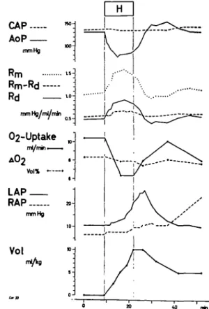
Effects of varying intravascular blood volume. Figures 6 and 7 illustrate experiments in which intravascular volume was increased in order to balance systemic arterial pressure. During halothane anaes-thesia (fig. 6) aortic pressure could not be prevented from falling even when the intravascular volume was increased by 10 ml/kg body weight. Left atrial pres-sure rose in this period from 10 to 20 mm Hg and continued to rise up to 25 mm Hg during the first five minutes after halothane had been stopped. As in
CAP AoP nrnHj Rm Rm-Rd Rd rnni Hg/mj/rren 02-Uptake A O2 Vol% • • LAP RAP mm Hg Vol nifa
FIG. 6. Effects of ventilation with halothane 1% in an experiment on the "working heart". Coronary artery pressure (CAP) is kept constant. Aortic pressure (AoP) decreases, even if intravascular volume is increased rapid-ly by 10 ml/kg body weight blood (Vol). Left atrial pres-sure (LAP) is climbing to pathological values. AOj= arterio-(coronary) venous difference in oxygen content Oj-uptake=oxygen consumption of the myocardium depending on the left coronary artery. RAP = right
Vol
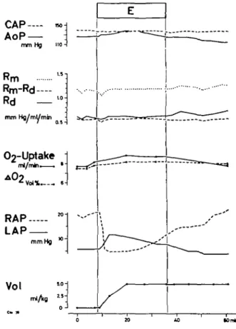
FIG. 7. Effects of breathing ether 10% in an experiment on the "working heart" (same experiment as in fig. 6). Coronary artery pressure (CAP) is kept constant. Due to the increase of intravascular volume by 5 ml/kg body weight (Vol) aortic pressure (AoP) is increasing by 10 trim Hg. Left atrial pressure rises from 6 to 12 mm Hg. See text. (Same abbreviations used as in figs. 5 and 6.) the experiments with constant intravascular volume, coronary arteriovenous oxygen difference fell. Since coronary artery pressure was kept constant and total coronary resistance rose, oxygen uptake was also falling. During the increase of left atrial pressure, the "non-vascular" component of resistance rose insignificantly while the vascular component of resistance increased distinctly. The effects were reversible after halothane had been stopped; but in spite of the withdrawal of the additional intravas-cular volume, the right atrial pressure increased during this period. When, 2 hours later, 10% ether was administered to the same animal (fig. 7), right atrial pressure which was elevated, quickly returned to normal levels. An increase of intravascular volume by 5 ml/kg body weight raised the left atrial pres-sure from 6 to 12 mm Hg. In this situation aortic
pressure rose by 10 mm Hg. All other values were essentially unaffected by ether.
The expected fall in aortic pressure caused by ether was prevented by augmenting the intravascular volume. In this situation left atrial pressure increased, but not to abnormal levels. The decrease in right atrial pressure confirms the dilatation in the peri-pheral vascular system. During halothane anaes-thesia, augmentation of intravascular volume caused left atrial pressure to rise to abnormal levels, with-out preventing the aortic pressure from falling. In spite of the rise in left atrial pressure, right atrial pressure remained the same. This suggests that the effect of halothane was predominantly on left ventri-cular function.
Group B
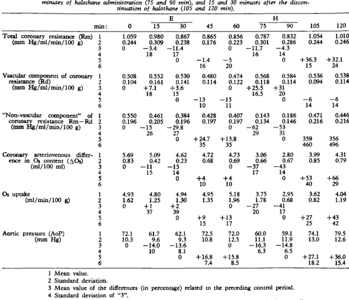
Experiments on the empty beating heart are illus-trated in figures 8 and 9, with figure 10 presenting the mean values of all experiments in this group and table I the mean values, including their standard deviations. Additionally the mean values of the differences (in per cent) and their standard devia-tions are listed in table I.
The effects of ether on the heart were relatively small compared to those of halothane. Total coro-nary vascular resistance decreased during ether and during halothane administration (5-10%), but in-creased after the end of halothane by more than 30% above the control value. The vascular component of the resistance was not changed by ether, but raised more than 20% by halothane. Its decrease after halothane was only about 5%, and control values were not reached in the observed 30-min period. The "non-vascular" component of resistance, there-fore, decreased little during ether, but markedly during halothane administration (more than 50% in the latter). After stopping halothane, the "non-vascu-lar" component of the resistance increased, until it was stabilized at 15% above the pre-halothane values.
The decrease in coronary arterio-venous oxygen difference during ether was small, and a variation in oxygen uptake was not detectable. On the other hand, halothane produced a spectacular decrease in coronary arteriovenous oxygen difference, a drop from 4.7 ml/100 ml to below 3 ml/100 ml, and increased again after the end of halothane admini-stration to 4.3 ml/100 ml. Oxygen consumption thus dropped during halothane inhalation from 5 to 3 ml/min/100 g, despite the decrease in total vascular resistance with elevated coronary mean flow. After
TABLE I. Effects of ether and halothane in the seven experiments in the empty beating heart (Group B): control values (0 miri) after 15 and 30 minutes of ether administration (IS and 30 miri) 15 and 30 minutes after the discontinuation of ether (45 and 60 min), after 15 and 30 minutes of halothane administration (75 and 90 min), and 15 and 30 minutes after the
discon-tinuation of halothane (105 and 120 miri).
E H
Total coronary resistance (Rrn) (mm Hg/ml/min/100 g)
Vascular component of coronary resistance (Rd)
(ram Hg/ml/min/100 g)
"Non-vascular component" of coronary resistance Rm—Rd
(mm Hg/ml/min/100 g)
Coronary arteriovenous differ-ence in Oi content (AOi)
(ml/100 ml)
OB uptake
(ml/min/100 g)
Aortic pressure (AoP) (mm Hg) min: 1 2 3 4 5 6 1 2 3 4 5 6 1 2 3 4 5 6 1 2 3 4 5 6 1 2 3 4 5 6 1 2 3 4 5 6 0 1.059 0.244 0 0.508 0.104 0 0.550 0.196 0 5.69 0.83 0 4.93 1.62 0 72.1 10.3 0 15 0.980 0.309 - 3 . 4 18 0.552 0.161 + 7.1 18 0.461 0.205 - 1 5 26 5.09 0.42 — 11 15 4.80 1.25 + 137 61.7 9.6 -14.0 10 30 0.867 0.238 -11.4 17 0 0.530 0.141 + 3.6 15 0 0.384 0.196 -29.8 27 0 4.62 0.23 - 1 5 14 0 4.94 1.30 + 2 39 0 62.1 9.3 -13.6 8.1 0 45 0.865 0.176 - 1 . 4 16 0.480 0.114 - 1 3 10 0.428 0.197 + 24.7 35 4.72 0.68 +4 10 4.95 1.35 + 9 15 72.5 10.8 + 16.8 7.4 60 0.856 0.223 0 - 5 20 0.474 0.122 0 - 1 5 11 0.407 0.197 0 + 13.8 35 4.73 0.69 0 + 4 10 5.18 1.96 0 + 13 17 72.0 12.5 0 + 15.8 8.5 75 0.787 0.301 -11.7 16 0.568 0.118 + 25.5 16.5 0.143 0.134 - 6 2 29 3.06 0.66 - 3 7 17 3.75 1.78 - 2 7 20 60.0 11.1 -16.3 6.3 90 0.832 0.286 - 4 . 3 14 0 0.584 0.114 + 31 20 0 0.186 0.146 - 5 3 31 0 2.80 0.67 - 4 3 14 0 2.95 0.68 - 4 1 17 0 59.1 11.9 -14.8 6.5 0 105 1.054 0.244 + 36.3 15 0.536 0.094 - 6 14 0.471 0.216 359 460 3.99 0.85 + 53 40 3.62 0.82 + 27 25 74.1 13.0 + 27.1 18.2 120 1.010 0.246 + 32.1 24 0.538 0.114 - 6 14 0.446 0.216 356 496 4.31 0.79 + 66 29 4.04 1.19 + 43 42 79.5 12.6 + 36.0 15.4 1 Mean value. 2 Standard deviation.
3 Mean value of the differences (in percentage) related to the preceding control period. 4 Standard deviation of "3".
5 Mean value of the differences (in percentage) related to the preceding value in the preceding anaesthesia.
6 Standard deviation of "5".
the end of halothane anaesthesia oxygen consump-tion increased up to 4 ml/min/100 g, the result of a rise in total vascular resistance, the latter rising above control values during this period. With either anaesthetic the systemic arterial pressure fell by about 15%. Both pressure drops were reversible.
Since the perfusion flow of the cardiopulmonary bypass was kept constant throughout the experiment with the empty beating heart and systemic blood
pressure fell during the administration of either anaesthetic agent, systemic vascular resistance must have decreased under ether and halothane by the same amount. Both anaesthetics reduced total coro-nary vascular resistance. In both situations the vas-cular component of coronary arteriolar resistance rose, while both anaesthetics reduced systolic myo-cardial pressure (Rm-Rd) to such a level that the increase in coronary arteriolar tone did not result in
BP CAP sec CAFphas 100-100 o - J 2 0 0 - 1 h 100 0 ," (I •< ' t ' " • ; r •< £ V V V 0 9 4 18 0 t • A , 4 i ii v w Ls •' u
e »
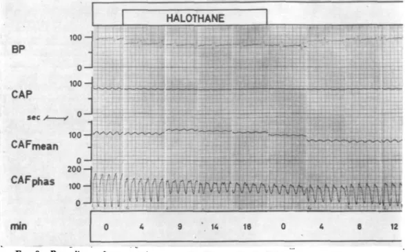
mmFIG. 8. Recordings of an experiment on the "empty beating" heart (group B), during the administration of halothane 2% for 30 min. Coronary artery pressure and die perfusion volume of the animal are kept constant. BP = systemic arterial pressure (mm Hg), the pulse pressure corresponds to the revolutions of die occlusive pump of die extracorporeal circulation. CAP = coronary artery pressure (mm Hg). CAFmean=mean flow of bodi coronary arteries (ml/min). CAFphas = phasic flow of bodi coronary arteries (ml/min) (see text), min=minutes after the
start or stop of the administration of halothane.
BP
CAP
100 — 0 1 0 0-ETHER
secCAFmean
CAFphas
min
100 0 200-100 0 -s t t ^LJ::.I !-:
-H
.-l-l-t-f i •: . i• f
1TTT:
t • » ; • • • > • i r > 12 18 0 5 9 15FIG. 9. Recordings of an experiment on the "empty beating heart" (group B) during the
mmHg/m^mln/iOOg
O2-Uptake—.
ml/min/IOdg
AoP
lHg
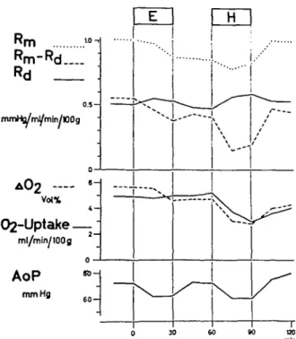
FIG. 10. Mean values of the seven experiments on the "empty beating heart" (group B). Control values — ether (for 30 rain) — control values (for 30 min) — hHiothane (for 30 min) — control values (30 min). Coronary artery pressure and perfusion volume of the animal are kept constant. Rm = total coronary resistance. Rd = vascular component of coronary resistance. Rm—Rd = non-vascular component of coronary resistance. AOi = (coro-nary) arteriovenous oxygen difference. Oa uptake = oxygen consumption of the total myocardium. AoP = systemic blood pressure, which changes depending on
systemic resistance.
an increase in total coronary resistance. The varia-tions were very small during ether, but marked dur-ing halothane administration. In the empty beatdur-ing heart, halothane leads to a state characterized by reduced myocardial oxygen consumption, coronary arteriolar vasoconstriction and some myocardial depression with lowered systolic intramural pres-sure. Since in the experiments on the empty beating heart, external heart work was not changed and kept at zero, the observed variations cannot have been caused by changes in the systemic circulation. Con-sequently the effects must have been caused by a direct influence of halodiane on the heart.
DISCUSSION
Since the coronary arteriovenous oxygen difference decreased under halothane despite the coronary vasoconstriction, the latter obviously does not cause
hypoxia and myocardial depression. Halothane must, therefore, act as a direct myocardial depressant with a subsequent decrease of oxygen consumption and an increase in coronary vasomotor tone.
The effects observed on the working heart during halothane anaesthesia may be explained by the same mechanisms. First, systemic vascular resistance and heart work are lowered: myocardial oxygen uptake is diminished as a result Coronary vasoconstriction occurs. Ether has the same qualitative effect on the heart, but to a very much smaller extent. Halothane diminishes myocardial contractility which did not improve even when left atrial pressure was elevated by an increase in intravascular volume. This situa-tion—left atrial pressure being raised by 20 mm Hg and aortic pressure still below 80 mm Hg —demonstrates that cardiac work under halothane has not diminished merely because of a fall in sys-temic vascular resistance or because cardiac output is stabilized at a lower level. Halothane diminished the myocardial response to its physiological stimulus, namely elevation of the left ventricular end-diastolic pressure or left atrial pressure. In other words, halo-thane leads to a loss of contractility sufficient to provoke myocardial failure.
The question arises whether the decrease in oxygen uptake and the increase in coronary vaso-motor tone caused by halothane are two independent effects, or whether one is responsible for the other. This question was investigated by experiments in which the coronary artery pressure was varied. The marked increase in the vascular component of resis-tance brought about by halothane administration only occurred when coronary artery pressure was kept constant. The fact that the vascular component of resistance is not elevated by halothane, when coro-nary artery pressure is lowered parallel with the decrease in aortic pressure, proves that coronary vasoconstriction and loss of myocardial contractility are not necessarily combined effects. Rather it sug-gests that halothane influences primarily the myo-cardium by decreasing its contractility and oxygen consumption. The vascular component of resistance rises only if coronary artery pressure is not dimini-shed. This may be due to an autoregulatory mechanism preventing "unnecessary" hyperfusion.
Since a constant coronary artery pressure must be maintained if the vascular component of resistance is to be increased, it is very unlikely that a variation in catecholamines is responsible for the coronary vasoconstriction: catecholamines are released from outside the heart and coronary artery pressure
would not be expected to influence this. Finally, one can now explain further the results of our previous experiments (Wolff, Gradel and Niederer, 1968). By changing from halothane to ether, a strong myo-cardial depressant is replaced by a weak one (and vice versa). In this situation, ether appears to exert a positive inotropic effect. As a consequence of the smaller reduction in myocardial work during ether administration, systolic intramural myocardial pres-sure and the "non-vascular" component of resistance increase and the rise in oxygen consumption is followed by coronary vasodilatation.
REFERENCES
Denison, A. B., and Green, H. D. (1958). Effects of auto-nomic nerves and their mediators on the coronary circulation and myocardial contraction. Circulai. Res.,
6. 633.
Weaver, P. C , Bailey, J. S., and Preston, T. D. (1970). Coronary artery blood flow in the halothane-depresscd canine heart. Brit. J. Anaesth., 42, 678.
Wolff, G., Gradel, E., and Niederer, W. (1968). Changes of coronary arteriolar tone, mean coronary flow and aortic pressure under halothane and ether anaesthesia in the dog. Brit. J. Anaesth., 40, 810.
REGULATION DU FLUX SANGUIN CORONARffiN DURANT L'ANESTHESIE A
L'ETHHR ET HALOTHANE SOMMAIRE
Lcs effets d'ether (6-10%) et halothane (1-2%) sur la regulation du flux coronarien ont ete etudies chez le chien.
Dans un groupe d'experiments, on proccda a une per-fusion mecanique de l'artere coronaire gauche; la pression de perfusion coronarienne etait soit tenue constante soit ajustee a la pression aortiquc et le coeur meme devait propulser le sang vers toutes les autres arteres. La pre-charge du coeur fut modifiee en variant le volume intra-vasculaire.
Dans un autre groupe, on appliqua une perfusion mecanique sous pression constante aux deux coronaires; les autres artires etaient egalement perfusees mecaniquc-ment par un bypass cardiopulmonaire. On fit les determi-nations dans ce groupe dans un coeur pulsant a vide. Halothane exerca un effet direct sur le coeur avec reduc-tion de la contractilitc du myocarde et diminureduc-tion du travail cardiaque et de la consommation d'oxygene par le myocarde, et par consequence done on nota une vaso-constriction coronarienne. Les effets de l'ether sur le coeur furent principalement les memes que ceux d'halothane, mais a moindre intensity. La vasoconstriction coronarienne causee par halothane ne produisit pas d'hypoxie du myo-carde. Cette vasoconstriction des coronaires survient comme uri mecanisme autoregulateur qui empeche une hyperperfusion "inutile" aussi longtemps que le travail du coeur et la consommation d'oxygene sont reduits. Ether et halothane ont tous deux reduit la resistance vasculaiie systemique.
UBER DIE REGULATION DER KORONARDURCHBLUTUNG WAHREND
ATHER- UND HALOTHAN-NARKOSE ZUSAMMENFASSUNG
Die Wirkung von Ather (6-10%) und Halothan (1-2%) auf die Regulierung der Koronardurchblutung wurde an Hunden untersucht. Bei einer Versuchsgruppe wurde die linke Koronararterie mechanisch perfundiert; der koro-nare Perfusionsdruck wurde dabei entweder konstant gehalten oder nach dem Aortendruck ausgerichtet. Das Herz selbst musste das Blut in alle anderen Arterien pumpen. Die Vorbelastung des Herzens wurde durch unterschiedh'che vasculare Volumina yerandert. Bei einer anderen Versuchsgruppe wurden beide Koronararterien unter konstantem Druck mechanisch perfundiert. Auch die anderen Arterien wurden mittels eines cordiopulmo-nalen Bypasses mechanisch perfundiert In dieser Gruppe wurden die Messungen am schlagenden, leeren Herzen vorgenommen. Halothan hatte einen direkten Einfluss auf das Herz; die myocardiale Kontraktionskraft wurde reduziert, die cardiale Lcistung und der myocardiale Sauerstoff-Verbrauch herabgesetzt. Die Folge war eine Konstriktion der Koronargefasse. Der Effekt von Ather war im Prinzip dem von Halothan gleich, nur weniger ausgepragL Die durch Halothan veriirsachte Konstriktion der Koronargefasse fflhrte nicht zu myocardialer Hypoxie. Die koronare Vasokonstriktion tritt als autoregulatorischer Mechanisms auf, urn eine "unnotige" Hyperperfusion zu verhindern, solange die Herzleistung und der Sauerstoff-verbrauch herabgesetzt sind. Sowohl Ather, als auch Halo-than semen die allgemeine vasculare Resistenz herab.
REGULACION DEL FLUJO SANGUINEO CORONARIO DURANTE LA ANESTESIA
POR ETER Y HALOTANO RESUMEN
Fueron estudiados los efectos del iter (6-10%) y halo-tano (1-2%) sobre la regulaci6n del flujo coronario en perros. En un grupo de experimentos fue perfundida mecanicamente la arteria coronaria izquierda, manteniendo la presi6n de perfusidn coronaria constante o ajustada a la presi6n adrtica y teniendo el coraz6n que bombear la sangre por si mismo hacia todas las arterias. La pre-carga sobre el coraz<5n fue modificada mediante variaci6n del volumen intravascular. En otro grupo, fueron perfun-didas mtcanicamente ambas arterias coronarias bajo pre-si6n constante y las otras arterias tambien fueron perfun-didas mecanicamente mediante un bypass cardiopulmonar. En este grupo fueron llevadas a cabo mediciones en el coraz6n contrayendose en vacfo. El halotano tuvo un efecto directo sobre el corazonj fue reducida la contrac-tilidad miocardica, fueron disminuidos el trabajo cardlaco y el consumo miocirdico de oxigeno y se produjo vaso-constricci6n coronaria. Los efeaos del eter sobre el cora-z6n fueron principalmente iguaks a los debidos al halo-tano, pero dt menor intensidad. La vasoconstricci6n coro-naria causada por el halotano no produjo hipoxia miocar-dica. La vasoconstriccid'n coronana ocurri6 en forma de un mecanismo autorregulador que impide un hiperperfu-si6n "innecesaria" siempre que habia disminuci6n del trabajo cardiaco y del consumo de oxigeno. Tanto el 6ter como el halotano redujeron la resistencia vascular sistemica.