REVIEW
Ammonia toxicity to the brain
Olivier Braissant&Valérie A. McLin&Cristina CudalbuReceived: 5 July 2012 / Revised: 19 September 2012 / Accepted: 25 September 2012 / Published online: 30 October 2012 # SSIEM and Springer Science+Business Media Dordrecht 2012
Abstract Hyperammonemia can be caused by various ac-quired or inherited disorders such as urea cycle defects. The brain is much more susceptible to the deleterious effects of ammonium in childhood than in adulthood. Hyperammonemia provokes irreversible damage to the developing central nervous system: cortical atrophy, ventricular enlargement and demyelin-ation lead to cognitive impairment, seizures and cerebral palsy. The mechanisms leading to these severe brain lesions are still not well understood, but recent studies show that ammonium exposure alters several amino acid pathways and neurotransmit-ter systems, cerebral energy metabolism, nitric oxide synthesis, oxidative stress and signal transduction pathways. All in all, at the cellular level, these are associated with alterations in neuro-nal differentiation and patterns of cell death. Recent advances in imaging techniques are increasing our understanding of these processes through detailed in vivo longitudinal analysis of neu-robiochemical changes associated with hyperammonemia. Fur-ther, several potential neuroprotective strategies have been put forward recently, including the use of NMDA receptor antago-nists, nitric oxide inhibitors, creatine, acetyl-L-carnitine, CNTF or inhibitors of MAPKs and glutamine synthetase. Magnetic
resonance imaging and spectroscopy will ultimately be a pow-erful tool to measure the effects of these neuroprotective approaches.
Introduction
Ammonia is produced by amino acid metabolism and intes-tinal urease-positive bacteria. In physiological conditions, it is mostly present as ammonium (NH4
+
) in serum. The urea cycle, which is fully expressed in the liver exclusively, serves to converts NH4+ to urea prior to renal excretion
and to maintain low serum concentrations (50–150 μM in preterm neonates, 50–75 μM in term neonates, and <50 μM in adults). Although the brain cannot convert NH4+to urea,
NH4 +
is also maintained at low levels in the central nervous system (CNS) by the astrocytic enzyme glutamine synthe-tase (GS), which synthesizes glutamine (Gln) from gluta-mate (Glu) and NH4+(see Cagnon and Braissant2007, and
references therein).
Excessive NH4+ is toxic for the CNS. In adults, liver
failure results in hyperammonemia which in turn leads to the potentially severe neuropsychiatric disorder hepatic en-cephalopathy (HE) characterized by altered mental status and coma. In the absence of irreversible cerebral edema, HE symptoms in adults are largely reversible when NH4+
returns to normal levels (Flint Beal and Martin 1998). In children, hyperammonemia can be caused by numerous inherited or acquired disorders (Leonard and Morris2002), among which the best known are inherited urea cycle dis-orders (UCD) (Braissant 2010a; Gropman et al 2007; Tuchman et al2008). The susceptibility of the developing brain to hyperammonemia leads to severe cognitive impair-ment, seizures and cerebral palsy (Enns2008). Neonates and infants with important hyperammonemia develop cortical at-rophy, ventricular enlargement, demyelination or gray and white matter hypodensities (Enns 2008; Gropman et al
2007; Tuchman et al2008). The extent of irreversible brain damage depends on its maturational stage and on the
Communicated by: Pascale de Lonlay
Presented at the 26th APS Meeting, Fulda, Germany, 7–9 March 2012. O. Braissant (*)
Service of Biomedicine, Lausanne University Hospital, Avenue Pierre-Decker 2, CI 02/33,
CH-1011 Lausanne, Switzerland e-mail: [email protected] V. A. McLin
Pediatric Gastroenterology Unit, Division of Pediatric Specialties, Department of Child and Adolescent,
University Hospitals of Geneva (HUG), Geneva, Switzerland
C. Cudalbu
Laboratory for Functional and Metabolic Imaging (LIFMET), Ecole Polytechnique Fédérale de Lausanne (EPFL),
magnitude and duration of NH4+ exposure. Damage may
become irreversible in case of prolonged hyperammonemia or when blood NH4+reaches levels between 200 and 500μM
during the two first years of life (Bachmann2003; Enns2008; Msall et al1984; Tuchman et al2008; Uchino et al1998).
This review focuses on the most recent advances in understanding NH4+ toxicity to the brain, with emphasis
on novel tools, experimental models, therapies and neuro-protective strategies.
Hyperammonemia in humans
Hyperammonemia and its consequences on the brain develop secondary to various congenital or acquired causes (Cagnon and Braissant2007). Examples include congenital portosystemic shunts (Kim et al2012), extrahepatic portal vein obstruction (Pietrobattista et al2010), and cirrhosis with portal hyperten-sion. However, much of what is understood concerning ammo-nia neurotoxicity stems from patients with UCD.
Non-specific symptoms common to the immature and mature CNS
Non-specific symptoms are common in most UCD patients presenting in the neonatal period (poor feeding, vomiting, somnolence, irritability, tachypnoea) (Braissant2010a). As NH4+rises in serum, hypothermia, lethargy and coma
prog-ress rapidly (Summar2001). In cases of partial UCD, clin-ical presentation can occur as late as months or years post-natally and are often triggered by illness or catabolic stress. In this case hyperammonemia is generally less severe and the symptoms are usually milder than in newborns (Takanashi et al2002). Patients with late-onset hyperammonemia can pres-ent with loss of appetite, cyclic vomiting, lethargy or behav-ioral abnormalities (Harada et al2006; Smith et al 2005). Patients with partial defects tend to sponteaneously avoid protein, especially female patients with ornithine transcarba-mylase (OTC) deficiency (Scaglia et al2002). Mental retar-dation and learning difficulties are frequent.
Cerebral edema: a common feature of the NH4+-exposed
CNS
In response to elevated serum NH4+, the developing and
the mature CNS respond similarly: Gln content in astro-cytes rises through increased GS activity, and astroastro-cytes swell. Under high NH4+levels, osmoregulation is insufficient
and cerebral edema develops, affecting all areas of the brain. In its most severe form, increased intracranial pressure even-tually leads to brain herniation (Cordoba and Blei 1996; Norenberg et al2005). In advanced cerebral edema, seizures, abnormal posture and neuromuscular irritability are frequent
(Butterworth1998). CNS edema first causes hyperventilation and respiratory alkalosis later progressing to hypoventilation and apnea (Brusilow and Maestri1996). Without any treat-ment most infants will die. In survivors of infantile hyper-ammonemia, mental retardation is the norm (Bachmann2003; Krivitzky et al2009; Tuchman et al2008).
Edema associated with HE can be followed by mag-netic resonance spectroscopy (MRS) and magmag-netic reso-nance imaging (MRI) (Gropman 2010; Gropman et al
2010; Grover et al 2006; Oldham et al 2010). In the acute setting, serum NH4+ levels and cerebral edema are
correlated with psychomotor performance (Foerster et al
2009; Yadav et al 2010).
Hyperammonemia in the adult brain does not provoke significant neuronal loss or structural damage to neurons, in contrast with what is observed in the developing CNS (Butterworth2003).
Irreversible effects of ammonia on the developing brain Irreversible damage to the developing brain results in mental retardation in most surviving children with UCD (Gropman et al
2007; Krivitzky et al2009; Tuchman et al2008). Neonatal onset leads to the most severe brain damage and the least IQ score, with significant volume loss of different parts of the developing brain as assessed by later MRI. Diffuse cortical atrophy, lesions in basal ganglia and thalamus, myelination delay and injury of the oligodendro-axonal unit are frequent (Majoie et al 2004; Takanashi et al2003; Yamanouchi et al2002). Cerebral MRI in UCD neonates suggest that some of these lesions might already be acquired in utero (Filloux et al1986; Harding et al1984; Majoie et al2004; Takeoka et al2001).
If hyperammonemia is diagnosed before irreversible ce-rebral insults, patients may have a normal neurodevelop-ment (Kurihara et al2003). Many however remain mentally retarded or have learning difficulties (Smith et al 2005). Brain MRIs of late-onset UCD patients show cortical injury including acute ischemia, ventricular dilatation and myeli-nation defects (Call et al1984; Choi et al2006; de Grauw et al1990; Gropman et al2010; Kim et al2006; Kurihara et al
2003; Oldham et al 2010; Scaglia and Lee 2006). Similar lesions are found in patients with propionic acidemia or hyperammonemia-hyperornithinemia-homocitrullinuria syndrome who followed an unremarkable neonatal course (Harding et al1991; Salvi et al2001).
Only a few UCD cases have been analyzed by autopsy. Findings included microcephaly, shrinkage of hemispheres coupled to multiple cysts, ventricular dilatation, atrophy or necrosis of various brain nuclei or myelination defects (Dolman et al1988; Takeoka et al2001; Yamanouchi et al
2002). Microscopically, spongious brain tissue with exten-sive neuronal loss (in cortex and hippocampus particularly) was observed, together with gliosis and astrocytes with
water-clear, oval nuclei characteristic of Alzheimer’s type II astrocytes.
Not all encephalopathies in children are due to UCD or primary gene defects affecting the liver, and there is increas-ing understandincreas-ing that behavioral and neurological changes are frequent in patients with chronic liver failure (CLF) or portocaval shunts (PCS) even in absence of any significant hyperammonemia (Caudle et al 2010, 2012; Yadav et al
2010). Clinical signs and electro-encephalogram findings in children with acute liver failure (ALF) are relatively well characterized. In contrast, while children affected with CLF display neurocognitive deficits at an early age, the subtleties of their CNS alterations are much less understood (Bajaj et al2011; Caudle et al2010; Yadav et al 2010). In adults, minimal hepatic encephalopathy (MHE) is often hidden and only unmasked by specialized tests (Bajaj et al2012; Felipo et al2012). Recent data show that adults with MHE present cortical thinning that parallels their cognitive impairment associated with early stages of liver disease (Montoliu et al
2012). Although children with liver disease or PCS can also display T1 hyperintensity of the pallidi by MR, the clinical, biological, and imaging subtleties of pediatric MHE still need to be defined.
Available treatment options
Neonatal hyperammonemia and UCDs
The rapid removal of NH4+should be the immediate
thera-peutic goal in neonatal hyperammonemia (Walker 2009). Protein restriction is the cornerstone of therapy, particularly in severe cases. Dialysis (hemodialysis, hemodiafiltration or continuous veno-venous hemofiltration) is indicated for hyperammonemia which does not correct rapidly or which is refractory to conservative measures (Leonard et al2008). Intravenous glucose (Glc) is essential to reverse catabolism, together with careful use of insulin to avoid fluctuations in serum Glc levels (Summar 2001). Intravenous administra-tion of sodium benzoate and sodium phenylacetate, both nitrogen scavengers, is an alternative and frequently used approach to achieve sufficient nitrogen excretion in acute phases (including neonatal) (Enns 2010; Shih 2007; Tuchman et al 2008). Long term control of NH4+ makes
use of the same compounds or oral sodium phenylbutyrate, in combination with a low-protein diet (Batshaw et al2001; Berry and Steiner2001; Cederbaum et al2010; Enns et al
2007; Scaglia2010). In UCD, large doses of arginine (Arg) for argininosuccinate synthetase (ASS) and argininosucci-nate lyase (ASL) deficiencies, or citrulline for carbamyl-phosphate synthetase 1 (CPS-1) and OTC deficiencies, further promote nitrogen excretion (Brusilow et al 1979; Leonard and Morris2002). Orthotopic liver transplantation
is currently the only option for severe, uncontrollable UCD (Lee and Goss 2001). However, cell-based rather than organ-based gene therapy is the ultimate goal and is an area of intense research (Meyburg and Hoffmann2010).
Early diagnosis and intensive treatment are often insuffi-cient to prevent death, and neurological problems are fre-quent in survivors (Bachmann2003). High resolved proton spectroscopy (1H-MRS) may prove to be a useful tool in tailoring the care for patients with hyperammonemia regard-less of the underlying cause. In patients with CLF for example, spectroscopic findings consistent with HE/MHE may be a new indication for liver transplant or for the prescription of the same nitrogen scavengers as those used in UCD.
Chronic hepatic encephalopathy
In adult patients with suspected or diagnosed chronic HE, the mainstay of management is the reduction in gut-derived ammonia. Historically, the drug of choice was lactulose which decreases luminal pH, thereby favoring the transfor-mation of non-resorbable NH4+ produced by enteric
com-mensals and thus decreasing NH4+ in portal venous blood
and nitrogen load to the liver (Als-Nielsen et al2004; Patil et al1987). More recently, the oral non-absorbable antibiotic rifaximin has become the drug of choice. In a pivotal trial in patients with CLF, rifaximin prevented HE relapse more efficiently than placebo (Bass et al 2010). A recent meta-analysis suggests that it is at least as effective as other oral treatments such as disaccharides and other antibiotics. It further suggests that it may have fewer side effects than previously used agents, and that it may in fact improve performance on psychometric tests (Eltawil et al2012).
Nitrogen scavengers are also used in HE patients. L-ornithine-L-aspartate (LOLA) is used to supply ornithine (Orn) to the urea cycle, thereby favoring NH4+conversion
to urea in residual periportal hepatocytes and Gln synthesis from Glu and NH4+in skeletal muscle, which in liver failure
is an important metabolic alternative for the breakdown of NH4
+
. In rodent models, LOLA significantly increased urea production and blood Gln levels, and decreased CNS NH4+
while slowing the rise in brain Gln (Rose et al 1998). Results from human trials are controversial however: both fasting and post-prandial serum NH4+falls within 7-days of
treatment initiation in parallel with improved psychomotor performance and overall well-being (Kircheis et al 1997). However, serum Gln rises contributing in turn to ammonia-genesis, rebound hyperammonemia and severe HE follow-ing LOLA withdrawal (Olde Damink et al2002). No studies have been done in children, although anecdotal, unpub-lished reports suggest that daytime drowsiness and attention deficits may improve in LOLA-treated older ones. L-ornithine-phenylacetate (OP) is an elegant alternative as it
is thought to act both on NH4+transformation and
elimina-tion. Its Orn moiety uses muscle GS activity to convert NH4+into Gln, while phenylacetate combines with Gln as
phenylacetylglutamine (Jalan et al 2007). OP showed a beneficial effect in rat models of CLF with acute HE de-compensation. In lipopolysaccharide-induced HE exacerba-tion in BDL rats, OP intraperitoneal administraexacerba-tion reduced brain edema and rate of progression to coma (Wright et al
2012). This study is auspicious of benefiting human subjects since OP was administered once the rats were diagnosed with HE. Conversely another study showed that pre-emptive OP limited HE severity in PCS rats in which HE exacerba-tion was triggered by administraexacerba-tion of blood in the gastro-intestinal tract (Oria et al2012). Although it can be argued that patients at risk of severe gastrointestinal bleeds could benefit from primary OP prophylaxis, this study seems less clinically significant as the cost-effectiveness of such an approach might be prohibitive. However, at this time it is too early to formally conclude since human studies using this newer compound are forthcoming.
Experimental models to study NH4+toxicity to the brain
In vivo: Spf mice, KO mice and rat models of hyperammonemia
Sparse-fur (Spf) mice have a single point substitution in the OTC gene, with X-linked transmission, mimicking the human disease (Veres et al1987). Hepatic OTC activity is 5–13 % of
that in normal mice. Adult Spf/Y mice show NH4+blood and
brain levels increased by 1.5- and five-fold respectively (Rat-nakumari et al1992). Neuropathologic studies in Spf mice show similar brain alterations as those observed in OTC patients: brain size reduction (including decreased striatum volume) and ventricular enlargement (Hopkins et al1998). Several knock-out (KO) mice have been engineered to model UCDs (Deignan et al2008): ASS, ASL, arginase I and argi-nase II KO mice, as well as double KO mice for argiargi-nases I+II. ASS-/-and ASL-/-mice die a few days after birth, with plasma NH4+increased four-fold. Arginase I and arginases I+II KO
mice die from hyperammonemia 14 days postnatally, with a ten-fold increase in plasma NH4
+
.
Different rat models exist to analyze the effects of hyper-ammonemia on the CNS. For example, pregnant rats can be fed a diet containing NH4-acetate from day 1 of pregnancy until
weaning, followed by feeding the pups after weaning with high NH4+-containing diet. NH4+levels in the brain of these animals
is 1.4 times higher as compared to control (Aguilar et al2000). Alternatively, hyperammonemia can be induced in adult rats by intraperitoneal injections of NH4+-acetate, continuous iv
infu-sion of NH4Cl, iv urease infusion (Robinson et al 1992b),
administration of a NH4+-acetate containing diet (Azorin et al
1989) or surgical PCS (Song et al2002). The following are the most valid in vivo models of ALF: hepatic-devascularized rats, thioacetamide-treated rats, NH4+-treated portocaval-shunted
rats and galactosamine-treated rats (see Butterworth et al2009
for a review; Bosoi et al2012; Cauli et al2011; Chavarria et al
2010; Cudalbu et al2012b; Kanamori et al1993; Shen et al
1998; Zwingmann et al2003).
In vitro: monotypic brain cell cultures and organotypic mixed-cell cultures
NH4 +
toxicity has been studied in monotypic primary cul-tures of neurons or astrocytes (Jayakumar et al 2006; Klejman et al 2005) as well as in organotypic cultures of hippocampal rat brain slices (Chepkova et al2006). These models provide several clues regarding the mechanisms of cellular NH4+toxicity, but they do not allow for the analysis
of the effects of hyperammonemia on the developing CNS, especially with respect to the relationships between devel-oping neurons and glia.
To this end, we have developed 3D primary reaggregated brain cell cultures as a valid experimental model to study the effects of NH4+ on the developing CNS (Braissant et al 2002,2008). These cultures, which are classified as organo-typic, are prepared from the brain of rat embryos, contain all types of brain cells (neurons, astrocytes, oligodendrocytes, microglia) and grow in a manner resembling in vivo CNS (Honegger and Monnet-Tschudi2001). In this model hyper-ammonemia is mimicked by treating cultures with NH4Cl.
Compared to classical monotypic cultures, 3D brain cell cultures allow for the analysis of irreversible NH4+toxicity
in a model that mimics brain complexity at different matu-rational stages. These cultures are also a useful tool to examine the effects of hyperammonemia in isolation, devoid of the confounding variables found in animal models owing to the secondary effects of hyperammonemia.
Mechanisms of CNS ammonium toxicity Amino acids disturbances
By synthesizing Gln from NH4 +
and Glu, the astrocytic en-zyme GS is the major CNS pathway of NH4+removal.
Ac-cordingly, hyperammonemia with high NH4+levels increase
Gln in brain cells, as seen in OTC patients (Connelly et al
1993), Spf mice (Inoue et al 1987), organotypic brain cell cultures (Bachmann et al 2004) and in NH4+-infused rat
(Figs.1and3). Gln is osmotically active and its NH4+-induced
increase leads to cytotoxic edema by astrocyte swelling. According to the“Trojan horse” hypothesis, astrocyte swell-ing under NH4+exposure may be subsequent to Gln transport
entry into mitochondria, thereby producing reactive oxygen species and inducing the mitochondrial permeability tran-sition (MPT) (see below) (Albrecht and Norenberg 2006; Albrecht et al 2010; Rama Rao et al 2012). In ALF, Gln trapping in astrocytes affects adjacent glutamatergic neu-rons, decreasing excitatory transmission and increasing neuroinhibition (Desjardins et al 2012).
Furthermore, astrocyte swelling can cause a secondary release of Glu into the intercellular space which, coupled to the conversion of Glu and NH4+to Gln, can decrease
intra-cellular pools of Glu, and induce the death of glutamatergic neurons (Hertz and Kala2007; Qureshi and Rao 1997). In-deed, Glu is significantly decreased in the cerebral cortex of Spf mice and NH4+-exposed brain cell 3D cultures (Bachmann
et al2004; Ratnakumari et al1994a). NH4 +
excess can also be detoxified by convertingα-ketoglutarate to Glu by glutamate dehydrogenase (GDH), albeit its activity being much lower than that of GS in astrocytes. The consequence is depletion of α-ketoglutarate from the tricarboxylic acid (TCA) cycle.
Patients with UCD (except arginase I deficiency) present with decreased plasma Arg, hence the indication for Arg sup-plementation (Leonard and Morris2002; Scaglia et al2004; Scaglia and Lee2006). Arg is the precursor for nitric oxide (NO) and creatine (Cr) synthesis. Consequently, decompen-sated UCD is associated with disturbances in the citrulline-NO cycle and in Cr metabolism both in the brain and periph-erally (see below). Spf mice display deficient Arg synthesis, not unlike what is seen in the CNS of OTC patients (Ratnakumari et
al 1996b). In contrast, intracellular Arg increases when brain cells with a normal Arg supply are exposed to NH4+. This has
been shown repeatedly in brain cell organotypic cultures (Bachmann et al2004), in rat cerebellar synaptosomes (Rao
2002) and in rat primary astrocytes (Zielinska et al2012). The NH4+-induced expression of ASS and ASL in astrocytes may
also contribute to this process (Braissant et al1999b). Finally, large neutral amino acids (tryptophan Trp, tyro-sine, phenylalanine, methionine, histidine) accumulate in the CNS of Spf mice (Bachmann and Colombo1984; Inoue et al 1987). Tryptophan accumulation may lead to distur-bances in serotoninergic neurotransmission.
Alterations in neurotransmission systems
If amino acid metabolism is altered in hyperammonemia, it follows that neurotransmission should be affected. NH4+
exposure leads to astrocyte swelling, pH- and Ca++ -depen-dent Glu release from astrocytes, inhibition of Glu re-uptake by astrocytes (inhibition of GLAST transporter) and excess depolarization of glutamatergic neurons (Cagnon and Braissant2007; Chan et al2000; Rose2006). These abnor-malities, in turn, induce excess extracellular Glu accumula-tion. Increased Glu release by brain cells is observed in Spf mice, in rabbit models of acute hyperammonemia and in primary astrocytes exposed to NH4+(de Knegt et al1994;
Rao and Qureshi 1999; Rose et al 2005). Excessive extra-cellular Glu is excitotoxic, essentially through
N-methyl-D-NH4+ Oxidative stress NO GLAST increased extracellular glutamate Neuro n Astro cy te glutamine Energy deficit TCA cycle NMDA receptors Respiratory chain NO NOS inhibitors iNOS GS Energy deficit Cell death MPT NMDA antagonists Oxidative stress NH4+ Creatine Acetyl-L-carnitine Creatine NH4 Creatine + nNOS
Fig. 1 NH4+toxicity for the central nervous system. Toxic effects of
NH4+to neurons and astrocytes are shown in red. In particular, NH4+
exposure generates oxidative stress, energy deficit and cell death in CNS through disturbances of the NO pathway, inhibition of the TCA cycle, opening of the mitochondrial permeability transition and sec-ondary creatine deficiency. Protective effects of creatine,
acetyl-L-carnitine, NMDA antagonists and NOS inhibitors against NH4+
toxic-ity are shown in green. GLAST: NA+-dependent Glu/Asp transporter; GS: glutamine synthase; iNOS: inductible nitric oxide synthase or NOS2; MPT: mitochondrial permeability transition; NMDA: N-methyl-D-aspartate; nNOS: neuronal nitric oxide synthase or NOS1; NO: nitric oxide
aspartate (NMDA) receptor activation. NMDA receptor acti-vation in turn leads to an array of metabolic alterations affect-ing NO metabolism and Na+/K+-ATPase. ATP shortage, mitochondrial dysfunction, free radical accumulation and ox-idative stress ultimately ensue (see below) and lead to cell death (Fig.1) (Braissant2010a; Rodrigo et al2009). AMPA and mGluR receptors are also affected by NH4+ exposure.
Finally, NH4+can also alter other neurotransmission systems
via its effect on glutamatergic excitotoxicity (e.g., activation of GABA or benzodiazepine receptors) (Cauli et al2009).
HE is characterized by an imbalance in cholinergic sys-tem activity in humans, BDL rats and rats fed with NH4
+
(Garcia-Ayllon et al2008). A significant decrease of cho-linergic neurons is observed in the forebrain of Spf mice (Ratnakumari et al 1994b) as well as in brain cell 3D cultures exposed to NH4+ (Braissant et al 2002). In the
CNS of Spf mice choline acetyltransferase (ChAT) activity decreases immediately after weaning and reaches signifi-cantly lower levels in adult animals. Likewise, cholinergic muscarinic M1 (postsynaptic) and M2 (presynaptic) receptors are altered (Michalak and Butterworth1997; Ratnakumari et al 1996a). These data suggest that hyperammonemia can severely impair cholinergic neurotransmission.
Trp and 5-hydroxyindoleacetic acid (respectively precur-sor and metabolite of serotonin) are increased in Spf mice brain and in the CSF of UCD children (Bachmann and Colombo1984; Hyman et al1987). Receptor binding stud-ies of Spf mice revealed a significant loss of 5HT2receptor
and a concomitant increase in 5HT1Areceptor (Robinson et
al 1992a). These hyperammonemia-induced alterations of the serotoninergic system may be involved in anorexia and sleep disturbance observed in UCD (Hyman et al1986). Cerebral energy deficit
Spf mice show decreased ATP in their brain (Ratnakumari et al1992), together with a lower cytochrome C oxidase ex-pression and activity, suggesting that ATP reduction might be due to a slowing of the electron transport chain enzymes (Fig.1) (Rao et al1997). The deficit in brain energy metab-olites under hyperammonemia might also be due to TCA cycle inhibition via α-ketoglutarate dehydrogenase (see above) (Hertz and Kala2007; Lai and Cooper1986). How-ever ATP depletion alone is not enough to induce cell death in CNS (Marcaida et al1995).
The Cr/phosphocreatine/creatine kinase system is essen-tial for cellular energy through buffering and regeneration of ATP, both systemically and in the brain (Béard and Braissant
2010; Braissant 2012; Braissant et al 2011; Brosnan and Brosnan 2010; Brosnan et al 2007). Spf mice show de-creased brain Cr (Ratnakumari et al 1996b), and NH4+
exposure generates a secondary Cr deficiency in brain cells (Braissant et al2008,2002).
Primary astrocyte cultures, but not neuronal cultures, exposed to NH4+ show opening of MPT (Alvarez et al 2011; Bai et al2001), leading to altered oxidative phosphor-ylation, cessation of ATP synthesis, production of reactive oxygen species and cell death (Fig.1). Increased superoxide production and decreased antioxidant enzyme activity were also observed in the brain of NH4+-infused rats (Kosenko et
al1997). This was prevented both by nitroarginine-mediated NOS inhibition and by NMDA receptor antagonists, sug-gesting that NH4+-induced oxidative stress is at least in part
due to increased NO formation through excessive NMDA receptor activation (Kosenko et al1999,1998). In keeping with this hypothesis, free radical production is enhanced in primary cultures of astrocytes exposed to NH4+ (Murthy et
al2001; Reinehr et al2007). Alteration of nitric oxide synthesis
NO is produced from Arg by nitric oxide synthase (NOS) which constitutes the citrulline-NO cycle in concert with ASS and ASL. The citrulline-NO cycle is well expressed in CNS, together with cationic amino acid transporters (CATs, y+LATs) allowing for a steady Arg supply to brain cells (Braissant et al1999a,2001; Wiesinger2001).
In neurons under Arg normal supply, the activation of NMDA receptors by NH4+ exposure activates neuronal
NOS (nNOS or NOS1) and stimulates NO synthesis. This finding has been demonstrated in PCS rats, in the CNS of rats infused with NH4+-acetate, and in primary
cultures of cortical neurons (Kosenko et al 1998; Rao
2002; Rodrigo et al 2005). Likewise, in glial cells, in-ducible NOS (iNOS or NOS2) activity can generate high concentrations of NO. NH4+ exposure induces iNOS
expression and enhances NO synthesis in primary astro-cyte culture (Schliess et al 2002). This is also coherent with the observation that brain cell cultures exposed to NH4
+
increase their Arg content and induce expression of ASS and ASL in astrocytes, thereby stimulating the citrulline-NO cycle (Bachmann et al 2004; Braissant et al 1999b; Zielinska et al 2011).
NH4+ exposure leads to excessive formation of NO
which can impair mitochondrial respiration by interacting with superoxide anions leading to formation of highly toxic peroxynitrites. It therefore follows that neuronal and glial death ensues from secondary ATP depletion, increased free radicals and oxidative stress (Rodrigo et al2009). Moreover, NH4+-induced production of NO can inhibit GS thus
poten-tiating the consequences of hyperammonemia on the CNS (Rose and Felipo2005).
Recent data also suggest that ammonia may alter blood– brain-barrier (BBB) permeability through a mechanism involv-ing increased NO and oxidative stress in the brain microcapil-lary endothelium, thus contributing to vasogenic edema
induced in ALF conditions (Jayakumar et al2012; Skowronska et al2012).
NH4+effects on NO are different in UCD. Except for
argi-nase I deficiency, other UCDs are associated with Arg shortage (see above), thus impairing the citrulline-NO cycle. According-ly, NOS activity and NO synthesis are decreased in CNS of Spf mice (Ratnakumari et al1996b). In OTC deficiency, plasma and urinary NO metabolites (as markers of NO synthesis) are below the normal range, suggesting decreased NO synthesis (Nagasaka et al2004). Arginase I deficiency presents with elevated plasma Arg levels, thereby inducing an upregulation of NO metabolism (Scaglia et al2004; Scaglia and Lee2006). In summary, brain NO metabolism is affected in a num-ber of ways by NH4+ exposure. Effects vary depending on
whether the exposure is acute or chronic, on brain cell type, and whether Arg supply is normal or decreased.
Impairment of axonal and dendritic growth
Cortical atrophy, ventricular enlargement, and gray and white matter hypodensities are characteristic neuroimaging findings in children having suffered from hyperammonemia, suggesting neuronal fiber loss or defects in neurite outgrowth. In layer V neurons of frontoparietal cortex, Spf mice show a decreased complexity of dendritic arborescence as well as dendritic spine density (Hopkins et al1998). Alteration of neurite outgrowth by hyperammonemia might be triggered by dysregulation of cyto-skeletal elements. Rats fed with NH4+-acetate develop
de-creased phosphorylation of the dendritic protein microtubule associated protein 2 (MAP-2), together with an increase of MAP-2 binding to microtubules (Felipo et al 1993). NH4+
exposure of 3D developing brain mixed-cell primary cultures decreases medium weight neurofilament (NFM) expression and phosphorylation and inhibits axonal growth (Braissant et al
2002). This occurs only in developing brain cells but not after neuronal differentiation, in line with the clinical differences between pediatric and adult patients (see above).
Cell death and signaling transduction pathways
Irreversible damage caused by NH4+exposure on the
devel-oping CNS is consistent with brain cell death, which we showed in neurons and oligodendrocytes of NH4
+
-exposed organotypic brain cell cultures (Braissant 2010a; Cagnon and Braissant 2007, 2008). In particular, NH4+ induces
neuronal apoptosis through activation of caspases and cal-pain. We further showed that NH4+-induced calpain
activa-tion cleaves the cdk5 activator p35 to p25, which induces neurodegeneration.
NH4+ exposure may also trigger endogenous protective
mechanisms to prevent or limit brain damage. We showed that ciliary neurotrophic factor (CNTF), an injury-associated surviv-al factor expressed by astrocytes, is up-regulated by NH4+
through p38 mitogen-activated protein kinase (MAPK) activa-tion (Cagnon and Braissant2009), with secondary roles of the two other MAPKs, SAPK/JNK and Erk1/2 in oligodendrocytes and neurons respectively. Erk1/2, SAPK/JNK and p38 are activated in primary astrocytes by NH4+, and phosphorylation
of Erk1/2 and p38 appears responsible for NH4+-induced
astro-cyte swelling, while phosphorylation of SAPK/JNK and p38 is involved in NH4+-induced inhibition of Glu uptake by
astro-cytes (Jayakumar et al2006; Moriyama et al2010; Schliess et al
2002). P53, a downstream target of MAPKs, is activated in NH4+-exposed astrocytes, contributing to astrocyte swelling
and Glu uptake inhibition (Panickar et al2009). Channels and transporters
Brain edema due to hyperammonemia is thought to occur essentially through astrocyte swelling. In recent studies both in primary astrocyte cultures exposed to NH4
+
and in thioacetamide-treated rats (an in vivo model of ALF), the Na+-K+-Cl-cotransporter-1 (NKCC1) was activated in response to NH4+ exposure, thus increasing water entry in astrocytes
(Jayakumar and Norenberg 2010; Jayakumar et al 2011). It was shown that connexin 43 (Cx43), aquaporin 4 (Aqp4) and the astrocytic inward-rectifying K+channels Kir4.1 and Kir5.1 are decreased in astrocytes of Spf/GFAP-EGFP mice (Lichter-Konecki et al2008). Kir4.1 is also down-regulated in the cortex of rats with liver failure (Obara-Michlewska et al2011). In a rat model of ALF, Aqp4 is increased in the astrocytic feet lining BBB (Rama Rao et al 2010), where these channels are co-localized and regulate K+and water transport. NH4+is known
to cross some aquaporins, which might link cerebral metabo-lism to volume control (Holm et al 2005). Astrocytes may respond to elevated blood NH4+by inducing a protective
down-regulation of Cx43, Aqp4, and Kir4.1/Kir5.1, thus slowing NH4+ influx and decreasing water and K+ efflux. Increased
brain extracellular K+and water may come with a price, how-ever: brain edema, a phenomenon which has been observed in UCD patients (Lichter-Konecki et al2008).
Impairment of cognitive performance
Consistent with what is seen in patients, several animal models with hyperammonemia show impaired cognitive performance. Spf mice show deficits in cognition during hyperammonemic episodes (D’Hooge et al2000). Prenatal and neonatal exposure to NH4
+
in rats appears to impair memory or conditioned learning, while no such effect is observed when NH4+exposure
occurs in adults (Aguilar et al2000). Long term potentiation (LTP), which is considered as the molecular basis of some forms of memory and learning, is significantly decreased in hippocampal slices from rats prenatally and neonatally exposed to ammonia (Munoz et al2000). LTP impairment in hyper-ammonemia might be responsible for at least some of the
cognitive alterations found in hyperammonemic rats and Spf mice, and could be involved in some aspects of mental retar-dation in pediatric patients exposed to NH4+.
Neuroprotection strategies
The progressive discovery of the various toxic effects of NH4+
on the CNS, as described above, has led in the last years to the proposal of various neuroprotective strategies. They all have the following aims: 1) restoring CNS energy status 2) allow-ing normal brain cell development and 3) protectallow-ing them from cell death. These different approaches were developed both in in vitro and in vivo models, but have yet to be tested in human subjects. Their potential lies in their association with treatments aimed at decreasing NH4+levels, the idea being to
protect neurons all the while reducing the insult. Preventing NH4
+
-induced excitotoxicity through NMDA receptors and excessive production of NO was proposed by using NMDA receptor antagonists such as MK-801 and 2-amino-5-phosphonovaleric acid (APV), which appear to improve neuron survival in primary cortical neurons of newborn rats exposed to NH4+ (Klejman et al 2005).
NMDA receptor antagonists have also demonstrated their neuroprotective properties in a galactosamine-injected rat model of ALF (Rodrigo et al2009). Moreover, APV dimin-ishes the NH4+-induced impairment of LTP in rat hippo-campal slices (Izumi et al 2005). NMDA antagonists and NOS inhibitors such as nitroarginine are potential candi-dates to counter the deleterious effects of NH4+-induced
NO upregulation (Klejman et al2005; Kosenko et al1998,
1999), as they can prevent increased superoxide production and decreased antioxidant enzyme activity in the brain of NH4+-infused rats (Kosenko et al1997).
Cr and acetyl-L-carnitine have been proposed to protect from NH4+-induced cerebral energy deficits. NH4+ exposure
can lead to a secondary Cr deficiency (Braissant et al2008). However, Cr co-treatment under NH4+exposure is
neuropro-tective. Cr appears to protect axonal growth in NH4+-exposed
organotypic cultures of rat brain cells, where it also restores NFM expression and phosphorylation in a glial cell-dependent manner (Fig.2) (Braissant et al2002; Braissant2010b). In the same model, Cr also prevent the loss of cholinergic neurons. Cr is also neuroprotective by inhibiting MPT opening (Dolder et al
2003). Using Cr as a neuroprotective agent may be facilitated by the NH4
+
-induced activity of the Cr transporter SLC6A8 both at BBB and in surrounding astrocytes (Bélanger et al2007; Braissant et al2008; Braissant2012). Offspring of Spf mice treated with acetyl-L-carnitine from day 1 of conception dis-played a significant restoration of ChAT activity when exposed to NH4+, suggesting a neuroprotective role for
acetyl-L-carnitine (Ratnakumari et al1995). Its neuroprotective mecha-nisms may include restoration of cytochrome C oxidase activity
(Rao et al1997) or free-radical scavenger action to protect cells against oxidative stress (Zanelli et al2005).
Protecting NH4+-exposed brain cells by modulating
intra-and extracellular signaling pathways may also be effective. We have shown that roscovitine, a cdk5 inhibitor, protects neurons from NH4+-induced death. However, as roscovitine
also impairs axonal growth probably through inhibition of the remaining cdk5/p35 activity, cdk5 appears as a promising therapeutic target to treat hyperammonemic newborns or infants provided that one can selectively inhibit cdk5/p25 (Cagnon and Braissant2008). Specifically inhibiting the p38 MAPK pathway, which is activated under NH4
+
-exposure (Cagnon and Braissant2009), appears to be neuroprotective in portacaval shunted rats (Agusti et al2011). We also dem-onstrated that co-treatment with exogenous CNTF protects oligodendrocytes from NH4+ toxicity through SAPK/JNK
(Cagnon and Braissant 2009), suggesting that CNTF may have therapeutic implications to counteract demyelination in hyperammonemic patients.
Finally, attenuating NH4+-induced edema in the CNS by
inhibiting GS activity through the use of methionine sulfoxi-mine (MSO) has long been proposed. MSO was demonstrated to decrease NH4+-induced astrocyte swelling and brain edema
in various in vivo rat models (Tanigami et al2005; Willard-Mack et al 1996), and was recently shown to divert NH4+
detoxication from Gln synthesis (GS activity) to alanine (Ala) through GDH activity in co-cultures of astrocytes and neurons (Dadsetan et al2011). MSO also appeared beneficial in mice under ALF by promoting their survival, however in that case through CNS-independent mechanisms (Jambekar et al2011). As MSO is not a specific inhibitor of GS, but also affects other targets such asγ-glutamyl cysteine synthetase, further work is needed in search of more specific GS inhibitors to evaluate the potential of brain GS inhibition in NH4+exposure.
In conclusion, much more work is needed to establish the best neuroprotective strategies, which may use the above-mentioned molecules alone or in combination, and coupled to NH4-lowering agents.
In vivo investigation of the brain during HE Understanding the pathophysiology of HE and NH4
+
toxicity to the brain requires experimental approaches focusing on the CNS in its cellular and molecular complexity (Braissant
2010a; Butterworth 2012; Cagnon and Braissant2007). Al-though there is extensive research examining the biochemistry of ammonia-induced neurotoxicity in various in vitro systems varying from primary monolayer cultures (neuronal or glial) to organotypic cultures (including mixed ones neuronal + glial) (see above), in vivo data are often missing. In the last decades, MRI and MRS have become powerful and reliable diagnostic tools with the unique advantages of being applicable in vivo,
non-invasively and longitudinally to monitor disease progres-sion or effect of treatments (Cudalbu et al2012a).
In vivo1H magnetic resonance spectroscopy
and spectroscopic imaging (SI) to study brain osmolytes, energy metabolism and neurotransmission in HE
Proton MRS (1H MRS) is a powerful tool to non-invasively investigate in vivo brain metabolism of rodents and humans. Very high magnetic field strengths (≥7T) combined with the possibility of acquiring spectra at very short echo time (TE) (< 10 ms) have dramatically increased the number of brain metab-olites detectable in vivo. At present, this neurochemical profile
comprises about 20 metabolites and neurotransmitters (Mekle et al2009; Mlynarik et al2008a; Pfeuffer et al1999; Tkác et al
1999,2009). They are involved in: myelination/cell prolifera-tion (phosphocholine, glycerophosphocholine, phosphoetha-nolamine, N-acetylaspartate NAA, N-acetylaspartylglutamate NAAG), energy metabolism (Glc, lactate Lac, Cr, phosphocre-atine, Ala), osmoregulation (taurine, myo-inositol), neurotrans-mission (Glu, Gln, aspartate,γ-aminobutyrate GABA, glycine) and antioxidants (ascorbate, glutathione). While 1H MRS allows signal detection from a well-defined single volume in the brain (Fig.3a), proton spectroscopic imaging (1H SI) allows the simultaneous detection of multiple spectra from different brain regions and thus the study of spatial metabolite
NH
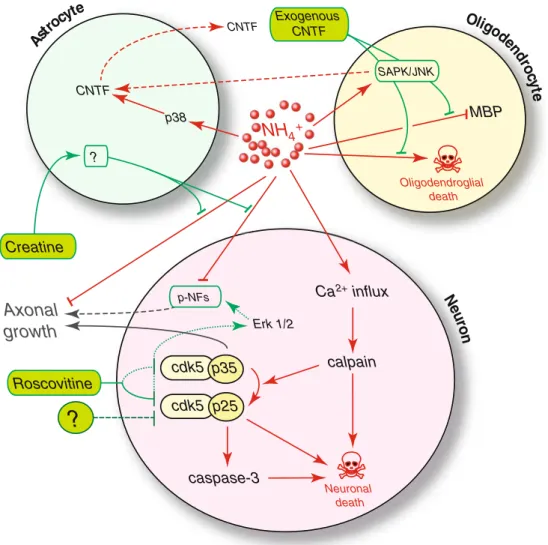
4+ MBP Ast rocy te Oligodendroglial death Oligod end ro cy te Erk 1/2 CNTF p38 CNTF calpain cdk5 p35 cdk5 p25 caspase-3 Ca influx2+Axonal
growth
Neuronal death?
Roscovitine N eu ro n SAPK/JNK Exogenous CNTF p-NFs Creatine ?Fig. 2 Proposed mechanisms leading to brain cell death under NH4+
exposure, and effects of NH4+exposure on CNS intracellular and
extra-cellular signaling pathways. Toxic effects of NH4+are shown in red,
while protective effects of creatine, roscovitine and exogenous CNTF are shown in green. NH4+activates calpain, which can induce neuronal death
directly. Activated calpain also cleaves p35 to p25 and activates caspase-3, causing neuronal death. Roscovitine decreases neuronal death by inhibiting cdk5/p25 and the subsequent caspase-3 activation. By inhibit-ing cdk5, roscovitine activates the Erk1/2 pathway, which stimulates the phosphorylation of neurofilaments. However, roscovitine also inhibits
axonal growth through inhibition of cdk5/p35. Targeting cdk5 to inhibit NH4+-induced neuronal death should thus be focused on the specific
inhibition of cdk5/p25. Creatine protects axonal growth under NH4+
exposure in a glial cell-dependent way. NH4+activates MAPKs in brain
cells, and particularly p38 in astrocytes, which increases their release of CNTF. Exogenous CNTF exerts a protective effect on oligodendrocytes, through SAPK/JNK. CNTF: ciliary neurotrophic factor; Erk1/2: extra-cellular signal regulated kinases 1/2; MAPKs: mitogen-activated protein kinases; p38: p38 kinase; SAPK/JNK: stress-activated protein kinase or c-Jun NH2-terminal kinase
distribution in various regions of the brain (Fig.3b) (Cudalbu et al2010; Mlynarik et al2008b). In vivo1H MRS and1H SI were used to study brain metabolism in animal models of hyper-ammonemia without liver failure (continuous infusion or single i.p. injection of ammonia) (Cauli et al2007; Cudalbu et al
2012b; Fitzpatrick et al1989) or ALF (galactosamine injection, portacaval anastomosis followed by hepatic artery ligation) (Cauli et al 2011; Chavarria et al2010; Nyberg et al1998) and in human studies of ALF and CLF (Chavarria et al2011; Rovira et al2008; Spahr et al2002). The main finding in all of these studies is the increase in brain Gln concentration as shown in Fig.3aandb.
In a recent in vivo study using continuous infusion of NH4Cl
we showed that Gln increased immediately after the initiation of NH4
+
exposure and continued to increase linearly over time (2.3 ± 0.4μmol/g before infusion, reaching 17.7 ± 4.0 μmol/g at the end of infusion) (Fig. 3a), suggesting that no delay in Gln accumulation occurred (Cudalbu et al2012b). No significant differences in the total concentrations of all other metabolites were observed. The linear and continuous increase of total Gln under NH4Cl infusion observed in our in vivo1H MRS data
implies an increased anaplerosis coupled to the NH4+
detoxifi-cation pathway (Berl et al1962; Shen et al1998; Zwingmann
2007). Furthermore, we mapped regional brain metabolism using1H SI (Cudalbu et al 2010) in the same rat model of hyperammonemia. Figure3billustrates the metabolic maps of Gln, Glu, Ins, NAA+NAAG and Lac superimposed on the anatomical T2w images and acquired at different time points
during NH4+infusion. As for1H MRS data, the Gln increase at
different time points was apparent from the maps with no significant differences for the concentration of other brain metabolites. Additionally, the Gln increase was higher in the cortex than in the hippocampus (16.2±2.7 mmol/kgww in the cortex and 11.5±1.2 mmol/kgww in the hippocampus after 5.5 h of NH4+infusion, p00.03). Consequently, these results showed
a higher net Gln synthesis flux in cortex than in hippocampus. Studies performed on animal models of ALF (e.g., galac-tosamine injection, portocaval anastomosis followed by he-patic artery ligation) reported additional alterations in brain Lac concentration at later stages and the presence of brain edema. The mechanisms leading to Lac increase are not clear, but may indicate brain energy impairment secondary to ammonia metabolism and brain edema (Chavarria et al
2010). CLF is associated with an additional drop in brain osmolytes (Ins, tCho and Tau) (Chavarria et al2011; Rovira et al2008; Spahr et al2002) probably reflecting an osmo-regulatory response to Gln increase. The differences in brain osmolytes may partially explain the differential frequency of brain edema between ALF and CLF (Cordoba 1996). We recently characterized for the first time the in vivo and longitudinal progression of HE in a rat model of CLF by BDL by using1H SI and diffusion tensor imaging (McLin et al2012). Gln was increased at all time points after BDL.
Among other brain osmolytes, Ins, tCho and Tau decreased significantly over time. We concluded that prior to the appearance of severe neurological signs in CLF, the osmotic imbalance created by continuous increase of Gln is likely to be compensated by a concomitant decrease of other idio-genic osmolytes resulting in minimal brain edema.
In vivo13C MRS to study neuroglial energy metabolism Cerebral metabolism is compartmentalized between neurons and glia (Gruetter 2002). Glc is the primary substrate for cerebral energy production, while Lac exchange between astrocytes and neurons is not excluded under specific con-ditions (Magistretti et al1999). In vivo 13C MRS together with administration of [1,6-13C]-Glc and an appropriate mathematical model of neuronal-glial metabolism is a unique technique to non-invasively investigate compart-mentalized cerebral energy metabolism (Gruetter2002). In particular, we measured 13C incorporation into different carbon positions of Glu and Gln to determine fluxes through
Fig. 3 a Representative in vivo1H MR spectra acquired at 9.4T in the rat brain during ammonium infusion, adapted from Cudalbu et al (2012b) (echo time 2.8 ms, 160 averages, voxel size of 5×7×7 mm3; continuous infusion of NH4Cl at 4.5 mmol/h per kilogram). From
bottom to top: spectrum acquired before infusion and spectra acquired during infusion (200, 300 and 500 min of infusion); only Gln is marked in the last spectrum since it is the only metabolite changing its con-centration, hereby increasing under NH4+infusion. Asp: aspartate; Cr:
creatine; GABA: γ-aminobutyrate; Gln: glutamine; Glu: glutamate; GPC: glycerophosphocholine; Ins: myo-inositol; Lac: lactate; NAA: N-acetylaspartate; PCho: phosphocholine; PCr: phosphocreatine; Tau: taurine. b In vivo metabolic maps of Gln, Glu, Ins, NAA+ NAAG (N-acetyl-aspartyl-glutamate) and Lac, superimposed on the anatomi-cal T2w images and acquired at different time points during NH4+
infusion at 9.4T, using1H SI (echo time 2.8 ms). The increase in the Gln pool at different time points during NH4+infusion is apparent from
the maps. c In vivo13C MRS spectra acquired in the rat brain during continuous infusion of [1,6-13C]-glucose: c1) Time course showing the incorporation of 13C into different carbon positions of Glu and Gln (only the first 90 min are shown) in a bile duct-ligated (BDL) rat; c2) In vivo brain13C MRS spectra acquired in control and BDL rats during the last hour (5th hour) of [1,6-13C]-glucose infusion. Only the metab-olites used for modeling are marked in the figure (Glu and Gln labeled at positions C4, 3 and 2). In red are shown the metabolites which are changing in the BDL rat as compared with control (Gln at positions C4, 3 and 2). d Representative series of in vivo15N spectra acquired under
infusion of15N-enriched ammonium at 9.4T in a rat brain at different
time points during infusion, adapted from Cudalbu et al (2012b). The
15
N labeling incorporation of NH4+at position 5N of Gln ([5-15N]Gln)
allows the direct measurement of glutamine synthetase flux, whereas the incorporation of15N into [2-15N]Gln+Glu provides further insight into the net flux through glutamate dehydrogenase. e In vivo diffusion tensor images (DTI) acquired at 9.4T in an animal model of hyper-ammonemia (continuous infusion of NH4Cl): e1) b0 image (b 0
0 mm2/s), diffusion tensor trace (ADC) and fractional anistropy (FA) maps acquired before and during NH4+infusion; e2) images acquired
before infusion, with the diffusion coefficient maps. b0image (b0
Control rat BDL rat Glu C4 Gl u C 2 Gl u C 3 Gln C4 Gln C 3 Gl n C 2 Gln C4 Gl n C 3 Gl n C 2 ppm 60 50 40 30 20 Gl u C 4 5 16 27 38 49 60 71 82 Chemical sh (ppm) Gl u C 2 Gl u C 3 Gl n C 4 Gl n C 3 Gl n C2 60 50 40 30 20
a
e
d
c
<0
3h
6h
C2
C1
E2
E1
b
important pathways involved in energy metabolism includ-ing: glycolysis, neuronal and astrocytic TCA cycle, malate-aspartate shuttle activity and glial anaplerotic pyruvate car-boxylation (Fig. 3c). In addition, the Glu/Gln neurotrans-mitter cycle within the neuron-astrocyte functional unit can be measured. The rate of13C label incorporated as a func-tion of time is related to metabolic rate thereby permitting the measurement of absolute metabolic fluxes. For example, the accumulation of13C label in Glu at the position C4 is indicative of both the neuronal and glial TCA cycle fluxes, whereas the labeling on Gln at the same position reveals the Glu-Gln neurotransmission flux. Further separation of the glial and neuronal TCA cycle activities is possible when measuring the C3 and C2 positions of Glu and Gln, due to the glial-specific activity of pyruvate carboxylase, diluting the carbon position 3 and labeling the position 2 of glial Glu.
In rat models of hyperammonemia by continuous infusion of NH4
+
without liver failure, in vivo13C MRS has been used to measure neuroglial metabolism in conjunction with Glc infusion labeled at different positions, i.e., [1,6-13C], [1-13C] or [2-13C]-Glc. Lanz et al (2011) and Sibson et al (1997,2001) reported that anaplerosis appears to be the major NH4+
detox-ification pathway, as measured in our1H MRS and15N NH4+
studies (Cudalbu et al2010,2012b), emphasizing the contri-bution of astrocytes in cerebral NH4+ processing. Neuronal
metabolism appears less affected, as reflected by GluC4 and GluC3 fractional enrichment time courses.
Only a few studies have tried to measure energy metabolism in animal models of ALF. These were performed ex vivo using brain extracts, and reported increased Lac and alanine synthesis as well as stimulated pyruvate carboxylation. These findings suggest that a deficit in brain Glc metabolism rather than Gln accumulation is the major cause of cerebral complications in this model of ALF (Chatauret et al2003; Zwingmann2007).
We have recently characterized in vivo brain energy me-tabolism of rats with CLF (BDL) by using13C MRS together with administration of [1,6-13C]-Glc and by following the kinetics of13C incorporation in Glu and Gln at positions C4, C3 and C2 over 5 h of [1,6-13C]-Glc infusion (Fig. 3c1). Continuous acquisition in live animals showed that13C incor-poration in Gln at positions C4, C3 and C2 was higher in BDL rats than in controls (Fig.3c), suggesting an increase in glial TCA cycle activity. In addition,13C incorporation in position C2 of Gln was higher than in positions C4 or C3, indicating increased activity of glial-specific pyruvate carboxylase flux as compared with controls. Additional data from these dy-namic studies are forthcoming and promise to shed important mechanistic information on metabolic fluxes during HE.
15
N MRS to study glutamate-glutamine metabolism
15
N MRS is an alternative approach to 13C MRS to study Glu-Gln metabolism under hyperammonemia, which
provides a more direct interpretation. In vivo 15N MRS using 15N-labeled NH4+infusion was first used to analyze
the incorporation of15N-NH4+into [5-15N]Gln and measure
the GS flux (Kanamori et al 1993; Shen et al 1998). The incorporation of15N into [2-15N]Gln+Glu was further use to study the net flux through GDH (Kanamori and Ross1995). We recently developed a new15N pulse sequence to simul-taneously detect [5-15N]Gln and [2-15N]Gln+Glu in vivo (Fig.3d) (Cudalbu et al2012b). Mathematical modeling of in vivo1H and15N MRS data, acquired interleaved on the same animal, allowed to reduce the number of assumptions and provided reliable determination of GS (0.30 ± 0.050 μmol/g/min), apparent neurotransmission (0.26 ± 0.030μmol/g/min), GDH (0.029 ± 0.002 μmol/g/min) and net Gln accumulation (0.033 ± 0.001 μmol/g/min). Our in vivo measurements allowed to clearly show the increase of brain GS activity and net Gln accumulation under hyper-ammonemia conditions, supporting the concept of their implication in cerebral NH4+detoxification.
In vivo MR diffusion to study brain edema
MR diffusion techniques (diffusion weighted or tensor imag-ing) (Fig.3e) are used to investigate brain edema by measuring the relative translational motion of water molecules which is expressed as the apparent diffusion coefficient (ADC) (Le Bihan1995). Changes in ADC reflect the presence of edema, which can be divided into cytotoxic (intracellular) and vaso-genic (extracellular) edema. Most human and animal models have shown cytotoxic edema in ALF (Cauli et al 2011; Chavarria et al2011,2010; Ranjan et al2005), while some studies proposed the coexistence of cytotoxic and vasogenic edema (Cauli et al2011). A limited number of human studies on brain edema in CLF speak in support of the presence of mild vasogenic edema (Kale et al2006).
As shown in the present review, several pathogenic mechanisms involved in HE can be explored in vivo using MRS and MRI. However, we need to emphasize that addi-tional in vivo MRS and MRI studies are needed to assess the relationship between plasma NH4
+
concentrations, brain Gln accumulation, osmoregulation, brain energy metabo-lism and brain edema in HE.
Conclusion and future directions
Hyperammonemia during brain development is associated with neuronal cell loss and cerebral atrophy leading to mental retardation and cerebral palsy in pediatric patients. In survi-vors, the pathogenic mechanisms of NH4+toxicity to the brain
involve alterations in amino acids pathways, neurotransmis-sion systems, cerebral energy, NO synthesis, axonal and den-dritic growth or signal transduction pathways (Figs.1and2).
These disturbances can lead to cytotoxic brain edema, cell death, impairment of neurite outgrowth, defects in nerve cell migration, or hypomyelination, in turn leading to brain tissue atrophy, ventricular enlargement, gray or white matter hypo-densities and demyelination. These toxic effects of NH4+are
specific to the developing brain, as neuronal damage is not observed in CNS of adult patients with hyperammonemia due to liver failure. In the mature brain, the main effect of NH4+
toxicity is the rise of Gln in astrocytes while osmoregulation is insufficient and cerebral edema develops, affecting CNS areas. Why the developing brain is so vulnerable to fluctua-tions in serum NH4
+
levels remains to be elucidated. MRS promises to be a powerful tool both to characterize the mo-lecular modifications characterizing the pathobiology in the developing brain and to monitor the effects of potential neuro-protective therapies.
Apart from the use of NH4+ scavengers such as Na+
-benzoate, Na+-phenylacetate, Na+-phenylbutyrate, OP or LOLA, new neuroprotective strategies have been proposed, making use of NMDA receptor antagonists, NOS inhibitors, Cr, acetyl-L-carnitine, inhibition of CDK5/p25, CNTF or inhibitors of MAPKs and GS (Figs.1and2).
Understanding the pathophysiology of ammonia toxicity to the CNS, or unraveling new therapeutic targets to protect CNS from hyperammonemia, requires experimental approaches fo-cusing on the brain in its cellular complexity, examining neurons and glia together (in vivo mouse and rat models; ex vivo CNS organotypic cultures; in vitro primary 3D brain cell cultures in aggregates). The extraordinary development of in vivo CNS imaging technologies (MRI, MRS, Fig.3) should contribute significantly to directing future investigations, in particular by focusing on intra- and extra-cellular metabolic and signaling pathways disturbed in the brain during NH4+exposure. Acknowledgments Olivier Braissant is supported by the Swiss Na-tional Science Foundation (grants n° 3100A0-100778 and 31003A-130278); Cristina Cudalbu is supported by the Centre d’Imagerie BioMédicale (CIBM - UNIL/UNIGE/HUG/CHUV/EPFL - Switzer-land) as well as by the Leenaards and Jeantet Foundations; Valérie McLin is supported by the Department of Pediatrics, University of Geneva Medical School. The authors thank Dr B. Lanz for his help and expertise in13C MRS, and Drs N. Kunz and Y. van de Looij for their help with DTI acquisitions.
Conflict of interest None.
References
Aguilar MA, Minarro J, Felipo V (2000) Chronic moderate hyperammo-nemia impairs active and passive avoidance behavior and condition-al discrimination learning in rats. Exp Neurol 161:704–713 Agusti A, Cauli O, Rodrigo R, Llansola M, Hernandez-Rabaza V, Felipo
V (2011) p38 MAP kinase is a therapeutic target for hepatic enceph-alopathy in rats with portacaval shunts. Gut 60:1572–1579
Albrecht J, Norenberg MD (2006) Glutamine: a Trojan horse in am-monia neurotoxicity. Hepatology 44:788–794
Albrecht J, Zielinska M, Norenberg MD (2010) Glutamine as a medi-ator of ammonia neurotoxicity: a critical appraisal. Biochem Pharmacol 80:1303–1308
Als-Nielsen B, Gluud LL, Gluud C (2004) Non-absorbable disacchar-ides for hepatic encephalopathy: systematic review of randomised trials. Br Med J 328:1046
Alvarez VM, Rama Rao KV, Brahmbhatt M, Norenberg MD (2011) Inter-action between cytokines and ammonia in the mitochondrial perme-ability transition in cultured astrocytes. J Neurosci Res 89:2028–2040 Azorin I, Minana MD, Felipo V, Grisolia S (1989) A simple animal
model of hyperammonemia. Hepatology 10:311–314
Bachmann C (2003) Outcome and survival of 88 patients with urea cycle disorders: a retrospective evaluation. Eur J Pediatr 162:410–416 Bachmann C, Colombo JP (1984) Increase of tryptophan and
5-hydroxyindole acetic acid in the brain of ornithine carbamoyl-transferase deficient sparse-fur mice. Pediatr Res 18:372–375 Bachmann C, Braissant O, Villard AM, Boulat O, Henry H (2004)
Ammonia toxicity to the brain and creatine. Mol Genet Metab 81 (Suppl 1):S52–S57
Bai G, Rama Rao KV, Murthy CR, Panickar KS, Jayakumar AR, Norenberg MD (2001) Ammonia induces the mitochondrial per-meability transition in primary cultures of rat astrocytes. J Neuro-sci Res 66:981–991
Bajaj JS, Cordoba J, Mullen KD et al (2011) Review article: the design of clinical trials in hepatic encephalopathy–an International Society for Hepatic Encephalopathy and Nitrogen Metabolism (ISHEN) con-sensus statement. Aliment Pharmacol Ther 33:739–747
Bajaj JS, Pinkerton SD, Sanyal AJ, Heuman DM (2012) Diagnosis and treatment of minimal hepatic encephalopathy to prevent motor vehicle accidents: a cost-effectiveness analysis. Hepatology 55:1164–1171 Bass NM, Mullen KD, Sanyal A et al (2010) Rifaximin treatment in
hepatic encephalopathy. N Engl J Med 362:1071–1081 Batshaw ML, MacArthur RB, Tuchman M (2001) Alternative pathway
therapy for urea cycle disorders: twenty years later. J Pediatr 138: S46–S54
Béard E, Braissant O (2010) Synthesis and transport of creatine in the CNS: importance for cerebral functions. J Neurochem 115:297–313 Bélanger M, Asashima T, Ohtsuki S, Yamaguchi H, Ito S, Terasaki T (2007) Hyperammonemia induces transport of taurine and crea-tine and suppresses claudin-12 gene expression in brain capillary endothelial cells in vitro. Neurochem Int 50:95–101
Berl S, Takagaki G, Clarke DD, Waelsch H (1962) Metabolic compart-ments in vivo. Ammonia and glutamic acid metabolism in brain and liver. J Biol Chem 237:2562–2569
Berry GT, Steiner RD (2001) Long-term management of patients with urea cycle disorders. J Pediatr 138:S56–S60
Bosoi CR, Yang X, Huynh J et al (2012) Systemic oxidative stress is implicated in the pathogenesis of brain edema in rats with chronic liver failure. Free Radic Biol Med 52:1228–1235
Braissant O (2010a) Current concepts in the pathogenesis of urea cycle disorders. Mol Gen Metab 100(Suppl 1):S3–S12
Braissant O (2010b) Ammonia toxicity to the brain: effects on creatine metabolism and transport and protective roles of creatine. Mol Genet Metab 100(Suppl 1):S53–S58
Braissant O (2012) Creatine and guanidinoacetate transport at blood– brain and blood-cerebrospinal fluid barriers. J Inherit Metab Dis 35:655–664
Braissant O, Gotoh T, Loup M, Mori M, Bachmann C (1999a) L-arginine uptake, the citrulline-NO cycle and arginase II in the rat brain: an in situ hybridization study. Mol Brain Res 70:231–241 Braissant O, Honegger P, Loup M, Iwase K, Takiguchi M, Bachmann
C (1999b) Hyperammonemia: regulation of argininosuccinate synthetase and argininosuccinate lyase genes in aggregating cell cultures of fetal rat brain. Neurosci Lett 266:89–92
Braissant O, Gotoh T, Loup M, Mori M, Bachmann C (2001) Differ-ential expression of the cationic amino acid transporter 2(B) in the adult rat brain. Mol Brain Res 91:189–195
Braissant O, Henry H, Villard AM et al (2002) Ammonium-induced impairment of axonal growth is prevented through glial creatine. J Neurosci 22:9810–9820
Braissant O, Cagnon L, Monnet-Tschudi F et al (2008) Ammonium alters creatine transport and synthesis in a 3D-culture of develop-ing brain cells, resultdevelop-ing in secondary cerebral creatine deficiency. Eur J Neurosci 27:1673–1685
Braissant O, Henry H, Beard E, Uldry J (2011) Creatine deficiency syndromes and the importance of creatine synthesis in the brain. Amino Acids 40:1315–1324
Brosnan JT, Brosnan ME (2010) Creatine metabolism and the urea cycle. Mol Genet Metab 100(Suppl 1):S49–S52
Brosnan ME, Edison EE, da Silva R, Brosnan JT (2007) New insights into creatine function and synthesis. Adv Enzym Regul 47:252–260
Brusilow SW, Maestri NE (1996) Urea cycle disorders: diagnosis, pathophysiology, and therapy. Adv Pediatr 43:127–170 Brusilow SW, Valle DL, Batshaw M (1979) New pathways of nitrogen
excretion in inborn errors of urea synthesis. Lancet 2:452–454 Butterworth RF (1998) Effects of hyperammonaemia on brain
func-tion. J Inherit Metab Dis 21(Suppl 1):6–20
Butterworth RF (2003) Hepatic encephalopathy. Alcohol Res Health 27:240–246
Butterworth RF (2012) Brain edema and encephalopathy in acute liver failure: a primary neurogliopathy? Neurochem Int 60:661 Butterworth RF, Norenberg MD, Felipo V et al (2009) Experimental
models of hepatic encephalopathy: ISHEN guidelines. Liver Int 29:783–788
Cagnon L, Braissant O (2007) Hyperammonemia-induced toxicity for the developing central nervous system. Brain Res Rev 56:183–197 Cagnon L, Braissant O (2008) Role of caspases, calpain and cdk5 in
ammonia-induced cell death in developing brain cells. Neurobiol Dis 32:281–292
Cagnon L, Braissant O (2009) CNTF protects oligodendrocytes from ammonia toxicity: intracellular signaling pathways involved. Neurobiol Dis 33:133–142
Call G, Seay AR, Sherry R, Qureshi IA (1984) Clinical features of carbamyl phosphate synthetase-I deficiency in an adult. Ann Neurol 16:90–93
Caudle SE, Katzenstein JM, Karpen SJ, McLin VA (2010) Language and motor skills are impaired in infants with biliary atresia before transplantation. J Pediatr 156:936–940
Caudle SE, Katzenstein JM, Karpen S, McLin V (2012) Developmen-tal assessment of infants with biliary atresia: Differences between males and females. J Pediatr Gastroenterol Nutr 55(4):384-389 Cauli O, Lopez-Larrubia P, Rodrigues TB, Cerdan S, Felipo V (2007)
Magnetic resonance analysis of the effects of acute ammonia intoxication on rat brain. Role of NMDA receptors. J Neurochem 103:1334–1343
Cauli O, Rodrigo R, Llansola M et al (2009) Glutamatergic and gabaergic neurotransmission and neuronal circuits in hepatic en-cephalopathy. Metab Brain Dis 24:69–80
Cauli O, Lopez-Larrubia P, Rodrigo R et al (2011) Brain region-selective mechanisms contribute to the progression of cerebral alterations in acute liver failure in rats. Gastroenterology 140:638–645
Cederbaum S, Lemons C, Batshaw ML (2010) Alternative pathway or diversion therapy for urea cycle disorders now and in the future. Mol Genet Metab 100:219–220
Chan H, Hazell AS, Desjardins P, Butterworth RF (2000) Effects of ammonia on glutamate transporter (GLAST) protein and mRNA in cultured rat cortical astrocytes. Neurochem Int 37:243–248 Chatauret N, Zwingmann C, Rose C, Leibfritz D, Butterworth RF
(2003) Effects of hypothermia on brain glucose metabolism in
acute liver failure: a1H/13C-nuclear magnetic resonance study. Gastroenterology 125:815–824
Chavarria L, Oria M, Romero-Gimenez J, Alonso J, Lope-Piedrafita S, Cordoba J (2010) Diffusion tensor imaging supports the cytotoxic origin of brain edema in a rat model of acute liver failure. Gastroenterology 138:1566–1573
Chavarria L, Alonso J, Rovira A, Cordoba J (2011) Neuroimaging in acute liver failure. Neurochem Int 59:1175–1180
Chepkova AN, Sergeeva OA, Haas HL (2006) Taurine rescues hippo-campal long-term potentiation from ammonia-induced impair-ment. Neurobiol Dis 23:512–521
Choi JH, Kim H, Yoo HW (2006) Two cases of citrullinaemia present-ing with stroke. J Inherit Metab Dis 29:182–183
Connelly A, Cross JH, Gadian DG, Hunter JV, Kirkham FJ, Leonard JV (1993) Magnetic resonance spectroscopy shows increased brain glutamine in ornithine carbamoyl transferase deficiency. Pediatr Res 33:77–81
Cordoba J (1996) Glutamine, myo-inositol, and brain edema in acute liver failure. Hepatology 23:1291–1292
Cordoba J, Blei AT (1996) Brain edema and hepatic encephalopathy. Semin Liver Dis 16:271–280
Cudalbu C, Mlynárik V, Lanz B, Frenkel H, Costers N, Gruetter R (2010) Imaging glutamine synthesis rates in the hyperammonemic rat brain. Proc Intl Soc Magn Reson Med 18:3324
Cudalbu C, Mlynárik V, Gruetter R (2012a) Handling macromolecule signals in the quantification of the neurochemical profile. J Alzh Dis 31:S101-115
Cudalbu C, Lanz B, Duarte JM et al (2012b) Cerebral glutamine metabolism under hyperammonemia determined in vivo by local-ized1H and15N NMR spectroscopy. J Cereb Blood Flow Metab
32:696–708
D’Hooge R, Marescau B, Qureshi IA, De Deyn PP (2000) Impaired cognitive performance in ornithine transcarbamylase-deficient mice on arginine-free diet. Brain Res 876:1–9
Dadsetan S, Bak LK, Sørensen M et al (2011) Inhibition of glutamine synthesis induces glutamate dehydrogenase-dependent ammonia fixation into alanine in co-cultures of astrocytes and neurons. Neurochem Int 59:482–488
de Grauw TJ, Smit LM, Brockstedt M, Meijer Y, van der Klei-van Moorsel JM, Jakobs C (1990) Acute hemiparesis as the presenting sign in a heterozygote for ornithine transcarbamylase deficiency. Neuropediatrics 21:133–135
de Knegt RJ, Schalm SW, van der Rijt CC, Fekkes D, Dalm E, Hekking-Weyma I (1994) Extracellular brain glutamate during acute liver failure and during acute hyperammonemia simulating acute liver failure: an experimental study based on in vivo brain dialysis. J Hepatol 20:19–26
Deignan JL, Cederbaum SD, Grody WW (2008) Contrasting features of urea cycle disorders in human patients and knockout mouse models. Mol Genet Metab 93:7–14
Desjardins P, Du T, Jiang W, Peng L, Butterworth RF (2012) Patho-genesis of hepatic encephalopathy and brain edema in acute liver failure: role of glutamine redefined. Neurochem Int 60:690–696 Dolder M, Walzel B, Speer O, Schlattner U, Wallimann T (2003)
Inhibition of the mitochondrial permeability transition by creatine kinase substrates. Requirement for microcompartmentation. J Biol Chem 278:17760–17766
Dolman CL, Clasen RA, Dorovini-Zis K (1988) Severe cerebral damage in ornithine transcarbamylase deficiency. Clin Neuropathol 7:10–15 Eltawil KM, Laryea M, Peltekian K, Molinari M (2012) Rifaximin vs. conventional oral therapy for hepatic encephalopathy: a meta-analysis. World J Gastroenterol 18:767–777
Enns GM (2008) Neurologic damage and neurocognitive dysfunction in urea cycle disorders. Semin Pediatr Neurol 15:132–139 Enns GM (2010) Nitrogen sparing therapy revisited 2009. Mol Genet