Review
Vascular consequences of menopause and hormone therapy: Importance
of timing of treatment and type of estrogen
Raghvendra K. Dubey
a,b,c,*, Bruno Imthurn
a,b, Matthias Barton
d, Edwin K. Jackson
a,c,e aCenter for Clinical Pharmacology, University of Pittsburgh, Pittsburgh, PA, United States
b
Department of Obstetrics and Gynecology, Clinic for Endocrinology (D217, NORD-1), University Hospital Zurich, Frauenklinikstrasse-10, CH-8051, Zurich, Switzerland
c
Department of Medicine, University of Pittsburgh, Pittsburgh, PA, United States
dMedical-Policlinic, Department of Internal Medicine, University Hospital Zurich, Zurich, Switzerland eDepartment of Pharmacology, University of Pittsburgh, Pittsburgh, PA, United States
Received 8 September 2004; received in revised form 15 December 2004; accepted 20 December 2004 Time for primary review 15 days
Abstract
Premenopausal women have a lower risk for cardiovascular events, and mortality due to coronary vascular disease (CVD) in premenopausal women is rare. These facts suggest that endogenous estrogens, such as estradiol, protect the cardiovascular system, and several observational studies and a few small clinical studies conducted in healthy and younger postmenopausal women support this hypothesis. In contrast, two large randomized clinical trials (RCTs), using conjugated equine estrogens and conducted in older women with established CVD or without overt CVD, failed to demonstrate protection against CVD by exogenous estrogens. These divergent findings have resulted in confusion with regard to the association between estrogen deficiency and CVD in postmenopausal women. In order to reconcile these contradictory findings, it is necessary to examine the pathophysiology associated with age-dependent changes within the vessel wall and to compare the pharmacology of different types of estrogens. Understanding age-dependent changes in vascular pathology and the pharmacology of different estrogens may facilitate the development of therapeutic strategies for hormone replacement therapy (HRT) that would be effective in delaying vascular remodeling leading to CVD following menopause. In this review we provide an overview of the impact of menopause and estrogen deficiency on vascular remodeling and emphasize the importance of timing and type of estrogen to achieve maximum benefits with regard to reducing the risk of CVD.
D 2004 European Society of Cardiology. Published by Elsevier B.V. All rights reserved.
Keywords: Aging; Menopause; Hormone therapy; Estrogen; Cardiovascular disease
1. Evidence for a protective action of endogenous estradiol
Endogenous estrogens may attenuate CVD. Age-associ-ated CVD lags by 10 years in premenopausal women
compared with men (Fig. 1). Between 45 and 64 years of
age, the prevalence of CVD in men is several times that of
age-matched women [1,2]. Following menopause and loss
of endogenous estradiol (major ovarian estrogen), these
gender-based differences narrow [3,4]. Most women who
enter menopause are asymptomatic for CVD, and 95% of the women who develop CVD do so after menopause.
Early loss of endogenous estradiol is associated with CVD. Most autopsy studies demonstrate increased CVD
in young oophorectomized women [5]. The Framingham
Study shows that women entering menopause early naturally or surgically have a greater likelihood of developing CVD compared to age-matched premenopausal women. The Nurses’ Health Study reports an increased risk of CVD in women with bilateral oophorectomy not
0008-6363/$ - see front matterD 2004 European Society of Cardiology. Published by Elsevier B.V. All rights reserved. doi:10.1016/j.cardiores.2004.12.012
* Corresponding author. Department of Obstetrics and Gynecology, Clinic for Endocrinology (D217, NORD-1), University Hospital Zurich, Frauenklinikstrasse-10, CH-8051, Zurich, Switzerland. Tel.: +41 1 255 8608; fax: +41 1 255 4439.
E-mail address: [email protected] (R.K. Dubey).
receiving hormone replacement therapy (HRT) [6]. Younger age at natural menopause is associated with
CVD [7] and CVD mortality [8,9]. A recent
population-based study demonstrates an inverse relationship between age at menopause and prevalence and extent of carotid
atherosclerosis [10].
In premenopausal women, endogenous estradiol may abrogate age-related vascular remodeling. Age-associated vascular remodeling involves endothelial dysfunction, enhanced growth of intimal smooth muscle cells (SMCs),
and increased prevalence of vascular plaques (Fig. 1). The
same cellular processes participate in atherosclerosis [11].
Although it is difficult to separate vascular changes due to aging from those due to menopause, comparisons between men versus age-matched postmenopausal women and between age-matched premenopausal versus postmeno-pausal women suggest that endogenous estradiol delays
CVD [1–4,12,13]. In support of this conclusion, estradiol inhibits many processes involved in age-associated vascu-lar remodeling, including SMC proliferation and endothe-lial dysfunction, and lowers cholesterol and improves
vascular tone (reviewed in Refs. [14,15]).
Time since menopause, rather than type of menopause (natural or surgical), is the major factor associated with
elevated subclinical atherosclerosis [16]. In women 35
years of age, only fatty streaks and minimal atherosclerotic
plaques occur in coronary arteries [17]. Active progression
of atherosclerotic lesions transpires in the following decade (45–55 years of age) when menopause usually occurs, and these lesions start to develop complications when women
are z65 years of age [18,19]. In contrast, pathologic
findings in younger premenopausal women indicate mild vascular disease with low-density fibrous tissue compared with high-density fibrous tissue found at later stages of
atherosclerosis development [20]. Marked endothelial
dysfunction and intimal thickening also occur following
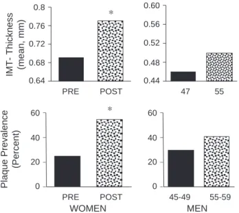
menopause [21]. A significant increase in the prevalence
of plaques and intima-media thickening (IMT) arises 5–8
years after menopause (Fig. 2;[21]). A significant increase
in total and LDL-cholesterol occurs within 3 years of
natural menopause [22], and in women undergoing
oophorectomy an increase in total and LDL-cholesterol
occurs in the first 6 weeks after oophorectomy [23].
Menopause is associated with endothelial dysfunction, decreased endothelial-dependent relaxation, and a decline
in flow-mediated dilation[24]. Ovariectomy blunts forearm
vasodilation in response to intrabrachial acetylcholine [25],
and sympathetic nerve activity increases during the early
phases of menopause [26]. Men Women Male Male Female Male Female Acceleration around menopause Female 30 20 30 40 50 60 70 80 90 100 10 0 0.12 0.10 0.08 0.08 0.04 0.02 0.00 100 50 0 < 35 35-3940-4445-49 50-5455-5960-6464-69 69-7475-84 35-44 45-54 56-64 Age Age (years) Age (years)
Coronary Artery Disease Annual Incidence per 1000
Common Carotid Intimal Medial Thickness (cm)
Plaque Prevelance (Percent) A B C 65-74 75-84
Fig. 1. (A) Age-dependent incidence of coronary artery disease (CAD) in men and women (Framingham Heart Study). (B) Age-dependent intimal-medial thickening of common carotid in healthy male and female volunteers (Baltimore Longitudinal Study on Aging; with permission,
[55]). (C) Prevelance of carotid atherosclerosis by age and sex (with permission[59]).
0.8 0.76 0.72
IMT- Thickness (mean, mm) 0.68 0.64 PRE POST * 60 40 20 Plaque Prevalence (Percent) WOMEN 0 PRE POST * 60 40 20 MEN 0 45-49 55-59 0.60 0.56 0.52 0.48 0.44 47 55
Fig. 2. Prevalence and degree of carotid atherosclerosis in premenopausal women and women 5–8 years after menopause, and in age-matched men after 8 years. The upper panel shows intima-media thickness and the lower panel shows prevalence of plaques. * significant increase versus premeno-pausal (PRE). (with permission[21,55,59]).
Hypertension may contribute to acceleration of athero-sclerosis following menopause. Postmenopausal women N60 years old make up the majority of hypertensives and
are more likely to develop CVD [27]. Surgical menopause
following oophorectomy increases peripheral vascular resistance and blood pressure, and this effect is abrogated
by HRT [28,29]. Sex hormones influence mechanisms
involved in regulating blood pressure [30–32]. Compared
to premenopausal women, the activity and synthesis of pro-hypertensive factors are increased, whereas the syn-thesis of blood pressure-lowering factors is decreased
[30,32]. Studies demonstrate the blood pressure-elevating effects of androgens and suggest that increases in the androgen-to-estradiol ratio activate pro-hypertensive
mech-anisms [30]. Estrogens, but not androgens, induce
favorable effects on the kidney, the organ that determines long-term levels of arterial blood pressure. There is increased salt sensitivity in postmenopausal women which
may contribute to hypertension [33], suggesting that
changes in renal function following menopause contribute to the increased risk of atherosclerosis. The effects of menopause on hypertension, and subsequently atheroscle-rosis, may vary between subjects. For example, the effects may be enhanced in women with diabetes mellitus
(associated with increased testosterone levels) [34] or
obesity [30].
2. Evidence that exogenous estrogens reduce menopause-associated vascular consequences
Cross-sectional and prospective studies demonstrate significant reductions in CVD in women taking conjugated
equine estrogens (CEEs) [5], suggesting that exogenous
estrogens prevent CVD. A meta-analysis of observational studies shows that HRT is associated with a one-third
reduction in fatal CVD [35]. Use of unopposed CEE and
CEE plus a progestin shows a relative CVD risk of 0.70 and
0.66, respectively[36].
The Nurses’ Health Study was conducted in 121,700
female nurses aged 30–55 years [12,13]. The latest report
involving 70,533 postmenopausal women followed for 20 years indicates that the overall relative CVD risk in current users of estrogens is 0.61 after adjustment for age and
common cardiovascular risk factors [13]. The Nurses’
Health Study suggests increased risk for ischemic stroke in HRT users.
3. Results of RCTs are inconsistent with observational studies
Randomized clinical trials (RCTs) were launched to verify that HRT reduces CVD. Two large placebo-controlled RCTs, the Heart and Estrogen/Progestin Replacement Study
(HERS[37]) and the Women’s Health Initiative Study (WHI
[38]) tested the effects of HRT in secondary and primary
prevention, respectively. 3.1. Secondary prevention trials
HERS enrolled 2763 women (mean age, 67 years) with documented CVD. Subjects were administered daily CEE (0.625 mg) and medroxyprogesterone (MPA; 2.5 mg) or placebo. HERS, after approximately 4.1 years of follow-up, found no difference in primary CVD outcome (nonfatal MI plus CVD death), even though HRT significantly reduced
LDL and increased HDL [37]. A significant increase in
adverse CVD events was found in the HRT group during the first year of treatment (52% excess cardiovascular events), and no protective effects were evident after an additional 2.7
years of follow-up[39].
The HERS findings were unexpected, but consistent with several smaller RCTs conducted for secondary prevention
[40,41]. These smaller studies also showed either no protection or a slight increase in CVD events during the first year of HRT. Early adverse effects (MI and throm-boembolic events) were also observed in the Coronary Drug Project conducted with CEEs (2.5 mg or 5.0 mg) in men
[42]. The Women’s Estrogen for Stroke Trial (WEST)
examined the effects of estradiol on stroke rates in
postmenopausal women and found no effect[43].
3.2. Primary prevention trials
The WHI study [38], conducted in healthy
postmeno-pausal women between 50 and 79 years of age, was initiated to evaluate whether HRT was effective in primary preven-tion. It was a double-blind randomized study with two arms, one studying the impact of CEE (0.626 mg/day) plus MPA (2.5 mg/day) or placebo in 16,608 women with a uterus and the second studying the impact of CEE (0.625 mg/day) alone or placebo in 10,739 women without a uterus. The outcome of WHI was that neither estrogen nor estrogen plus progestin decreased CVD.
The WHI findings are supported by some, but not all, smaller primary prevention trials. A pooled analysis of smaller RCTs conducted in mostly young women poten-tially devoid of unrecognized CVD at baseline supported the WHI findings with no significant cardiovascular benefits of
HRT [44]. In the Postmenopausal Estrogen/Progestin
Interventions (PEPI) trial there was a non-significantly higher incidence of cardiovascular and thrombotic events
among women assigned HRT[45].
The Estrogen in the Prevention of Atherosclerosis Trial
(EPAT) [46] demonstrated that oral administration of
unopposed estradiol (1 mg/day) significantly reduced the progression in carotid artery atherosclerosis in healthy women (average age, 61 years). Similar to EPAT, unop-posed estradiol therapy slowed the progression of athero-sclerosis in the Asymptomatic Carotid Artery Progression
desogestrel or CEE plus cyclic norgestrel [48] tended to decrease carotid IMT in healthy perimenopausal women (age 40–60 years).
In the Women’s Estrogen–Progestin Lipid Lowering Hormone Atherosclerosis Regression Trial (WELL–HART)
[49], 226 postmenopausal women (mean age, 63.5 years)
who had at least one coronary artery lesion were randomly assigned to placebo, estradiol, or estradiol plus MPA. After a median of 3.3 years of follow-up, the change in stenosis was measured using quantitative coronary angiography. In
contrast to EPAT[46], no significant effects of estradiol or
estradiol plus progestin on the progression of atherosclerosis were found in older postmenopausal women with estab-lished coronary artery atherosclerosis. The different out-comes of the EPAT versus WELL–HART may largely be due to the subject population studied, i.e., healthy subjects versus those with established CVD.
Other primary prevention trials include the Puget Sound
Group Health Cooperative (PSGHC; [50]), the
Postmeno-pausal Hormone Replacement against Atherosclerosis
(PHOREA; [51]), and the Women’s International Study of
Long Duration Oestrogen after Menopause (WISDOM). In the PSGHC trial, similar to the Nurses’ Health Study, the relative risk for myocardial infarction in healthy women was increased more than twofold in new users of HRT as
compared to women using HRT for 1 to 2 years[50]. In the
PHOREA trial, which investigated HRT (1 mg/day estradiol plus 0.025 mg gestodene) in healthy postmenopausal women (age 40–70 years), no significant difference in
carotid IMT was observed after 2 years[51]. The WISDOM
trial is a primary prevention trial investigating the use of CEE (plus MPA in women with uterus) over a 10-year period, and similar to WHI it will assess the risk of coronary events. This study is ongoing and the results will help in assessing the cardioprotective role of estrogens in women. Finally, results should be available soon from the Estrogen and Graft Atherosclerosis Research study (EAGAR; secon-dary prevention trial), which is examining the effects of estradiol plus MPA to prevent graft occlusion in postmeno-pausal women who have recently undergone coronary artery bypass surgery.
4. The basis for discrepancies between observational studies and RCTs
4.1. Role of timing of HRT
In primates, CEE-induced vascular protection was attenuated when treatment was initiated after atherosclerosis
was already established [52]. A 70% protection was
observed when HRT was initiated simultaneously with an atherosclerotic diet in ovarectomized primates. A marginal delay in initiation of HRT, i.e., after moderate atheroscle-rosis, resulted in only a 50% protection. When HRT was begun 2 years into an atherosclerotic diet, no protection was
observed [52]. Similar to the findings in primates,
admin-istration of estradiol prior and during, but not seven days after, balloon injury resulted in inhibition of neointima
formation in rats [53]. Delayed delivery also failed to
prevent neointima formation in rabbits[54].
The Nurses’ Health Study supports the hypothesis that time when HRT is initiated influences cardiovascular benefit from HRT. Women who participated in this study were between 30 to 55 years of age, and c80% initiated HRT
within 2 years of the onset of menopause[12]. In contrast to
the Nurses’ Health Study, women in HERS were 67 years of age and had been postmenopausal for many years at the time of enrollment. In HERS, the interval from menopause to
randomization was 23 years versus 13 years in EPAT[46].
Although WHI was a primary prevention trial, similar to HERS, the participants in WHI were older (50–79 years) with only 10% of the participants between 50 and 54 years and 20% between 54 and 59 years. In women assigned HRT in WHI, 36% had hypertension, 49% were current or past smokers, and 34% were obese. The contention that the participants were healthy should be reconsidered.
The progression rate of vascular disease may depend on age at menopause and status of subclinical atherosclerosis.
As shown in Fig. 3, intimal thickening increases with age
and CVD extent [55]. In women who underwent bilateral
oophorectomy, intimal thickening increases with years since menopause and reaches significance 15 years after
meno-pause (Fig. 4) [16]. McGarth et al.[56]demonstrated that
age-dependent progression of carotid intimal thickening in postmenopausal women is significantly reduced by HRT (Fig. 5). These findings suggest that estrogen plays a role in regulating intimal thickening. In addition to the impact of estrogen on intimal growth, estrogen reduces prevalence of
No CAD Age (years) Common Carotid Intimal-Medial Thickness (mm) (17) (33) (89) (81) (72) (10) (68) (37) (4) (4) 1.0 0.8 0.6 0.4 0.2 0.0 30 40 50 60 70 80 90 (4) (22) (4) (5)(12) (8) (8) * Possible CAD-1 Possible CAD-2 Definite CAD
Fig. 3. Common carotid intimal-medial thickness as function of age, stratified by coronary artery disease (CAD) classification CAD2N CAD1NCAD (with permission[55]).
plaque [21,57,58], and the time-curve for prevalence of
plaques is shifted to the right in women (Fig. 1) [59]. In
contrast to the linear increase observed in men, a plateau in
the incidence of plaques is evident around perimenopause (40–50 years age) in women, and after 50 years of age (around menopause) a steep increase in the incidence of
plaque is seen (Fig. 1). These observations suggest that
estrogen slows vascular processes associated with CVD. Because in the absence of estrogen the progression of atherosclerosis may be rapid, the timing of initiation of treatment, with respect to onset of menopause, may have important ramifications on the therapeutic efficacy of HRT in preventing or delaying the progression of atherosclerosis and CVD. In WHI, the relative risk for non-fatal myocardial infarction increased as a function of years since menopause (Fig. 4).
4.2. Role of type of estrogen used for HRT
Both HERS and WHI were conducted with CEEs. CEEs are a mixture of estrogens extracted from horse urine and also contain progestins, androgens, and other substances (Table 1). The main constituents of CEEs are specific equine estrogens and weak estrogenic molecules such as estrone, estrone sulfate, and estriol. In contrast to CEE, the major
Control HRT 1.3 1.2 1.1 1.0 0.9 0.8 0.7 0.6 0.5 0.4 45 50 55 60 Age IMT (mm) 65 70 75
Fig. 5. Hormone replacement therapy (HRT) slows age-associated increase in intimal-medial thickness (IMT) in postmenopausal women (with permission[56]).
Table 1
Various constituents of conjugated equine estrogen Estrogens Sodium-estrone sulfate Sodium-Equilinsulfate Sodium-17a-Dihydroequilinsulfate Sodium-17a-Estradiolsulfate Sodium-17h-Dihydroequilinsulfate Sodium-17a-Dihydroequileninsulfate Sodium-17h-Hydroequileninsulfate Sodium-Equileninsulfate Sodium-17h-Estradiolsulfate Sodium-delta 8,9-Dehydroestronsulfate Progestins 5a-Pregnane-3h, 20h-diol 5a-Pregnane-3h, 16a, 20h-triol 5a-Preg-16-en-3h-ol-20-one 5a-Pregnane-3 h-ol-20-one
Sodium-4-Pregene-20-ol-3-one-Sulfate
3h-Hydroxy-5(10), 7-estradiene 17-one-3-Sulfate Androgens 5a-Androstane-3h, 17a-diol 5a-Androstane-3h, 16h-diol 5a-Androstane-3h, 16a-diol 5a-Androstane-3h-ol, 16-one Other substances 5,7,9 (10) Estratriene-3h, 17h-diol 17a-Dihydro-delta 8,9-Dehydroestrone 17h-Dihydro-delta 8,9-Dehydroestrone 5,7,9,(10) Estratriene-3h-ol-17-one 2-Hydroxyestrone 2-Methoxyestrone 0.84 0.8 0.76 0.72 0.68 0.64 0.6 > 0-5 > 15
A
5-10Years Since Menopause
WHI data
§ Intima-Media Thickness (mm; mean
± SEM) Relatve Risk Non-fatal MI or CRD 10-15 1.8 1.5 1.2 0.9 0.6 0.3 0 < 10 > 20
B
19-20Years Since Menopause
Fig. 4. (A) Time-dependent and early changes in intimal-medial thickening following menopause (with permission [17]). (B) Effect of time since menopause on incidence of myocardial infarction in women receiving HRT in WHI[38]. CRD, coronary related death.
endogenous estrogen lost during menopause is estradiol,
and this is not present in CEE[45].
The effects of CEEs or other estrogens may be very different than those of estradiol. Binding affinity, selectivity for estrogen receptor (ER) subtypes, ER activation, and
metabolism vary markedly among different estrogens[14].
Because ER dependent and independent mechanisms play a role in mediating the biological actions of estradiol on the cardiovascular system, CEEs and other estrogens may not mimic the cardiovascular effects of estradiol. For example, ethinyl estradiol, an estrogen compound, is known to induce
deleterious effects on the cardiovascular system [14]. In a
follow-up study in Uppsala, Sweden[60], the authors found
a reduced risk of myocardial infarction for medium-potency estrogens (48%, estradiol [predominantly 2 mg]; 15.2% CEEs [predominantly 0.625 mg]; and 36.8% for both) compared with low-potency estrogens (oral estriol [predom-inantly 1 mg] or vaginal estriol/dienoestrol) (relative risk, 0.75), with a similar decrease in the subgroup that took estrogens with progestin (relative risk, 0.69).
In in vitro studies using human aortic SMCs, we demonstrated that in contrast to estradiol, estrone, estriol, estrone sulfate (major components of CEEs) were less potent in inhibiting mitogen-induced SMC growth and
mitogen-activated protein kinase activity [61]. Because
abnormal growth of SMCs plays a role in the remodeling processes associated with CVD, antiproliferative efficacy and potency of an estrogen may play an important role in defining the vascular protective actions of that estrogen. Indeed, in a non-human primate model, administration of CEE was shown to inhibit the progression of atheroscle-rosis, but had no effect on intimal hyperplasia after balloon
injury[62]. Importantly, in EPAT, administration of estradiol
to postmenopausal women with no evidence of CVD significantly reduced the progression of intimal thickening
[46].
Most animal studies conducted to elucidate the vascular protective actions of estrogen therapy used estradiol rather than CEEs. Importantly, studies conducted in animals provide strong support that estradiol protects against CVD. In almost all the models of atherosclerosis and neointimal thickening estradiol prevents pathological
vas-cular remodeling processes and neointima formation[14].
Several studies indicate that the vascular protective effects of estradiol are mediated in part by ER-independent
actions [63–66] and, therefore, the vascular protective
effects of estradiol would not be mimicked by non-estradiol-based estrogens with a different pharmacological profile. Our studies indicate that metabolism of estradiol to methoxyestradiols is responsible for the anti-mitogenic effects of estradiol on vascular SMCs, cardiac fibroblasts,
and glomerular mesangial cells [67]. Importantly, these
effects of estradiol on cell growth are ER-independent[67].
Increased proliferation of these cell types leads to hyper-tension, vascular disease, left ventricular hypertrophy, and glomerulosclerosis. Thus, some of the cardiovascular and
renal protective effects of estradiol may be mediated via their conversion to methoxyestradiols. Direct evidence for this hypothesis comes from our finding that the anti-mitogenic effects of estradiol are lost in aortic SMCs cultured from COMT-knockout mice that cannot form
methoxyestradiols[68]. The importance of estradiol
metab-olites in vasoprotection is further supported by our finding that in male obese ZSF1 rats that exhibit the metabolic syndrome, treatment with 2-hydroxyestradiol (precursor of 2-methoxyestradiol) decreases body weight, improves vas-cular endothelial function, decreases nephropathy, exerts antidiabetic actions, and lowers blood pressure and blood
cholesterol[69].
Changes in the androgen-to-estradiol ratio following menopause may also play a role in defining the deleterious effects of menopause on the cardiovascular system. The decline in estradiol levels during menopause leads to a higher androgen-to-estradiol ratio in postmenopausal
women; moreover, this ratio is lower in HRT users [70].
Androgens induce vasoconstriction and SMC growth and exacerbate diet-induced atherosclerosis, plaque formation, and pro-atherosclerotic arterial remodeling. These findings suggest that the increase in the androgen-to-estradiol ratio in postmenopausal women may be another mechanism which contributes to the acceleration of atherosclerosis observed in
postmenopausal women [30–32]. Indeed, in women, high
testosterone levels are associated with dyslipidemia [71],
type-2 diabetes mellitus [34], and hypertension [72],
suggesting that increases in androgen levels may accelerate CVD in postmenopausal women. In a case control study, compared to women with normal ovaries, more extensive atherosclerosis was observed in women with polycystic ovary syndrome, a condition associated with elevated androgen levels. Increased testosterone levels correlate with the degree of coronary atherosclerosis measured by
angiog-raphy [73], and in a recent nested case-control prospective
study a trend towards increased CVD risk was observed in
women with higher androgen-to-estradiol ratios [74].
Together, the above findings suggest that acceleration of atherosclerosis in postmenopausal women may in part be due to unopposed actions of androgens due to decline in estradiol levels.
4.3. Role of polymorphisms (single nucleotide polymor-phism [SNP])
Polymorphisms in the gene for the ER-a receptor are associated with: increased incidence of premature CVD in a
man [75]; CVD in postmenopausal women [76] and older
men [77]; pro-atherosclerotic profile of serum lipids in
women with CVD[78]; and CVD in patients with familial
hypercholesterolemia [79]. The incidence of in-stent
reste-nosis is significantly increased in women with a
poly-morphism in the ER-a gene[80]. Polymorphisms in the
ER-h gene in healthy postmenopausal women are associated
found an association between polymorphisms in ER-a and ER-h genes and cardiovascular risk, there is also evidence for lack of an association. Matsubara showed that three polymorphisms in ERs were not associated with prevalence and severity of CVD and that these polymorphisms were
unrelated to serum lipid levels [82]. An alternative
mechanism that may be involved in regulating ER-depend-ent protection of the cardiovascular system is age-dependER-depend-ent
methylation of ER[83].
The importance of SNPs in governing biological effects of estrogen and defining therapeutic efficacy of HRT has
been reviewed by Herrington [84]. In a hallmark-study
using subjects from the ERA trial, the authors demonstrated that ER-a polymorphisms can alter the response to HRT
[78]. Subjects from both active arms of ERA (oral CEE and
oral CEE plus MPA) and placebo were characterized with respect to selected ER-a polymorphisms. After adjusting for potential confounders, the 18.9% of women who had IVSI-401 C/C genotype in the active-treatment arms showed increases in HDL cholesterol more than twice the increase observed in other women. This effect was found in both groups, i.e., women receiving estrogen or estrogen plus progestin, and the effects were observed across racial groups
[78]. A similar pattern of response was observed for the
HDL-3 subfraction, and a trend for greater increase in
apolipoprotein A-1 was evident[78]. In a subsequent report,
women in the active treatment arms with ER-a IVS1-401 C/ C genotype showed greater reductions in E-selectin but not CRP, suggesting that the effects were due to selective gene–
drug interaction[85].
SNPs of other genes can also modulate the actions of estrogen or HRT by influencing drug–gene interactions. Estrogens increase the risk of venous and arterial thrombotic events, and this is associated with genetic polymorphisms
[86]. Genetic mutations, such as Factor-V Leiden and
prothrombin 20210A, may increase the risk of venous and arterial thrombotic events when estrogen levels are
increased [87]. Recent examination of data from HERS
revealed that women with factor-V Leiden polymorphism who were assigned HRT had substantially higher rates of venous thromboembolic events than women receiving HRT who lacked factor-V Leiden polymorphism, or women with factor-V Leiden polymorphism who were assigned placebo
[88]. Presence of the prothrombin 20210GYA mutation in
women with hypertension was found to be a significant
factor for myocardial infarction in a case control study[87].
Women with the mutant prothrombin allele who were using HRT had almost 11-fold greater risk of nonfatal myocardial infarction, compared with current HRT users without the
prothrombin variant[87].
Polymorphisms in other genes of the coagulation/ fibrinolytic cascade may also play an important role in regulating estrogen induced thrombosis via altered drug– gene interaction. The sequence variants in Factor-VII gene, the prothrombin gene, factor-V, fibrinogen, and the gene for
plasminogen activator inhibitor may be important[84,86].
Finally, since estradiol is metabolized to multiple bio-logically active endogenous metabolites, mutations in CYP450 isozymes or COMT may play a role in defining estradiols cardiovascular protective effects. Polymorphisms of CYP1B1 are associated with estrogen-induced breast
cancer [89], suggesting that screening of patients for SNPs
may identify patients with increased risk of estrogen-induced cancer.
4.4. Role of progestin
Progestins are administered sequentially or continuously with estrogen. The progestins/gestagens used clinically vary in chemical properties and possess varying degrees of
androgenic and glucocorticoid activity[90]. Via
glucocorti-coid receptors, progestins can potentiate vascular procoa-gulant effects of thrombin by increasing thrombin receptors
in SMCs [90]. Hence, the negative findings of HERS and
one arm of WHI may have been due in part to concomitant use of MPA. In support of this idea, in the PEPI trial, CEE caused beneficial effects on LDL and HDL levels that were
attenuated by MPA [45]. Because increased LDL and
decreased HDL are associated with cardiovascular disease, the interpretation is that MPA may abrogate the protective effects of estrogens on the cardiovascular system. However, this interpretation is not supported by the observations that CEE and CEE plus MPA are equipotent in inhibiting
atherosclerosis in non-human primates [91]. Similar to
MPA, the anti-atherosclerotic effects of CEE were not
abrogated by progesterone in cynomolgus monkeys [91].
However, in contrast to these studies, administration of CEE, but not CEE plus MPA, caused anti-atherosclerotic
effects in monkeys [91].
The effects of progestins in attenuating the protective actions of estrogens on atherosclerosis are unclear. In cynomolgus monkeys given continuous estradiol or estra-diol plus cyclically administered progesterone for 30
months, the anti-atherosclerotic effects were similar [91].
Loss of protective effects was observed in monkeys administered CEE plus MPA (no protection) as compared to those treated with CEE alone (72% reduction in coronary
artery atherosclerosis[91]). In rabbits the protective actions
of CEE or estradiol on atherosclerosis were not reversed by MPA nor by other progestins (norethindrone acetate and
hydroxyprogesterone caproate;[91]). In most observational
studies with positive outcomes, including the Nurse’s
Health Study [13], comparable reductions in CHD risk
were found with HRT regardless of whether HRT included a progestin.
Intimal thickening plays a role in vascular remodeling and is associated with atherosclerosis. In a rat model, MPA abrogated the ability of estradiol to attenuate balloon-injury
induced intimal thickening [32]. In contrast to these
findings, progesterone and MPA inhibited mitogen-induced proliferation of SMCs in vitro. Also, in the Atherosclerosis Risk in Communities Study, the reductions in
intimal-medial thickness were similar in women receiving estrogen
alone or estrogen plus MPA[92].
The above findings suggest that factors other than MPA are involved in the lack of protective actions observed in RCTs. The termination of the estrogen alone arm of WHI supports the notion that factors other than MPA are involved.
4.5. Role of interactions with lipoproteins
Ovarian dysfunction at the onset of menopause and increased CVD incidence are associated with increases in
LDL, and decreases in HDL and estradiol [93]. Thus
interactions between hormones and lipoproteins may par-ticipate in maintaining vascular homeostasis.
Estrogens influence the vascular effects of LDL choles-terol. Estradiol, which is a phenol with anti-oxidant proper-ties, prevents the oxidation of LDL and VLDL to oxLDL and oxVLDL, and protects the vasculature against the
deleterious effects of ox-lipids [14]. Estradiol prevents
compromise of the endothelial barrier mediated by mmLDL and attenuates accumulation of mmLDL and oxLDL in the
artery wall [94]. TNF-a-mediated oxidation and
accumu-lation of LDL in the artery wall is prevented by estradiol
[95]. Estradiol increases the catabolism of LDL, apo-B100,
and beta VLDL via LDL-R-dependent and -independent
mechanisms[14,96]. Also, estrogen: increases expression of
LDL-R, increases clearance of VLDL, decreases LDL production, decreases LDL particle size, increases clearance of light and dense LDL, increases expression of VLDL and LDL-R in left ventricles of the heart, induces HMG-CoA reductase activity, and induces sterol-27-hydroxylase
activ-ity which decreases LDL production [14,96,97].
Estrogen-induced removal of VLDL is associated with increased activities of hepatic lipase, lipopotein lipase, and expression
of LDL-R [14]. There is evidence for cross talk between
ERs and LDL-R. Up-regulation of ERs is associated with an increase in LDL-R expression, and this effect can be
blocked by the ER antagonist, tamoxifen[14].
Foam cell/fatty streak formation is involved in plaque pathogenesis and the interaction of LDL, macrophages, and SMCs. Estrogens can interfere with several biochemical events associated with these processes. Incubation of human THP-1 macrophages with estradiol reduces the uptake and
metabolism of 125I-labeled human acLDL, suggesting that
estrogen reduces degradation of oxLDL via scavenger
receptors[14]. In vivo studies in primates demonstrate that
the rate of LDL degradation is decreased in arteries in response to estradiol. Estrogen reduces cholesterol accumu-lation and esterification, an important step in foam cell
formation[14]. Pharmacological concentrations of estradiol
inhibit migration of monocytic cells stimulated with oxidized-LDL by inhibiting secretion of MCP-1 by
mono-cytic cells[14]. Estradiol inhibits oxLDL-induced adhesion
of monocytes to endothelial cells [14]. Because estradiol
downregulates the expression of adhesion molecules, it is
feasible that estradiol inhibits the adhesion process by preventing oxLDL induced expression of VCAM-1 and
ICAM-1 [14].
HDL-C is more closely related to cardiovascular disease in women than LDL-C and HDL-C is the best predictor of CVD risk in women. Apo-AI accounts for most of the protein in HDL, and plasma concentrations of apo-AI are increased in premenopausal women and in postmenopausal
women treated with estrogens[98]. Clinical studies provide
evidence for estrogen-induced increases in the rate of production of HDL subfractions and decreases in the plasma
clearance of HDL [98]. Studies in rodents showed that
estradiol increases hepatic apo-AI mRNA levels and
increases hepatic rates of apo-AI transcription [14]. In
hepatoblastoma cells, pharmacological concentrations of estradiol increase apo-AI secretion and apo-AI mRNA levels by increasing the rate of apo-AI mRNA transcription
without effecting apo-AI mRNA stability [14]. Similar to
HDL, estrogen induces synthesis of apolipoprotein-E [99],
which may confer vasoprotection. In apo-E-deficient mice, estradiol prevents both fatty streak formation and
athero-sclerotic lesions [100]. Therefore, mechanisms other than
apo-E synthesis are involved in mediating the estradiol-induced vasoprotective effects.
Estradiol via ERs induces the expression of
ATP-binding cassette-A1 (ABC-A1) transporter [101], and thus
estradiol may facilitate reverse-cholesterol transport responsible for the movement of cholesterol from periph-eral cells, including macrophage-derived foam cells in the arterial wall, back to the liver, where cholesterol is
catabolized [102]. This action of estradiol may attenuate
formation of foam cells, early players in the formation of arterial lesions. Acquisition of cholesterol and phospholi-pid protects the nascent liphospholi-pid-poor apo-A1 particles from rapid catabolism, thus enabling their transition to mature HDL[102]. Patients with Tangier disease are heterozygous for ABC-A1 mutations and have dramatically low levels of HDL and low cholesterol efflux capacity. This suggests that the ABC-A1-mediated lipid secretory pathway corre-sponds to a rate limiting step in the production of HDL
[102]. Hence, ABC-A1 acts as a gatekeeper for cholesterol flux from tissues. The recent reports that cholesterol can directly induce foam cell like phenotype in vascular SMCs
[103] and that ABC-A1 is expressed and participates in
reverse-cholesterol transport [102] suggest that
estradiol-induced ABC-A1 expression in these cells protects against endothelial damage and atherosclerosis. Thus, estrogen-mediated up-regulation of the ABC-A1 expression may contribute to accelerated removal of cholesterol from peripheral tissues, and inhibit progression of
atheroscle-rosis[104]. It should be noted that it is the functional HDL
pool, not necessarily the total amount of HDL, that is important in removing arterial cholesterol. Decreased levels of HDL may result from fast turnover of smaller HDL particles. Tibolone, a hormone therapy drug widely used in Europe, reduces total HDL levels by almost 30%
without changing the cholesterol efflux potential[104]and without increasing the incidence of atherosclerosis.
Estrogen down regulates scavenger receptor-B1 (SR-B1) which is involved in the transfer of HDL2 particles to the liver, suggesting that estrogen-induced increases in HDL are associated with impaired rather than improved elimination of
HDL-cholesterol[105]. Also, estrogens reduce hepatic lipase
gene expression and activity [106], which would result in
increases in HDL concentration and production of larger HDL particles. Oral, but not transdermal, estrogen therapy increases levels of serum amyloid-A (SAA) and alters HDL
composition to contain higher SAA levels [107]. Because
elevated levels of SAA predict adverse prognosis in healthy postmenopausal women, this mechanism could interfere with estrogen’s protective effect. This suggests that the route of HRT plays an important role in minimizing adverse effects of estrogen. As pointed out by Herrington and Parks, increases in HDL levels may not always result in cardiovascular
protection[108]. Because ERs play a major role in mediating
the effects of estrogen on HDL synthesis [84], factors
in-fluencing ER function can significantly influence the effects
of HRT on HDL. A study by Herrington and colleagues[78]
demonstrates that a polymorphism of the ER-a can dramat-ically influence the effects of estrogen on HDL levels and may play a critical role in defining the protective effects of HRT in individual subjects receiving HRT.
5. Conclusion and future directions for treating vascular consequences of menopause
In conclusion, it is likely that the timing of the initiation of HRT and the status of cardiovascular health determines in part whether HRT will protect against CVD. Also, it is likely that transdermal estradiol, rather than oral CEEs, would be more effective in achieving cardiovascular protection. Finally, polymorphism/SNPs may importantly influence the outcome of HRT via altered drug–gene interactions, and, in the future diagnostic SNP-screening may help tailor HRT for the individual’s needs, thereby increasing efficacy and safety.
Acknowledgement
Supported by Swiss National Science Foundation grant 32-64040.00 and 3200B0-106098/1 and NIH grant HL69846.
References
[1] Isles CG, Hole DJ, Hawthorne VM, Lever AF. Relation between coronary risk and coronary mortality in women of the Renfrew and Paisley survey: comparison with men. Lancet 1992;339:702 – 6. [2] Tracy RE. Sex difference in coronary disease: two opposing views.
J Chronic Dis 1996;19:1245 – 51.
[3] Barrett-Connor E. Sex differences in coronary heart disease. Why are women so superior? The 1995 Ancel keys lecture. Circulation 1997;95:252 – 64.
[4] Maxwell SR. Women and heart disease. Basic Res Cardiol 1998;93(Suppl-2):79 – 84.
[5] Barrett-Connor E, Bush TL. Estrogen and coronary heart disease in women. JAMA 1991;265:1861 – 7.
[6] Kannel WB, Hjortland MC, McNamara PM, Gordon T. Menopause and risk of cardiovascular disease: the Framingham study. Ann Intern Med 1976;85:447 – 52.
[7] Hu FB, Grodstein F, Hennekens CH, Colditz GA, Johnson M, Manson JE, et al. Age at natural menopause and risk of cardiovascular disease. Arch Intern Med 1999;159:1061 – 6. [8] Jacobsen BK, Nilssen S, Heuch I, Kvale G. Does age at natural
menopause affect mortality from ischemic heart disease? J Clin Epidemiol 1997;50:475 – 9.
[9] van der Schouw YT, van der Graaf GY, Steyerberg EW, Eijkemans JD, Banga JD. Age at menopause as a risk factor for cardiovascular mortality. Lancet 1996;347:714 – 8.
[10] Joakimsen O, Bonaa KH, Stensland-Bugge E, Jacobsen BK. Population-based study of age at menopause and ultrasound assessed carotid atherosclerosis: the Tromso study. J Clin Epidemiol 2000;53:525 – 30.
[11] Lakatta EG. Arterial and cardiac aging: major shareholders in cardiovascular disease enterprises: Part III. Cellular and molecular clues to heart and arterial aging. Circulation 2003;107:490 – 7. [12] Grodstein F, Stampfer MJ, Manson JE, Colditz GA, Willett WC,
Rosner B, et al. Postmenopausal estrogen and progestin use and the risk of cardiovascular disease. N Engl J Med 1996;335:453 – 61. [13] Grodstein F, Manson JE, Colditz GA, Willett WC, Speizer FE,
Stampfer MJ. A prospective, observational study of postmenopausal hormone therapy and primary prevention of cardiovascular disease. Ann Intern Med 2000;133:933 – 41.
[14] Dubey RK, Jackson EK. Estrogen-induced cardiorenal protection: potential cellular, biochemical, and molecular mechanisms. Am J Physiol, Renal Physiol 2001;280:F365 – 88.
[15] Mendelsohn ME, Karas RH. The protective effects of estrogen on the cardiovascular system. N Engl J Med 1999;340:1801 – 11. [16] Mack WJ, Slater CC, Xiang M, Shoupe D, Lobo RA, Hodis HN.
Elevated subclinical atherosclerosis associated with oophorectomy is related to time since menopause rather than type of menopause. Fertil Steril 2004;82:391 – 7.
[17] Strong JP, Malcom GT, McMahan CA, Tracy RE, Newman III WP, Herderick EE, et al. Prevalence and extent of atherosclerosis in adolescents and young adults: implications for prevention from the pathobiological determinants of atherosclerosis in youth study. JAMA 1999;281:727 – 35.
[18] McGill Jr HC, Stern MP. Sex and atherosclerosis. Atheroscler Rev 1979;4:157 – 242.
[19] Tejada C, Strong JP, Montenegro MR, Restrepo C, Solberg LA. Distribution of coronary and aortic atherosclerosis by geographic location, race, and sex. Lab Invest 1968;18:509 – 26.
[20] Dollar AL, Kragel AH, Fernicola DJ, Waclawiw MA, Roberts WC. Composition of atherosclerotic plaques in coronary arteries in women b40 years of age with fatal coronary artery disease and implications for plaque reversibility. Am J Cardiol 1991;67: 1223 – 7.
[21] Sutton-Tyrrell K, Lassila HC, Meilahn E, Bunker C, Matthews KA, Kuller LH. Carotid atherosclerosis in premenopausal and postme-nopausal women and its association with risk factors measured after menopause. Stroke 1998;29:1116 – 21.
[22] Peters HW, Westendorp ICD, Hak AE, Grobbee DE, Stehouwer CD, Hofman A, et al. Menopausal status and risk factors for cardiovas-cular disease. J Intern Med 1999;246:521 – 8.
[23] Farish E, Fletcher C, Hart D, Smith M. Effects of bilateral oophorectomy on lipoprotein metabolism. Br J Obstet Gynaecol 1990;97:78 – 82.
[24] Celermajer DS, Sorensen KE, Spiegelhalter DJ, Georgakopoulos D, Robinson J, Deanfield JE. Aging is associated with endothelial dysfunction in healthy men years before the age-related decline in women. J Am Coll Cardiol 1994;24:471 – 6.
[25] Virdis A, Ghiadoni L, Pinto S, Lombardo M, Petraglia F, Gennazzani A, et al. Mechanisms responsible for endothelial dysfunction associated with acute estrogen deprivation in normotensive women. Circulation 2000;101:2258 – 63.
[26] Sudhir K, Esler MD, Jennings GL, Komesaroff PA. Estrogen supplementation decreases norepinephrine-induced vasoconstriction and total body norepinephrine spillover in perimenopausal-women. Hypertension 1997;30:1538 – 43.
[27] Welty FK. Preventing clinically evident coronary heart disease in the postmenopausal woman. Menopause 2004;11:484 – 94.
[28] Mercuro G, Zoncu S, Saiu F, Mascia M, Melis GB, Rosano GMC. Menopause induced by oophorectomy reveals a role of ovarian estrogen on the maintenance of pressure homeostasis. Maturitas 2004;47:131 – 8.
[29] Weiss NS. Relationship of menopause to serum cholesterol and arterial pressure: the United States health examination survey of adults. Am J Epidemiol 1972;96:237 – 41.
[30] Reckelhoff JF, Fortepiani LA. Novel mechanisms responsible for postmenopausal hypertension. Hypertension 2004;43:918 – 23. [31] Von Eckardstein A, Wu FCW. Testosterone and atherosclerosis.
Growth Horm IGF Res 2003;13:S72 – 84.
[32] Dubey RK, Oparil S, Imthurn B, Jackson EK. Sex hormones and hypertension. Cardiovasc Res 2002;53:688 – 708.
[33] Peche`re-Bertschi A, Burnier M. Female sex hormones, salt and blood pressure regulation. Am J Hypertens 2004;17:994 – 1001. [34] Oh JY, Barrett-Connor E, Wedick NM, Wingard DL. Rancho
Bernardo study. Endogenous sex hormones and the development of type 2 diabetes in older men and women: the Rancho Bernardo study. Diabetes Care 2002;25:55 – 60.
[35] Grady D, Rubin SM, Petitti DB, Fox CS, Black D, Ettinger B, et al. Hormone therapy to prevent disease and prolong life in postmeno-pausal women. Ann Intern Med 1992;117:1016 – 37.
[36] Barrett-Connor E, Grady D. Hormone replacement therapy, heart disease, and other considerations. Annu Rev Public Health 1998;19:55 – 72.
[37] Hulley S, Grady D, Bush T, Furberg C, Herrington D, Riggs B, et al. Heart and Estrogen/Progestin Replacement Study (HERS) Research Group. Randomized trial of estrogen plus progestin for secondary prevention of coronary heart disease in postmenopausal women. JAMA 1998;280:605 – 13.
[38] Rossouw JE, Anderson GL, Prentice RL, LaCroix AZ, Kooperberg ML, Stefanick ML, et al. Risks and benefits of estrogen plus progestin in healthy postmenopausal women: principal results from the women’s health initiative randomized controlled trial. JAMA 2002;288:321 – 33.
[39] Grady D, Herrington D, Bittner V, Blumenthal R, Davidson M, Hlatky M, et al. Cardiovascular disease outcomes during 6.8 years of hormone therapy: Heart and Estrogen/progestin Replacement Study follow-up (HERS-II). JAMA 2002;288:49 – 57.
[40] Barrettt-Connor E. An epidemiologist looks at hormones and heart disease in women. J Clin Endocrinol Metab 2003;88:4031 – 42. [41] Dubey RK, Imthurn B, Zacharia LC, Jackson EK. Hormone
replacement therapy and cardiovascular disease: what went wrong and where do we go from there? Hypertension 2004;44:789 – 95. [42] Wenger NK, Knatterud GL, Canner PL. Early risks of hormone
therapy in patients with coronary heart disease. JAMA 2000;284: 41 – 3.
[43] Viscoli CM, Brass LM, Kernan WN, Sarrel PM, Suissa S, Horwitz RI, et al. A clinical trial of estrogen-replacement therapy after ischemic stroke. N Engl J Med 2001;345:1243 – 9.
[44] Hemminki E, McPherson K. Impact of postmenopausal hormone therapy on cardiovascular events and cancer: pooled data from clinical trials. BMJ 1997;315:149 – 53.
[45] The Writing Group for the PEPI Trial. Effects of estrogen or estrogen/progestin regimens on heart disease risk factors in postmenopausal women. The Postmenopausal Estrogen/Progestin Interventions (PEPI) Trial. JAMA 1995;273:199 – 208.
[46] Hodis HN, Mack WJ, Lobo RA, Shoupe D, Sevanian A, Mahrer PR, et al. Estrogen in the prevention of atherosclerosis. A randomized, double-blind, placebo-controlled trial. Ann Intern Med 2001;135: 939 – 53.
[47] Espeland MA, Applegate W, Furberg CD, Lefkowitz D, Rice L, Hunninghake D. Estrogen replacement therapy and progression of intimal-medial thickness in the carotid arteriesnof postmenopausal women. Asymptomatic carotid atherosclerosis prevention study. Am J Epidemeol 1995;142:1011 – 9.
[48] DeKleijn MJJ, Bots ML, Bak AAA, Westendorp IC, Planellas J, Coelingh Bennink HJ, et al. Hormone replacement therapy in perimenopausal women and 2-year change of carotid intima thick-ness. Maturitas 1999;32:195 – 204.
[49] Hodis HN, Mack WJ, Azen SP, Lobo RA, Shoupe D, Mahrer PR, et al. Women’s Estrogen–Progestin Lipid-Lowering Hormone Atherosclerosis Regression Trial Research Group. Hormone therapy and the progression of coronary-artery atherosclerosis in postmeno-pausal women. N Engl J Med 2003;349:535 – 45.
[50] Heckbert SR, Weiss NS, Koepsell TD, Lemaitre RN, Smith NL, Siscovick DS, et al. Duration of estrogen replacement therapy in relation to the risk of incident myocardial infarction in postmeno-pausal women. Arch Intern Med 1997;157:1330 – 6.
[51] Angerer P, Stork S, Kothny W, Schmitt P, von Schacky C. Effect of oral postmenopausal hormone replacement on progression of atherosclerosis. A randomised controlled trial. Arterioscler Thromb Vasc Biol 2001;21:262 – 8.
[52] Mikkola TS, Clarkson TB. Estrogen replacement therapy, athero-sclerosis, and vascular function. Cardiovasc Res 2002;53:605 – 19. [53] Mori T, Durand J, Chen Y-F, Thompson JA, Bakir S, Oparil S.
Effects of short-term estrogen treatment on the neointimal response to balloon injury of rat carotid artery. Am J Cardiol 2000;85:1276 – 9. [54] Finking G, Krauss N, Rfmer S, Eckert S, Lenz C, Kamenz J, et al. h-estradiol, gender independently, reduces atheroma development but not neointimal proliferation after balloon injury in the rabbit aorta. Atherosclerosis 2001;154:39 – 49.
[55] Nagai Y, Metter J, Earley CJ, Kemper MK, Becker LC, Lakatta EG, et al. Increased carotid artery intimal-medial thickness in asympto-matic older subjects with exercise-induced myocardial ischemia. Circulation 1998;98:1504 – 9.
[56] McGrath BP, Liang YL, Teede H, Shiel LM, Cameron JD, Dart A. Age-related deterioration in arterial structure and function in postmenopausal women. Impact of hormone replacement therapy. Arterioscler Thromb Vasc Biol 1998;18:1149 – 56.
[57] Le Gal G, Gourlet V, Hogrel P, Plu-Bureau G, Touboul PJ, Scarabin PY. Hormone replacement therapy use associated with a lower occurrence of carotid atherosclerotic plaques but not with intima-media thickness progression among postmenopausal women. The vascular aging (EVA) study. Atherosclerosis 2003;166:163 – 70. [58] Burke AP, Farb A, Malcom G, Virmani R. Effect of menopause on
plaque morphologic characteristics in coronary atherosclerosis. Am Heart J 2001;141:S58 – 62.
[59] Joakimsen O, Bonaa KH, Stensland-Bugge E, Jacobsen BK. Age and sex differences in the distribution and ultrasound morphology of carotid atherosclerosis. The Tromso-study. Arterioscler Thromb Vasc Biol 1999;19:3007 – 13.
[60] Grodstein F, Stamfer MJ, Falkeborn M, Naessen T, Persson I. Postmenopausal hormone therapy and risk of cardiovascular disease and hip fracture in a cohort of Swedish women. Epidemeology 1999;10:476 – 80.
[61] Dubey RK, Gillespie DG, Zacharia LC, Zacharia LC, Imthurn B, Keller PJ. Clinically used estrogens differentially inhibit human aortic smooth muscle cell growth and MAP kinase activity. Arterioscler Thromb Vasc Biol 2000;20:964 – 72.
[62] Geary RL, Adams MR, Benjamin ME, Williams JK. Conjugated equine estrogens inhibit progression of atherosclerosis but have no effect on intimal hyperplasia or arterial remodeling induced by balloon catheter injury in monkeys. J Am Coll Cardiol 1998;31: 1158 – 64.
[63] Iafrati MD, Karas RH, Aronovitz M, Kim S, Sullivan Jr TR, Lubahn DB, et al. Estrogen inhibits the vascular injury response in estrogen receptor alpha-deficient mice. Nat Med 1997;3:545 – 8.
[64] Karas RH, Hodgin JB, Kwoun M, Krege JH, Aronovitz M, Mackey W, et al. Estrogen inhibits the vascular injury response in estrogen receptor beta-deficient female mice. Proc Natl Acad Sci U S A 1999;96:15133 – 6.
[65] Levine RL, Chen SJ, Durand J, Chen YF, Oparil S. Medroxypro-gesterone attenuates estrogen-mediated inhibition of neointima formation after balloon injury of the rat carotid artery. Circulation 1996;94:2221 – 7.
[66] Pare G, Krust A, Karas RH, Dupont S, Aronovitz M, Chambon P, et al. Estrogen receptor-alpha mediates the protective effects of estrogen against vascular injury. Circ Res 2002;90:1087 – 92. [67] Dubey RK, Tofovic SP, Jackson EK. Cardiovascular
pharmacol-ogy of estradiol metabolites. J Pharmacol Exp Ther 2004;308: 403 – 9.
[68] Zacharia LC, Gogos JA, Karayiorgou M, Jackson EK, Gillespie DG, Barchiesi F, et al. Methoxyestradiols mediate the antimitogenic effects of 17beta-estradiol: direct evidence from catechol-O-methyl-transferase-knockout mice. Circulation 2003;108:2974 – 8. [69] Tofovic S, Dubey RK, Jackson EK. 2-Hydroxyestradiol attenuates
the development of obesity, the metabolic syndrome, and vascular and renal dysfunction in obese ZSF1-rats. J Pharmacol Exp Ther 2001;299:973 – 7.
[70] Rexrode KM, Manson JE, Lee I-M, Ridker PM, Sluss PM, Cook NR, et al. Sex hormone levels and risk of cardiovascular events in postmenopausal women. Circulation 2003;108:1688 – 93.
[71] Haffner SM, Newcomb PA, Marcus PM, Klein BE, Klein R. Relation of sex hormones and dehydroepiandrosterone-sulfate (DHEA-SO4) to cardiovascular risk factors in postmenopausal women. Am J Epidemiol 1995;142:925 – 34.
[72] Phillips GB, Jing TY, Laragh JH. Serum sex hormone levels in post-menopausal women with hypertension. J Hum Hypertens 1997;11: 523 – 6.
[73] Birdsall MA, Farquhar CM, White HD. Association between polycystic ovaries and extent of coronary artery disease in women having cardiac catheterisation. Ann Intern Med 1997;126:32 – 5. [74] Phillips GB, Pinkernell BH, Jing TY. Relationship between serum
sex hormones and coronary artery disease in postmenopausal women. Arterioscler Thromb Vasc Biol 1997;17:695 – 701. [75] Sudhir K, Chou TM, Chatterjee K, Smith EP, Williams TC, Kane JP,
et al. Premature coronary artery disease associated with a disruptive mutation in the estrogen receptor gene in a man. Circulation 1997;96:3774 – 7.
[76] Shearman AM, Cupples LA, Demissie S, Peter I, Schmid CH, Karas RH, et al. Association between estrogen receptor-a gene variation and cardiovascular disease. JAMA 2003;290:2263 – 70.
[77] Lehtimaki T, Kunnas TA, Mattila KM, Perola M, Penttila A, Koivula T, et al. Coronary artery wall atherosclerosis in relation to the estrogen receptor-1 gene polymorphism: an autopsy study. J Mol Med 2002;80:176 – 80.
[78] Herrington DM, Howard TD, Hawkins GA, Reboussin DM, Xu J, Zheng SL, et al. Estrogen-receptor polymorphisms and effects of estrogen replacement on high-density lipoprotein cholesterol in women with coronary disease. N Engl J Med 2002;346:967 – 74. [79] Lu H, Higashikata T, Inazu A, Nohara A, Yu W, Shimizu M, et al.
Association of estrogen receptor-alpha gene polymorphisms with coronary artery disease in patients with familial hypercholesterole-mia. Arterioscler Thromb Vasc Biol 2002;22:817 – 23.
[80] Ferrero V, Ribichini F, Matullo G, Guarrera S, Carturan S, Vado A, et al. Estrogen receptor-a polymorphisms and angiographic
out-come after coronary artery stenting. Arterioscler Thromb Vasc Biol 2003;23:2223 – 8.
[81] Ogawa S, Emi M, Shiraki M, Hosoi T, Ouchi Y, Inoue S. Association of estrogen receptor-beta (ESR2) gene polymorphism with blood pressure. J Hum Genet 2000;45:327 – 30.
[82] Matsubara Y, Murata M, Kawano K, Zama T, Aoki N, Yoshino H, et al. Genotype distribution of estrogen receptor polymorphisms in men and postmenopausal women from healthy and coronary populations and its relation to serum lipid levels. Arterioscler Thromb Vasc Biol 1997;17:3006 – 12.
[83] Post WS, Goldschmidt-Clermont PJ, Wilhide CC, Heldman AW, Sussman MS, Ouyang P, et al. Methylation of estrogen receptor gene is associated with aging and atehrosclerosis in cardiovascular system. Cardiovasc Res 1999;43:985 – 91.
[84] Herrington DM. Role of estrogen receptor-a in pharmacogenetics of estrogen action. Curr Opin Lipidol 2003;14:145 – 50.
[85] Herrington DM, Howard TD, Brosnihan KB, McDonnell DP, Li X, Hawkins GA, et al. Common estrogen receptor polymorphism augments effects of hormone replacement therapy on E-selectin but not c-reactive protein. Circulation 2002;105:1879 – 82.
[86] Braunstein JB, Kershner DW, Bray P, Gerstenblith G, Schulman SP, Post WS, et al. Interaction of hemostatic genetics with hormone therapy: new insights to explain arterial thrombosis in postmeno-pausal women. Chest 2002;121:906 – 20.
[87] Psaty BM, Smith NL, Lemaitre RN, Vos HL, Heckbert SR, LaCroix AZ, et al. Hormone replacement therapy, prothrombotic mutations, and risk of incident nonfatal myocardial infarction in postmeno-pausal women. JAMA 2001;285:906 – 13.
[88] Herrington DM, Vittinghoff E, Howard TD, Major DA, Owen J, Reboussin DM, et al. Factor-V leiden, hormone replacement therapy, and risk of venous thromboembolic events in women with coronary disease. Arterioscler Thromb Vasc Biol 2002;22:1012 – 7. [89] Liehr JG. Genotoxicity of steroidal oestrogens oestrone and
oestradiol: possible mechanism of uterine and mammary cancer development. Hum Reprod Updat 2001;7:273 – 81.
[90] Herkert O, Kuhl H, Sandow J, Busse R, Schini-Kerth VB. Sex steroids used in hormonal treatment increase vascular procoagulant activity by inducing thrombin receptor (PAR-1) expression. Role of the glucocorticoid receptor. Circulation 2001;104:2826 – 31. [91] Clarkson TB, Appt SE. MPA and postmenopausal coronary artery
atherosclerosis revisited. Steroids 2003;68:941 – 51.
[92] Antony MS. Effects of hormone replacement therapy on athero-sclerosis. thesis, Wake Forest University, Winston-Salem, NC; 2001. [93] Nabulsi AA, Folsom AR, White A, Patsch W, Heiss G, Wu KK, et al. Association of hormone-replacement therapy with various cardio-vascular risk factors in postmenopausal women. The atherosclerosis risk in communities study investigators. N Engl J Med 1993;328:1069 – 75.
[94] Gardner G, Banka CL, Roberts KA, Mullick AE, Rutledge JC. Modified LDL-mediated increases in endothelial layer permeability are attenuated with 17beta-estradiol. Arterioscler Thromb Vasc Biol 1999;19:854 – 61.
[95] Walsh BA, Mullick AE, Walzem RL, Rutledge JC. 17beta-estradiol reduces tumor necrosis factor-alpha-mediated LDL accumulation in the artery wall. J Lipid Res 1999;40:387 – 96.
[96] Colvin PLJ. Estrogen increases low-density lipoprotein receptor-independent catabolism of apolipoprotein B in hyperlipidemic rabbits. Metabolism 1996;45:889 – 96.
[97] Wolfe BM, Huff MW. Effects of combined estrogen and progestin administration on plasma lipoprotein metabolism in postmenopausal women. J Clin Invest 1989;83:40 – 5.
[98] Walsh BW, Li H, Sacks FM. Effects of postmenopausal hormone replacement with oral and transdermal estrogen on high density lipoprotein metabolism. J Lipid Res 1994;35:2083 – 93.
[99] Srivastava RA, Srivastava N, Averna M, Lin RC, Korach KS, Lubahn DB, et al. Estrogen up-regulates apolipoprotein-E (ApoE) gene expression by increasing ApoE mRNA in the translating pool via
the estrogen receptor alpha-mediated pathway. J Biol Chem 1997;272:33360 – 6.
[100] Elhage R, Arnal JF, Pieraggi MT, Duverger N, Fie´vet C, Faye JC, et al. 17beta-estradiol prevents fatty streak formation in apolipoprotein E-deficient mice. Arterioscler Thromb Vasc Biol 1997;17:2679 – 2684. [101] Srivastava RA. Estrogen-induced regulation of the ATP-binding
cassette transporter-A1 (ABCA1) in mice: a possible mechanism of atheroprotection by estrogen. Mol Cell Biochem 2002;240:67 – 73. [102] Zannis VI, Cohen J. Old and new players in the lipoprotein system.
Curr Opin Lipidol 2000;11:101 – 3.
[103] Rong JX, Shapiro M, Fisher EA. Transdifferentiation of mouse aortic smooth muscle cells to a macrophage-like state after cholesterol loading. Proc Natl Acad Sci U S A 2003;100:13531 – 6.
[104] Mikkola TS, Antony MS, Clarkson TB, St Clair RW. Serum cholesterol efflux potential is an independent predictor of coronary artery atherosclerosis. Atherosclerosis 2003;170:318.
[105] Stangl H, Graf A, Yu L, Cao G, Wyne K. Effect of estrogen on scavenger receptor-B1 expression in the rat. J Endocrinol 2002;175:663 – 72.
[106] Srivastava N, Chowdhury PR, Averna M, Srivastava RA. Estrogen increases hepatic lipase levels in inbred strains of mice: a possible mechanism for estrogen-dependent lowering of high density lip-oprotein. Mol Cell Biochem 2001;220:87 – 93.
[107] Abbas A, Fadel PJ, Wang Z, Arbique D, Jialal I, Vongpatanasin W. Contrasting effects of oral versus transdermal estrogen on serum amyloid-A (SAA) and high density lipoprotein-SAA in postmeno-pausal women. Arterioscler Thromb Vasc Biol 2004;24:e164 – 7. [108] Herrington DM, Parks JS. Estrogen and HDL. All that glitters is not
![Fig. 3. Common carotid intimal-medial thickness as function of age, stratified by coronary artery disease (CAD) classification CAD2 N CAD1 N CAD (with permission [55]).](https://thumb-eu.123doks.com/thumbv2/123doknet/14885663.646699/4.892.476.826.724.1055/common-carotid-thickness-function-stratified-coronary-classification-permission.webp)
![Fig. 4. (A) Time-dependent and early changes in intimal-medial thickening following menopause (with permission [17])](https://thumb-eu.123doks.com/thumbv2/123doknet/14885663.646699/5.892.69.418.713.1054/dependent-changes-intimal-medial-thickening-following-menopause-permission.webp)