Original article
Overexpression of the p73 gene is a novel finding in high-risk B-cell chronic
lymphocytic leukemia
U. Novak,
1T. J. Grob,
1G. Baskaynak,
2U. R. Peters,
2S. Aebi,
1D. Zwahlen,
1M. P.Tschan,
1K.-A. Kreuzer,
2E. Oppliger Leibundgut,
1J.-F. Cajot,
1A.Tobler
1& M. F. Fey
11 University and Inselspilal. Department of Clinical Research. Medical Oncology/Haematology, Berne. Switzerland; 2Charite/Virchow-Clinic,
Humboldt-Unh'crsity. Department of Haematology'/Oncology, Berlin. Germany
Summary
The p73 protein shares structural and functional similarities with the tumour-suppressor p53, but its role in neoplastic transformation is unknown. Alternative splicing leads to the expression of at least nine p73 C-terminal mRNA splice variants (a, (3, y, 5, e, £,, x\, r\ 1, 8). In this survey, we analyse the expression of p73 by real-time quantitative RT-PCR, its known C-terminal variants with an RT-PCR-Southern tech-nique and by Western blot in samples of 51 patients with B-CLL, normal B lymphocytes from eight individuals, and five haematopoetic cell lines. p73a protein expression posi-tively correlated with higher risk B-CLL stages (P = 0.046).
Total p73 mRNA expression was higher (P = 0.01) and p73ot protein more frequently detected (P = 0.008) in B-CLL com-pared with normal CD19+-B-lymphocytes. p73 C-terminal mRNA variants were expressed both in B-CLL and in normal B-lymphocytes, but their expression was biased since the y {P - 0.041), the 0 (P < 0.001), and the r\ variant (P - 0.033) prevailed in normal B-lymphocytes. In summary, we con-clude that the accumulation of p73, the expression pattern of particular p73 variants and its link to progression may play a distinct role in the molecular pathology B-CLL.
Key words: apoptosis, B-CLL, overexpression, p53, p73
Introduction
B-cell chronic lymphocytic leukaemia (B-CLL), the most frequent adult leukaemia in Western countries, results from slow clonal accumulation of neoplastic, and mature appearing B lymphocytes. Most cells are in the G0/G1 phase of the cell cycle and therefore do not proliferate [1]. B-CLL is a heterogeneous disease, mainly characterized by failed programmed cell death [2]. So far, only few genes involved in its molecular pathology have been identified [3]. In B-CLL, p53-inactivating mutations are associated with an excess of prolympho-cytes, poor response to treatment with purine ana-logues, and poor survival. Such mutations have been reported for a small percentage of B-CLL patients only p73 is a gene with structural and functional similar-ities to p53 [5]. At least nine different p73 C-terminal mRNA splice variants (a, p, y, 5, e, £,, r\, r\\, 9) are known. The variants P to 9 result in truncated forms of the C-terminal full length p73oc protein; variants P, y, 5, E, r\\, and 0 use an alternative reading frame leading to the formation of different C-termini compared with p73oc [6-8]. The p73 C-terminal mRNA splice variants differ in vitro in their potential to activate p53-respon-sive genes such as p21Wafl/ciP' and BAX [6]. The
struc-tural homology of p73 with p53 as well as its local-ization on chromosome Ip36.1, a region frequently
deleted in various tumours [5] initially suggested that p73 might also act as a tumour suppressor. Indeed, overexpression of p73 in vitro can induce irreversible cell cycle and growth arrest [5, 6, 9], and promote apoptosis, even in the absence of p53 [10]. Despite these similarities, several lines of evidence suggest a func-tional difference between p73 and p53. The genes differ in their interaction with viral oncoproteins [11]. Mdm2 inhibits p73, but in contrast to p53 without degradation [12]. DNA damage induces p73, albeit through a dis-tinct, p53-independent pathway [11]. In contrast to p53, mutations of the p73 gene are very rare in human cancers including haematological malignancies [11, 13-15], and p73-knockout-mice do not develop spontane-ous tumours [16]. Finally, p73 mRNA expression is increased rather than absent in a variety of tumours relative to their homologous normal tissues [11]. The overexpression of the p73 gene may be a mechanism of p53-inactivation in p53 wild-type tumours through competition for DNA binding sites [17]. Although the role of p73 in tumorigenesis is unknown, it was shown that the high expression of p73 is positively correlated with poor prognosis in hepatocellular carcinoma [18] and with progression of bladder tumours [19]. Data on p73 expression in B-CLL have not yet been pub-lished. We have therefore studied the expression of p73 including its known C-terminal splice variants in B-CLL patients.
Materials and methods
Patient samples, normal controls, and cell lines
Peripheral blood samples from 51 patients with typical B-CLL accord-ing to standard clinical and laboratory findaccord-ings [20] includaccord-ing a minimal Matutes-Score of four [21] were prospectively collected with informed consent. The mean age was 65 years (range 38-85) and the " male_to female ratio was 1.83:1. B-CLL were classified according to the modified Rai-stage [22], and patients with autoimmune anaemia and/or thrombocytopenia were classified separately as high risk/im-mune cases [23]. Twelve of fifty-one (23.5%) patients were in the high risk, 6 of 51 (11.8%) in the high risk/immune, 27 of 51 (52.9%) in the intermediate risk, and 6 of 51 (11.8%) in the low risk group. At the time of sampling. 18 of 51 patients (35.3%) had never been treated. 25 of 51 patients (49%) had received treatment > two months prior to sample collection, and 8 of 51 patients (15%) were under treatment with different regimens. Malignant B-lymphocytes were isolated by density gradient centrifugation on Lymphoprep (Nycomed Pharma AS: Oslo. Norway). A purity of > 9 0 % mononuclear cells was obtained as assessed microscopically. Normal CD19+-B-lymphocytes were iso-lated from peripheral blood (Figure 2. nos. 1-3, 5). normal spleens (nos. 4,6 and 7) and tonsils (no 8) (spleen and tonsil samples were kind gifts from S. Miescher, Berne, Switzerland). Positive selection was performed using magnetic beads coated with anti-CDI9 antibodies according to the manufacturer's protocol (Dynal A.S. Oslo. Norway) with a purity of > 9 0 % measured by flow cytometry Three lymphoid (JVM-2 (B-CLL). RPMI-8402 (T-ALL). the Jurkat (T-cell leukaemia) and two myeloid leukaemic cell lines (Kasumi-1 (AML). K562 (CML in blast crisis) were obtained from the German Collection of Micro-organisms and Cell Cultures (DSMZ: Braunschweig, Germany) and were grown in RPMI-1640 (Sigma: St. Louis. Missouri) with 10% FCS.
RNA extraction, RT-PCR and real-time quantitative PCR
Total RNA was extracted using the RNeasy Mini Kit® (Qiagen. Basel. Switzerland). Synthesis of first strand cDNA and the real-time quanti-tative RT-PCR were performed as previously described [24. 25], Briefly, total p73 was amplified by PCR using primers in exons 5 and 6 of p73. The TaqMan probe hybridized to a sequence of exon 6. The (5-actin gene was used for normalization. All measurements were performed twice, and the arithmetic mean was used for further calcu-lations.
Determination of C-terminal p73 mRNA splice variants
C-tcrminal mRNA splice variants of p73 were detected as previously described [24, 26]. Briefly, all known p73 C-terminal mRNA splice variants were amplified using a forward primer in exon 8 and a reverse primer in exon 14. The PCR products were Southern blotted, hybri-dized to a PCR generated, digoxigenin labeled probe and identified by length. As a control, the human glyceraldehyde-3-phosphate dehydro-genase (GAPDH) was amplified in parallel
Protein analysis
The protein extraction was performed as described [24]. 120 ug total cellular protein was Western blotted, and a polyclonal antibody against p73 (a kind gift from D Caput, Labege, France [5]) was used [24, 25]. Equal loading and transfer were assessed by Ponceau staining The cell line K562 was used as a positive, and Kasumi-1 as a negative control [24, 25]. The p73 isoforms a and p were synthesized in vitro by TNT T7 Quick Coupled Transcription/Translation System according to the manufacturer's protocol (Promega.Wallisellen. Switzerland) from p73a-and (5-HA-pcDNA3 plasmids (kind gifts from V. Delaurenzi. Rome. Italy [6]), and were used as standards for the Western blots. To assess for protein quality of the patient samples, 50 ug of protein was Western blotted and detected with a rabbit polyclonal antibody against actin (A2066. Sigma, Buchs, Switzerland).
Statistical analysis
The expression of p73 was compared by Mests and analysis of variance of the log-transformed ratios of p73 and P-actin. The data were tested for normality by constructing Q-Q-plots and with the Komolgorov-Smirnov procedure, and for equal variances with the Levene test. Tukey's least significant difference was used for the post-hoc compar-ison of individual risk groups Categorical data were analysed in contingency tables and tested for significant differences by x2 and by Fisher's exact test as appropriate. Spearmen's rank correlation was used for correlation coefficients. All high risk/immune cases were excluded from the correlation analysis since this group can not be ordered appropriately according to the modified Rai staging system [23, 27]. SPSS for Windows 8.0 was used for the calculations.
Results
Totalp73 mRNA expression is significantly increased in B-CLL
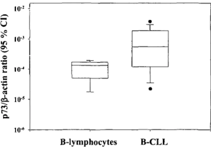
Total p73 mRNA expression was measured quantita-tively by real-time RT-PCR using p73 primers spanning exon 5 and 6, a region without any known splice sites [6-8]. The expression of total p73 mRNA was signifi-cantly higher in the 51 B-CLL patient samples compared to a population of normal CD19+-B-lymphocytes ob-tained from eight individuals (Figure 1, P - 0.01). Among particular B-CLL risk groups, the expression of total p73 mRNA was higher in the intermediate risk
(P = 0.013) and in the high risk B-CLL group (P =
0.007) than in normal B-lymphocytes, but there was no difference between the low risk and the high risk/im-mune group (data not shown in Figure 1). The cell lines JVM-2 and K562 expressed high levels of p73 mRNA whereas expression was very low or undetectable in RPMI-8402, Kasumi-1 and Jurkat (data not shown).
a.
S5 a 10J io-3 1 0J io-5 1(H -T1
_l_ B-lymphocytes B-CLLFigure I. Absolute total p73 and p-actin RNA transcript numbers as
assessed by real-time quantitative RT-PCR in a population of normal CD19.+-B-lymphocytes and B-CLL samples. Results are depicted as the 95% confidential interval (CI) of the ratio of log values of the mean. Total p73 mRNA expression in B-CLL (all risk groups) is significantly higher than in normal B-lymphocytes (P = 0.01).
Normal
B-lymphocytes
B-CLL
low risk
B-CLL
high risk
00 Vi Vi o o o o o o o o Z Z Z Z Z Z Z Z Z Z Z Z Z 7.o o o o o o o o o o o o ?: Z Z Z Z ?-.ttvi
p73u ap73 C-terminal
mRNA splice variants
GAPDH mRNA
p73 protein analysis
actin
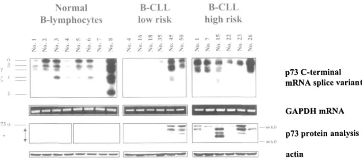
Figure 2. Expression of p73 C-terminal mRNA splice variants and p73 protein analysis of a population of normal CD19+-B-lymphocytes and
representative samples of patients with B-CLL from two different risk groups. Each lane represents p73 mRNA and protein data from a particular sample. By increasing the exposure time, all p73 C-terminal mRNA splice variants outlined in Table 1 can be detected. 'Additional proteins other than p73a.
Table I. Expression of the p73 gene in a population of normal CD19+-B-lymphocytes. patients with B-CLL. and cell lines. See 'Materials and
methods' for details about different B-CLL risk groups.
i
Normal control B-lymphocytes B-CLL Low risk Intermediate risk High risk/immune High risk Total B-CLL Cell lines JVM-2 RPM1-8402 Jurkat Kasumi-1 K562n
p73 C-terminal mRNA splice variants No. of samples 8 6 27 6 12 51 ota 7 4 24 4 12 44 X X X X X pa 8 4 24 3 11 42 X
o
o
X X Ya 8 4 15 2 9 30 Xo
o
o
X Ea 7 3 20 2 10 35 Xo
o
o
X 1 0 2 1 7 10 Xo
o
o
o
0" 4 0 0 0 2 2 O Oo
o
o
aa 5 1 6 1 8 16 Xo
Xo
o
n"
4 1 3 0 3 7 X Oo
o
o
p73 protein analysis No. of samples 8 6 27 6 12 51 p73aa 1 2 17 4 10 33 X Xo
o
X Addi-tional15 8 3 22 4 12 41 X Oo
o
X"' Number of samples in which a particular C-terminal mRNA splice variant or protein was detected: cell lines- X - detected; O - not detected; b In addition to p73a as shown in Figure 2. p73 mRNA C-terminal splice variants y (P = 0.041). G (P < 0.001). and the r) (P = 0.033) prevailed in normal B-lymphocytes. Prevalent detection of the p73a protein in B-CLL samples compared to normal B-lymphocytes (P = 0.008) and positive correlation with higher risk B-CLL stages (P = 0.046).
The expression ofp73 C-terminal mRNA splice variants is biased
The investigation of p73 C-terminal mRNA splice var-iants by RT-PCR showed that many different varvar-iants were expressed in normal and malignant samples. The majority of both B-CLL and normal B-lymphocyte samples expressed the larger p73 C-terminal splice var-iants oc, p, and e (Table 1 and Figure 2). The C, variant was detected in a subset of B-CLL (10 of 51) and normal B-lymphocytes (1 of 8) only. The y variant was more frequently expressed in normal B-lymphocytes than in B-CLL (P = 0.041) as were the shorter mRNA splice
variants 6 (P < 0.001) and r\ {P = 0.033). Table 1 summarises the expression of p73 mRNA C-terminal splice variants in the lymphoid and the myeloid cell lines included in our study. In contrast to published data [14], RPM1-8402 was found to express p73a mRNA, and Kasumi-1 expressed the p73 mRNA variants a and [3 (Table 1).
p73a protein expression is positively correlated with higher risk B-CLL stages
p73 protein expression was investigated by Western blot using a rabbit polyclonal antibody [5]. We detected the
p73a protein in 33 of 51 B-CLL patient samples, but only in one of eight samples of normal B-lymphocytes
(P = 0.008). Ten of twelve samples from high risk, but
only two of six samples from low risk B-CLL patients expressed detectable levels of the p73a protein (Table 1 and Figure 2). Sixteen of 51 B-CLL and 7 of 8 B-lym-phocyte samples expressed total p73 mRNA but no p73a protein, and in 2 of 51 B-CLL samples, neither was found. Detection of the p73oc protein was positively correlated with higher risk B-CLL stages (P = 0.046).
Western blot analysis revealed a complex pattern of proteins (Table 1, Figure 2). The p73oc protein was identified by its comigration with the in vitro translated p73a product (data not shown). None of the additional smaller bands comigrated with in vitro translated p73(3 which is also detected by the polyclonal antibody used (data not shown). In normal B-lymphocyte samples, a band of approx. 56 kD was present and was the only signal in 7 of 8 B-lymphocyte samples (Figure 2). This band was not exclusive to normal B-lymphocytes, but was also detected in B-CLL samples. In a subset of B-CLL samples of different risk groups, additional bands were detected in the absence of p73a (data not shown). The polyclonal antibody did not detect any proteins in the K.asumi-1 and the Jurkat cell lines which both express p73 mRNA at very low or undetectable levels as measured by real-time RT-PCR (data not shown).
Discussion
The results of this survey on the expression of p73 in B-CLL and normal CD19+- B-lymphocytes have re-vealed several novel features. In B-CLL, total p73 mRNA expression is significantly higher than in a nor-mal B-lymphocyte population. A detailed analysis of the p73 C-terminal mRNA splice variants yields a biased expression pattern since the shorter p73 variants y, 9, and r| prevail in the normal B-lymphocyte population studied. p73a protein expression is significantly more frequent in B-CLL samples than in normal B-lympho-cytes. Importantly, the expression of the p73a protein is positively correlated with higher risk B-CLL stages.
It was suggested that p73 acts as a tumour suppressor gene in ALL and Burkitt's lymphoma and loss of its function would contribute to the development and the progression of such lymphoid neoplasms [8, 13, 14]. In contrast to these reports and in agreement with prior investigations in hepatocellular carcinoma [18] and bladder cancer [19], we have shown that in B-CLL, p73 expression is positively correlated with disease progres-sion. This supports a possible oncogenic role of p73 in B-CLL.
The differences in the expression pattern of p73 be-tween B-CLL, B-NHL and ALL may reflect differences in the biology of these groups of lymphoid malignancies. A link of the expression of the p73 gene to lymphoid differentiation is possible. The p73 gene is overexpressed
in progressive B-CLL which is characterized by an accumulation of non-proliferating, senescent cells with disrupted apoptosis [2, 28]. In mature ALL, lack of p73 expression was suggested to contribute to the high pro-liferation rate [8]. p73 overexpression was initially shown to induce cell cycle and growth arrest as well as apoptosis in a series of cell lines [5, 6, 10]. Interestingly, recent reports on other cell lines confirmed the induc-tion of cell cycle and growth arrest by overexpression of p73, but without apoptosis and in the presence of repli-cative senescence [9, 29]. Thus, the effect of p73 expres-sion may be tissue-specific [5, 6, 30]. This may partially explain the apparent paradox that this gene is overex-pressed in a variety of malignancies with disrupted apoptosis including B-CLL whilst inducing apoptosis in other cell types.
Earlier reports on malignancies other than B-CLL suggested that instability in the splicing of p73 exons preferentially occurs in cancer cells [11, 26]. We found that various p73 mRNA splice variants are expressed in both B-CLL and normal B-lymphocyte samples, but in different patterns. Shorter p73 mRNA splice variants prevailed in normal B-lymphocytes, although none of the p73 variants was restricted to either normal or malignant samples in our series. The shorter p73 splice variants such as y, e, 5 are less effective in activating p53-responsive genes than p73a [6]. The recently identi-fied p73 variants 0, r\, and r)l lack the p73 C-terminal transactivation domain and the region of homology with the p53 oligomerization domain [8]. Some of the p73 variants exhibit p53-like functions or inhibit p53 while others may carry out yet unknown p73-specific functions [31]. In addition to the C-terminal splice var-iants, 2 different N-terminal p73 mRNA variants have recently been described [5, 16]. In mice, one of them, the ANp73 variant, was shown to act as a dominant neg-ative inhibitor of p73a and p53 [16, 32]. These data suggest that the balance of expressed p73 variants may determine the function of the protein. E2F-1 is a tran-scriptional factor which can act as an oncogene or a tumour-suppressor gene [33]. The recently reported interactions of p73 with E2F1 [33] will help to clarify the role of p73 as a pro- or antiapoptotic actor in malignant diseases.
Earlier investigations on p73 mRNA expression in clinical samples have shown that p73 is overexpressed in a variety of malignancies with respect to their normal cellular counterparts [11, 25, 26]. Our data suggest that this is true for B-CLL with respect to normal CD19+-B-lymphocytes. Expression of the p73 gene may differ between various other subpopulations of B cells including CD5+-B-cells, which have been purported to represent normal counterparts of B-CLL cells [34, 35]. A compre-hensive survey of p73 expression in all normal B-cell subtypes as well as other B-cell malignancies expressing the CD5 marker such as mantle cell lymphomas would be of some interest and warrant further investigation. Overexpression of the p73 gene in B-CLL is unlikely to be linked to treatment since only a minority of our
patients (8 of 51) were under treatment at the time of sample collection and the regimens did not contain drugs known to up-regulate p73 [11].
In agreement with prior reports [5, 12, 24, 25], p73 protein western analysis revealed the presence of several additional protein bands in all types of samples included in our series (Table 1, Figure 2). To investigate the nature of these additional proteins, we treated three different cell lines (JVM-2, MCF-7, and Calu-1) with N-acetyl-leucinyl-leucinyl-norleucinal (LLnL, MG-101, Sigma; Buchs, Switzerland) which inhibits ubiquitin-dependent protein degradation by the proteasome [12]. Evidence for protein degradation was only found in the JVM-2 cell line. The treatment with LLnL led to a transient band shift in JVM-2 as previously shown for other cell lines [12] (data not shown). The additional protein bands seen are currently under investigation, and may repre-sent either p73 variants or posttranscriptional modifica-tions of p73. New antibodies are needed to identify all known p73 variants.
In summary, we conclude that the accumulation of p73, the expression pattern of particular p73 variants and its link to progression may play a distinct role in the molecular pathology B-CLL.
Acknowledgements
We express our appreciation to Mrs. M. Oestreicher and B. Hiigli for excellent technical assistance, and to A. Marti and J. Schwaller for critical review of the manuscript. We thank D. Caput for the anti-p73 rabbit polyclonal serum, V. Delaurenzi for the p73 plasmids, and S. Miescher for the spleen and tonsil samples. This work was supported by grants from the Swiss National Foundation (3100-043458.95/1), from the Swiss Cancer League (KFS 156-9-1995), the Ursula-Hecht-Stiftung and the Deutsche Jose Carreras-Leukamie-Stiftung.
References
1. Reed JC. Molecular biology of chronic lymphocytic leukemia: Implications for therapy. Semin Hematol 1998; 35' 3-13 2. Dohner H, Stilgenbauer S, Benner A et al. Genomic aberrations
and survival in chronic lymphocytic leukemia. N Engl J Med 2000; 343: 1910-6.
3. Cordone 1, Masi S, Mauro FR et al p53 expression in B-cell chronic lymphocytic leukemia: A marker of disease progression and poor prognosis. Blood 1998; 91: 4342-9
4. Kaghad M, Bonnet H, Yang A, et al. Monoallehcally expressed gene related to p53 at Ip36, a region frequently deleted in neuro-blastoma and other human cancers. Cell 1997; 90: 809-19. 5. De Laurenzi V, Costanzo A, Barcaroli D et al. Two new p73 splice
variants, gamma and delta, with different transcriptional activity J Exp Med 1998; 188: 1763-8.
6. De Laurenzi VD, Catani MV, Terrinoni A et al. Additional complexity in p73: Induction by mitogens in lymphoid cells and identification of two new splicing variants c and {,. Cell Death Differ 1999, 6. 389-90.
7 Scaruffi P, Casciano I, Masiero L et al. Lack of p73 expression in mature B-ALL and identification of three new splicing variants
restricted to pre B- and C-ALL indicate a role of p73 in B cell ALL differentiation. Leukemia 2000; 14: 518-9.
8. Fang L, Lee SW. Aaronson SA. Comparative analysis of p73 and p53 regulation and effector functions. J Cell Biol 1999: 147: 823-30.
9. Jost CA. Marin MC. Kaelin WG, Jr. p73 is a simian p53-related protein that can induce apoptosis. Nature 1997; 389: 191-4. 10. Levrero M. De Laurenzi V, Costanzo A et al. The p53/p63/p73
family of transcription factors: Overlapping and distinct func-tions. J Cell Sci 2000; 113: 1661-70.
11 Balint E, Bates S. Vousden KH. Mdm2 binds p73 alpha without targeting degradation. Oncogene 1999; 18: 3923-9.
12. Corn PG. Kuerbitz SJ. van Noesel MM et al. Transcriptional silencing of the p73 gene in acute lymphoblastic leukemia and Burkitt's lymphoma is associated with 5' CpG island methylation. Cancer Res 1999; 59- 3352-6.
13. Kawano S, Miller CW. Gombart AF et al. Loss of p73 gene expression in leukemias/lymphomas due to hypermethylalion. Blood 1999; 94: 1113-20.
14. Stirewalt DL, Clurman B. Appelbaum FR et al. p73 mutations and expression in adult de novo acute myelogenous leukemia. Leukemia 1999: 13: 985-90.
15. Yang A, Walker N. Bronson R et al. p73-deficient mice have neurological, pheromonal and inflammatory defects but lack spontaneous tumours. Nature 2000: 404: 99-103.
16. Vikhanskaya F. D'lncalci M, Broggini M. p73 competes with p53 and attenuates its response in a human ovarian cancer cell line. Nucleic Acids Res 2000: 28. 513-9.
17. Tannapfel A.Wasner M. Krause K el al. Expression of p73 and its relation to histopathology and prognosis in hepalocellular carci-noma. J Natl Cancer Inst 1999; 91: 1154-8.
18. Chi SG, Chang SG. Lee SJ et al. Elevated and biallelic expression of p73 is associated with progression of human bladder cancer. Cancer Res 1999. 59:2791-3.
19. Cheson BD. Bennett JM. Grever M et al. National Cancer Institute-sponsored working group guidelines for chronic lym-phocytic leukemia. Revised guidelines for diagnosis and treat-ment. Blood 1996; 87: 4990-7.
20 Matutes E. Owusu-Ankomah K. Monlla R ct al. The immuno-logical profile of B-cell disorders and proposal of a scoring system for the diagnosis of CLL. Leukemia 1994; 8: 1640-5. 21. Rai K.R. A critical analysis of staging in CLL. In Gale RP,
Rai KR (eds)1 Chronic Lymphocytic Leukemia. Recent Progress and Future Directions. New York: Wiley-Liss 1987; 253ff. 22. Geisler C. Hansen MM. Chronic lymphocytic leukaemia: A test
of a proposed new clinical staging system. Scand J Haematol 1981; 27-279-86.
23. Zwahlen D. Tschan MP. GrobTJ et al. Differential expression of p73 splice variants and protein in benign and malignant ovarian tumours, hit J Cancer 2000: 88: 66-70.
24. Peters (JR. Tschan MP, Kreuzer KA et al. Distinct expression patterns of the p53-homologue p73 in malignant and normal hematopoiesis assessed by a novel real-time reverse transenption-polymerase chain reaction assay and protein analysis. Cancer Res
1999; 59: 4233-6.
25. Tschan MP, GrobTJ. Peters UR et al. Enhanced p73 expression during differentiation and complex p73 isoforms in myeloid leu-kemia. Biochem Biophys Res Commun 2000: 277: 62-5. 26. Mauro FR, Foa R, Cerretti R et al. Autoimmune hemolytic
anemia in chronic lymphocytic leukemia: Clinical, therapeutic, and prognostic features. Blood 2000; 95: 2786-92.
27. Trentin L. Ballon G. Ometto L et al. Telomerase activity in chronic lymphoproliferative disorders of B-cell lineage. Br J Haematol 1999: 106: 662-8.
28. De Laurenzi V. Raschella G, Barcaroli D et al. Induction of neuronal differentiation by p73 in a neuroblastoma cell line. J Biol Chem 2000: 275: 15226-15231.
29 Loiseau H, Arsaut J. Demotes-Mainard J. p73 gene transcripts in human brain tumors: Overexpression and altered splicing in ependymomas. Neurosci Lett 1999; 263. 173-6.
30 Mann MC. Kaelin WG. p63 and p73: Old members of a new family. Biochim Biophys Acta 2000; 1470: M93-M100.
31. Pozniak CD. Radinovic S.Yang A et al. An anti-apoptotic role for the p53 family member. p73, during developmental neuron death. Science 2000; 289: 304-6.
32. Stiewe T, Piitzer BM Role of the p53-homologue p73 in E2F1-induced apoptosis. Nat Genet 2000; 26: 464-9.
33. Caligans-Cappio F. Hamblin TJ. B-cell chronic lymphocytic leukemia: A bird of a different feather. J Clin Oncol 1999; 17-399-408.
34. Lydyard PM. Jewell AP. Jamin C et al. CD5 B cells and B-cell malignancies Curr Opin Hematol 1999; 6: 30-6.
Received 2 November 2000: accepted 26 March 2001.
Correspondence to:
M. F. Fey. MD
Institute of Medical Oncology University of Berne, Inselspital 3010 Berne
Switzerland