HAL Id: hal-02384917
https://hal.archives-ouvertes.fr/hal-02384917
Submitted on 8 Feb 2021
HAL is a multi-disciplinary open access
archive for the deposit and dissemination of
sci-entific research documents, whether they are
pub-lished or not. The documents may come from
teaching and research institutions in France or
abroad, or from public or private research centers.
L’archive ouverte pluridisciplinaire HAL, est
destinée au dépôt et à la diffusion de documents
scientifiques de niveau recherche, publiés ou non,
émanant des établissements d’enseignement et de
recherche français ou étrangers, des laboratoires
publics ou privés.
Further Studies on the Role of Metabolites in
(
±)-3,4-Methylenedioxymethamphetamine-Induced
Serotonergic Neurotoxicity
Melanie Mueller, Jie Yuan, Anne Felim, Anne Neudörffer, Frank Peters, Hans
Maurer, Una Mccann, Martine Largeron, George Ricaurte
To cite this version:
Melanie Mueller, Jie Yuan, Anne Felim, Anne Neudörffer, Frank Peters, et al.. Further Studies on the
Role of Metabolites in (
±)-3,4-Methylenedioxymethamphetamine-Induced Serotonergic Neurotoxicity.
Drug Metabolism and Disposition, American Society for Pharmacology and Experimental
Therapeu-tics (ASPET), 2009, 37 (10), pp.2079-2086. �10.1124/dmd.109.028340�. �hal-02384917�
Further Studies on the Role of Metabolites in (
ⴞ)-3,4-Methylenedioxymethamphetamine-Induced
Serotonergic Neurotoxicity
Melanie Mueller, Jie Yuan, Anne Felim, Anne Neudo¨rffer, Frank T. Peters, Hans H. Maurer,
Una D. McCann, Martine Largeron, and George A. Ricaurte
Departments of Neurology (M.M., J.Y., G.A.R.) and Psychiatry and Behavioral Sciences (U.D.M.), Johns Hopkins University School of Medicine, Baltimore, Maryland; Unite´ Mixte de Recherche 8638 Associe´e au Centre National de la Recherche
Scientifique, Universite´ Paris Descartes, Paris, France (A.F., A.N., M.L.); and Department of Experimental and Clinical Toxicology, Institute of Experimental and Clinical Pharmacology and Toxicology, Saarland University, Homburg, Germany
(M.M., F.T.P., H.H.M.)
Received April 30, 2009; accepted July 21, 2009
ABSTRACT:
The mechanism by which the recreational drug ( ⴞ)-3,4-methyl-enedioxymethamphetamine (MDMA) destroys brain serotonin (5-HT) axon terminals is not understood. Recent studies have implicated MDMA metabolites, but their precise role remains unclear. To further evaluate the relative importance of metabolites versus the parent compound in neurotoxicity, we explored the relationship between pharmacokinetic parameters of MDMA, 3,4-methylenedioxyam-phetamine (MDA), 3,4-dihydroxymetham3,4-methylenedioxyam-phetamine (HHMA), and 4-hydroxy-3-methoxymethamphetamine (HMMA) and indexes of serotonergic neurotoxicity in the same animals. We also further evaluated the neurotoxic potential of 5-(N-acetylcystein-S-yl)-HHMA (5-NAC-5-(N-acetylcystein-S-yl)-HHMA), an MDMA metabolite recently implicated in 5-HT neurotoxicity. Lasting serotonergic deficits correlated strongly with pharmacokinetic parameters of MDMA (Cmax and
area under the concentration-time curve), more weakly with those
of MDA, and not at all with those of HHMA or HMMA (total amounts of the free analytes obtained after conjugate cleavage). HHMA and HMMA could not be detected in the brains of animals with high brain MDMA concentrations and high plasma HHMA and HMMA concentrations, suggesting that HHMA and HMMA do not readily penetrate the blood-brain barrier (either in their free form or as sulfate or glucuronic conjugates) and that little or no MDMA is metabolized to HHMA or HMMA in the brain. Repeated intraparen-chymal administration of 5-NAC-HHMA did not produce significant lasting serotonergic deficits in the rat brain. Taken together, these results indicate that MDMA and, possibly, MDA are more important determinants of brain 5-HT neurotoxicity in the rat than HHMA and HMMA and bring into question the role of metabolites (including 5-NAC-HHMA) in MDMA neurotoxicity.
Despite much research, the mechanism by which ( ⫾)-3,4-methyl-enedioxymethamphetamine (MDMA, Ecstasy) destroys brain seroto-nin (5-HT) axon terminals remains unknown. One hypothesis that has recently drawn considerable attention is that a drug metabolite is involved (Capela et al., 2009; Perfetti et al., 2009). Over the years, various metabolites of MDMA and related drugs have been evaluated for possible 5-HT neurotoxic activity (Fig. 1). Steele et al. (1991), for example, assessed the neurotoxic potential of
3,4-dihydroxymetham-phetamine (HHMA; which they designated ␣-methylepinine) and concluded that it alone was not responsible for MDMA neurotoxicity. Likewise, McCann and Ricaurte (1991) evaluated the neurotoxic properties of␣-methyldopamine and 3-O-methyl-␣-methyldopamine [alternatively designated as 3,4-hydroxyamphetamine (HHA) and 4-hydroxy-3-methoxyamphetamine, respectively] and reached similar conclusions.
Based on theoretical considerations and the identification of MDMA metabolites that are analogs of the well established neuro-toxin 6-hydroxydopamine (Lim and Foltz, 1991a,b), the neurotoxico-logical properties of 6-hydroxy-3,4-methylenedioxymethamphet-amine and 2,4,5-trihydroxymethamphetamine (THMA) were investigated (Johnson et al., 1992; Zhao et al., 1992). 6-Hydroxy-3,4-methylenedioxymethamphetamine, administered intraventricularly and intraparenchymally, was found to be without effect. In contrast, THMA (same routes of administration) produced substantial
deple-This work was supported by the National Institutes of Health National Institute on Drug Abuse [Grants DA05707, DA01796401]; and jointly by the Mission Interministe´rielle de Lutte contre la Drogue et la Toxicomanie (MILDT) and Institut National de la Sante´ et de la Recherche Me´dicale (INSERM) [Appel a` projets commun 2007 MILDT-INSERM “Recherche sur les drogues et la toxicomanie”]. Article, publication date, and citation information can be found at http://dmd.aspetjournals.org.
doi:10.1124/dmd.109.028340.
ABBREVIATIONS: MDMA, 3,4-methylenedioxymethamphetamine; 5-HT, serotonin; HHMA, 3,4-dihydroxymethamphetamine; HHA,
dihy-droxyamphetamine; THMA, 2,4,5-trihydroxymethamphetamine; SKF-525A, 2-diethylaminoethyl 2:2-diphenylvalerate hydrochloride; MDA, 3,4-methylenedioxyamphetamine; 5-NAC-HHMA, 5-(N-acetylcystein-S-yl)-3,4-dihydroxymethamphetamine; HMMA, 4-hydroxy-3-methoxymetham-phetamine; 5-HIAA, 5-hydroxyindol acetic acid; 5,7-DHT, 5,7-dihydroxytryptamine; SMBS, sodium metabisulfite; LC-MS, liquid chromatography-mass spectrometry; AUC, area under the curve; aCSF, artificial cerebrospinal fluid.
DRUGMETABOLISM ANDDISPOSITION Vol. 37, No. 10
Copyright © 2009 by The American Society for Pharmacology and Experimental Therapeutics 28340/3517523
DMD 37:2079–2086, 2009 Printed in U.S.A.
tions of 5-HT and dopamine that lasted for at least 5 to 7 days beyond drug administration (Johnson et al., 1992; Zhao et al., 1992). Given the known selectivity of MDMA for 5-HT neurons, results with THMA were deemed to be inconclusive but suggestive of the possi-bility that THMA may play a role in MDMA neurotoxicity, because the effect on dopamine may have been related to the route of THMA administration (Zhao et al., 1992).
As mentioned above, there has recently been a resurgence of interest in the possibility that MDMA metabolites might play a role in MDMA neurotoxicity. A role for systemically formed MDMA me-tabolites is often inferred from the observation that direct injection of MDMA into brain fails to reproduce the 5-HT neurotoxic effects of peripherally administered MDMA (Schmidt and Taylor, 1988; Es-teban et al., 2001). A report that cytochrome P450 modulators (SKF-525A and phenobarbital) influence MDMA-induced 5-HT depletions (Gollamudi et al., 1989) is also often cited to support the role of a drug metabolite. However, in that study, SKF-525A and phenobarbital altered acute (3 h) but not lasting effects of MDMA on brain 5-HT neurons.
Hiramatsu et al. (1990) were the first to report metabolism of MDMA to a reactive quinone, which formed a glutathione adduct that might be responsible for MDMA neurotoxicity. More recently, other glutathione and N-acetylcysteine conjugates of catechol metabolites of MDMA and MDA have been identified and implicated in MDMA neurotoxicity (Miller et al., 1997; Bai et al., 1999; Jones et al., 2005; Capela et al., 2007; Pizarro et al., 2008). Of these,
5-(N-acetylcystein-S-yl)-N-methyl-␣-methyldopamine [here designated as
5-(N-acetyl-cystein-S-yl)-HHMA (5-NAC-HHMA)] has been the metabolite most strongly implicated (Jones et al., 2005; Erives et al., 2008).
MDMA metabolism proceeds mainly through two pathways at different rates in different species (Meyer et al., 2008). The first pathway involves O-demethylenation of MDMA to HHMA, followed by O-methylation to 4-hydroxy-3-methoxymethamphetamine (HMMA), with subsequent O-conjugation with sulfate or glucuronic acid. The second pathway involves initial N-demethylation to MDA, followed by deamination and oxidation to the corresponding benzoic acid deriva-tives conjugated with glycine. As mentioned above, catechol metabolites of MDMA and MDA (HHMA and HHA) can be further oxidized to their corresponding quinones, which can then form adducts with glutathione and other thiol-containing compounds (Hiramatsu et al., 1990; Monks et al., 2004).
The purpose of the present study was severalfold: 1) to assess the relative importance of the parent compound (MDMA) versus its major metabolites (HHMA, HMMA, and MDA) in MDMA neurotoxicity;
2) to determine which pharmacokinetic parameter of MDMA or its metabolites best predicts subsequent 5-HT neurotoxicity; and 3) to further assess the 5-HT neurotoxic potential and selectivity of the catechol thioether, 5-NAC-HHMA.
Materials and Methods
Animals. Male Sprague-Dawley rats (Harlan, Indianapolis, IN) that were 49
to 69 days of age and weighed 200 to 299 g were used for all experiments. Animals were housed three per cage (except during drug treatment and after surgical cannula implantation, when they were housed singly) in standard polypropylene cages (17 inches⫻ 10 inches ⫻ 8 inches) at 22 ⫾ 2°C ambient temperature (except during drug treatment, when the ambient temperature was 25°C), with free access to food and water. Animals were maintained on a 12:12-h light/dark cycle. The facilities for housing and care of the animals are accredited by the American Association for the Assessment and Accreditation of Laboratory Animal Care. Animal care and experimental manipulations were approved by the Institutional Animal Care and Use Committee at the Johns Hopkins University School of Medicine and were in accordance with the National Institutes of Health Guide for the Care and Use of Laboratory
Animals (Institute of Laboratory Animal Resources, 1996).
Study Design. To assess the relationship between MDMA and its major
metabolites (MDA, HHMA, and HMMA) and brain 5-HT neurotoxicity, pharmacokinetic parameters of MDMA, HHMA, HMMA, and MDA were measured during the period of drug exposure and related to indexes of brain 5-HT neurotoxicity (depletions of 5-HT and 5-HIAA) measured 1 week later in the same animals. The reason for the 1-week delay was that MDMA and metabolites are known to alter 5-HT and 5-HIAA levels acutely (by inducing 5-HT release, by blocking its reuptake and, possibly, by blocking metabolism of 5-HT by monoamine oxidase). Thus, “acute” depletions of 5-HT and 5-HIAA may not necessarily reflect neurotoxicity. Indeed, Chu et al. (1996) have already shown that there is no relationship between acute depletions of 5-HT and brain MDMA and metabolite levels, probably because at least some of the perturbations seen in 5-HT levels while MDMA is still in the tissue are related to pharmacological (rather than toxic) effects of MDMA on the 5-HT neuron. In contrast, depletions of 5-HT and 5-HIAA documented at least 1 week after drug exposure (when drug and metabolites are no longer on board) are known to be related to 5-HT axon loss (Molliver et al., 1990; Ricaurte et al., 1992). To further assess the 5-HT neurotoxic potential of 5-NAC-HHMA, the compound was administered directly into the striatum at a dose and frequency previously reported to produce lasting 5-HT deficits. Possible in-volvement of the 5-HT transporter in the anticipated 5-HT deficits was as-sessed with fluoxetine, a 5-HT uptake blocker that is known to protect against MDMA neurotoxicity (Schmidt, 1987).
Drugs and Reagents. Racemic MDMA hydrochloride was obtained
through the National Institute on Drug Abuse (Rockville, MD). Racemic HHMA hydrochloride and methanolic solutions (1000 mg/l) of racemic MDMA hydrochloride and racemic MDA hydrochloride were purchased from Lipomed (Cambridge, MA). Methanolic solutions (1000 mg/l) of racemic
FIG. 1. Metabolites of MDMA and related drugs that have been evaluated for 5-HT neurotoxic potential:1Ricaurte et al. (1985), 2
Zhao et al. (1992),3
Johnson et al. (1992),4
Elayan et al. (1992),
5Steele et al. (1991),6Escobedo et al. (2005), and7McCann and
Ricaurte (1991). With the exception of MDA, the only other MDMA metabolite of the various metabolites shown known to have 5-HT neurotoxic potential is THMA. However, recent efforts in our laboratory to identify THMA in the brain of rats given neurotoxic doses of MDMA (20 – 60 mg/kg p.o.) have been unsuccessful, even though THMA given intracerebroventricularly can be readily mea-sured in the rat brain for up to 3 h after administration using LC-MS methods (M. Mueller and G. Ricaurte, unpublished observations). 6-OHMDMA, 6-hydroxy-3,4-methylenedioxymethamphetamine; HMA, hydroxymethamphetamine.
HMMA and methanolic solutions (100 mg/l) of racemic MDMA-d5 and
MDA-d5were obtained from Cerilliant Corporation (Round Rock, TX).
Flu-oxetine, 4-hydroxymethamphetamine (pholedrine), 4-methylcatechol, EDTA, 5,7-dihydroxytryptamine (5,7-DHT), and glucuronidase type HP-2 from Helix
pomatia (glucuronidase activity ⱖ100,000 units/ml and sulfatase activity
⬍7500 units/ml) were obtained from Sigma-Aldrich (St. Louis, MO). Sodium metabisulfite (SMBS) was obtained from Merck (Darmstadt, Germany). Per-chloric acid was obtained from Mallinckrodt Baker, Inc. (Phillipsburg, NJ). Xylazine was obtained from Butler Animal Health Supply (Dublin, OH). Ketamine was supplied by Phoenix Pharmaceuticals (St. Joseph, MO). 5-NAC-HHMA was synthesized as described recently (Felim et al., 2007). The authenticity of the MDMA, HHMA, HMMA, MDA, and 5-NAC-HHMA samples used in the present studies was confirmed using liquid chromatogra-phy-mass spectrometry (LC-MS) methods to determine the corresponding pseudomolecular ions and at least one fragment ion for each compound. Analysis was performed in full scan (mass range from 100 to 1000) to check for presence of possible impurities.
Drug Treatment. MDMA was given orally (by gavage) at a dose of 20
mg/kg at an ambient temperature of 25°C. For studies involving intrastriatal administration of 5-NAC-HHMA, rats received four consecutive doses of either 21 or 42 nmol of the compound, with a 12-h interval between each dose. This particular dose regimen was selected because it is the same one that was used by Bai et al. (1999), who first reported on the neurotoxic potential of 5-NAC-HHMA. In an additional experiment involving 5-NAC-HHMA, rats were pretreated with 10 mg/kg fluoxetine (i.p.) 15 min before each intrastriatal injection of 21 nmol of 5-NAC-HHMA. In the latter experiment, the well established selective 5-HT neurotoxin, 5,7-dihydroxytryptamine (5,7-DHT) was used as a positive control and was also given intrastriatally, at a dose of 52 nmol. Doses refer to the base form of all drugs.
Blood Sampling and Plasma Preparation. For determination of plasma
concentrations of MDMA (and its metabolites) and their pharmacokinetic profiles, blood was sampled at various times after MDMA administration. Blood collection times were selected to allow for accurate determinations of drug pharmacokinetic parameters. For logistic reasons, blood was sampled at 0.75, 1.5, 3, 6, and 12 h after MDMA administration in one group of animals (n⫽ 15); in a second group of rats (n ⫽ 9), blood was collected at 1, 3, 6, 8, 9, and 24 h after MDMA treatment. At each time point, approximately 0.2 ml of blood was collected by means of retro-orbital bleeding. One week after MDMA treatment, all animals were sacrificed for regional brain 5-HT and 5-HIAA determinations, as detailed below. A third group of animals (n⫽ 8 at each time point) was used for determination of plasma and brain concentra-tions of MDMA (and its metabolites). In this group of animals, the major point of interest was the relationship between plasma and brain MDMA and metab-olite concentrations. Blood sampling in this experiment occurred at 1, 3, 6, 8, and 24 h MDMA treatment. Blood samples were dispensed into 2-ml BD Vacutainer hematology tubes, containing 4 mg of K3 EDTA solution (BD Biosciences, Franklin Lakes, NJ) and stored on ice for up to 30 min until centrifuged. Samples were centrifuged at 1100g for 10 min. Plasma was withdrawn using a 5-ml 3/4 Pasteur pipette and decanted into a 1.5-ml polypropylene tube, and SMBS (250 mM) was added at a volume of 30l/ml plasma to minimize oxidation of the compounds of interest. Plasma samples were stored at⫺20°C until assay.
Measurement of Plasma MDMA and Metabolite Concentrations.
Plasma MDMA, MDA, HHMA, and HMMA concentrations were determined as described recently (Mueller et al., 2007). In brief, aliquots (100l) of rat plasma were preserved with 20l of SMBS (250 mM) and 10 l of EDTA (250 mM). After addition of 100l of an aqueous solution of the racemic internal standards MDMA-d5, MDA-d5, and pholedrine (1.0g/ml each) and
10l of glucuronidase solution, samples were mixed (15 s) on a rotary shaker and left at 50°C for 90 min to perform conjugate cleavage. After cooling to room temperature, 20 l of 4-methylcatechol (1 mg/ml) was added, and samples were briefly vortexed. Perchloric acid (10l) was then added, and the samples were mixed again on a rotary shaker for 15 s to perform protein precipitation. The samples were centrifuged (16,000g for 5 min), and the supernatant was transferred to autosampler vials. Aliquots (5l) were injected into an LC-MS system and amounts of MDMA and metabolites were deter-mined. The linear range for each analyte was 20 to 1000 ng/ml MDMA, HHMA, HMMA, and 10 to 500 ng/ml MDA. Method accuracy was greater
than 80%. The lowest point of the calibration curve was the limit of quanti-fication of the method (20 ng/ml for MDMA, HHMA, and HMMA each and 10 ng/ml for MDA).
Measurement of Brain MDMA and Metabolite Concentrations. For
determination of brain concentrations of MDMA, HHMA, HMMA, and MDA, samples were prepared and analyzed according to a recently published LC-MS method (Mueller et al., 2008). Values for HHMA and HMMA represent total amounts (i.e., amounts measured after cleavage of sulfate and glucuronic acid conjugates). In particular, aliquots of rat cortices (approximately 100 mg) were weighed and for each microgram of tissue, 10l of internal standards solution were added. After homogenization with a Polytron homogenization unit (model PT 10-35, 15 s, setting 6; Kinematica Inc., Bohemia, NY), 10l of glucuronidase solution were added, and the samples were briefly mixed (15 s) on a rotary shaker and left at 50°C on a waterbath for 90 min to perform conjugate cleavage. After cooling to room temperature, the samples were centrifuged (16,000g for 10 min), and the supernatant was transferred to autosampler vials. Aliquots (5l) were injected into the LC-MS system. The linear range for each analyte was 2 to 100g/g MDMA, 1 to 50 g/g MDA, and 0.1 to 5g/g HHMA and HMMA. Method accuracy was greater than 90%. The lowest point of the calibration curve was defined as the limits of quantitation of the method (2g/g for MDMA, 1 g/g for MDA, and 0.1 g/g for HHMA and HMMA). Values for HHMA and HMMA represent free amounts (i.e., amounts measured after cleavage of sulfate and glucuronic acid conjugates).
Calculation of Pharmacokinetic Parameters. Peak plasma concentrations
(Cmax), times of peak plasma concentration (Tmax), area under the
concentra-tion-time curve (AUC), and the elimination half-lives (t1/2) were obtained
using the pharmacokinetic functions for Microsoft Excel (developed by J. L. Usansky, A. Desai, and D. Tang-Liu, http://www.boomer.org/pkin/xcel/pkf/ pkf.doc).
Surgical Cannula Implantation. Animals were anesthetized with xylazine
(25 mg/kg, i.p.) and ketamine (35 mg/kg i.p.). Guide cannulae (20 gauge; Plastic One, Roanoke, VA) were surgically implanted into the right striatum [anteroposterior, 0.4 mm; mediolateral,⫺3.0 mm; dorsoventral, 4.0 mm (Paxi-nos and Watson, 1986)]. Cannulae were fixed to the skull with dental acrylic (Ortho-Jet, Lang Dental, Wheeling, IL) and two stainless steel screws. Dummy cannulae were placed in the guide cannulae, and animals were individually housed and allowed a 7-day recovery period.
Intrastriatal Administration of 5-NAC-HHMA. The dummy cannulae
were replaced with internal cannulae (24 gauge; Plastic One) connected to PE 20 tubing that in turn were connected to a 1-l Hamilton 7000 series glass syringe (Hamilton Co., Reno, NV) containing the various injection solutions. Artificial cerebrospinal fluid (aCSF) served as a vehicle control and was prepared as described previously by Miller et al. (1997). In a first experiment, rats received either 1 l of aCSF (control group, n ⫽ 8), 21 nmol of 5-NAC-HHMA (n⫽ 10), 42 nmol of 5-NAC-HHMA (n ⫽ 5), or 52 nmol of 5,7-DHT (positive control group, n⫽ 4). In a second experiment, animals were pretreated with either saline (0.3 ml) or fluoxetine (15 mg/kg i.p.) 15 min before intrastriatal injections. After pretreatment, either 1l of aCSF or 21 nmol of 5-NAC-HHMA were injected into the striatum (total four groups, n⫽ 6 in each group). One microliter of the drug solution was injected manually into the striatum [anteroposterior, 0.4 mm; mediolateral,⫺3.0 mm; dorsoven-tral, 5.0 mm (Paxinos and Watson, 1986)] at a rate of 0.2l over 5 min for a total of four consecutive doses, with each dose administered 12 h apart. After the injection was completed, the internal cannulae were left in the striatum for an additional 2 min. Animals were awake but gently restrained during the injections. After injection, the dummy cannulae were replaced. Animals were sacrificed 2 weeks later for determination of 5-HT and 5-HIAA levels, as described below.
Determination of Brain 5-HT and 5-HIAA Concentrations. Samples of
cortex and striatum were analyzed for their content of 5-HT and 5-HIAA 1 or 2 weeks after drug treatment, as described previously (Mechan et al., 2006).
Statistics. The significance of differences between means was determined
using a two-tailed Student’s t test or analysis of variance followed by Tukey’s multiple comparison test. Correlations were explored using Pearson’s product moment correlation. Statistical analyses were performed using Prism (version 3.02; GraphPad Software Inc., San Diego, CA). Differences and correlations were considered significant if p⬍ 0.05.
Results
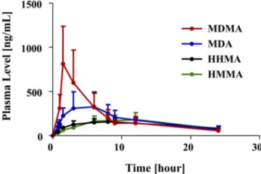
Plasma profiles of MDMA and its major metabolites after a single neurotoxic dose of MDMA (20 mg/kg p.o.) are shown in Fig. 2 and pharmacokinetic parameters are specified in Table 1. As shown in Fig. 2 and Table 1, MDMA had the highest peak plasma concentrations (Cmax), followed by MDA and then HHMA and HMMA. In particu-lar, the Cmaxof MDMA was approximately 2-fold higher than that of MDA and approximately 4 times and 3 times higher than that of HHMA and HMMA, respectively.
Relative proportions of MDMA and metabolites were somewhat different when AUC, instead of Cmax, values were considered. In particular, the AUC of MDMA was only 1.14 higher than that of MDA and only approximately 2-fold higher than that of HHMA and HMMA.
The t1/2of MDMA after oral administration was 5.8⫾ 3.5 h. If a biphasic decay process is assumed, the estimated decay rate of the first phase was 3.0 h and the estimated decay rate of the second phase was 10.5 h. The t1/2of MDA, HHMA, and HMMA could not be computed because, within the time window of measurement (0.75–24 h), there were insufficient data points in the terminal elimination phase of the plasma profiles of MDA, HHMA, and HMMA (Fig. 2).
Rats treated with a single 20 mg/kg oral dose of MDMA showed a significant depletion of brain 5-HT 1 week later (Fig. 3). On average, cortical 5-HT was reduced by 38%. There were comparable depletions of 5-HIAA (Fig. 3). The 20 mg/kg dose of MDMA produced a 0.5 to 1°C elevation in core temperature (data not shown).
Figure 4 shows results of analyses exploring the relationship be-tween the Cmaxof the parent compound and its various metabolites and cortical 5-HT deficits. It is noteworthy that these were within-subject analyses, as plasma drug concentrations and subsequent brain 5-HT deficits were measured in the same animal. Significant relation-ships were observed between the Cmax of MDMA and MDA and
subsequent 5-HT depletions, such that animals with the highest peak plasma concentrations of MDMA and MDA had the largest depletions of brain 5-HT (Fig. 4). In contrast, there were no significant relation-ships between peak plasma concentrations of HHMA or HMMA and brain 5-HT depletions 1 week later (Fig. 4).
Because the relative proportions of MDMA to MDA, HHMA, and HMMA varied depending on whether their respective Cmaxor AUC values were considered (see above), we also explored the relationship between the AUC of the parent compound (MDMA) and its various
metabolites (MDA, HHMA, and HMMA) and subsequent 5-HT de-pletions. Only the AUC of MDMA correlated significantly with subsequent cortical 5-HT deficits (Fig. 5).
Given that brain concentrations of MDMA and/or metabolites are, in all likelihood, more proximate causes of brain 5-HT neurotoxicity than plasma concentrations of the various compounds, we next mea-sured brain concentrations of MDMA and its various metabolites (MDA, HHMA, and HMMA) in the brains of rats treated with the same dose of MDMA used in the previous experiment (20 mg/kg p.o.). Brain concentrations of MDMA and metabolites in this study were determined at various times after MDMA administration (1, 3, 6, 8, and 24 h), necessarily in different groups of animals at each time point (n⫽ 8 at each time point). As shown in Fig. 6, top, and Table 2, only MDMA and MDA were detected in the brain at all time points examined. There was a high correlation between brain and plasma concentrations of MDMA and MDA (r ⫽ 0.88 and 0.98, respec-tively). HHMA and HMMA were not detectable in the brains of animals that had high concentrations of HHMA and HMMA in plasma and high concentrations of MDMA in brain. The limit of detection for HHMA and HMMA in brain tissue was 0.1g/g.
Because previous research has implicated the catechol thioether metabolite of MDMA, 5-NAC-HHMA, in MDMA neurotoxicity (Jones et al., 2005; Erives et al., 2008), we also performed studies to further assess the 5-HT neurotoxic potential of 5-NAC-HHMA and its selectivity. In these studies, we administered 5-NAC-HHMA directly into the striatum, at two different doses (21 and 42 nmol). The established 5-HT neurotoxin, 5,7-DHT (also administered directly into the striatum), served as a positive control. As anticipated, 5,7 DHT produced a sizable depletion of striatal 5-HT 2 weeks later. In contrast, 5-NAC-HHMA produced a modest, nonsignificant decrease in striatal 5-HT content that was neither dose-related (Fig. 7) nor influenced by fluoxetine (Fig. 8). No significant differences were observed in 5-HIAA levels in controls and rats treated with 5-NAC-HHMA groups, with or without fluoxetine pretreatment (data not shown).
FIG. 3. 5-HT (left panel) and 5-HIAA (right panel) concentrations in rats treated with saline (n⫽ 17) or a single oral dose of MDMA (20 mg/kg) (n ⫽ 24) 1 week previously.ⴱ, p ⬍ 0.05 (two-tailed Student’s t test).
FIG. 2. Plasma profile of MDMA and its metabolites (MDA, HHMA, and HMMA) in rats (n⫽ 24) given a single oral dose of MDMA (20 mg/kg). Concentrations of HHMA and HMMA represent total amounts of free HHMA and HMMA obtained after conjugate cleavage, as detailed under Materials and Methods.
TABLE 1
Pharmacokinetic parameters of MDMA and its metabolites in plasma of rats given a single oral dose of 20 mg/kg MDMA
Values represent the mean⫾ S.D. (n ⫽ 24).
Analyte Cmax AUC Tmax t1/2
ng/ml ng/ml䡠 h h MDMA 652⫾ 368.7 4469⫾ 1694.2 2.5⫾ 1.7 5.8⫾ 3.5 MDA 361⫾ 174.1 3926⫾ 1348.1 5.7⫾ 2.0 N.D. HHMA 170⫾ 46.9 2383⫾ 897.8 7.2⫾ 2.4 N.D. HMMA 201⫾ 67.6 2409⫾ 846.2 7.7⫾ 2.1 N.D. N.D., not determined.
2082
MUELLER ET AL.Discussion
The potential role of metabolites in MDMA neurotoxicity has been a topic of recent interest (Capela et al., 2009; Perfetti et al., 2009). This is the first study to assess the relationship between pharmacoki-netic parameters (Cmaxand AUC) of MDMA and its major
metabo-lites (HHMA, HMMA, and MDA) and 5-HT neurotoxic effects in the same animal. Results indicate that MDMA-induced 5-HT neurotox-icity is most closely related to concentrations of MDMA, with a weaker relationship to concentrations of MDA, and no relationship to
concentrations of HHMA or HMMA. Indeed, whereas levels of MDMA and MDA in brain were 5- to 10-fold higher than those in plasma, brain HHMA and HMMA could not be detected, despite high plasma HHMA and HMMA concentrations in the same animals. These results, which are consistent with those of Escobedo et al. (2005), suggest that HHMA and HMMA do not readily penetrate the blood-brain barrier (either in their free form or as sulfate or glucuronic conjugates) and indicate that there is little or no brain metabolism of MDMA to HHMA or HMMA. Taken together, these observations and
FIG. 4. Relationship between Cmaxof MDMA,
MDA, HHMA, or HMMA and cortical 5-HT depletion. Rats received a single oral dose of MDMA (20 mg/kg). One week later, cortical 5-HT levels were determined. The figures re-flect within-subject analyses, as drug plasma levels and cortical 5-HT levels were measured in the same animal (n⫽ 24). R (Pearson cor-relation coefficient) and p values are shown.
FIG. 5. Relationship between AUC of MDMA and its various metabolites and cortical 5-HT depletion in rats given a single oral dose of MDMA (20 mg/kg) and sacrificed 1 week later. The results reflect within-subject analyses, as drug plasma levels and cortical 5-HT levels were measured in the same animal (n⫽ 24). R (Pearson correlation coefficient) and p values are shown.
those of others (Steele et al., 1991; Escobedo et al., 2005) cast doubt on the view that HHMA and HMMA are directly involved in MDMA neurotoxicity (Goni-Allo et al., 2008) but leave open the possibility that MDA or a catechol-thioether metabolite of MDMA might be involved.
Although pharmacokinetic parameters of both MDMA and MDA
were found to be significantly associated with subsequent 5-HT neurotoxicity, the association with MDMA appeared to be more robust. In particular, both the Cmaxand AUC of MDMA were signif-icantly and highly correlated with subsequent 5-HT deficits, whereas only the Cmaxof MDA was correlated with 5-HT loss (at a lower significance level). Potential reasons that only MDA Cmax, but not AUC, are related to subsequent 5-HT depletion are as follows: 1) pharmacokinetic parameters for MDA AUC may not be sufficiently precise, because of insufficiently long sampling times; 2) Cmaxmay be
the relevant pharmacokinetic parameter for predicting neurotoxicity; and 3) MDA may not be involved in MDMA neurotoxicity. The current results do not permit definitive conclusions regarding the relative importance of MDMA and MDA in the neurotoxic process as it occurs in rats, as the pharmacokinetic parameter that best predicts 5-HT neurotoxicity is unknown. At least in rats (see below), both MDMA and MDA may contribute in an additive or synergistic fash-ion to 5-HT neurotoxicity, because they interact with many of the same neuronal systems and elements, and MDA is known to have 5-HT neurotoxic potential (Ricaurte et al., 1985).
Comparisons of the current data, collected in rats, to pharmacoki-netic data collected in primates (squirrel monkeys and humans) may also shed light on the relative importance of the parent compound (MDMA) and MDA in 5-HT neurotoxicity. In particular, in squirrel monkeys, MDA is a minor metabolite (3–5%), yet this species also develops MDMA-induced 5-HT neural injury. Although within-sub-ject studies involving pharmacokinetic and neurotoxicity measures have not been conducted in humans, the pharmacokinetics of MDMA in humans are similar to those in squirrel monkeys and demonstrate relatively low levels of MDA production (Kolbrich et al., 2008). A growing body of data indicates that human recreational MDMA users are susceptible to MDMA neurotoxicity (McCann et al., 1998, 2005; Kish et al., 2009) and, taken together with the pharmacokinetic data in humans, argue against a major role for MDA in MDMA-induced neurotoxicity, at least in primates.
As alluded to above, it is not known which pharmacokinetic pa-rameter (Cmax, AUC, or other) of MDMA (or MDA) most influences 5-HT neurotoxicity. However, there are clues in the literature that certain thresholds must be met for neurotoxicity to develop. In par-ticular, intravenous dosages of MDMA that engender high, but short-lived, peak concentrations of MDMA (Banks et al., 2007; M. Mueller and G. Ricaurte, unpublished observation) do not appear to be asso-ciated with neurotoxicity (Fantegrossi et al., 2004), presumably be-cause of an insufficiently long duration of drug action. Likewise, repeated low doses of MDMA that fail to achieve a certain threshold concentration would not be expected to produce neurotoxic effects, even though, when considered in aggregate, they would lead to high
FIG. 6. Plasma and brain profiles of MDMA, MDA, HHMA, and HMMA in rats given a single oral dose of MDMA (20 mg/kg) and sacrificed after 1, 3, 6, 8, and 24 h, respectively (n⫽ 8 at each time point). Determinations were made after conjugate cleavage, as detailed under Materials and Methods. HHMA and HMMA could not be detected in brain tissue. Limit of detection for HHMA and HMMA was 0.1g/g.
TABLE 2
Pharmacokinetic parameters of MDMA and MDA in brain of rats given a single oral dose of 20 mg/kg MDMA
n⫽ 8 at each time point.
Analyte Cmax AUC Tmax t1/2
ng/ml ng/ml䡠 h h MDMA 3315 386,839 1.0 14.3 MDA 1761 297,299 6.0 14.3
FIG. 7. Concentrations of 5-HT in the ipsilat-eral and contralatipsilat-eral striatum of rats that re-ceived direct unilateral intrastriatal injections of 5-NAC-HHMA at two different concentrations (21 or 42 nmol) 2 weeks previously. Each dose of 5-NAC-HHMA was injected four times, with a 12-h interval between each injection. 5-NAC-HHMA was dissolved in aCSF, at the concen-trations shown, shortly before each injection. Control animals received unilateral intrastriatal injections of an equivalent volume of aCSF. Treatment groups were aCSF (n⫽ 8), 21 nmol of 5-NAC-HHMA (n⫽ 10), and 42 nmol of 5-NAC-HMMA (n ⫽ 5). A positive control group consisted of animals that received a sin-gle intrastriatal injection of 52 nmol of 5,7-DHT (n⫽ 4). Only the effect of 5,7-DHT was significant.ⴱ, significant relationship.
AUC values. With respect to duration of action, coadministration of a selective 5-HT reuptake inhibitor (fluoxetine) up to 6 h after MDMA administration can protect from 5-HT neurotoxicity, suggesting that key events for the development of neurotoxicity take place within 6 h of drug administration (Schmidt, 1987). When these previously pub-lished data are considered along with the present findings, the most parsimonious explanation is that peak plasma drug concentrations must reach a threshold for a certain period of time (3– 6 h) for 5-HT neurotoxicity to develop. Stated differently, it is likely that both Cmax and AUC are important determinants of MDMA-induced 5-HT neurotoxicity.
Although precise threshold neurotoxic Cmax and AUC MDMA
values have yet to be determined, a working model of a potential mechanism underlying MDMA neurotoxicity can be proposed. This model, which emerges from data discussed above, relates the two principal outcome measures of the present study: pharmacokinetic parameters of MDMA and its metabolites during the period of drug (metabolite) exposure and 5-HT axonal markers (5-HT and 5-HIAA) measured 1 week later. The model assumes that, for neurotoxicity to occur, drug (or metabolite) must interact with the 5-HT transporter for 3 to 6 h. Furthermore, it assumes that a certain threshold drug level must be achieved and maintained during the 3 to 6 h that critical toxic drug/transporter interactions appear to take place. It is noteworthy that the model makes no assumption about serotonin or other monoamine or metabolite levels during the period of drug exposure. However, it does allow for a role of core temperature, with high temperatures facilitating and low core temperatures retarding toxic drug/transporter interactions (Malberg and Seiden, 1998).
It should be emphasized that correlation does not imply causa-tion, that the relationship between MDMA (and MDA) and 5-HT deficits could be coincidental, and that other drug effects may be the most important mediators of neurotoxicity [e.g., transporter-based ion dysregulation, as postulated for methamphetamine (Cal-lahan et al., 2001)]. As noted earlier (see Introduction), there are data indicating that when MDMA is injected directly into the brain, neurotoxicity does not develop. Although this may be viewed as incontrovertible evidence that MDMA is not the major mediator of
MDMA-induced 5-HT injury, it is possible that peripheral phar-macological effects not reproduced by central administration (e.g., increased temperature) are required for neurotoxicity to occur. In addition, it is likely that centrally administered MDMA is only toxic when its concentration and duration of action are similar to those after peripheral administration.
The thioether metabolite of HHMA, 5-NAC-HHMA, has been directly implicated in MDMA neurotoxicity (Jones et al., 2005; Erives et al., 2008). In the present study, 5-NAC-HHMA, when administered repeatedly and in large doses into the striatum did not lead to statis-tically significant 5-HT depletions. Moreover, the modest effect of 5-NAC-HHMA on striatal 5-HT was neither dose related nor blocked by the 5-HT uptake inhibitor, fluoxetine, which is known to protect against MDMA neurotoxicity (Schmidt, 1987). These observations argue against a pivotal role for 5-NAC-HHMA in MDMA-induced 5-HT neurotoxicity but leave open the possibility that it may work in conjunction with MDMA or MDA in the neurotoxic process. Alter-natively, 5-NAC-HHMA may require the presence of MDMA and/or elevated body temperature to be toxic, although an earlier study (McCann and Ricaurte, 1991) also suggested that the thioether ad-ducts of HHA are not likely to be responsible for serotonergic neu-rotoxicity.
The present findings with 5-NAC-HHMA are at odds with findings of a previous study showing that this compound produced dose-related depletions of 5-HT in rats (Jones et al., 2005). The reasons for this discrepancy are not entirely clear. We established the identity of 5-NAC-HHMA by high-performance liquid chromatography and NMR spectra [methods available in the supporting information for Felim et al. (2007)]. Furthermore, the stability of 5-NAC-HHMA was confirmed after each injection by using LC-MS to monitor the abun-dance of its molecular mass ion [MH⫹] and one fragment ion (m/z⫽ 343 and m/z⫽ 181, respectively). Another potential reason for dis-crepant findings is inadequate drug delivery of an unstable compound to target tissues. However, 5,7-DHT (which is also unstable and has a tendency to oxidize) was injected using identical methods and was found to produce robust 5-HT deficits. Finally, it may be relevant that 5-NAC-HHMA used in the present studies was prepared using a biomimetic electrochemical synthetic method (Felim et al., 2007), whereas 5-NAC-HHMA used by Jones et al. (2005) was prepared with mushroom tyrosinase, which yields a different ratio of 5-NAC-HHMA diastereoisomers (Pizarro et al., 2008). Additional research will be required to determine the basis for discrepant findings between the present study and that of Jones et al. (2005).
In conclusion, the present results indicate that MDMA-induced 5-HT neurotoxicity is most closely related to plasma and brain con-centrations of MDMA, with a weaker relationship to concon-centrations of MDA and no relationship to concentrations of HHMA or HMMA. The present results also indicate that the pharmacokinetic parameter of MDMA that best predicts subsequent 5-HT neurotoxicity is Cmax,
although AUC is also a good predictor and both peak levels and duration of action are likely to be important. It is noteworthy that neither HHMA nor HMMA could be detected in brain, despite high concentrations of these MDMA metabolites in plasma, indicating that HHMA and HMMA do not readily penetrate the blood-brain barrier. Because brain concentrations of MDMA in the same animals were 5-to 10-fold higher than those in plasma, the absence of measurable amounts of HHMA and HMMA in their brains also suggests that biotransformation of MDMA to HHMA and HMMA does not occur to any appreciable degree in the brain. Finally, repeated intrastriatal administration of 5-NAC-HHMA produced a modest, nonsignificant decrease in striatal 5-HT content that was neither dose-related nor influenced by fluoxetine. Taken together, these results favor the view
FIG. 8. Concentrations of 5-HT in the ipsilateral striatum of rats that received direct unilateral intrastriatal injections of 5-NAC-HHMA (21 nmol) alone or in combina-tion with fluoxetine (Fluox; 10 mg/kg; i.p., 15 min before 5-NAC-HHMA) 2 weeks previously. 5-NAC-HHMA was injected four times, with a 12-h interval between each injection. 5-NAC-HHMA was dissolved in aCSF shortly before each injection. Control animals received unilateral intrastriatal injections of an equivalent volume of aCSF. n⫽ 6 for each treatment group.
that MDMA and, possibly, MDA are the compounds that trigger brain 5-HT neurotoxicity in rats, and suggest that HHMA, HMMA, and the catechol thioether metabolite, 5-NAC-HHMA do not play a crucial role in MDMA-induced 5-HT neurotoxicity in vivo.
Acknowledgments. We thank George Hadtzidimitriou and Gian-luigi Tanda for help with these experiments.
References
Bai F, Lau SS, and Monks TJ (1999) Glutathione and N-acetylcysteine conjugates of ␣-meth-yldopamine produce serotonergic neurotoxicity: possible role in methylenedioxyamphet-amine-mediated neurotoxicity. Chem Res Toxicol 12:1150 –1157.
Banks ML, Sprague JE, Kisor DF, Czoty PW, Nichols DE, and Nader MA (2007) Ambient temperature effects on 3,4-methylenedioxymethamphetamine-induced thermodysregulation and pharmacokinetics in male monkeys. Drug Metab Dispos 35:1840 –1845.
Callahan BT, Cord BJ, Yuan J, McCann UD, and Ricaurte GA (2001) Inhibitors of Na⫹/H⫹and Na⫹/Ca2⫹exchange potentiate methamphetamine-induced dopamine neurotoxicity: possible role of ionic dysregulation in methamphetamine neurotoxicity. J Neurochem 77:1348 –1362. Capela JP, Carmo H, Remia˜o F, Bastos ML, Meisel A, and Carvalho F (2009) Molecular and cellular mechanisms of Ecstasy-induced neurotoxicity: an overview. Mol Neurobiol 39:210 – 271.
Capela JP, Macedo C, Branco PS, Ferreira LM, Lobo AM, Fernandes E, Remia˜o F, Bastos ML, Dirnagl U, Meisel A, et al. (2007) Neurotoxicity mechanisms of thioether ecstasy metabolites.
Neuroscience 146:1743–1757.
Chu T, Kumagai Y, DiStefano EW, and Cho AK (1996) Disposition of methylenedioxymeth-amphetamine and three metabolites in the brains of different rat strains and their possible roles in acute serotonin depletion. Biochem Pharmacol 51:789 –796.
Elayan I, Gibb JW, Hanson GR, Foltz RL, Lim HK, and Johnson M (1992) Long-term alteration in the central monoaminergic systems of the rat by 2,4,5-trihydroxyamphetamine but not by 2-hydroxy-4,5-methylenedioxymethamphetamine or 2-hydroxy-4,5-methylenedioxyamphet-amine. Eur J Pharmacol 221:281–288.
Erives GV, Lau SS, and Monks TJ (2008) Accumulation of neurotoxic thioether metabolites of 3,4-(⫾)-methylenedioxymethamphetamine in rat brain. J Pharmacol Exp Ther 324:284–291. Escobedo I, O’Shea E, Orio L, Sanchez V, Segura M, de la Torre R, Farre M, Green AR, and Colado MI (2005) A comparative study on the acute and long-term effects of MDMA and 3,4-dihydroxymethamphetamine (HHMA) on brain monoamine levels after i.p. or striatal administration in mice. Br J Pharmacol 144:231–241.
Esteban B, O’Shea E, Camarero J, Sanchez V, Green AR, and Colado MI (2001) 3,4-Methylenedioxymethamphetamine induces monoamine release, but not toxicity, when admin-istered centrally at a concentration occurring following a peripherally injected neurotoxic dose.
Psychopharmacology (Berl) 154:251–260.
Fantegrossi WE, Woolverton WL, Kilbourn M, Sherman P, Yuan J, Hatzidimitriou G, Ricaurte GA, Woods JH, and Winger G (2004) Behavioral and neurochemical consequences of long-term intravenous self-administration of MDMA and its enantiomers by rhesus monkeys.
Neuropsychopharmacology 29:1270 –1281.
Felim A, Urios A, Neudo¨rffer A, Herrera G, Blanco M, and Largeron M (2007) Bacterial plate assays and electrochemical methods: an efficient tandem for evaluating the ability of catechol-thioether metabolites of MDMA (“ecstasy”) to induce toxic effects through redox-cycling.
Chem Res Toxicol 20:685– 693.
Gollamudi R, Ali SF, Lipe G, Newport G, Webb P, Lopez M, Leakey JE, Kolta M, and Slikker W Jr (1989) Influence of inducers and inhibitors on the metabolism in vitro and neurochemical effects in vivo of MDMA. Neurotoxicology 10:455– 466.
Goni-Allo B, O Mathu´na B, Segura M, Puerta E, Lasheras B, de la Torre R, and Aguirre N (2008) The relationship between core body temperature and 3,4-methylenedioxymethamphetamine metabolism in rats: implications for neurotoxicity. Psychopharmacology (Berl) 197:263–278. Hiramatsu M, Kumagai Y, Unger SE, and Cho AK (1990) Metabolism of methylenedioxymeth-amphetamine: formation of dihydroxymethamphetamine and a quinone identified as its glu-tathione adduct. J Pharmacol Exp Ther 254:521–527.
Institute of Laboratory Animal Resources (1996) Guide for the Care and Use of Laboratory
Animals, 7th ed. Institute of Laboratory Animal Resources, Commission on Life Sciences,
National Research Council, Washington, DC.
Johnson M, Elayan I, Hanson GR, Foltz RL, Gibb JW, and Lim HK (1992) Effects of 3,4-dihydroxymethamphetamine and 2,4,5-trihydroxymethamphetamine, two metabolites of 3,4-methylenedioxymethamphetamine, on central serotonergic and dopaminergic systems.
J Pharmacol Exp Ther 261:447– 453.
Jones DC, Duvauchelle C, Ikegami A, Olsen CM, Lau SS, de la Torre R, and Monks TJ (2005) Serotonergic neurotoxic metabolites of ecstasy identified in rat brain. J Pharmacol Exp Ther
313:422– 431.
Kish SJ, Fitzmaurice PS, Boileau I, Schmunk GA, Ang LC, Furukawa Y, Chang LJ, Wickham DJ, Sherwin A, and Tong J (2009) Brain 5-HT transporter in human methamphetamine users.
Psychopharmacology (Berl) 202:649 – 661.
Kolbrich EA, Goodwin RS, Gorelick DA, Hayes RJ, Stein EA, and Huestis MA (2008) Plasma
pharmacokinetics of 3,4-methylenedioxymethamphetamine after controlled oral administra-tion to young adults. Ther Drug Monit 30:320 –332.
Lim HK and Foltz RL (1991a) In vivo formation of aromatic hydroxylated metabolites of 3,4-(methylenedioxy)methamphetamine in the rat: identification by ion trap tandem mass spectrometric (MS/MS and MS/MS/MS) techniques. Biol Mass Spectrom 20:677– 686. Lim HK and Foltz RL (1991b) Ion trap tandem mass spectrometric evidence for the metabolism
of 3,4-(methylenedioxy)methamphetamine to the potent neurotoxins 2,4,5-trihydroxymetham-phetamine and 2,4,5-trihydroxyam2,4,5-trihydroxymetham-phetamine. Chem Res Toxicol 4:626 – 632.
Malberg JE and Seiden LS (1998) Small changes in ambient temperature cause large changes in 3,4-methylenedioxy-methamphetamine (MDMA)-induced serotonin neurotoxicity and core body temperature in the rat. J Neurosci 18:5086 –5094.
McCann UD and Ricaurte GA (1991) Major metabolites of (⫾)3,4-methylenedioxyamphetamine (MDA) do not mediate its toxic effects on brain serotonin neurons. Brain Res 545:279 –282. McCann UD, Szabo Z, Scheffel U, Dannals RF, and Ricaurte GA (1998) Positron emission tomographic evidence of toxic effect of MDMA (“Ecstasy”) on brain serotonin neurons in human beings. Lancet 352:1433–1437.
McCann UD, Szabo Z, Seckin E, Rosenblatt P, Mathews WB, Ravert HT, Dannals RF, and Ricaurte GA (2005) Quantitative PET studies of the serotonin transporter in MDMA users and controls using [11C]McN5652 and [11C]DASB. Neuropsychopharmacology 30:1741–1750.
Mechan A, Yuan J, Hatzidimitriou G, Irvine RJ, McCann UD, and Ricaurte GA (2006) Pharmacokinetic profile of single and repeated oral doses of MDMA in squirrel monkeys: relationship to lasting effects on brain serotonin neurons. Neuropsychopharmacology 31:339 – 350.
Meyer MR, Peters FT, and Maurer HH (2008) The role of human hepatic cytochrome P450 isozymes in the metabolism of racemic 3,4-methylenedioxy-methamphetamine and its enan-tiomers. Drug Metab Dispos 36:2345–2354.
Miller RT, Lau SS, and Monks TJ (1997) 2,5-Bis-(glutathion-S-yl)-␣-methyldopamine, a puta-tive metabolite of (⫾)-3,4-methylenedioxyamphetamine, decreases brain serotonin concentra-tions. Eur J Pharmacol 323:173–180.
Molliver ME, Berger UV, Mamounas LA, Molliver DC, O’Hearn E, and Wilson MA (1990) Neurotoxicity of MDMA and related compounds: anatomic studies. Ann N Y Acad Sci
600:649 – 661.
Monks TJ, Jones DC, Bai F, and Lau SS (2004) The role of metabolism in 3,4-( ⫹)-methylenedioxyamphetamine and 3,4-(⫹)-methylenedioxymethamphetamine (ecstasy) toxic-ity. Ther Drug Monit 26:132–136.
Mueller M, Peters FT, Ricaurte GA, and Maurer HH (2007) Validated liquid chromatographic-electrospray ionization mass spectrometric assay for simultaneous determination of methylenedioxymethamphetamine and its metabolites methylenedioxyamphetamine, 3,4-dihydroxymethamphetamine, and 4-hydroxy-3-methoxymethamphetamine in squirrel monkey plasma. J Chromatogr B Analyt Technol Biomed Life Sci 855:262–270.
Mueller M, Peters FT, Ricaurte GA, and Maurer HH (2008) Liquid chromatographic-electrospray ionization mass spectrometric assay for simultaneous determination of methylenedioxymethamphetamine and its metabolites methylenedioxyamphetamine, 3,4-dihydroxymethamphetamine, and 4-hydroxy-3-methoxymethamphetamine in rat brain.
J Chromatogr B Analyt Technol Biomed Life Sci 874:119 –124.
Paxinos G and Watson C (1986) The Brain in Stereotaxic Coordinates, 2nd ed, Academic Press, Inc., New York.
Perfetti X, O’Mathu´na B, Pizarro N, Cuya`s E, Khymenets O, Almeida B, Pellegrini M, Pichini S, Lau SS, Monks TJ, et al. (2009) Neurotoxic thioether adducts of 3,4-methylenedioxymeth-amphetamine identified in human urine after Ecstasy ingestion. Drug Metab Dispos 37:1448 – 1455.
Pizarro N, de la Torre R, Joglar J, Okumura N, Perfetti X, Lau SS, and Monks TJ (2008) Serotonergic neurotoxic thioether metabolites of 3,4-methylenedioxymethamphetamine (MDMA, “Ecstasy”): synthesis, isolation, and characterization of diastereoisomers. Chem Res
Toxicol 21:2272–2279.
Ricaurte G, Bryan G, Strauss L, Seiden L, and Schuster C (1985) Hallucinogenic amphetamine selectively destroys brain serotonin nerve terminals. Science 229:986 –988.
Ricaurte GA, Martello AL, Katz JL, and Martello MB (1992) Lasting effects of ( ⫾)-3,4-methylenedioxymethamphetamine (MDMA) on central serotonergic neurons in nonhuman primates: neurochemical observations. J Pharmacol Exp Ther 261:616 – 622.
Schmidt CJ (1987) Neurotoxicity of the psychedelic amphetamine, methylenedioxymethamphet-amine. J Pharmacol Exp Ther 240:1–7.
Schmidt CJ and Taylor VL (1988) Direct central effects of acute methylenedioxymethamphet-amine on serotonergic neurons. Eur J Pharmacol 156:121–131.
Steele TD, Brewster WK, Johnson MP, Nichols DE, and Yim GK (1991) Assessment of the role of␣-methylepinine in the neurotoxicity of MDMA. Pharmacol Biochem Behav 38:345–351. Zhao ZY, Castagnoli N Jr, Ricaurte GA, Steele T, and Martello M (1992) Synthesis and neurotoxicological evaluation of putative metabolites of the serotonergic neurotoxin 2-(meth-ylamino)-1-[3,4-(methylenedioxy)phenyl] propane [(methylenedioxy)methamphetamine].
Chem Res Toxicol 5:89 –94.
Address correspondence to: Dr. George A. Ricaurte, Department of
Neurol-ogy, Johns Hopkins Medical Institutions, 5501 Hopkins Bayview Circle, Rm. 5B.71E, Baltimore, MD 21224. E-mail: ricaurte@jhmi.edu