ORIGINAL ARTICLE
The use of vacuum assisted closure (VAC
™
) in soft tissue
injuries after high energy pelvic trauma
Ludwig Labler&Otmar Trentz
Received: 1 February 2006 / Accepted: 20 July 2006 / Published online: 16 September 2006 # Springer-Verlag 2006
Abstract
Background Application of vacuum-assisted closure (VAC™) in soft tissue defects after high-energy pelvic trauma is described as a retrospective study in a level one trauma center.
Materials and methods Between 2002 and 2004, 13 patients were treated for severe soft tissue injuries in the pelvic region. All musculoskeletal injuries were treated with multiple irrigation and debridement procedures and
broad-spectrum antibiotics. VAC™ was applied as a
temporary coverage for defects and wound conditioning. Results The injuries included three patients with traumatic hemipelvectomies. Seven patients had pelvic ring fractures with five Morel–Lavallee lesions and two open pelviper-ineal trauma. One patient suffered from an open iliac crest fracture and a Morel–Lavallee lesion. Two patients sustained near complete pertrochanteric amputations of the lower limb. The average injury severity score was 34.1± 1.4. The application of VAC™ started in average 3.8± 0.4 days after trauma and was used for 15.5±1.8 days. The dressing changes were performed in average every 3 days. One patient (8%) with a traumatic hemipelvectomy died in the course of treatment due to septic complications. Conclusion High-energy trauma causing severe soft tissues injuries requires multiple operative debridements to prevent high morbidity and mortality rates. The application of VAC™ as temporary coverage of large tissue defects in pelvic regions supports wound conditioning and facilitates the definitive wound closure.
Keywords Open fractures . Pelvis . Soft tissue defect . Wound conditioning . Vacuum-assisted closure
Introduction
Pelvic fractures with critical soft tissue conditions are usually the result of high-energy trauma [1]. Open pelvic fractures are rare and amount to 2–4% of all pelvic fractures only [2,3]. Traumatic hemipelvectomy is an extreme form of a pelvic ring fracture and is defined as an unstable ligamentous or osseous hemipelvic injury with rupture of the pelvic neurovascular bundle [4, 5]. Other severe soft tissue injuries occur after traumatic paraarticular hip amputation. The local and systemic contraindications for replantation occur in 30 to 50% of all traumatic macro-amputations [6]. Traumatic amputations may be revised to a more proximal level after initial operative procedure [7,8]. The high mortality of open pelvic injuries is due to two complications: In the early phase, the patient is endangered by exsanguination and in the following course the sepsis determines the lethal outcome [9]. Significant soft tissue injuries often occur as a part of high-energy injuries of the pelvis and in order to minimize complications they must already be recognized and considered when the treatment plan is implemented. Because of a high risk of complica-tions and death, the patients having these injuries have to be managed aggressively [10]. Essential is exclusion of occult and readily apparent perforations of the genital, urinary, and gastrointestinal tracts. Recognition of open and closed degloving injury patterns and appropriate adherence to treatment guidelines optimize the outcome and avoid catastrophic complications [11]. Cultures from closed internal degloving injury of the pelvic region (Morel– Lavallee lesion) were positive in 46% of 24 patients with
DOI 10.1007/s00423-006-0090-0
L. Labler (*)
:
O. TrentzDivision of Trauma Surgery, Department of Surgery, University Hospital Zürich,
Rämistrasse 100, 8091 Zürich, Switzerland e-mail: ludwig.labler@usz.ch
these injuries [12]. The Morel–Lavallee lesion should be debrided early, either before or at the time of fracture fixation. The wound should be left open, and repeated surgical debridements of the injured tissue are recommended [12]. To prevent septic complications, similar surgical procedures are recommended in the case of complex perineal injuries as well [13–17]. Fecal diversion and rectal washout is recommended in the case of rectal injuries of open pelvic fractures [18–20].
The principles described for trauma on extremities are also valid for the management of the above-mentioned soft tissue injuries associated with a high-energy pelvic trauma. Open fractures require early aggressive debridement of the soft tissues followed by skeletal stabilization. Temporary
wound dressings should remain in place until definitive soft-tissue coverage is obtained. Definitive soft-tissue closure will be expedited by serial debridements performed every 48 to 72 h in a sterile environment [21].
The use of vacuum-assisted closure (VAC) as a temporary wound dressing in open fracture situations was described in the literature [22–24]. During the last 10 years, VAC™ (Kinetic Concepts, Inc., San Antonio, TX, USA) was suc-cessfully used in the management of problematic wounds. The VAC™therapy represents a temporary protection of soft tissue defects by means of polyurethane foam, which is sealed airtight by a polyvinyl foil. A negative topical pres-sure gradient is generated by a VAC™ unit. In the clinical practice, the VAC™ therapy increases quantitatively and
Table 1 Patients and clinical data
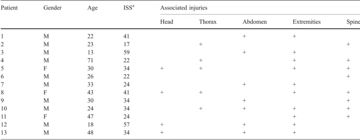
Patient Gender Age ISSa Associated injuries
Head Thorax Abdomen Extremities Spine
1 M 22 41 + + 2 M 23 17 + + 3 M 13 59 + + 4 M 71 22 + + + 5 F 30 34 + + + + 6 M 26 22 + 7 M 33 24 + + 8 F 43 41 + + + + 9 M 30 34 + + 10 M 24 34 + + + + 11 F 47 24 + + 12 M 18 57 + + + 13 M 48 34 + + +
M Male and F female
aInjury severity score [29]
Table 2 Pelvic fractures with associated soft tissue injuries and traumatic amputation
Patient Pelvic fracture Associated soft tissue injuries Traumatic amputation Aa Ba Ca Traumatic hemipelvectomy Open fractureb Genitourinary tract Intestinal tract Morel– Lavallee Penetrating trauma 1 + + + 2 + + + 3 + + + + + 4 + + 5 + 6 + + + 7 + 8 + + 9 + + + 10 + + + + 11 + + + 12 + + + + 13 + + + +
aAO classification of pelvic fractures [32]
qualitatively the granulation tissue formation on the wound surface [25–27]. Well-documented is a significantly improved microperfusion, an increased partial oxygen pressure in the tissue and a reduction of the bacterial colonization (bacterial clearance) after 4 days [28]. The increased microvessel density, which develops with time, results in an improved granulation formation and in the wound healing as well.
We report our experience with the application of VAC™ dressing in severe soft tissue injuries in the pelvic region caused by a high-energy trauma.
Materials and methods
Patients Thirteen patients with severe soft tissue injuries in the pelvic region were treated at a level one trauma center using the commercial VAC™Dressing System in the period between March 2002 and October 2004. The patients were identified by reviewing the hospital trauma registries and their age, associated injuries, injury severity score (ISS) [29], and intensive care unit (ICU) and hospital stay are summarized in Table1[29]. The mechanism of injury was related in ten cases to traffic accidents and in two cases to fall from great heights. One case was a working accident. Except one patient (patient 8) who was transferred 6 days after the accident, the remaining patients were admitted to our institution at the day of the accident. The patients were managed according to advanced trauma life support guide-lines [30] after arrival. The hemodynamically unstable patients were submitted to a damage control procedure in the operating room (OR) and transferred afterward to the ICU [31]. The patients under stable conditions were evaluated by CT scan before initial surgical procedures and were then transferred to the ICU. The pelvic ring fractures, their associated injuries, and the traumatic amputations are summarized in Table2 [32, 33]. Table 3 includes the damage control procedures performed. Technique The VAC™ system consisted of polyurethane soft sponge cut to fit the wound and placed into the cavity and of a transparent occlusive gas- and fluid-impermeable plastic film applied over the foam to create an airtight seal. A hole of 2 cm in diameter was cut into the film in the middle of the foam. A TRAC™pad was embedded over the hole and attached to an adjustable vacuum pump by means of a suction tube. A continuous topic negative pressure of 125 mmHg was used.
Results
All 13 patients were severely injured with an average ISS of 34.1±1.4 (range 17 to 59). In the group of three Table
3 Damage control interventions Patient Pelvic clamp Pelvic fixator ORIF Laparotomy Aortic clamping Exclusion of large vessels Abdominal packing Colostomy Reconstruction of urinary bladder Open abdomen W ound packing Amputation 1+ + + + + 2+ 3+ + + + + + + + 4 + 5 ++ 6 7 ++ 8+ 9+ + + + + 10 ++ 11 + 12 + + + + + + + + 13 + + + + ORIF Open reduction, internal fixation
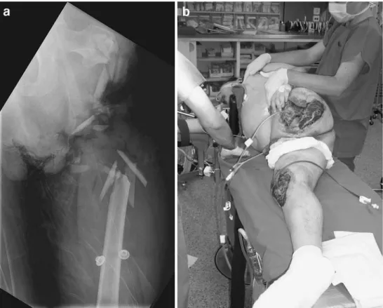
traumatic hemipelvectomies, one hemipelvectomy was complete (patient 1) and the large wound was left open after debridement. The other two patients (patients 5 and 7) suffered a nearly total proximal traumatic amputation in the trochanteric region and the wounds were left open after surgical amputation and debridement (Fig.1). The wounds were also left open after debridement in two patients (patients 4 and 13) with penetrating injuries in the iliac crest. Out of the remaining six patients with pelvic ring injuries, in only two cases of complex perineal injuries (patients 9 and 10) were the wounds left initially open
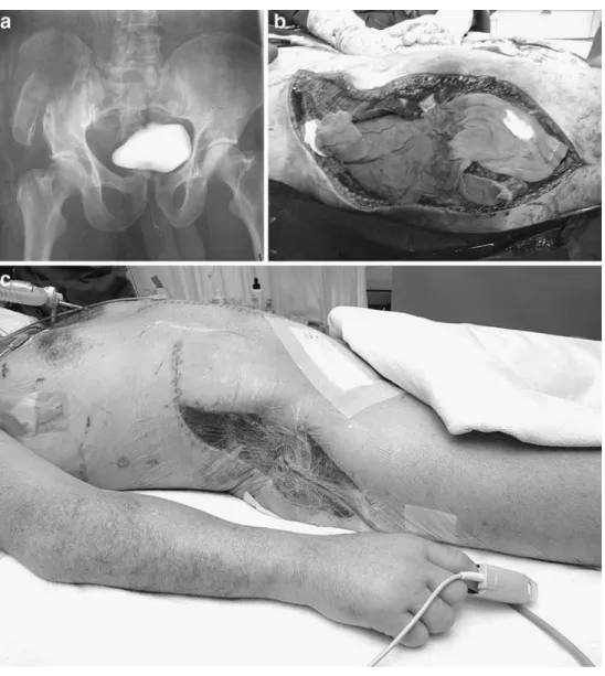
(Fig. 2). In all cases mentioned above the wounds were initially packed by surgical towels because of posttraumatic coagulopathy. An exception was patient 10 where VAC™ therapy was initially used. In the course of the first second-look operations 24 to 48 h after the trauma and a correction of the posttraumatic coagulopathy, the extended wounds of seven patients were then treated by VAC™ therapy (Fig.3 and Table 4). In five patients the wounds were then left open for 4 to 13 days after the trauma and were then covered temporarily by VAC™therapy (Table4).
Fig. 1 a X-ray of traumatic amputation in the trochanteric region on the left side after a rollover by train (patient 7). b The wound was left open after damage control procedure and then packed. During the first second-look operation VAC™ dressing was applied
Fig. 2 a X-ray after laparotomy, colostomy, rectal washout, and anterior plating of open pelvic ring fracture type C according AO classification with b perineal disrupture and lesion of rectal sphincter muscles (patient 9). c Local conditioning of the perineal cavity by VAC™dressing
Fig. 3 a X-ray of penetrating injury to the left iliac wing with open fracture of the iliac crest (patient 13). b After damage control by laparotomy, aortic clamping and ligation of the left superior gluteal artery when pa-tient was in shock. The wound was packed with surgical tow-els. c During first second-look operation and a new debride-ment, the wound was temporally closed by VAC™dressing and was conditioned until it was closed by mesh graft
Table 4 The course of therapy Patient ICU staya Therapy time period in totala VAC start after traumaa VAC perioda Number of VAC dressings VAC dressing changeda (range) Wound free of bacterial growth (day after start of VAC)
Definite closure Hospital staya Primary suture Primary suture with mesh grafting Mesh grafting Free flap 1 18 19 10 9 4 2.2 (2–3) 5 1 62 2 1 6 1 5 2 2.5 (2–3) 5 1 21 3 57 69 1 68 21 3.0 (1–7) 18 1 89 4 15 19 13 6 2 3.0 (2–4) 7 1 27 5 7 17 2 15 5 4.0 (2–4) 17 1 43 6 1 23 9 14 5 2.8 (2–3) 8 1 40 7 3 6 2 4 2 2.0 (1–3) 20 1 47 8 16 29 6 23 8 2.9 (2–4) 22 1 44 9 4 12 2 10 3 3.3 (3–4) 7 1 25 10 8 6 0 6 3 2.0 (1–3) 6 1 13 11 6 9 1 8 3 2.7 (1–4) 21 1 24 12 6b 6 2 4 3 1.3 (1–2) X X X X X 6 13 26 33 4 29 10 4.5 (3–7) 21 1 61 aDays bPatient died (X)
In two cases (patients 3 and 12), surgical hemipelvec-tomies were performed after damage control procedures 24 and 48 h later and the wounds were then left open (Fig.4). In four patients (patients 2, 6, 8, and 11) with pelvic ring
injuries with Morel–Lavallee lesion, the degloving injuries were opened in the course of the hospital stay after 4.5± 3.0 days (range 1 to13). The mostly large hematoma was evacuated and the wound cavities were conditioned by VAC™ therapy. An intravenous antibiotic therapy was administered in all patients and adapted to the bacterial results sampled from the open wounds.
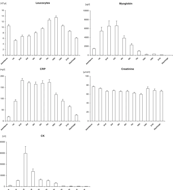
The wounds were left open and conditioned by VAC™ therapy for 15.1±1.8 days and dressing changes were performed in the OR during the planed second-look operations in 3.0±1.3 days (range 1 to 7 days) (Table 4). The exceptionally long treatment in patient 3 with traumatic hemipelvectomy (68 days) was due to severe systemic septic complication and a flap failure for covering the soft tissue defect. Bacterial samples were collected from the wounds in all patients during the reoperations. According to the bacterial clearance the wounds were free of bacterial growth after 13.1±7.2 days (range 5 to 22 days) (Table4). Results of leukocyte counts, C-reactive protein (CRP), creatine kinase (CK), myoglobin, and creatinine levels are summarized in Fig.5.
One patient (patient 12) with a traumatic hemipelvec-tomy died in the course of treatment in the ICU 6 days after the trauma due to septic complications. The open wounds of 9 out of the 12 patients who survived were closed by delayed primary suture. In one patient the wound closure was managed by delayed primary suture in combination with mesh grafting. In two patients the defects were covered in one case by mesh grafting and in the other by free flap (Table4).
One patient (patient 5) was transferred to his native country and was not available for a follow-up. In the remaining 11 patients the follow-up was possible within 19.8±1.0 months (range 7–38 months). In this group, complications such as infections or unstable wound scars were not found in wounds treated by the VAC therapy.
Discussion
In all cases reported in this study an early closure of the large wounds was not feasible. A reconstructive surgery with the aim to achieve early coverage of the large soft tissue defects was impossible due to the severity of the systemic injuries and to the time-consuming interventions of the plastic surgery. In view of the expected posttraumatic coagulopathy, it was helpful to pack the large wounds after staged debridement and surgical control of bleeding during the damage control procedures. A question already arose in the course of the first second-look operation and debridement of these large wounds. How the soft tissue defect should be temporarily covered before a definitive closure by a delayed primary suture, mesh grafting, or free flap is possible is another question.
Fig. 4 a Patient 24 h after damage control procedures by laparotomy, temporary aortic clamping, and pelvic clamp for traumatic hemi-pelvectomy (patient 12). b Planed surgical hemihemi-pelvectomy and debridement. c Temporary closure of the large wound by VAC™ dressing
Besides a variety of skin substitutes such as Epigard™ (Biovision GmbH, Ilmenau, Germany), VAC™therapy was introduced initially for treatment of chronic wounds [25, 34]. Its further application in the case of trauma settings was well documented for injuries of extremities [22–24, 35–37]. On the other hand, the use of VAC™ therapy in
extended wounds after severe injuries in the pelvic region was not yet reported so far. In our view, the advantage of this type of a temporary wound coverage, apart from the reported wound conditioning mechanism [28], consists in a reduction of the dead space and thus in a better control of potential infected wounds. Furthermore, we believe that this Leucocytes 0 2 4 6 8 10 12 14 16 Adm itanc e 1st 2nd 3rd 4th 5th 7th 10th 14th 21st Disch arge [103/µl] CRP 0 50 100 150 200 Adm itan ce 1st 2nd 3rd 4th 5th 7th 10th 14th 21st Dis char ge [mg/l] CK 0 10000 20000 30000 40000 Adm itan ce 1st 2nd 3rd 4th 5th 7th 10th 14th 21st Disc harge [U/l] Myoglobin 0 2000 4000 6000 8000 10000 Adm itanc e 1st 2nd 3rd 4th 5th 7th 10th 14th 21st Disch arge [µg/l] Creatinine 0 20 40 60 80 100 Adm itanc e 1st 2nd 3rd 4th 5th 7th 10th 14th 21st Disch arge [µmol/l]
temporary wound closure prevents wound exsiccation and in-hospital wound infections. The latter is crucial in combination with pelvic fractures or in cases of traumatic amputations near the pelvis. The time intervals between
dressing changes and the duration of VAC™ therapy
application, despite the substantial difference in the wound dimensions, according to our own and other authors’ [23, 24] experience, did not differ substantially from those currently used for the extremity injuries.
The use of VAC™ therapy was described early for coverage of dermatofasciotomy after compartment syn-drome in extremities [38]. The effect of an externally applied subatmospheric pressure on serum myoglobin levels after prolonged crush injury was studied in an animal model [39]. In our study, we found in serum increased levels of CK and myoglobin in the first 7 days after trauma (Fig. 5), dependent on the dimension of the soft tissue injury. On the other hand, no renal failure was observed during the course of treatment except one patient who died due to septic complications. Bacterial clearance was described in an animal model and defined as a significant reduction of bacterial counts in a wound after 4 days of VAC™ therapy [28]. Recently, two publications appeared describing a different behavior of a bacterial load in relation to VAC™ therapy [40, 41]. According to our own results the bacterial growth ceased in the VAC™-treated wounds after 12.6±0.8 days in average (range 5 to 22 days), supposedly in dependence on the wound dimension, the immune deficiency after severe injury, and on systematic inflammatory response syndrome. The above-mentioned results probably do not reflect the whole reality because only wound smears were collected and not wound tissue samples for the bacteriological examinations. Nevertheless, the data of the bacterial load in our study correlate quite well with the leukocyte counts and the measured CRP showing both fast decreasing levels 14 days after the trauma.
Even large traumatic wounds resulting after traumatic hemipelvectomy or very proximal traumatic amputations near to the hip are very suitable for the use of VAC™ therapy, as a temporary wound coverage provided that the bleeding in the wound is fully under surgical control.
Because of a high risk of a tissue necrosis, the wound in the case of Morel–Lavallee lesion should remain open after evacuation of hematoma [12] and a repeated debridement is also recommended. In no one Morel–Lavallee case, even after repeated debridements, could we observe any tissue necrosis during application of VAC™ therapy and could close the injuries secondary. The debrided wound cavities of perineal injuries occurring during disruption of the pelvic ring were also managed by VAC™ therapy and because colostomies and rectal washout were performed each time without exception, a good wound conditioning was the result [42].
Conclusion
In conclusion, to prevent high morbidity and mortality rates, a high-energy trauma causing severe soft tissues injuries requires multiple operative debridements and consequently an appropriate temporary dressing. In view of our results, the management of the large tissue defects in pelvic regions by
means of VAC™ as a temporary coverage positively
supports wound conditioning, reduces infectious complica-tions, and facilitates a definitive wound closure.
References
1. Giannoudis PV, Pape HC (2004) Damage control orthopaedics in unstable pelvic ring injuries. Injury 35:671–677
2. Grotz MR, Allami MK, Harwood P, Pape HC, Krettek C, Giannoudis PV (2005) Open pelvic fractures: epidemiology, current concepts of management and outcome. Injury 36:1–13 3. Brenneman FD, Katyal D, Boulanger BR, Tile M, Redelmaeier DA
(1997) Long-term outcomes in open pelvic fractures. J Trauma 42:773–777
4. Pohlemann T, Paul C, Gansslen A, Regel G, Tscherne H (1996) Traumatic hemipelvectomy. Experiences with 11 cases. Unfallchirurg 99:304–312
5. Rieger H, Dietl KH (1998) Traumatic hemipelvectomy: an update. J Trauma 45:422–426
6. Seiler H, Braun C (1997) Replantation of a leg after hip para-articular amputation. Unfallchirurg 100:683–688
7. Blazar PE, Dormans JP, Born CT (1997) Train injuries in children. J Orthop Trauma 11:126–129
8. Singer G, Thordarson D (1994) Train-versus-pedestrian injuries. Orthopaedic management. Orthop Rev (Suppl):30–34 (Feb) 9. Smektala R, Hahn MP, Henkel M, Muhr G (1995) Surgical
management of complications of open pelvic injuries. Zentralbl Chir 120:893–898
10. Collinge C, Tornetta P III (2004) Soft tissue injuries associated with pelvic fractures. Orthop Clin North Am 35:451–456 11. Kottmeier SA, Wilson SC, Born CT, Hanks GA, Iannacone WM,
DeLong WG (1996) Surgical management of soft tissue lesions associated with pelvic ring injury. Clin Orthop 329:46–53 12. Hak DJ, Olson SA, Matta JM (1997) Diagnosis and management
of closed internal degloving injuries associated with pelvic and acetabular fractures: the Morel–Lavallee lesion. J Trauma 42:1046–1051
13. Birolini D, Steinman E, Utiyama EM, Arroyo AA (1990) Open pelviperineal trauma. J Trauma 30:492–495
14. Davidson BS, Simmons GT, Williamson PR, Buerk CA (1993) Pelvic fractures associated with open perineal wounds: a surviv-able injury. J Trauma 35:36–39
15. Michel JM, Peter RE, Roche B, Vermeulen B, Morel P (1999) Primary surgical care of pelvic fractures associated with perineal laceration. Swiss Surg 5:33–37
16. Mosheiff R, Suchar A, Porat S, Smushkevich A, Segal D, Liebergall M (1999) The “crushed open pelvis” in children. Injury 30(Suppl 2):B14–B18
17. Kudsk KA, Hanna MK (2003) Management of complex perineal injuries. World J Surg 27:895–900
18. Woods RK, O’Keefe G, Rhee P, Routt ML Jr, Maier RV (1998) Open pelvic fracture and fecal diversion. Arch Surg 133:281–286 19. Jones AL, Powell JN, Kellam JF, Mccormack RG, Dust W, Wimmer P (1997) Open pelvic fractures. A multicenter retrospective analysis. Orthop Clin North Am 28:345–350
20. Pell M, Flynn WJ Jr, Seibel RW (1998) Is colostomy always necessary in the treatment of open pelvic fractures? J Trauma 45:371–373
21. Norris BL, Kellam JF (1997) Soft-tissue injuries associated with high-energy extremity trauma: principles of management. J Am Acad Orthop Surg 5:37–46
22. Fleischmann W, Strecker W, Bombelli M (1993) Vacuum sealing as treatment of soft tissue damage in open fractures. Unfallchirurg 96:488–492
23. Herscovici D Jr, Sanders RW, Scaduto JM, Infante A, DiPasquale T (2003) Vacuum-assisted wound closure (VAC therapy) for the management of patients with high-energy soft tissue injuries. J Orthop Trauma 17:683–688
24. Labler L, Keel M, Trentz O (2004) Vacuum assisted closure (V.A.C.™) for temporary coverage of soft tissue injury in type III open fracture of lower extremities. Eur J Trauma 30:305–312 25. Argenta LC, Morykwas MJ (1997) Vacuum-assisted closure: a
new method for wound control and treatment: clinical experience. Ann Plast Surg 38:563–576
26. Mullner T, Mrkonjic L, Kwasny O, Vescei V (1997) The use of negative pressure to promote the healing of tissue defects: a clinical trial using the vacuum sealing technique. Br J Plast Surg 50:194–199
27. Labler L, Oehy K (2002) Vacuum sealing of problem wounds. Swiss Surg 8:266–272
28. Morykwas MJ, Argenta LC, Shelton-Brown EI (1997) Vacuum-assisted closure: a new method for wound control and treatment: animal studies and basic foundation. Ann Plast Surg 38:553–562 29. Baker SP, O’Neill B, Haddon W Jr, Long WB (1974) The injury severity score: a method for describing patients with multiple injuries and evaluating emergency care. J Trauma 14:187–196 30. Collicott PE, Hughes I (1980) Training in advanced trauma
support. JAMA 293:1156–1159
31. Shapiro MB, Jenkins DH, Schwab CW, Rotondo MF (2000) Damage control: collective review. J Trauma 49:969–978 32. Pohlemann T (2000) Pelvic ring injuries: assessment and concepts
of surgical management. In: Ruedi TP, Murphy WM (eds) AO principles of fracture management. Thieme, Stuttgart, pp 391–412
33. Gustilo RB, Anderson JT (1976) Prevention of infection in the treatment of one thousand and twenty-five open fractures of long bones: retrospective and prospective analyses. J Bone Joint Surg Am 58:453–458
34. Alexander JW, Wheeler LM, Rooney RC, Mcdonald JJ, Macmillan BG (1973) Clinical evaluation of Epigard, a new synthetic substitute for homograft and heterograft skin. J Trauma 13:374–383
35. Mooney JFIII, Argenta LC, Marks MW, Morykwas MJ, DeFranzo AJ (2000) Treatment of soft tissue defects in paediatric patients using the V.A.C.™ system. Clin Orthop Relat Res 376:26–31
36. Greer S, Kasabian A, Thorne C, Borud L, Sims CD, Hsu M (1998) The use of a subatmospheric pressure dressing to salvage a Gustilo grade IIIB open tibial fracture with concomitant osteo-myelitis to avert a free flap. Ann Plast Surg 41:68
37. DeFranzo AJ, Argenta LC, Marks MW, Molnar JA, David LR, Webb LX, Ward WG, Teasdall RG (2001) The use of vacuum-assisted closure therapy for treatment of lower-extremity wounds with exposed bone. Plast Reconstr Surg 108:1184–1191 38. Fleischmann W, Lang E, Kinzl L (1996) Vacuum assisted
wound closure after dermatofasciotomy of the lower extremity. Unfallchirurg 99:283–287
39. Morykwas MJ, Howell H, Bleyer AJ, Molnar JA, Argenta LC (2002) The effect of externally applied subatmospheric pressure on serum myoglobin levels after a prolonged crush/ischemia injury. J Trauma 53:537–540
40. Moues CM, Vos MC, van den Bemd GJ, Stijnen T, Hovins SE (2004) Bacterial load in relation to vacuum-assisted closure wound therapy: a prospective randomized trial. Wound Repair Regen 12:11–17
41. Weed T, Ratliff C, Drake DB (2004) Quantifying bacterial bioburden during negative pressure wound therapy: does the wound VAC enhance bacterial clearance? Ann Plast Surg 52:276–279
42. Rexer M, Dittrich D, Rupprecht H (2004) Vakuumtherapie in der Bauchchirurgie-Über Grenzerfahrungen und Indikationsstellung. Zentralbl Chir 129:S27–S32