HAL Id: hal-03264607
https://hal.archives-ouvertes.fr/hal-03264607
Submitted on 21 Jun 2021
HAL is a multi-disciplinary open access
archive for the deposit and dissemination of sci-entific research documents, whether they are pub-lished or not. The documents may come from teaching and research institutions in France or abroad, or from public or private research centers.
L’archive ouverte pluridisciplinaire HAL, est destinée au dépôt et à la diffusion de documents scientifiques de niveau recherche, publiés ou non, émanant des établissements d’enseignement et de recherche français ou étrangers, des laboratoires publics ou privés.
multifunctional metallochaperone
Milica Denic, Evelyne Turlin, Valérie Michel, Frédéric Fischer, Mozhgan
Khorasani-Motlagh, Deborah Zamble, Daniel Vinella, Hilde de Reuse
To cite this version:
Milica Denic, Evelyne Turlin, Valérie Michel, Frédéric Fischer, Mozhgan Khorasani-Motlagh, et al.. A novel mode of control of nickel uptake by a multifunctional metallochaperone. PLoS Pathogens, Public Library of Science, 2021, 17 (1), pp.e1009193. �10.1371/journal.ppat.1009193�. �hal-03264607�
RESEARCH ARTICLE
A novel mode of control of nickel uptake by a
multifunctional metallochaperone
Milica DenicID1,2, Evelyne TurlinID1, Vale´rie MichelID1, Fre´de´ric FischerID3,
Mozhgan Khorasani-Motlagh4, Deborah Zamble4,5†, Daniel Vinella1*, Hilde de ReuseID1* 1 Institut Pasteur, De´partement de Microbiologie, Unite´ Pathogenèse de Helicobacter, CNRS UMR 2001, Paris, France, 2 Universite´ de Paris, Sorbonne Paris Cite´, Cellule Pasteur, Paris, France, 3 Ge´ne´tique Mole´culaire, Ge´nomique, Microbiologie, UMR 7156, CNRS, Universite´ de Strasbourg, Institut de Botanique, Strasbourg, France, 4 Department of Chemistry, University of Toronto, Toronto, Ontario, Canada, 5 Department of Biochemistry, University of Toronto, Toronto, Ontario, Canada
† Deceased.
*daniel.vinella@pasteur.fr(DV);hdereuse@pasteur.fr(HDR)
Abstract
Cellular metal homeostasis is a critical process for all organisms, requiring tight regulation. In the major pathogen Helicobacter pylori, the acquisition of nickel is an essential virulence determinant as this metal is a cofactor for the acid-resistance enzyme, urease. Nickel uptake relies on the NixA permease and the NiuBDE ABC transporter. Till now, bacterial metal transporters were reported to be controlled at their transcriptional level. Here we uncovered post-translational regulation of the essential Niu transporter in H. pylori. Indeed, we demonstrate that SlyD, a protein combining peptidyl-prolyl isomerase (PPIase), chaper-one, and metal-binding properties, is required for the activity of the Niu transporter. Using two-hybrid assays, we found that SlyD directly interacts with the NiuD permease subunit and identified a motif critical for this contact. Mutants of the different SlyD functional domains were constructed and used to perform in vitro PPIase activity assays and four different in
vivo tests measuring nickel intracellular accumulation or transport in H. pylori. In vitro, SlyD
PPIase activity is down-regulated by nickel, independently of its C-terminal region reported to bind metals. In vivo, a role of SlyD PPIase function was only revealed upon exposure to high nickel concentrations. Most importantly, the IF chaperone domain of SlyD was shown to be mandatory for Niu activation under all in vivo conditions. These data suggest that SlyD is required for the active functional conformation of the Niu permease and regulates its activ-ity through a novel mechanism implying direct protein interaction, thereby acting as a gate-keeper of nickel uptake. Finally, in agreement with a central role of SlyD, this protein is essential for the colonization of the mouse model by H. pylori.
Author summary
Metal ions are essential for the viability of all living organisms. Indeed, more than one-third of all proteins need metal cofactors for their function. Intracellular metal concentra-tions require tight control as non-physiological amounts are very toxic. In particular, a1111111111 a1111111111 a1111111111 a1111111111 a1111111111 OPEN ACCESS
Citation: Denic M, Turlin E, Michel V, Fischer F, Khorasani-Motlagh M, Zamble D, et al. (2021) A novel mode of control of nickel uptake by a multifunctional metallochaperone. PLoS Pathog 17(1): e1009193.https://doi.org/10.1371/journal. ppat.1009193
Editor: Steven R. Blanke, University of Illinois, UNITED STATES
Received: August 13, 2020
Accepted: November 26, 2020
Published: January 14, 2021
Copyright:© 2021 Denic et al. This is an open access article distributed under the terms of the
Creative Commons Attribution License, which permits unrestricted use, distribution, and reproduction in any medium, provided the original author and source are credited.
Data Availability Statement: All relevant data are within the manuscript and itsSupporting Informationfiles.
Funding: The study was supported by Projet transversal of the Institut Pasteur, grant number: PTR#73 -17 to HDR; Janssen donation to HDR; Agence Nationale de la Recherche (ANR16-CE18-0026-03) to HDR; Fondation de la recherche me´dicale for the grant DBF20161136767 to HDR and Canadian Institutes of Health Research (MOP-142421) to DBZ. The funders had no role in study,
nickel plays a unique role inHelicobacter pylori, a bacterial pathogen that colonizes the
stomach of about half of the human population worldwide and is associated with the development of gastric cancer. Nickel is essential forH. pylori as it is the cofactor of
ure-ase, an enzyme indispensable for resistance to the gastric acidity of the stomach and thus forin vivo colonization. To import nickel despite its scarcity in the human body, H. pylori
requires efficient uptake mechanisms. Till now, control of nickel uptake was only reported to rely on transcriptional regulators. In the present study, we uncovered a novel mecha-nism of regulation of nickel acquisition. SlyD, a multifunctional enzyme was found to control, by direct protein interaction, the activity of an essential nickel uptake system in
H. pylori. We revealed that the SlyD chaperone activity is mandatory for the active
confor-mation and thus functionality of the nickel permease.
Introduction
Metal ions are essential for the viability of all living organisms. Metals are known to be involved in over 40% of enzymatic reactions, and metal-binding proteins carry out at least one step in almost all biological pathways, notably in essential processes such as metabolism, respi-ration and photosynthesis [1]. In cells, the amount and distribution of each metal must be finely tuned, to prevent toxic effects of some metal ions and to ensure that metalloproteins bind their cognate metal ion, thereby preventing mis-metalation [2].
The allocation of transition metal ions has also been associated with bacterial virulence [3]. To protect themselves, the infected hosts combat bacterial colonization by limiting the bio-availability of metal ions, a process known as nutritional immunity [4]. Conversely, the hosts are well known to exploit the potential toxicity of metal ions to intoxicate invading pathogens [5]. To counter the host protective strategies and maintain proper cytoplasmic metal ion abun-dance, pathogenic bacteria have evolved a network of metalloregulatory processes that control import and efflux, as well as storage and intracellular trafficking. Many efforts have gone into understanding iron homeostasis and trafficking. Less information is known for nickel although it is a cofactor of at least nine enzymes important for metabolism or virulence [6,7]. We report here an original mechanism by whichHelicobacter pylori controls nickel import.
The bacteriumH. pylori is a pathogen that infects the stomach of about half of the human
population and is associated with the development of gastritis, peptic ulcer disease and adeno-carcinoma causing the death of approximately 800,000 people each year in the world [8].H. pylori is a model bacterium for the study of metal metabolism because its survival depends on
two nickel enzymes, urease and [NiFe]-hydrogenase, both essential for colonization of the stomach and important for the virulence of the bacterium [9]. Urease catalyzes the hydrolysis of urea to ammonium, which serves as a buffer that allowsH. pylori to survive the acidity of
the stomach. Urease represents about 6% of the soluble proteins ofH. pylori and contains 24
nickel ions per active complex [10]. The [NiFe]-hydrogenase is also essential for colonization by allowing the bacterium to utilize molecular hydrogen as an energy substrate [11]. Therefore,
H. pylori needs a constant and significant flow of nickel to ensure its survival, but in the host
stomach, the nickel concentration is very low (about 0.5 nM). Accordingly, we previously observed thatH. pylori and other gastric Helicobacter species have, over the course of
evolu-tion, acquired several genes that encode factors involved in the transport and the storage of nickel, highlighting the importance of nickel import in the adaptation of bacteria to the hostile environment of the stomach [12,13].
design, data collection and analysis, decision to publish, or preparation of the manuscript. Competing interests: The authors have declared that no competing interests exist. Author Deborah Zamble was unable to confirm their authorship contributions. On their behalf, the corresponding author has reported their contributions to the best of their knowledge.
InH. pylori, the import of nickel through the outer membrane is carried out by the FrpB4
TonB-dependent transporter (TBDT) [14]. The metal is then transported into the cytoplasm by one of the two sole nickel uptake systems ofH. pylori, NixA and Niu [13,15]. The NixA per-mease is a member of the NiCoT family that uses the physicochemical gradient of the cyto-plasmic membrane as an energy source. The NiuBDE (in short Niu) ABC transporter energized by NTP has recently been identified in our team [13,16]. For now, the only regula-tion of nickel uptake that was reported inH. pylori relies on transcriptional repression by
NikR, a pleiotropic nickel responsive regulator [17,18]. Both nickel transporters are required for efficient colonization of the mouse model, with Niu even being essential for the process [13]. To date, only one pathway of nickel export has been reported inH. pylori, the
proton-driven metal efflux pump,CznABC [19].H. pylori also expresses several unique proteins able
to bind nickel in the cytoplasm. First, HspA the sole homologue of the chaperone protein GroES inH. pylori, has a C-terminal histidine and cysteine-rich extension, absent from non-Helicobacter bacteria that behaves like a nickel sequestration domain [20]. Second,H. pylori
produces two small histidine-rich proteins, Hpn and Hpn-2 [21–23]. HspA and Hpn/Hpn-2 contribute to nickel storage but also to the control of hydrogenase and urease activity, respec-tively [12,20]. One other nickel-binding protein in theH. pylori cytoplasm is SlyD [24], but the physiological function of this protein was not defined and was investigated here.
SlyD is a member of the FK506-binding protein (FKBP) family with peptidyl-prolyl cis-trans isomerase activity (PPIase) [25,26]. These enzymes catalyze thecis/trans-isomerization of
peptidyl-prolyl (XAA-Pro) bonds which can occur spontaneously but is often a rate-limiting step for protein folding, thus modulating protein activity, interaction with protein partners or other protein signaling [27,28]. SlyD belongs to a subfamily of PPIases that is found in bacteria and archaea, characterized by the insertion of a chaperone domain into the FKBP domain "insert-in-flap (IF)" which enables it to also function as an efficient molecular chaperone, thus preventing protein aggregation [29,30]. Although distinct, the IF and the FKBP domains are mechanistically linked [31,32]. Furthermore, bacterial SlyD homologues have a C-terminal extension varying from 2 to 50 residues in length [24,33] which binds divalent cations. InE. coli, the extension contains 13 histidine and 6 cysteine residues, along with multiple
carboxyl-ate amino acids, and can bind a variety of divalent cations including up to 7 nickel ions per SlyD molecule [25,34]. Notably, nickel binding to the C-terminal region was shown to down regulate theE. coli SlyD PPIase activity [25,35]. The N-terminal domain was also found to har-bor an additional nickel binding site [33].
The SlyD protein was first studied inE. coli where it was characterized as a host factor
dur-ing bacteriophage FX174 infection, stabilizdur-ing the viral lysis protein E [36]. However, the extent of the cellular function and the physiological substrates of SlyD are still not clear. It has been shown that the chaperone IF domain can bind the TAT (Twin-Arginine Translocation) secretion signal sequences, facilitating the translocation of folded proteins from the cytoplasm to the periplasm [37].E. coli SlyD also plays a role in the maturation of the [NiFe]-hydrogenase
andslyD mutant strains display two-to-ten times lower [NiFe]-hydrogenase activity compared
to that of the wild-type bacteria [38,39]. Anin vitro interaction between the SlyD IF domain
and the HypB hydrogenase accessory protein appears to be required for nickel insertion and maturation of the hydrogenase complex, a process that depends on the C-terminal nickel-binding region [40–42].
Much less is known about the role of SlyD protein inH. pylori. The structure of its
C-termi-nus truncated form in solution has been determined [24]. TheH. pylori SlyD C-terminal
extension contains multiple metal-binding residues (5 histidines and 5 cysteines), allowing the purified protein to bind divalent cations, such as particularly nickel [24]. Previous interac-tomic studies suggested that SlyD is part of a complex comprising the UreA urease subunit
and the hydrogenase maturation accessory protein HypB [43,44]. It was reported later thatH. pylori SlyD binds, through its IF domain, to HypB and to the TAT signal peptide of the
peri-plasmic HydA [NiFe]-hydrogenase subunit [24,45]. Additionally, it has been shown that the concomitant deletion ofslyD and hypA resulted in diminished urease activity [46]. However, the actual role of SlyD in urease activity in this context is not fully understood.
In the present study, we explored for the first time the role of the SlyD protein in nickel metabolism and virulence ofH. pylori and demonstrated that SlyD is required for colonization
of a mouse model byH. pylori. By combining assays to measure metal resistance, nickel
trans-port and accumulation, we provide evidence that SlyD controls nickel uptake inH. pylori. In
addition, we show that SlyD directly interacts with the NiuD permease subunit of the NiuBDE ABC transporter. Altogether, the results indicate that SlyD performs an essential role inH. pylori, and support a model of a novel mode of regulation of nickel acquisition.
Results
Mutagenesis of the
Helicobacter pylori cytoplasmic SlyD protein
To study the role of SlyD inH. pylori, we first constructed a mutant with a complete deletion
of theslyD open reading frame in strain B128 [47,48] and a complemented strain in which the wild typeslyD gene was reintroduced at the native locus in the ΔslyD mutant. To dissect the
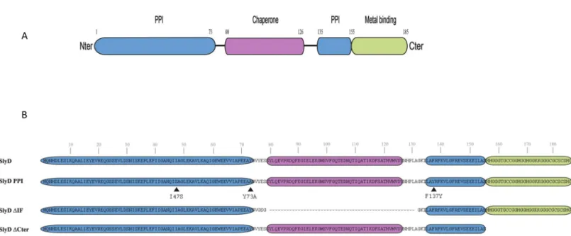
contributions of the different activities of the SlyD protein, a series of strains expressing mutant versions of SlyD from the native chromosomal locus under the control of the WT pro-moter was also constructed (Fig 1). Structural modeling based on the NMR structures of the SlyD proteins fromE. coli [49] and fromH. pylori strain 26695 [24] along with analysis of con-served residues in theH. pylori SlyD protein (S1A Fig), allowed us to predict the locations of the functional domains of SlyD (Figs1AandS1B). The mutants are represented inFig 1B. The first mutant designated SlyD-PPI, carries substitutions of three residues that are predicted to take part in the peptidyl-prolyl isomerase activity, I47S, Y73A and F137Y. The second mutant,
Fig 1. Illustration of theH. pylori SlyD wild type and mutant proteins. A. Schematic representation of the functional domains of the SlyD protein of H. pylori strain
B128, with indication of the corresponding encompassing residues. The regions required for the peptidyl-prolyl isomerase activity (PPIase) are colored in blue, the "inserted in Flap" IF chaperone domain is colored in pink and the C-terminal metal-binding region is colored in green. B. Illustration of the different SlyD mutants, SlyD-PPI, SlyD-ΔIF and SlyD-ΔCter. The three residues (I47S, Y73A and F137Y) changed in the SlyD-PPI mutant are marked by a black arrow.
designated SlyD-ΔIF carries a deletion of 56 residues encompassing the IF chaperone domain. In the last mutant strain, designated SlyD-ΔCter, the C-terminal His and Cys region-rich start-ing from residue 155 was removed. The strains expressstart-ing the SlyD mutants were viable and presented negligible growth and viability defects (S2A Fig).
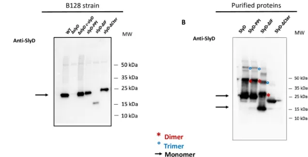
To evaluate the expression of each SlyD construct inH. pylori, we produced specific
anti-SlyD antibodies and performed Western analysis under reducing conditions (with DTT), revealing production of all SlyD variants, although at lower levels for the SlyD-ΔIF mutants (Fig 2A). The subcellular localization of SlyD was also analyzed by using a validatedH. pylori
fractionation procedure (S2B Fig). We found that SlyD protein is exclusively present in the sol-uble fraction strongly suggesting that it is a cytoplasmic protein.
Peptidyl-prolyl isomerase (PPIase) activity and nickel regulation of the
H.
pylori SlyD protein
To investigate the biochemical activities of theH. pylori SlyD variants, the corresponding
recombinant proteins were expressed and purified fromE. coli. Under non-reducing
condi-tions, we observed that the SlyD proteins form multimers (probably dimers and trimers) that are not observed upon analysis of the SlyD-ΔCter mutant (Fig 2B) or under reducing condi-tions (Fig 2A). These forms likely result from the spontaneous formation of disulfide bounds between the cysteine residues present in the C-terminal domain of SlyD (Fig 1B).
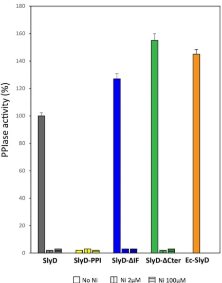
Circular dichroism spectroscopy analysis of the different SlyD proteins showed that the mutations did not result in major secondary structure changes (S3A Fig). Next, the PPIase activities of the purified wild type and mutant SlyD proteins were assayed by using anin vitro
assay in whichcis to trans prolyl isomerization of a tetrapeptide substrate is followed by
moni-toring the electronic absorption at 314 nm [50]. PurifiedE. coli SlyD protein served as a
posi-tive control (Fig 3). Our measurements revealed that the PPIase activity of wild typeH. pylori
Fig 2. Analysis of the production of SlyD wild type and mutant proteins inH. pylori. A. Western blot of equal amounts of total
extracts, under reducing conditions, fromH. pylori B128 WT strain and B128-derived mutants carrying the following mutations ΔslyD, ΔslyD c-slyD (complemented mutant), slyD-PPI, slyD-ΔIF, and slyD-ΔCter strain, that were probed with specific anti-SlyD polyclonal
antibodies prepared during this study. An arrow shows the position of the SlyD protein. B. Western blot of purified recombinant SlyD proteins (WT, SlyD-PPI, SlyD-ΔIF and SlyD-ΔCter) probed with specific anti-SlyD polyclonal antibodies. An arrow shows the position of the monomeric SlyD proteins, red stars and blue circles highlight SlyD dimers and trimers, respectively.
SlyD protein is similar (84%) to that of theE. coli protein. The H. pylori ΔIF and
SlyD-ΔCter mutants retained even higher activity than that of the WT protein (145% and 150%, respectively) (Fig 3). In contrast the SlyD-PPI mutant had, as anticipated, completely lost detectable PPIase activity.
In vitro PPIase activity of E. coli SlyD was reported to be negatively affected upon nickel
binding [25]. PurifiedH. pylori SlyD protein was previously reported to bind nickel ions with
aKdvalue of 2.74± 0.26 μM [24]. Therefore, the PPIase activities of purified SlyD proteins
(2μM) were assayed in the presence of 2 μM or 100 μM NiSO4. It was found that both nickel
concentrations totally inhibit the PPIase activity of all the SlyD variants (Figs3andS3B–S3C). These data established the PPIase activity of theH. pylori SlyD protein and validate the
importance of the three predicted active site residues. In addition, they demonstrate that nei-ther the chaperone domain nor the C-terminal region is required for PPIase activityin vitro
Fig 3.In vitro PPIase activity and nickel regulation of H. pylori wild type and mutant SlyD proteins. PPIase
activities of WT and mutant SlyD proteins, measured without or with 2μM or 100 μM NiSO4. The PPIase activity of
purifiedE. coli SlyD (EcSlyD) is also presented as a control. The data (S3B Fig) were fit to second-order rate equations and the PPIase activities are expressed as a percentage of the activity of the wild typeH. pylori SlyD protein. Grey bars
correspond to the wild type protein, yellow to the SlyD-PPI mutant, blue to the SlyD-ΔIF mutant and green to the SlyD-ΔCter mutant. The orange bar corresponds to the E. coli SlyD protein. The values are the averages from three replicates and error bars represent the standard deviation.
and that stoichiometric nickel inhibits this activity even in the absence of the C-terminal metal-binding region.
SlyD inactivation increases
H. pylori tolerance to high nickel
concentrations
The dependent regulation of the SlyD PPIase activity, along with its established nickel-binding properties [24], prompted us to examine whether this protein plays a role in nickel metabolism, transport and/or trafficking inH. pylori. We first tested whether SlyD is required
for the activation of the nickel-dependent enzymes ofH. pylori, urease and hydrogenase. We
found that urease and hydrogenase activities are not affected in theΔslyD mutant as compared to those of the wild type strain (S1 Table). In the closely related bacteriumCampylobacter jejuni, wild type hydrogenase activity was also measured in the ΔslyD mutant [51]. These results demonstrated that hydrogenase activity, in these two epsilon proteobacteria, does not require SlyD, in contrast to the situation inE. coli [41].
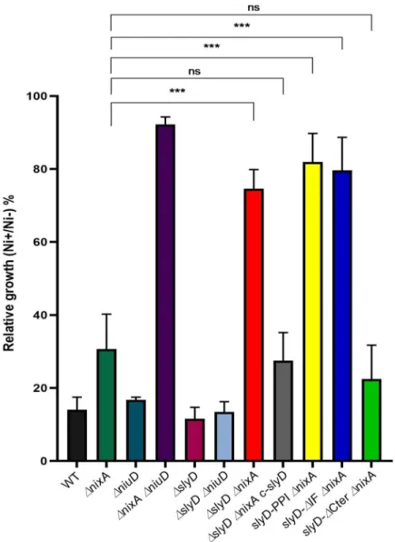
Then, the tolerance of the wild type strain andslyD mutants to toxic nickel exposure was
evaluated by measuring their growth at neutral pH in liquid BB medium after 24 h incubation with 1.5 mM NiCl2(Fig 4). Enhanced tolerance might result from several mechanisms, among
which decreased metal uptake, as we showed to be the case for anH. pylori mutant lacking
nickel transporters [13].
Here, we first found that theΔslyD mutant presented no difference in nickel tolerance. We next examined the effect of theΔslyD mutation in combination with deletions of the genes encoding the nickel uptake systems, NixA or NiuD (Fig 4). The single and double
ΔnixA and ΔniuD mutants behaved as we previously reported; the mutant deficient in
both nickel transporters being strongly tolerant to nickel [13]. The nickel tolerance of the
ΔslyD ΔniuD double mutant was similar to that of the single ΔniuD mutant. In contrast, it
was found that theΔslyD ΔnixA mutant was as highly tolerant to nickel as the ΔnixA
ΔniuD mutant, suggesting that nickel uptake is strongly impaired in the ΔslyD ΔnixA
strain. This phenotype could be complemented by the re-introduction of a wild typeslyD
copy (c-slyD), indeed the ΔslyD ΔnixA c-slyD strain recovered nickel tolerance levels
com-parable to that of the parentalΔnixA mutant (Fig 4). Then, the effect of mutations in the
slyD gene was tested in combination with ΔnixA (Fig 4). Deletion of the C-terminal domain of SlyD (strainslyD-ΔCter ΔnixA) did not impact the tolerance of bacteria to high
nickel concentrations. However, the mutants carrying either the SlyD-PPI mutation (slyD-PPI ΔnixA) or the deletion of its chaperone domain (slyD-ΔIF ΔnixA) were
insensi-tive to 1.5 mM nickel exposure just like theΔslyD ΔnixA mutant, attesting of the lack of activity ofslyD variants in this assay.
These results indicate thatH. pylori SlyD plays an essential role in nickel transport and/or
metabolism. In addition, under these test conditions, the C-terminal domain of SlyD is not essential for this activity but both the PPIase isomerase activity and chaperone domain of SlyD are required for its function related to nickel uptake or metabolism.
SlyD inactivation results in reduced intracellular nickel availability
One possible explanation for these results is that SlyD modulates nickel accumulation intoH. pylori. To test this hypothesis, we used an assay to indirectly evaluate the intracellular nickel
content of different mutants by measuring the expression offecA3, a gene known to be
repressed by the transcriptional regulator NikR, in response to intracellular nickel concentra-tions [18]. First, we introduced in our mutant strains, a stable plasmid carrying a reporter gene fusion PfecA3::lacZ that we previously validated as a reporter of intracellular nickel
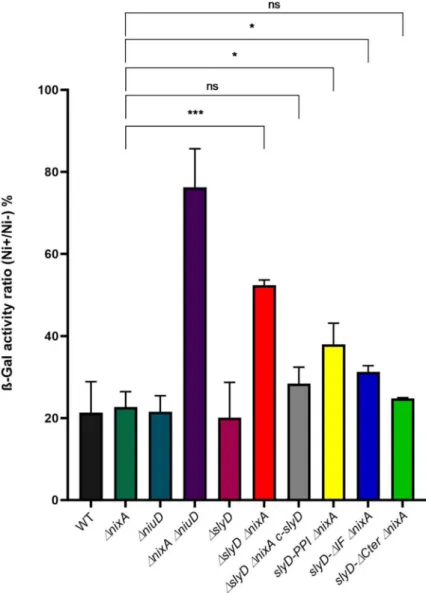
bioavailability [13].Fig 5presents the ratio of ß-galactosidase activities of strains grown for 24 h in media supplemented with 100μM NiCl2versus without additional nickel. Lower ratios
indicate stronger PfecA3::lacZ repression and thus higher intracellular nickel availability. The
wild type strain, as well as theΔslyD and ΔnixA individual mutants present ratios of about 15– 20%, attesting of proper nickel-dependent repression and therefore efficient nickel uptake whereas theΔniuD ΔnixA mutant presents a ratio of 70% as expected from its inability to import nickel [13]. TheΔslyD ΔnixA double mutant presents a ratio of 50% that is significantly
Fig 4. Tolerance ofH. pylori wild type and mutant strains to nickel exposure. Growth of H. pylori B128 wild type
strain, isogenicslyD mutants and a complemented strain (c-slyD) was measured after 24h in the presence of 1.5 mM
NiCl2or without added metal. The results are presented as the percentage of growth in the presence of nickel relative
to growth in its absence. In this figure as well as in Figs5,6and7, the same color codes were used for the bars corresponding to each strain or mutant which name is indicated below each bar. Black bars correspond to the wild type strain, dark green to theΔnixA mutant, dark blue to the ΔniuD mutant, violet to the ΔnixA ΔniuD mutant, dark
red to theΔslyD mutant, light blue to the ΔslyD ΔniuD mutant, bright red to ΔslyD ΔnixA mutant, light grey to the ΔslyD ΔnixA c-slyD mutant, yellow to the slyD-PPI ΔnixA mutant, bright blue to the slyD-ΔIF ΔnixA mutant and light
green to theslyD-ΔCter ΔnixA mutant. The data correspond to the mean value of three independent experiments.
Error bars represent the standard deviation. Statistics are presented only for the comparison with theΔnixA mutant:
���corresponds to p<0.001 and "ns" for non-significant.S9 Figpresents the complete statistical analysis of these data.
different from that of the singlenixA mutant, and could be complemented by the
reintroduc-tion of a wild typeslyD gene. The phenotype of the three targeted slyD mutants in combination
withΔnixA was next analyzed. The slyD-PPI ΔnixA and slyD-ΔIF ΔnixA mutants presented a weak reduction in the repression by nickel while the deletion of the SlyD C-terminal domain did not prevent its activity. In conclusion, these results revealed an essential contribution of SlyD to the accumulation of intracellular nickel and demonstrated that the C-terminal domain is not required for this activity.
Fig 5. Evaluation of intracellular nickel availability ofH. pylori wild type strain and isogenic mutants with a PfecA3::lacZ reporter fusion. ß-galactosidase activity of a PfecA3::lacZ reporter fusion expressed from a plasmid in
differentH. pylori B128-derived strains, after 24H exposure to 100 μM NiCl2. The expression of the fusion decreases in
a NikR-dependent manner with increasing intracellular nickel concentration. In medium without added nickel, the ß-galactosidase activities of the different strains were found to be comparable (about 6,000 miller units). ß-ß-galactosidase activities are presented as the ratio of activity measured in strains grown in the presence of 100μM NiCl2or in the
absence of nickel supplementation, expressed as a percentage. Color codes of the bars are as inFig 4. The data correspond to the mean value of three independent experiments (S5 Table). Error bars represent the standard deviation. Statistics are presented only for the comparison with theΔnixA mutant.�corresponds to p<0.05,���to
p<0.001 and "ns" for non-significant.S9 Figpresents the complete statistical analysis of these data.
Reduced intracellular nickel content of a
ΔslyD ΔnixA mutant
To evaluate the role of SlyD in nickel accumulation more precisely, we measured the total intracellular nickel content of our collection of mutants grown 24h in the presence of 100μM NiCl2, by Inductively-coupled plasma optical emission spectrometry (ICP-OES) as previously
reported [13] (Fig 6). Strains grown without supplemented nickel had metal levels below the detection limit. In agreement with our previous publication [13], the nickel content of the
ΔnixA and ΔnixA ΔniuD mutants was reduced 2.4-fold and 15-fold, respectively as compared
to the wild type strain. Consistent with the data presented above, theΔslyD mutant accumu-lated nickel as efficiently as the wild type strain. However, when theΔslyD mutation was com-bined withΔnixA, we measured a significant 1.5-fold and 3.5-fold reduction in nickel content
Fig 6. Measurement of the intracellular nickel content ofH. pylori wild type strain and isogenic mutants by ICP-OES. Nickel amounts were measured by Inductively Coupled Plasma Optical Emission Spectrometry (ICP-OES) and are expressed as the percentage of the ratio of nickel mass versus total sample mass. Strains were grown either without added nickel or with 100μM NiCl2. Color codes of the bars are as inFig 4. The measurement of each strain
under each condition was performed in triplicates in two experiments. Statistics are presented only for the comparison with theΔnixA mutant.�corresponds to p<0.05,���to p<0.001 and "ns" for non-significant.S9 Figpresents the
complete statistical analysis of these data.
as compared with theΔnixA single mutant and the wild type strain, respectively. This reduc-tion was clearly restored in the complementedΔslyD ΔnixA c-slyD strain. When compared to the full-length deletion ofslyD, only the slyD-IF mutant was significantly deficient in nickel
accumulation. These data confirm that SlyD is indeed involved in nickel accumulation inH. pylori and underline a major role of its chaperon function associated with the IF domain.
SlyD is required for the activity of the nickel ABC transporter Niu
Increased tolerance to nickel toxicity associated with decreased nickel availability and accumu-lation could result from impaired metal import, a reduction in intracellular nickel storage capacity or enhanced export. Concerning nickel efflux, the only published system is Czn [19]. However, in our strain and under our conditions, we did not detect any increase in nickel sen-sitivity in mutant strains lacking this system alone (ΔcznABC) or in combination with the
ΔslyD deletion (ΔcznABC ΔslyD). This excludes a role of the Czn efflux system in our nickel
tolerance phenotypes.
To define the role of SlyD in nickel import, the uptake rates of radioactive63NiCl2were
measured in the wild type and mutant strains at pH 5 during 30 min (Fig 7). The nickel uptake rate is moderately reduced in theΔnixA mutant and strongly decreased in the ΔnixA
ΔniuD mutant [13]. No significant change in the nickel uptake rate was seen in theΔslyD mutant compared to that of the wild type bacteria. However, theΔslyD ΔnixA mutant pre-sented a 50% reduction in63Ni uptake rate, which was significantly different from the rate of theΔnixA mutant and could be complemented with wild-type slyD. The three strains car-rying targetedslyD mutations in the ΔnixA background were also tested. The slyD-ΔIF ΔnixA strain exhibited a reduced uptake rate that phenocopied that of the ΔslyD ΔnixA
mutant. While theslyD-PPI ΔnixA behaved like the ΔnixA mutant, we observed that nickel
uptake of strainslyD-ΔCter ΔnixA was more robust than the ΔnixA mutant and closer to the
level of the WT strain.
These results demonstrate that, under these test conditions, SlyD is required for optimal nickel uptake by the Niu transporter and only its chaperone domain plays a crucial role in this process.
By which mechanism does SlyD impact the activity of the Niu transport
system?
The finding that a PPIase like SlyD regulates a metal transport activity has, to our knowledge, never been previously reported. Therefore, several tests were performed in order to investigate how SlyD regulates the Niu transport system. First, we examined whether SlyD controls the expression of theniu genes. Expression of the niuD and niuB1 genes (respectively, the first
gene of theniuDE operon and the first gene of the niuB1-niuB2 operon encoding the two
NiuB paralogous proteins [13]), as monitored by RT-qPCR, were similar in the wild type and
ΔslyD mutant (S4 Fig). This indicates that SlyD does not regulate the Niu transporter at the transcriptional level.
We then examined whether SlyD impacts the protein levels of components of the Niu sys-tem or their subcellular localization (S5 Fig). To this aim, the NiuB1 protein was purified and used to produce specific polyclonal antibodies. Contrary to NiuB1, attempts to produce anti-NiuD antibodies were unsuccessful. Therefore, a C-terminal fusion between theniuD gene
and a V5-tag was constructed and introduced at its original locus into the wild type strain and theΔslyD mutant. The amounts and localization of NiuD and NiuB were analyzed by Western blot after fractionation of crude extracts of bacteria grown without or with the addition of 100μM NiCl2, a non-toxic nickel amount (S5 Fig). As previously reported, production of the
NiuB and NiuD proteins was repressed by nickel [52,53]. Notably, SlyD impacted neither the amounts nor the localization of the two proteins. NiuB was found in both the soluble extract and inner membrane fractions in a NiuD-independent manner. As expected, NiuD was exclu-sively detected in the inner membrane.
Fig 7. SlyD is required for the uptake of radioactive nickel by the Niu nickel transporter. Measurements of radioactive nickel uptake rates inH. pylori B128 wild type strain and isogenic mutant strains in the presence of 10 μM
of63NiCl
2. Uptake rates are normalized to the rate of wild typeH. pylori strain. Color codes of the bars are as inFig 4.
Error bars represent the standard deviation. Statistics are presented only for the comparison with theΔnixA mutant.�
corresponds to p<0.05 and "ns" for non-significant.S9 Figpresents the complete statistical analysis of these data.
These results show that SlyD impacts neither the protein levels nor the subcellular localiza-tion of NiuD and NiuB. This suggests that SlyD might regulate the activity of the Niu trans-porter by direct protein interaction.
SlyD interacts with NiuD but not with NiuB or NiuE
We hypothesized that the control of the Niu transporter by SlyD might involve direct interac-tion between SlyD and one or more components of the Niu system. This possibility was exam-ined by using the Bacterial Adenylate Cyclase Two-Hybrid system (BACTH) and testing pairwise interactions between protein fusions inE. coli [54]. TheslyD gene was fused at its 5’
or 3’-extremity with a fragment encoding the T25 domain of theBordetella pertussis adenylate
cyclase into the pKT25 or pNKT25 vector, respectively. TheniuB1, niuE, and niuD genes were
fused at their 3’-extremities with the T18 fragment adenylate cyclase into the pUT18 vector. Fusions with the 5’-extremity of NiuD could not be obtained as they were toxic inE. coli.
No interaction was detected between SlyD and either the periplasmic nickel-binding pro-tein NiuB1 or the cytoplasmic NTP-binding propro-tein NiuE. In contrast, combinations of pNKT (slyD) with pUT18(niuD) (in which the 3’-extremity of niuD is fused to the T18 sequence)
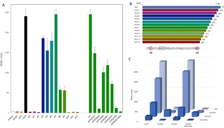
scored positive in the ß-galactosidase assay revealing an interaction between SlyD and the transmembrane permease subunit NiuD (Fig 8A). As a control, we verified that NiuD was
Fig 8. SlyD interacts with NiuD, the membrane permease of the Niu nickel uptake system. Bacterial two-hybrid BACTH was used to analyze, inE. coli strain
BTH101, the interaction between SlyD and the NiuD permease. The values and standard deviation for each strain and controls are available inS6 Table, and each measurement was performed three times. A. ß-galactosidase activities of pairwise combinations of WT SlyD with different truncated and mutant versions of the NiuD protein. B. Illustration of the truncated NiuD protein versions and sequence of the NiuD region surrounding the RWR motif in the region that is required for the interaction with SlyD. C. ß-galactosidase activities of pairwise combinations of wild type and mutant SlyD proteins (PPI,ΔIF and ΔCter) with wild type and mutant NiuD proteins.
correctly targeted to the inner membrane inE. coli (S6 Fig). To delineate the region of interac-tion of NiuD with SlyD, we constructed plasmids expressing T18 fusions of NiuD proteins with progressive C-terminal truncations (Figs8A and 8BandS7). The first NiuD deletions (Δ1, Δ2, Δ3) abolished its interaction with SlyD and larger deletions (Δ4, Δ5, Δ6, Δ7, Δ8, Δ9) restored the interaction with SlyD. These results can be interpreted in light of a predictive model of the NiuD protein folding and trans-membrane segments (S7 Fig). Indeed, this model suggests that theΔ1, Δ2, Δ3 fusions are predicted to expose the T18 fused domain into the peri-plasm, a localization that prevents its interaction with the cytoplasmic SlyD-T25 fusions. In contrast, the larger deletions (Δ4, Δ5, Δ6, Δ7, Δ8, Δ9) are compatible with a cytoplasmic expo-sure of the T18 domain. These interpretations also reinforce our confidence in the specificity of the NiuD-SlyD interaction. When we further deleted the NiuD protein (Δ10, Δ11, Δ12), the interaction was lost. The comparison betweenΔ9 (positive for interaction) and Δ10 (negative for interaction) allowed us to identify, at the NiuD C-terminus, a predicted cytoplasmic loop with three residues (Arg208-Trp209-Arg210) that are essential for NiuD interaction with SlyD.
To further determine which NiuD residues are important for its interaction with SlyD, we introduced mutations into plasmid pUT18(NiuDΔ7) that targeted the region defined above (Figs8A and 8BandS7). First, when we introduced a triple exchange of Gly for residues Arg208-Trp209-Arg210, the ß-galactosidase activity dramatically dropped, confirming the importance of this region for NiuD interaction with SlyD. In the cases of single mutants Pro203 to Val, and Trp209 to Phe or Gly, the ß-galactosidase activity decreased but was still significant, indicating that these residues are not crucial for the NiuD interaction with SlyD. In contrast, the interaction was abolished when we introduced into NiuD the Arg208 to Gln and Arg210 to Gly mutations, indicating that these two arginine residues are essential for the NiuD interaction with SlyD.
Finally, we checked the impact of SlyD mutations on the interaction with full-length NiuD andΔ7, which generated comparable results, and used Δ1 and the triple RWR mutant as nega-tive controls (Fig 8C). For the NiuD andΔ1 constructs, the interaction is maintained with the SlyD-PPI mutant and decreased with SlyD-IF. For the SlyD-Cter mutant, although the ß-galac-tosidase activity was diminished, the interaction was still detectable above baseline, and the reduced signal may be due to the reduced expression of this fusion given that the stability of this SlyD mutant protein is reduced (S6 Fig). Unexpectedly, the SlyD-IF mutant displayed sig-nificant interaction with the NiuDΔ1 and triple mutant negative controls suggesting that the IF chaperone region of SlyD contributes to the specificity to its protein interaction.
These data revealed a physical interaction between the SlyD protein and the NiuD permease subunit and defined regions of the transporter that are critical for this direct contact.
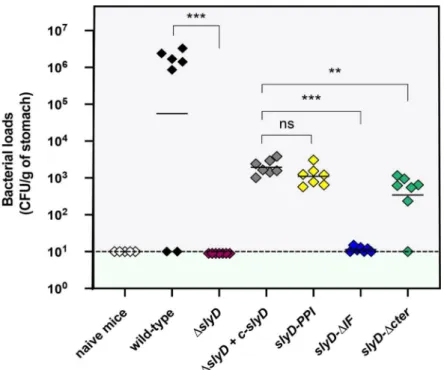
SlyD is essential for the mouse stomach colonization
The role of SlyD during gastric colonization was evaluated using the mouse model of infection by
H. pylori. Different slyD mutations were introduced into the H. pylori mouse-adapted strain SS1
[55]; these includedΔslyD, the complemented strain ΔslyD-c-slyD and the slyD-PPI, slyD-ΔIF and
slyD-ΔCter mutations. The expression of the SlyD protein in the WT and mutant strains was
vali-dated by western blot using anti-SlyD antibodies (S8 Fig). We orogastrically inoculated 109 bacte-ria of WT SS1 and every mutant strain in seven NMRI mice each. One month later, colonization was assessed by quantitative cultures of stomach homogenates. As presented inFig 9, theΔslyD mutant was completely deficient in its capacity to colonize the mouse stomach. The comple-mented strain SS1ΔslyD+c-slyD recovered partial capacity of colonization. This was also the case for the SS1slyD-PPI and SS1 slyD-ΔCter mutants. In contrast slyD-ΔIF mutant was completely
These results show thatslyD is indispensable for colonization of the mouse stomach. The
PPI and C-terminus domains are not essential forin vivo colonization. In contrast, the
chaper-one domain plays a major role in gastric colonization.
Discussion
Control of essential metal homeostasis is a complex and critical process. Bacteria have devel-oped two major strategies to maintain this equilibrium. First, bacteria produce in the cyto-plasm and/or the pericyto-plasm several "sequestering" proteins that specifically bind and store metals, preventing their association with unwanted targets [2]. The second strategy is control of metal import and/or efflux. In the vast majority of cases, expression of the metal transport systems is under the control of specific transcriptional regulators which have activities propor-tional to intracellular metal concentrations [2]. However, this type of regulation involves sev-eral steps as compared to post-translational regulation that was, to our knowledge, never reported for metal transporters in bacteria.
Here, such a novel mode of regulation was demonstrated; we found that the SlyD metallo-chaperone is required for the activity and thus active conformation of Niu, an essential nickel ABC transporter ofH. pylori. In addition, SlyD is crucial for colonization of a mouse model by
this pathogen. Our findings represent the first example of post-translational regulation of a metal transporter that relies on a peptidyl-prolyl isomerase (PPIase)-chaperone SlyD protein. To our knowledge, there is only one previous report ofcis-trans Proline isomerization being
required for activation of an ABC transporter [56]. In that instance, it was found that the peri-plasmic binding protein component of the system requires isomerization but the corresponding
Fig 9. SlyD is essential for mouse colonization byH. pylori SS1 strain. Each diamond corresponds to the
colonization load of one mouse one month after infection with theH. pylori strain indicated below. Each strain was
tested in a group of seven mice. The color codes for the different strains are as inFig 4. Horizontal bars represent the geometric means of the colonization load for the wild type bacteria, each mutant and the chromosomally
complementedΔslyD mutant (designated c-slyD). A dashed line shows the detection limit. The ΔslyD and isogenic mutants exhibited a statistically significant colonization defect when compared to the WT orΔslyD c-slyD strains, respectively (��p<0.01,���p<0.001 and "ns" non-significant).
PPIase was not identified. Enzymes that combine chaperone and PPIase activities have mainly been characterized inE. coli for which nine such proteins were reported [25]. Their general function is to accelerate protein folding, but actual substrates are mostly unknown. One excep-tion is the cytoplasmic Trigger factor (TF), a ribosome-associated molecular chaperone that, by acting as a foldase on numerous nascent polypeptides, protects them from misfolding and aggregation. TF mutants reported so far did not discriminate between the chaperone and PPIase activities and the PPIase activity is thought not to be central in bulk TF function [57]. InH. pylori, only three chaperones with PPIase activity are found, the periplasmic SurA homologue
(HP0175) [58] and two cytoplasmic proteins: a TF homologue [59] and SlyD [24].
Here, we explored thein vivo role of H. pylori SlyD, a multifaceted protein that combines
chaperone, PPIase and metal-binding properties. Using a collection ofH. pylori slyD mutants,
we characterized the function of this protein bothin vitro and in vivo. We found that, in vitro,
the SlyD PPIase activity was comparable to that ofE. coli SlyD, and validated three essential
active site residues. In our test, deletion of the IF chaperone domain did not affect PPIase activ-ity. This result contrasts with previous studies that found that the two domains act synergisti-cally, with the IF region being required for PPIase catalytic efficiency [24,60]. This discrepancy can be attributed to differences between the assays used. In these latter reports, protein refold-ing was measured, which reflects both chaperone and PPIase activities, while our test, usrefold-ing an isolated proline-containing tetrapeptide, specifically assays PPIase activity.
Using four differentin vivo tests, we consistently demonstrated that the SlyD protein is
important for the activity of the NiuBDE nickel ABC transporter. Using two-hybrid experi-ments, we found a direct interaction between the NiuD permease subunit and SlyD. Multi-component ABC transporters generally require molecular chaperones for correct insertion within membranes in order to prevent aggregation or accumulation of toxic "dead-end" inter-mediates [61]. However, we observed no significant change of NiuD protein levels in theH. pylori inner membrane of the ΔslyD mutant, suggesting that SlyD does not affect synthesis and
membrane insertion of NiuD but that it likely acts beyond this step. We found that the chaper-one activity of SlyD, located in the IF domain, is required for NiuD activity, probably by help-ing the permease to acquire an active conformation. Indeed, in our tests, the SlyD-ΔIF mutant phenocopied theΔslyD mutant with respect to nickel uptake; this is not due to a defect in PPIase activity, since deletion of the IF domain (SlyD-ΔIF) does not affect the PPIase activity
in vitro. In two-hybrid tests, the interaction between the SlyD-ΔIF mutant and NiuD was
reduced and the SlyD-ΔIF mutant presented, with negative controls, presumably non-specific interactions. We conclude that the SlyD IF domain is required for a productive NiuD-SlyD interaction and activation and, more generally, that the IF domain might be important for SlyD target recognition and discrimination. The function of the SlyD PPIase activity in the activation of Niu is more intriguing. Indeed, among the four tests that we performed, the
slyD-PPI mutant only robustly phenocopied the ΔslyD mutation in the assay of tolerance to
the high 1.5 mM nickel concentration. In the other tests, where nickel concentration was much lower (100μM and 10 μM), the SlyD-PPI mutant did not display a major loss of Niu activation. In addition, as suggested by the results of the two-hybrid assays, the nickel tolerance phenotype of the SlyD-PPI mutant is not a consequence of a loss of physical interaction between SlyD-PPI and NiuD. Thus, we conclude that the PPIase function of SlyD only becomes criticalin vivo under conditions of nickel overload, possibly through regulation of
the NiuD conformation under these conditions (for instance through isomerization of the bonds of two proline residues close to the interacting region that we defined by two-hybrid) but it might also result from a role in the function of other nickel-resistance factors.
We also investigated the role of the C-terminal nickel-binding region of SlyD in Niu activa-tion. This was particularly interesting since nickel binding at the SlyD C-terminal region was
reported to inhibit PPIase activity of theE. coli protein [25] and Chenget al. [24] observed conformational changes around the FKBP/PPIase domain after nickel binding toH. pylori
SlyD.In vitro, we found that removal of the SlyD C-terminal region resulted in increased in vitro PPIase activity. Furthermore, addition of an equivalent of nickel completely inhibited the
PPIase activity of the wild type SlyD protein and this inhibition was preserved in the SlyD mutant lacking a C-terminal region. This result indicates that down-regulation by nickel is not only relying on the C-terminal region but also on other binding sites in the SlyD N-terminal domains. In future experiments, it will be interesting to define which residue(s) of the N-ter-minal SlyD domains is(are) required for this C-terN-ter-minal independent inhibition.
Analysis of the phenotype of the SlyD C-terminal deletion mutant inH. pylori reveals that
this region of the protein is not required for the impact of SlyD on nickel metabolism. Thus, the diminished ß-galactosidase activity measured when SlyD-ΔCter was tested with NiuD in two-hybrid assays was unexpected. However, we could attribute this result to a lower level of expression of the ΔCter mutant under these test conditions. Interestingly, the SlyD-ΔCter mutant presented slightly higher radioactive nickel uptake rates. We speculate that the C-terminal SlyD region could negatively regulate its chaperone activity, an aspect that we intend to analyze in future studies.
The two-hybrid assays allowed us to define regions of NiuD that are important for its inter-action with SlyD (Figs8andS7). We defined a three amino-acid long motif of NiuD (RWR), predicted to be located within a cytoplasmic loop between transmembrane helices 6 and 7, that is essential for its interaction with SlyD. Interestingly, this loop (and this RWR motif) is surrounded by two proline residues (Pro203, Pro228) that might be substrates for the PPIase SlyD activity. We speculate that isomerization of the corresponding AA-proline bonds might induce conformational changes either in the cytoplasmic loop containing the RWR motif, or in the flanking transmembrane helices, which could positively impact NiuD activity.
Finally, we demonstrated that SlyD is essential for colonization of the mouse stomach byH. pylori. PPIases belonging to other families have been associated with the virulence of several
bacteria [62,63]. In the mouse model, we found that the chaperone function of SlyD is essential and observed minor effects of the PPI andΔCter mutations. We previously published that the Niu transporter is essential for colonization [13]. Thus, it is likely that the essentiality of SlyD
in vivo is, at least partially, due to defective nickel uptake by the Niu system. However, it is
pos-sible that additionalin vivo essential functions are disturbed in the absence of SlyD.
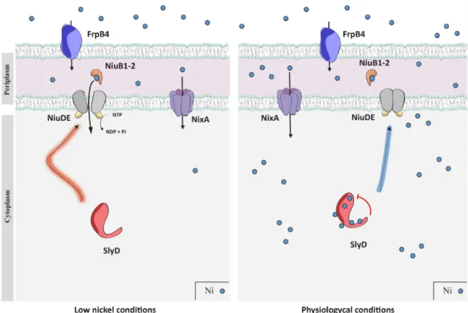
We propose a model for the regulation of the NiuD nickel permease by SlyD that implies direct protein interaction (Fig 10). The SlyD chaperone activity is essential for the correct fold-ing of NiuD or its association with its partners and thus for the functionfold-ing of the Niu trans-porter. SlyD could, in addition, play the role of a sensor of intracellular nickel concentration and act as a "gatekeeper" of nickel uptake allowing a shut down or activation of the Niu trans-porter when nickel bio-availability increases or decreases, respectively.
In conclusion, the role of SlyD inH. pylori is reminiscent of the multiple signaling roles of
eukaryotic PPIases in cellular processes including immune response, neuronal differentiation and cell cycle control. We anticipate that bacterial PPIases have similar important properties that have, till now, been largely underestimated.
Material and methods
Ethics statement
Experiments in mice were carried out in strict accordance with the recommendations in the Specific Guide for the Care and the Use of Laboratory Animals of the Institut Pasteur, accord-ing to the European Directive (2010/63/UE) and the correspondaccord-ing French law on animal
experimentation (Arrête´s de 1988). The protocol has been approved by the Committee of Cen-tral Animal Facility Board of the Institut Pasteur (#08–551). To follow the new European directives, the project was approved by the CETEA, Comite´ d’e´thique en Expe´rimentation Animale of the Institut Pasteur (#2013–0051) and by the Ministère de l’Enseignement Supe´r-ieur et de la recherche (#751501).
Bacterial strains and growth conditions
TheH. pylori strains used in this study are derivatives of B128 [47,48] and SS1 [55] (S2 Table). They were grown at 37˚C under microaerophilic conditions (6% O2, 10% CO2, 84% N2) on
Blood Agar Base 2 (Oxoid) plates supplemented with 10% defibrinated horse blood or Brucella broth agar (BD Difco) plates (designated BB) supplemented with 10% fetal calf serum (FCS, Eurobio). For liquid cultures, we used Brucella broth (BD Difco), designated BB, supple-mented with 10% fetal calf serum (Eurobio) or with 0.2%β-cyclodextrin (Sigma), designated BBß. All plates and liquid cultures were supplemented with the following antibiotics-anti-fun-gal cocktail: amphotericin B 2.5μg.mL-1, polymyxin B 0.31μg.mL-1, trimethoprim 6.25μg.mL
-1
and vancomycin 12.5μg.mL-1. Selection ofH. pylori mutants and transformants was
Fig 10. Model for the regulation of nickel uptake by the SlyD protein. InH. pylori, nickel ions (small blue dots) are transported across the
outer membrane by FrpB4 (blue), a TonB-dependent transporter. Once in the periplasm, uptake of nickel through the inner membrane can be performed by the NixA permease (violet) or the ABC-transporter, NiuBDE (orange, grey and yellow). NiuB1-2 (orange) are the periplasmic nickel shuttles that deliver the metal to the NiuD permease, which activity is energized by the NiuE NTPase. The function of the Niu transporter requires activation by SlyD (red), a regulation that relies on direct interaction between SlyD and the NiuD permease. This is illustrated on the left panel, presenting conditions of low nickel availability. On the right panel, when nickel is available, binding of nickel to SlyD regulates its PPIase activity. This allows SlyD to sense the intracellular nickel concentration and to act as a gate keeper to control nickel entry.
performed using kanamycin 20μg.mL-1, chloramphenicol 6μg.mL-1, streptomycin 10μg.mL-1 or apramycin 10μg.mL-1.Escherichia coli XL1-Blue bacteria grown on solid or liquid
Luria-Bertani (LB) medium was used for subcloning and as a host for the preparation of the plasmids employed to transformH. pylori. E. coli strain BTH101 was used for Bacterial Two Hybrid and
BL21(DE3)ΔslyD::apra for protein overexpression and purification (S2 Table). LB medium was supplemented with chloramphenicol 30μg.mL-1, ampicillin 100μg.mL-1or kanamycin 40μg.mL-1when required.
Molecular technics
Molecular biology experiments were performed according to standard procedures and the supplier (Fermentas) recommendations. NucleoBond Xtra Midi Kit (Macherey-Nagel) and QIAamp DNA Mini Kit (Qiagen) were used for plasmid preparations andH. pylori genomic
DNA extractions, respectively. PCR was performed either with DreamTaq DNA polymerase (ThermoFisher), Q5 DNA polymerase (Biolabs) or with PrimeSTAR Max DNA polymerase (Takara) when the product required high fidelity polymerase. The pGEMT vector (Promega,
S3 Table) was used to construct, inE. coli, the suicide plasmids that served for mutagenesis in H. pylori.
Construction of
H. pylori slyD mutants and of the niuD-V5 fusion strain
An unmarkedslyD deletion mutant of H. pylori strain B128 (S2 Table) was constructed by alle-lic exchange as previously described [13]. We used aH. pylori suicide plasmid derived from
pGEMT, in which about 500 bp of the 5’-end and the 3’-end regions immediately flanking the open reading frame ofslyD gene were cloned on each side of a difH-cat-rpsL-difH cassette
amplified with primers difH-rpsL-cat1 and difH-rpsL-cat2 (primers are listed inS4 Table). This plasmid (S3 Table) was used to naturally transformH. pylori strain B128 that we made
Streptomycin resistant. The insertion of the cassette by homologous recombination was selected on blood agar plates containing chloramphenicol 6μg.mL-1. Removal of the cassette was achieved by plating the CmRclones on blood agar plates containing streptomycin 10μg. mL-1. The Gibson assembly method was used to obtain a PCR product designed to introduce a
slyD deletion in H. pylori strain SS1 (S2 Table). This PCR product carried a Cm resistance cas-sette flanked by 500 bp upstream and downstreamslyD PCR fragments. Deletion of the slyD
gene was verified in both genetic backgrounds by PCR and sequencing of the gene region. Plasmids used for these constructs are listed inS3 Table. TheslyD deletions of strains B128
and SS1 were complemented by reintroducing a wild typeslyD copy at its original locus on the
chromosome under the control of its own promotor. For that, a PCR product was generated with the Gibson assembly procedure. This PCR product comprised theslyD gene followed by
the apramycin resistance cassette and flanked by the 500 bp upstream and downstream flank-ing region of theslyD gene. The final PCR-amplified product was used to directly transform in
B128ΔslyD or SS1 ΔslyD::Km strain resulting in a strain in which the wild type slyD gene and theapra cassette integrated by homologous recombination between flanking regions of the slyD locus. The same procedure was used to introduce slyD versions carrying mutations (PPI,
ΔIF, ΔCter) in strain B128 and SS1. Sequencing was performed in both genetic backgrounds to verify the deletions and reintroduction of wild type or mutated versions ofslyD at the correct
locus. The expression of the mutated SlyD versions in B128 and SS1 was validated by western blot.
The NiuD-V5 fusion was obtained by gene synthesis (Eurofins). A PCR fragment carrying this fusion, a kanamycin resistance cassette and 500 bp downstream theniuD gene were fused
naturally transformed intoH. pylori and allelic exchange was selected on kanamycin. Correct
insertion of the fusion and cassette were verified by PCR and sequencing.
Purification of recombinant
H. pylori SlyD proteins and circular dichroism
spectroscopy analysis
SlyD wild-type (WT) and mutants (PPI,ΔIF and ΔCter) proteins were expressed according to the method previously described [38] in aΔslyD::apra BL21(DE3) strain of E. coli using pET28 (a)+ vector (Novagen). SlyD proteins were purified by using a nickel-nitrilotriacetic acid (Ni-NTA, Qiagen) column and the presence of SlyD in the protein fractions collected at each step was verified by SDS-PAGE analysis. Cleavage with thrombin was initiated by the addition of 0.3 U thrombin protease (Novagen) per mg target protein. The purified proteins were dialyzed overnight in thrombin cleavage buffer (20 mM Tris-HCl, pH 8.4, 150 mM NaCl, 2.5 mM CaCl2) for 12 hours at 4˚C. This step was followed by anion exchange on a MonoQ column
(GE Healthcare) in Tris-HCl 20mM + 200mM NaCl, 1mM TCEP buffer. Selected fractions were pooled and used for verification by Mass Spectrometry analysis (Department of Chemis-try, University of Toronto) and further activity assays.
CD spectra were recorded on an Olis rapid scanning monochromator at room temperature. Approximately 15 or 25μM protein was used in 5 mM Phosphate buffer, pH 7.6. CD spectra were collected by scanning the wavelength range of 200–260 nm using a step size of 1 nm and an integration time of 2 s. Five scans were averaged for each sample, and each CD spectrum was normalized to mean residue ellipticity [θ]mre(deg cm2dmol–1) using the equation [θ]mre=
[(MM/N– 1) × θ]/(c × l × 10)], where MM is the molecular mass of the protein in Da, N is the
number of residues,θ is the measured ellipticity (degrees), c is the total protein concentration in g/mL, andl is the cell path length. The averaged spectra were smoothed by using a
three-period moving average. The concentrations of the SlyD samples were confirmed after the scans.
Peptidyl-prolyl isomerase activity assays
An uncoupled protease-free assay was used to measure the PPIase activity of SlyD and the vari-ants [40,50]. The substrate, succinyl-Ala-Phe-Pro-Phe-4-pNa (Bachem Bioscience), was dis-solved in trifluoroethanol (dried over sieves) and 0.47 M LiCl (dried in 220˚C oven overnight). The reactions contained 35 mM HEPES, pH 7.6, and 2μM of each purified SlyD protein (wild type or mutants) and were incubated at 10˚C before the addition of 71μM substrate with/out 2 or 100μM of NiSO4. Isomerization was monitored at 314 nm at 10˚C on a Cintra 404
spec-trophotometer and fit to a single exponential decay. The time course of the reversible first-order prolyl isomerization was measured during 180 seconds, and the apparent second-first-order rate constant was calculated.
Metal sensitivity assay and evaluation of intracellular nickel content
The effect of metal exposure onH. pylori growth was tested by inoculating bacteria at OD6000.1, in 10 mL liquid medium (BB with FCS) without or with 1.5 mM NiCl2. Bacterial growth
was monitored 24 hours later by measuring their OD600. The data correspond to at least three
independent experiments.
For the evaluation of the intracellular nickel content, we used a PfecA3promoter fusion as a
reporter, as previously validated [13]. The PfecA3promoter is under the control of the
nickel-responsive transcriptional regulator ofH. pylori, NikR and its activity is thus proportional to
the intracellular nickel concentration. A pILL2157(PfecA3::lacZ) fusion plasmid (S3 Table) was
gene,H. pylori bacteria were grown on blood agar plates for 24 hours, then inoculated at
OD6000.05 in BB FCS liquid medium and grown overnight. This preculture was used to
inocu-late the bacteria at OD6000.1 in liquid BB FCS without or with the addition of 100μM NiCl2.
After 24h, theβ-galactosidase activity of these cultures was measured to monitor the response to nickel of the reporter fusion, and thus NikR activity (the values are available inS5 Table).
Nickel content measurements by Inductively Coupled Plasma Optical
Emission Spectrometry (ICP-OES)
Overnight liquid cultures ofH. pylori strain were diluted and grown until OD6000.1 at 37˚C in
15 ml Brucella-Broth with FCS, then 100μM NiCl2were added and the cultures were left to
grow until OD6006 after 24h. Then, the cultures were washed once with PBS-1X prior to be
resuspended in cold PBS-1X with EDTA 1mM and adjusted at OD60010. Six mL of this culture
preparation were centrifuged at 4,000 g at 4˚C for 25 min through 400μL of a 1:2 mixture of the silicone oils AR20/AR200 (Wacker) in order to separate the cells from the medium. Pellet were dried by speed-vac for 2h at 60˚C. Ten mg of the pellet were mineralized overnight with a solution mix of 500μL nitric acid 69% (EMSURE) and 500 μL sulfuric acid 96% (Alfa Aesar). After mineralization, MiliQ water was added in each sample to a final volume of 20 mL. Nickel content was measured by ICP-OES with an Agilent 720 Series with axially-viewed plasma and with a Ni calibration curve of 10–1,000 ppb at “Institut Lavoisier de Versailles”. The content of Ni(II) was determined using a curve established with certified ICP grade nickel-standards. The measurement of each strain under each condition was performed in triplicates in two experi-ments. The results are presented as the percentage of the ratio of nickel mass versus total sam-ple mass.
Transport of radioactive nickel
The procedure was adapted from our previously published protocol [13]. The preculture of B128 wild type and isogenic mutants was used to inoculate, at OD6000.1, 10 mL of fresh BB
medium supplemented with 10% FCS and incubated under microaerophilic conditions with shaking at 37˚C. When the cultures reached OD6000.5, cells were harvested, washed and
resus-pended in the same volume of BBβ and shaken during 20 min under microaerophilic condi-tions at 37˚C. Radioactive63NiCl2(3.953 mCi/mL), was isotopically diluted 10-fold with cold
NiCl2and added at a final concentration of 10μM. 63
NiCl2was supplied by the Eckert &
Zieg-ler Isotope Products (Valencia, CA USA). Kinetics were performed for 30 minutes. Aliquots of 1 mL were withdrawn, immediately vacuum filtered through 0.45μm pore-size cm filters (diameter = 2.5; Millipore) and washed with 10 mL of 50 mM HEPES buffer (pH 7.0), 1 mM cold NiCl2. Two series of experiments were performed and each time point was measured in
duplicates. Uptake rates were calculated as CPM of accumulated63Ni as a function of time.
Urease and [NiFe] hydrogenase activity measurements in
H. pylori strains
Urease activity of wholeH. pylori cells was assayed by measuring the ammonia production
using the Ammonia-Assay kit (Sigma) as described [13]. The NH3concentration in the
super-natant was measured with the ammonia-assay kit according to the manufacturer’s (Sigma) instructions. Hydrogen uptake activity was determined spectrophotometrically at 604 nm by following the color change of methyl viologen (MV) from a colorless oxidized form to a dark-violet reduced form as described in [20]. The data correspond to at least three independent experiments with two technical replicates each time.
Bacterial Two-Hybrid assays
The Bacterial Two-Hybrid (BACTH) test is based on the reconstitution of adenylate cyclase activity in acya-E. coli strain as a result of the interaction between two proteins: a bait and a
prey fused to two separate catalytic domains (T18 and T25) of theBordetella pertussis
adenyl-ate cyclase. Empty pNKT25 an pUT18 vectors served as controls of background adenyladenyl-ate cyclase activity [54]. To detect interactions between the SlyD and Niu proteins,slyD and niu
genes were amplified by PCR using primers listed inS4 Tableand chromosomal DNA from B128H. pylori strain as a template.
Several plasmids were constructed (S3 Table) expressing either an N-terminal or a C-termi-nal fusion of these proteins with the T25 catalytic domain (derived from vectors pKNT25 and pKT25, respectively) or either a N-terminal or a C-terminal fusion with the T18 catalytic domain (derived from vectors pUT18 and pUT18C, respectively). All inserts were digested by
XhoI and EcoRI, and were then cloned into plasmids pUT18, pNKT25, pUT18C and pKT25
(PCR primers listed inS4 Table). The two plasmids expressing fusions to be tested were co-transformed inE. coli strain BTH101 and transformants were selected in Luria-Bertani agar
plates containing kanamycin and ampicillin at 30˚C. To avoid toxic effect of transformation of the plasmid pUT18(niuD+) into the recipient strain BTH101, 1% glucose was added to the media. Five mL of LB medium supplemented with antibiotics and IPTG 10−3M were inocu-lated with the transformants clones and incubated overnight at 30˚C. Quantification of the interactions in strains carrying each plasmid combination was obtained by measurement of theβ-galactosidase activity expressed in Miller units that was performed in at least 5 replicates as in [54] (S6 Table).
RNA extraction and cDNA synthesis
A total of 30 ml of three independent cultures grown at pH7 for 24 h with/out 100μM NiCl2
was centrifuged for 15 min at 4000g, treated with RNA protect solution (Qiagen) and stored at
-80˚C. Cells were lysed and RNA was extracted with the Nucleospin miRNA kit (Macherey-Nagel). RNA was incubated for 30 minutes at 37˚C with 2 U/μL of Turbo DNase-free enzyme (Ambion). Synthesis of cDNA reactions were carried out following the manufacturer’s proto-col using SuperScript IV First-Strand Synthesis System (ThermoFisher), starting with 1μg total RNA. cDNA was final diluted to 10 ng/μl in Nuclease-free water. qRT-PCR Mix was per-formed with Power SYBR Green PCR Master Mix (Applied Biosystems), 900 nM of each primer (S4 Table), and 30 ng of total cDNA. PCR products were amplified and detected with an Applied Biosystem (Thermofisher) machine. The cycling conditions were as follows: one cycle at 95˚C for 10min, 45 cycles at 95˚C for 15 s and 60˚C for 2 min, and 80 cycles at 55˚C for 30 s with a 0.5˚C increase every 30 s. The transcript levels were normalized to the level of the housekeepingppK (encoding polyphosphate kinase, HP1010) as previously validated [18]. The data correspond at least two independent experiments with two technical replicates each time.
Fractionation and western blots
RecombinantH. pylori SlyD protein was purified from E. coli and used to raise polyclonal
anti-bodies that were validated by Western blot under reducing conditions (with DTT) (Fig 2A). The cellular fractionation protocol was adapted from [64].H. pylori cells were grown to an
OD600of 0.8–1, then harvested by centrifugation and washed twice in PBS prior to be
resus-pended at OD60010 and disrupted by sonication in a lysis buffer containing 10 mM Tris-HCl
pH7.5 (buffer A) and Complete Protease Inhibitor Cocktail (Roche). Cell debris was removed by centrifugation at 20,000g at 4˚C for 15 minutes and supernatants were collected as total