HAL Id: hal-02959591
https://hal.archives-ouvertes.fr/hal-02959591
Submitted on 6 Oct 2020
HAL is a multi-disciplinary open access
archive for the deposit and dissemination of
sci-entific research documents, whether they are
pub-lished or not. The documents may come from
teaching and research institutions in France or
abroad, or from public or private research centers.
L’archive ouverte pluridisciplinaire HAL, est
destinée au dépôt et à la diffusion de documents
scientifiques de niveau recherche, publiés ou non,
émanant des établissements d’enseignement et de
recherche français ou étrangers, des laboratoires
publics ou privés.
Perturbation of the circadian clock and pathogenesis of
NAFLD.
Atish Mukherji, Mayssa Dachraoui, Thomas F. Baumert
To cite this version:
Atish Mukherji,
Mayssa Dachraoui,
Thomas F. Baumert.
Perturbation of the
circa-dian clock and pathogenesis of NAFLD..
Metabolism, clinical and experimental, 2020,
Perturbation of the circadian clock and pathogenesis of NAFLD
Atish Mukherji
a,⁎
,1,
Mayssa Dachraoui
a,1, Thomas F. Baumert
a,b,⁎
aUniversité de Strasbourg, Inserm, Institut de Recherche sur les Maladies Virales et Hépatiques INSERM, UMR_S 1110, Strasbourg, France
b
Pôle Hépato-Digestif, Institut Hospitalo-Universitaire, Hôpitaux Universitaires de Strasbourg, Strasbourg, France
a b s t r a c t
a r t i c l e i n f o
Article history: Received 25 May 2020
Received in revised form 24 July 2020 Accepted 26 July 2020
Available online xxxx
All living organisms including humans, experience changes in the light exposure generated by the Earth's rota-tion. In anticipation of this unavoidable geo-physical variability, and to generate an appropriate biochemical re-sponse, species of many phyla, including mammals have evolved a nearly 24-hour endogenous timing device known as the circadian clock (CC), which is self-sustained, cell autonomous and is present in every cell type. At the heart of the ‘clock’ functioning resides the CC-oscillator, an elegantly designed transcriptional-translational feedback system. Notably, the core components of the CC-oscillator not only drive daily rhythmicity of their own synthesis, but also generate circadian phase-specific variability in the expression levels of thousands of target genes through transcriptional, post-transcriptional and post-translational mechanisms. Thereby, this ‘clock’-system provides proper chronological coordination in the functioning of cells, tissues and organs. The CC governs many physiologically critical functions. Among these functions, the key role of the CC in maintaining metabolic homeostasis deserves special emphasis. Indeed, the several features of the modern lifestyle (e.g. travel-induced jet lag, rotating shift work, energy-dense food) which, force disruption of circadian rhythms have re-cently emerged as a major driver to global health problems like obesity, cardiovascular disease and metabolic liver disease such as non-alcoholic fatty liver disease (NAFLD). Here we review, the CC-dependent pathways in different tissues which play critical roles in mediating several critical metabolic functions under physiological conditions and discuss their impact for the development of metabolic disease with a focus on the liver. © 2020 The Authors. Published by Elsevier Inc. This is an open access article under the CC BY-NC-ND license (http://
creativecommons.org/licenses/by-nc-nd/4.0/).
Contents
1. Introduction . . . 0
2. Mammalian circadian clock: anatomic and molecular organization . . . 0
3. Cross talk between clock and feeding cycles . . . 0
4. Pathophysiology of the non-alcoholic fatty liver disease (NAFLD) . . . 0
5. Peripheral circadian clocks: regulation of metabolism and impact on pathogenesis of NAFLD . . . 0
5.1. Liver . . . 0
5.2. Pancreas . . . 0
5.3. Intestine and microbiota . . . 0
5.4. Immune system . . . 0
6. Circadian clock-related therapeutic interventions . . . 0
7. Conclusion . . . 0
. . . 0
References . . . 0
1. Introduction
Diurnal alteration between the light (day) and the darkness (night) is unavoidable, and to adopt to this environmental variable in a manner which best suits to physiology, circadian rhythms have evolved over Metabolism Clinical and Experimental xxx (xxxx) xxx
⁎ Corresponding authors at: Inserm U1110, Institut de Recherche sur les Maladies Virales et Hépatiques, 3 Rue Koeberlé, 67000 Strasbourg, France.
E-mail addresses:mukherji@unistra.fr(A. Mukherji),thomas.baumert@unistra.fr
(T.F. Baumert).
1
Atish Mukherji and Mayssa Dachraoui contributed equally.
YMETA-154337; No of Pages 9
https://doi.org/10.1016/j.metabol.2020.154337
0026-0495/© 2020 The Authors. Published by Elsevier Inc. This is an open access article under the CC BY-NC-ND license (http://creativecommons.org/licenses/by-nc-nd/4.0/).
Contents lists available atScienceDirect
Metabolism Clinical and Experimental
billions of years, and are displayed by nearly all living organisms. This clear separation between light-dark periods induces active- and rest-phases in various phyla including mammals. The circadian clock (CC) an endogenous‘time-keeper’ is the key link between these environ-mental variable factors and organismal physiology, as it sets an adaptive rhythm for physiological mechanisms, as it allows them to be antici-pated [1–6]. The idea of CC regulating critical functions was noted as early as the 18th century regarding the diurnal movement of plant leaves. In 1959, Franz Hallberg coined the term‘circadian rhythm’ (latin origin: about a day) to acknowledge the periodicity of these bio-logical rhythms. Subsequently, landmark investigations conducted in Drosophila provided thefirst evidence of genes controlling the circadian rhythm [7]. Since then, numerous studies have established plethora of molecular mechanisms which generate and maintain these ~24 h rhythms. The importance of investigations on the CC was exemplified by the award of the Nobel Prize in Medicine and Physiology (2017) to professors Jeffrey C. Hall, Michael Rosbash and Michael Young. The CC-rhythms have allowed mammals to anticipate changes in the external environment (e.g. day-night), and to respond by adjusting cellular CC-machinery driven numerous physiological functions, e.g. metabolism and endocrine functions [1–6]. Accordingly, recent molecular- and genetic-studies have demonstrated that, in mammals, the expression of numerous genes in different organs display circadian rhythmicity, thus enabling control of both anabolism and catabolism. As an example, food absorption, processing, assimilation and oxidative burning of nu-trients all display through circadian variations, thus enabling their tem-poral adjustment with food availability and bio-energetic need of the organism [1–6]. Under physiological conditions these ‘metabolic rhythms’ are generated and maintained by the dynamic interactions be-tween the CC and timing cues e.g. light and food (eating time and its quality). Importantly, in our modern industrialized world, various human behaviors and activities such as shift work, jet lag, energy-dense fatty foods and sleep deprivation often interfere with these rhythms and disrupt CC-functioning. Unsurprisingly then that disrup-tion of CC funcdisrup-tioning has recently emerged as a major contributor to different metabolic diseases, as well as carcinogenesis [1–5,8–16]. Therefore, detailed comprehension of the mechanistic basis of the CC-control on gene expression is critical to develop novel therapeutics for metabolic disorders whose therapeutic efficacy may be administration time-of -day dependent.
In this review, we lay emphasis on how the CC regulates metabolism in different peripheral organs to maintain metabolic homeostasis and provide overview of how the disruption of these CC-regulated processes could lead to the development of NAFLD. Furthermore, we also briefly discuss the potential of chronopharmacology in therapeutics.
2. Mammalian circadian clock: anatomic and molecular organization Well-known light receptors (rods and cones) converts the energy of the light signal to electrical impulses and relays them to the brain by uti-lizing retinal ganglion cells (RGC). It has been demonstrated that the melanopsin (photopigment) expressing RGCs directly relay the photic signal to a group of neurons in the anterior hypothalamus known as the suprachiasmatic nucleus (SCN;8–9), which by anatomical design acts as the‘central/master’ CC. The SCN-CC in turn, by utilizing barely understood humoral and neuronal mechanisms transmits the‘time in-formation’ (a. k. a; ‘Zeitgeber’; ZT) to other peripheral organs (synchro-nization of peripheral CCs; 1–6,8–9).
The molecular architecture of the CC-functioning has been discov-ered over the 30 years [1–6,17]. Remarkably, the SCN-CC and PCCs are constituted by identical molecular components which regulate them-selves using a similar transcriptional-translational feedback loops (TTFL). At the core of the molecular CC functioning (in mammals) re-sides the heterodimer of transcription factors BMAL1 and CLOCK, which acts as the trans-activator of genes containing E-box DNA binding sequences (DBS). BMAL1/CLOCK-drives the transcription of the Period
(PER1/2) and the Cryptochrome (CRY1/2) genes, whose protein prod-ucts heterodimerise to inhibit the transcriptional functions of the BMAL1/CLOCK-complex [1–6], thereby leading to the reduction in their own expression, thus constituting the so-calledfirst loop of the CC-oscillator (Fig. 1). During the early rest phase, high transcriptional activity of BMAL1/CLOCK drives the accumulation of its products CRY1/2 and PER1/2 (in late rest phase) which subsequently dimerise and inhibit BMAL1/CLOCK-activity during the active phase [17]. In the ‘second’ loop of the oscillator, BMAL1/CLOCK- activates the transcrip-tion of the nuclear receptors (NR) Rev-Erbα and Rev-Erbβ [18] during the rest phase [1–5]. Molecularly, REV-ERBs act as transcriptional re-pressors by binding to the ROR-response elements (RORE) present in numerous target genes including themselves. Importantly, REV-ERBs by repressing the transcription of Bmal1 and Clock genes reduce their own expression, thus closing this second loop. Importantly, during the active phase another set of NRs RORα/γ are recruited to these same RORE-DBSs to activate the expression of Bmal1 and Clock genes [1–5]. This phase-specific recruitment and accumulation of RORα/γ (activa-tors) and REV-ERBα/β (repressors) induces rhythmicity in Bmal1 and Clock expression, thus generating a variability in not only CC-oscillator functions but also in the transcription of numerous RORE-DBS contain-ing target genes which are transcribed exclusively durcontain-ing the active phase (Fig. 1; [1–5]). Moreover, REV-ERBs repress and RORα/γ induce the E4BP4 repressor, which in turn represses the transcription of D-box DBS-containing CC-controlled genes (CCGs) in active phase. While the BMAL1/CLOCK-induced DBP transactivates these D-box CCGs strictly during the rest phase (Fig. 1). Moreover, post-translational mod-ifications of CC-components as well as epigenetic modifications-induced by the recruitment of CC-components on their respective DBSs also generates further regulation of the CC-functioning. Alto-gether, by utilizing several sophisticated molecular mechanisms the CC-oscillator drives a time of the day-dependent gene expression pro-gram, which lies at the heart of producing distinct critical biochemical outputs in different organs (Fig. 2). Importantly, this CC-governed tem-poral coordination in gene-expression between organs is the major driver of metabolic homeostasis.
3. Cross talk between clock and feeding cycles
Feeding cycles are one of the most prominent zeitgeber for peripheral tissues [4,5,19,20], and investigations have uncovered the existence of multi-layered cross-talk between metabolism and the CC, and the num-ber of ways through which metabolism and CC influence each other are rapidly increasing [1–5]. Not only the CC exerts a remarkable control of metabolism, but also the information about metabolic state is transmitted back to the CC, thus creating a crosstalk between metabolic and circadian cycles. In this regard, the‘clock’ receives information (e.g. changes in feeding time or composition) from a range of metabolic sensors which can modify PCCs rhythms. Specifically, the importance of feeding time on the hepatic-CC was demonstrated in Cry1/2 mutant mice, in which an imposed night-time only feeding largely restored the circadian gene expression pattern [21]. Changing the feeding time from the active phase to the rest phase in mice is known to shift peripheral CCs by nearly 12 h [19,20]. At the molecular level, this change is orchestrated by meta-bolic alterations which induce the activity of well-known transcription factors PPARα and CREB [22]. One highly relevant physiological setting of CC-metabolism crosstalk is exemplified by BMAL1/CLOCK-dependent expression of the nicotinamide phosphoribosyl transferase (Nampt) gene, which is the rate-limiting enzyme in NAD+synthesis [23,24]. ‘Clock’-gated NAMPT transcription generates a rhythmicity in NAD+ syn-thesis which in turn dictates the biochemical activities of NAD+ -dependent proteins, e.g. the SIRT1 deacetylase and the PARP-1. Remark-ably, SIRT1-activity is known to determine: (i) the functioning of BMAL1/ CLOCK-complex and, (ii) the‘half-life’ of PER2 protein, which in unison maintain CC-oscillator functioning [1–3]. In accordance, genetic ablation of Sirt1 or its pharmacological inhibition is known to desynchronize
E-Box BMAL1 CLOCK Per 1/2 Cry 1/2 Dbp Rev-Erbα/β E-CCGs D-Box DBP D-CCGs RORE REV-ERBα/β Bmal1 Clock E4bp4 R-CCGs E-Box BMAL1 CLOCK Per 1/2 Cry 1/2 Dbp Rev-Erbα/β E-CCGs PER1/2 CRY1/2 D-Box E4BP4 D-CCGs RORE RORα/γ Bmal1 Clock E4bp4 R-CCGs
Fig. 1. Model of the molecular‘clock’: The BMAL1/CLOCK-heterodimer binds to the E-Box DBS present in the promoter-enhancer elements of numerous CCGs, including their inhibitors Periods (Per1/2) and Cryptochromes (Cry1/2) and increases their expression during the rest phase. Subsequently, PERs and CRYs proteins dimerize to inhibit (in the active phase) the transcriptional activity of BMAL1/CLOCK. Additionally, BMAL1/CLOCK-dependent expression of Rev-Erbα/β, leads to the trans-repression of several RORE-DBS-containing CCGs including, Bmal1, Clock and E4BP4 during the rest phase. A reduction in REV-ERBs levels (during active phase) permit the RORα/γ-dependent RORE-mediated activation of CCGs including Bmal1 and Clock, which enables the turning of the circadian clock. DBP expression during the rest phase leads to the expression of D-Box DBS containing CCGs, which are transcriptionally repressed by E4BP4 during the active phase. CCGs-Clock Controlled Genes, DBP-D-Box binding protein, E4BP4-E4 promoter binding protein 4, E-CCGs: E-Box DBS-containing CCGs, R-CCGs: RORE-DBS-containing CCGs, D-CCGs: D-Box-DBS-containing CCGs. See text for details.
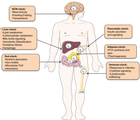
Fig. 2. Coordinated regulation of metabolic physiology by central and peripheral clocks: The light-entrained central SCN-clock not only governs rest-activity and feeding-fasting cycle but also synchronizes peripheral tissue clocks. Indicated in the boxes are some of the major peripheral clocks and the critical physiological functions they perform. Importantly, deregulations in the functioning of peripheral clock-regulated pathways are often encountered in NAFLD. SCN-Supra Chiasmatic Nucleus. See text for details.
circadian rhythmicity. Another highly relevant feedback regulation exists between the CC and heme biosynthesis and activity has also been uncov-ered [25,26]. Altogether, extensive investigations have unraveled multi-faceted CC-metabolism crosstalk as a tuning fork for the CC-oscillator functioning, which has systemic repercussions as: (i) a change in the feeding time in mice to the“rest” phase leads to features resembling met-abolic syndrome [27] and, (ii) high-fat diet (HFD)-induced reprogramming of the hepatic CC-functioning in mice can be largely prevented by restricting the food access to the circadian active phase [28,29].
4. Pathophysiology of the non-alcoholic fatty liver disease (NAFLD) Over the last decades, lifestyle modifications have shifted the health care priorities worldwide from infectious to metabolic diseases [30–34]. In the context of liver disease, availability of vaccines and antiviral ther-apies have started to reduce the disease burden caused by hepatotropic viruses such as chronic hepatitis B and C and their complications [35–39]. In contrast, the prevalence of metabolic liver disease such as non-alcoholic fatty liver disease (NAFLD) and non-alcoholic steatohepatitis (NASH) have increased dramatically. Indeed, with an es-timated worldwide prevalence of ~25%, NAFLD has emerged as the most common chronic liver disease [30–34]. This increase in global preva-lence of NAFLD is closely associated with the world-wide epidemic in the incidence of other metabolic disorders e.g. type 2 diabetes and obe-sity. Importantly, 20–25% of fatty liver patients progress to develop NASH, which is a major aetiology of liver transplantation required by cirrhotic and hepatocellular carcinoma (HCC) patients [30,31,40]. “Fatty liver” is a complex spectrum of disease and considering the cur-rent knowledge of the pathology and the understanding of patient het-erogeneity, the scientific community has recently [34] suggested metabolic (dysfunction) associated fatty liver disease (MAFLD) to be more appropriate. NAFLD generally initiates with the accumulation of excessive triglyceride (TG) in hepatocytes, a largely benign state com-monly referred as simple steatosis [30–33]. Importantly, persistent fatty liver drives simple steatosis to steatohepatitis (NASH), which is characterized by simultaneous presence of both inflammation and hepatocytic damage (a.k.a ballooning). Furthermore, NASH proceeds tofibrosis, which can eventually progress to cirrhosis and hepatocellu-lar carcinoma (HCC) [30–33,40]. Like metabolic syndrome, develop-ment of NAFLD is highly complex and has been extensively reviewed elsewhere [30–34,40–43]. Despite large research and development ef-forts, there are no approved drugs specifically targeting metabolic liver disease and compounds in late stage of development are fre-quently characterized by limited efficacy [30,32,33].
The pathogenesis of“fatty liver” was initially postulated in 1998 [44] to be“a tale of two hits”-first involving excessive hepatic triglyceride (TG) accumulation which was followed by secondary insults such as ox-idative stress. However, recent investigations in chronic metabolic dis-eases have now clearly established that pathogenesis of NAFLD is a complex multi-step metabolic disorder [30–34,40–43]. Several studies have indeed uncovered crucial roles for deregulations in the functioning of pancreas, intestine, adipose tissue and immune system in NAFLD de-velopment ([30–33,40–43];Fig. 3). Physiologically, mammalian bioener-getics is maintained by intricate intra- and inter-organ communications and deregulations of which lie at the core of metabolic disease, including NAFLD. At the basic level NAFLD arises due to the inability of the hepato-cytes to effectively metabolize carbohydrates and free fatty acids (FFA). Mechanistically, NAFLD is a consequence of an imbalance between adipocytic FFA supply, hepatic de novo lipogenesis and FFA utilization through mitochondrialβ-oxidation and production of ketone bodies, andfinally disposal through secretion of TGs in very low-density lipopro-tein (VLDL) particles [1,30–33]. Fat accumulation in the liver can be traced to either an increased incidence of de novo lipogenesis or overwhelming of the capacity to oxidize FFA. Additionally, mitochondrial dysfunction could impair fatty acidβ-oxidation and cause lipid accumulation, which
usually precedes NAFLD [45–47]. Furthermore, excessive TG is transported out of the hepatocytes by binding to liver-produced VLDL and, with impairedβ-oxidation or TG transport, the capacity of the liver to clear accumulated TG is compromised, which further contributes to the development of NAFLD [30,31,45]. As discussed above, the complexi-ties of NAFLD pathogenesis and its progression to steatohepatitis are barely understood with both genetic and environmental factors playing crucial roles. Importantly, due to the overwhelming role of the CC in maintaining metabolic homeostasis (Fig. 2), it can be postulated that dis-ruption in the CC-functioning can drive NAFLD [1–3,15]. In the subse-quent chapters we describe some of these functions to further illustrate the link between CC and NAFLD.
5. Peripheral circadian clocks: regulation of metabolism and impact on pathogenesis of NAFLD
5.1. Liver
Unarguably, the liver plays a central role in governing whole-body physiology (Fig. 2). Considering its preeminent role in metabolism several genomic studies have utilized time-course in mouse models to uncover circadian cistrome [48–51], transcriptome [52,53], proteome [54–56], and lipidome [57,58]. Analyses of circadian gene expression have revealed two broad time-periods of transcription in liver, which correspond to the periodic transition between alternating active and rest phases [1–4]. These two‘peaks’ reflect the highly differential physiological require-ments, such as in energy demand or detoxification activity, as per their necessity during the periods of activity or rest [1–5]. Analyses of CC-components and epigenetic factors binding [48–51] uncovered that these two circadian phase-specific distinct mRNA pools are generated due to the intrinsic rhythmicity of the CC-oscillator. Furthermore, CC is also known to post-transcriptionally control cellular processes like DNA repair, ribosome biogenesis, autophagy, ER-stress [54–56].
Mammalian gluconeogenesis is principally controlled by the liver. Indeed, along with several other organs (brain, pancreas, muscle), the liver-CC largely contributes to maintain homeostatic blood glucose levels [59]. In a critical genetic study it was demonstrated that Bmal1 ab-lation in the liver reduces expression of the glucose transporter (Glut2), which lead to a decreased post-feeding glucose uptake in mutant mice, thus revealing a role for the liver-CC-oscillator in glucose metabolism [60]. Remarkably, the liver-CC also regulates glucose metabolism post-hepatocytic entry at multiple levels, by controlling expression of gluco-kinase (Gck; regulator of glycogen synthesis) [1–5]. By controlling either the expression or the activity of several gluconeogenic transcription fac-tors e.g. Klf10 [61], Hnf4α [18,62], CREB [63], Pgc1α [64], the liver-CC thoroughly controls glucose metabolism.
Along with its influence on carbohydrate metabolism, several ge-netic studies have established that liver CC as a predominant regulator of lipid metabolism [65–67]. These investigations have established plasma levels of FFA, TG and cholesterol display diurnal variations, and are altered upon mutations of CC-genes. To illustrate, liver-restricted mutation of Rev-Erbα/β profoundly increased plasma levels of FFA, TG and cholesterol [67]. Mechanistically, the hepatic-CC regulates either the expression or the activity of enzymes that are critically involved in regulating multiple critical steps of lipid metabolism. As an example, TG synthesis in liver is a multistep process and requires the activity of several enzymes (Gpat2, Agpat1/2, Lipin1/2 and Dgat2) expression of which are CC-controlled [56], thereby leading to a prominent crest and trough of hepatic TG levels (in mice) during the rest and active phases, respectively [56]. Furthermore, REV-ERBα by regulating the transcription of Insig2 controls the activity of SREBP1c (master regulator of lipogenesis; [68]). Additionally, the hepatic CC-oscillator also partici-pates in: (i) fatty acid synthesis by controlling the expression of Elovl3, Elovl6, Fas etc. [1–3,6], (ii) regulatingβ-oxidation and ketone-body pro-duction [69,70] and, (iii) determining the expression of key lipid-responsive NRs LXRs, PPARα and δ [1–3,18]. Recent studies have
established BA-signaling as a major regulator of TG, cholesterol and glu-cose homeostasis [71,72]. BA synthesis is controlled by a transcriptional feed-back loop consisting of the NRs FXR and SHP and intestinal hor-mone FGF15 (FGF19 in humans; [71–72]). Importantly, CC-regulates the expressions of both FXR and SHP [18,62] as well as FGF15 secretion [73], thereby controlling the diurnal expression of cholesterol 7 α-hydroxylase (Cyp7a1), the rate-limiting enzyme in BA synthesis. Addi-tionally, both REV-ERBα and DBP (CC-output regulator) are known to control Cyp7a1 transcription [74,75]. Furthermore, in mice an essential molecular feedback exists between SHP and the neuronal PAS domain protein 2 (NPAS2; Clock gene paralog) which not only contributes to-wards their own circadian rhythmicity but also enables to maintain he-patic lipid, BA and lipoprotein metabolism [76]. Taken together, these mechanisms combine to generate circadian rhythmicity in BA levels which is also observed in humans [77]. To further illustrate the intimate connection between the lipid-and BA-metabolism and CC-functioning, it has been noted that atorvastatin (routinely to treat hyperlipidemia) administration in mice alters the expression of not only Cyp7a1 but also of key CC-components e.g. Bmal1 and Npas2 [78]. The liver CC is also well known to regulate several cellular processes e.g. autophagy, ER stress and oxidative stress [79–83], all of which have been implicated in pathogenesis of NAFLD and has been extensively described elsewhere [30,31].
5.2. Pancreas
The pancreas is well known to play a critical role in maintaining glu-cose homeostasis through production of hormones insulin and glucagon (Fig. 2). Pancreatic function is controlled by both the central SCN-clock as well as Pancreatic CC-oscillator and aligns biochemical activities in pancreatic islets as per the metabolic demands [2,3,84,85]. The‘clock’ is known to regulate both the exocrine [84] and endocrine [86] func-tions of the pancreas. The presence of an autonomous circadian pancre-atic clock has been demonstrated not only in rodents [85–87], but also in human islets and dispersed human islet cells [87]. The pancreatic clock is synchronized to the light-dark cycle via signals derived from
the SCN-clock that include autonomic neuronal system, melatonin and glucocorticoids [88]. The pancreatic CC-oscillator inβ cells drives highly rhythmic oscillation of insulin secretion which is strictly aligned with the expression of genes encoding insulin secretion and signaling [85]. Mechanistically, pancreatic CC-components helped in the spatiotempo-ral recruitment of key transcription factor PDX1 to specific enhancers to regulate transcription of insulin and other genes of insulin signaling pathway [85]. Importantly,β-cell-specific mutation of either Bmal1 or Clock leads to wide-spread changes in the transcriptome, and speci fi-cally reduces genes encoding cell cycle, synaptic assembly and secretion of insulin, thereby leading to diabetes in mutant mice [86]. Notably, non-alcoholic fatty pancreas disease (NAFPD) a recently described dysmetabolic phenotype (akin to NAFLD), has been shown to perturb the expression of several CC-components in murine pancreas which correlates with pancreatic inflammation and fibrosis development [89]. As insulin-resistance often accompanies NAFLD [30], deregulation of the pancreatic-CC-controlled insulin signaling could play a critical role in the predisposing to fatty liver development (Fig. 3).
5.3. Intestine and microbiota
Several critical aspects of the intestinal physiology e.g. motility, in-testinal permeability, hormone secretion, nutrient absorption, cell pro-liferation and interactions with microbiota are CC-controlled and have been thoroughly reviewed [90–92]. However, in recent years the rela-tionship between intestine and resident microbiota has gained spotlight as a major regulator of metabolic health and disease including, NAFLD [93–96]. Indeed, obesity has been shown to not only alter the composi-tion of gut microbiota (dysbiosis) composicomposi-tion but also perturbs their nature of interactions with the host (intestinal epithelial cells; IEC), both of which have been suggested as an etiological agent in the patho-genesis of metabolic diseases, including NAFLD [93–96]. One of the pro-posed mechanisms through which dysbiosis could induce NAFLD is by augmenting lipopolysaccharide (LPS) production and delivery to the liver via the portal circulation, a consequence of increased intestinal permeability [97,98]. This abnormal presence of microbiota-associated
Fig. 3. Model of NAFLD pathogenesis: The scheme depicts an overview of how alterations in the circadian clock-controlled functions/pathways and processes in different peripheral tissues may predispose to NAFLD pathogenesis and contribute to therapeutic intervention. See text for details.
LPS in liver perturbs lipid metabolism by affecting the generation of short-chain fatty acids and altering the BA pool composition which may influence intestinal and hepatic FXR activity, thus affecting both glucose and lipid homeostasis [97].
The‘clock’ and the microbiota intersect at many levels. Most notably, the IEC CC has been demonstrated to regulate the circadian expression of microbial pattern recognition receptors (e.g. TLRs, NOD2) which cre-ates a‘temporal window’ for the microbiota-signals to regulate gene ex-pression in IEC to maintain homeostasis [99]. Importantly, absence of this IEC CC-microbiota crosstalk leads to metabolic disorders [99]. Inter-estingly, it was also demonstrated that the gut microbiota undergoes circadian oscillations in composition and function [100,101]. These mi-crobiota oscillations were found to be controlled by the timing of food intake and the diet composition. Furthermore, it was also demonstrated that the gut microbiota undergoes circadian oscillations in biogeograph-ical localization and metabolome patterns which in turn determine the diurnal exposure of the intestinal epithelium to different bacterial spe-cies and their metabolites [102,103]. Importantly, this circadian varia-tions in microbial behavior in turn regulates the transcriptome and metabolome of both gut and distant tissues e.g. liver [102,103]. Most importantly, dysbiosis-induced by‘clock’ perturbations (either through genetic ablation of CC-components or jet lag) lead to and development of metabolic pathologies [93,94,103].
5.4. Immune system
The immune system is heavily influenced by time-of-day cues, both under steady-state conditions and in response to inflammatory chal-lenges. Indeed, diurnal host responses to endotoxins were noted more than 6 decades back [104]. Importantly, several inflammatory diseases e;g. myocardial infarction, rheumatoid arthritis and asthma are known exhibit pronounced circadian rhythmicity in their pathology [105–108]. Recently, molecular evidence has started emerging to reveal that numerous aspects of immune functions including lymphocyte traf-ficking, host-pathogen interactions, cytokine secretion and activation of innate and adaptive immunity are thoroughly controlled by the CC [107–110]. Taken together, investigations have established the CC operates to as a gating mechanism to control the magnitude of immune response in a diurnal fashion and has been described [107–110]. The role of the deregulated immune system and fatty liver disease have been extensively reviewed [41–43]. Here we briefly discuss the immune components which are known to be controlled by CC under physiolog-ical conditions [107–110].
Like every other aspect of the metabolic syndrome, pathogenesis of `fatty liver` is strongly linked with inflammation, and both innate and adaptive branches of immunity have been implicated in this process [31,32,41–43]. However, the innate immune system has received more attention. Although in initial studies focused on Kupffer cells, more recent investigations have revealed that several specialized im-mune cells (resident and infiltrating) participate in hepatosteatosis [42]. Kupffer cells are activated by a variety of stimuli including FFA, peroxidized lipids, microbiota-derived LPS and ROS [43]. Importantly, both FFA and LPS drive Kupffer cell stimulation through TLR2 and TLR4, which leads to perpetual activation of inflammatory signaling pathways like ASK-1, JNK, IL-6 etc. thereby enabling sustained induction of NF-κB and STATs, thus augmenting cytokine production (TNF-α, IL-1β etc.). In murine models, reducing the number of Kupffer cells through clodronate administration considerably ameliorates NASH pa-thology [111]. In addition, the inflammasome which can both sense and be activated by danger-associated molecular patterns (DAMPs) such as FFA and pathogen-associated molecular patterns (PAMPs) e.g. LPS, has recently emerged as a critical molecular link between metabolic stress and fatty liver development [31]. In animal models of NAFLD, trig-gering inflammasome activity enhances the expression of the pro-inflammatory cytokines IL-1β and IL-18 which subsequently through caspase-1 promote cell death in liver [111]. Recent studies have also
shed light on the role of the IL17-secreting Th17 cells in metabolic diseases including NAFLD [112]. It has been observed that the obesity-induced dysbiosis elevates IL-17 production [113,114] and, in the set-ting of NAFLD, this cytokine drives neutrophil and monocyte infiltration in the liver, thereby potentiating hepatic insulin resistance and steatosis progression [115]. Consistently, abrogation of IL17-induced signaling activity in a diet-induced murine model of NASH reduces steatosis [116]. Taken together, these studies indicate how possibly deregulated CC-functioning in immune cells could predispose towards fatty liver development.
6. Circadian clock-related therapeutic interventions
In the past few years, many studies have investigated the effects of the timing of drug treatment on the circadian appearance or exacerba-tion on of disease symptoms, leading to the development of a concept known as chronomedicine [117–119]. Chronomedicine is described as the approach employed to maximize the efficacy and minimize the side effects when drugs are administered in accordance with the CC as ‘timing’ of drug-administration is of crucial but still a less-appreciated factor in drug efficacy considerations [117–119]. This is not surprising considering that to a large extent CC control over pharmacology arises from its ability to thoroughly regulate almost all steps of xenobiotic de-toxification in the liver, including absorption, biotransformation and elimination [[118],120–122]. Thereby, CC-controls pharmacological pa-rameters such as pharmacokinetics and pharmacodynamics [118,119]. Remarkably, 56 of the top 100 best-selling drugs in the USA are known to target the product of a circadian gene [122]. Until now, this approach of chronomedicine has been evaluated for several diseases, such as hypertension [123,124] and cancers [125,126]. The most impor-tant example is that of the circadian hormone melatonin that has been used in combination with cancer therapy to minimize toxicity or en-hance chemotherapeutic viability in clinical and laboratory settings [126]. To further illustrate, influenza vaccine when administered in the morning produces higher titers of antibodies than when given in the evening [127].
Pharmacological therapies are not yet available for NASH [30–34], although several compounds are in preclinical and clinical develop-ment, including obeticholic acid (INT-747; [128]) which activates FXR and, elafibranor (currently in phase 3 trial; [129]) which activates NRs PPAR-α/β. Notably, physiological targets of potential NASH-modulating compounds [30–32], e.g. resveratrol (SIRT1-agonist) and inhibitors of acetyl-CoA carboxylase1 (ACC1) are also CC-regulated [1–4], thereby further strengthening the CC-connection to the develop-ment of novel therapeutics. Considering, the role of the CC in regulating the expression and activities of FXR, PPARs, SIRT1 and ACC chronopharmacology could very well dictate the efficacy of these ap-proaches. Circadian“misalignment” between central and peripheral CCs has been found to be a core feature of almost every dietary or envi-ronmental model of metabolic disease including NAFLD. For therapeutic treatment of metabolic diseases like NAFLD a strategy could be to give to patients more scheduled eating habits, the so-called chrononutrition. In this regard, time-restricted feeding (TRF), a behavioral approach where feeding is solely restricted to the circadian active phase not only pre-vents circadian misalignment but also has been shown to correct several metabolic pathologies in animal models [130–132]. TRF is distinct from intermittent fasting and when applied to humans, the amount of calory ingested is not relevant [130–132]. Importantly, several small-scale human TRF investigations have indicated its usefulness in improving outcomes in patients with metabolic syndromes [133–135], however, the usefulness of TRF on NAFLD endpoints are yet to be ascertained. 7. Conclusion
As the prevalence and economic burden of the metabolic syndrome and NAFLD/NASH/MAFLD continues to rise worldwide, the knowledge
about the mechanisms contributing to the development of this disease has been progressively increasing over the last two decades. Circadian misalignment has been associated with increased incidence of meta-bolic and cardiovascular disorders in various human studies [136–142]. These discoveries have led to the recognition of CC rhythms as an essential piece of the complex puzzle that depicts our physiologic homeostasis. The understanding of the multi-faceted role of the‘clock’ in the pathogenesis of fatty liver (Fig. 3), is not only crucial to advance scientific knowledge, but also to improve public health by identifying new therapeutic targets and life-style modifications. Disruption of the CC has been shown to play an important role in the increasing incidence of metabolic homoeostasis with a key contribution to the metabolic syndrome and NAFLD. Hence, it is necessary to investigate in detail the CC-controlled pathways and elucidate how they are linked with the development of fatty liver disease (Fig. 3).
Expanding our knowledge about the genetic and environmental risk factors making individuals more susceptible to metabolic dysfunction combined with the discovery of new therapeutic approaches to restore the perturbed circadian machinery will ultimately contribute to im-prove the outcome of this rapidly growing pandemic of metabolic liver disease.
Declaration of competing interest
The authors declare no conflict of interest Acknowledgements
The authors apologize to all colleagues whose work could not be cited due to space limitations.
Author contributions
AM designed the concept, focus areas and overall structure of the manuscript. MD prepared thefigures. TFB conceptualized the section on the liver disease and therapeutic implications. AM, MD and TFB discussed, wrote and edited the different versions of the manuscript. Funding
This work was supported by the European Union: ERC-AdG-2014-671231-HEPCIR, EU H2020-667273-HEPCAR, EU-InfectEra HepBccc (T.F.B.), the National Institute of Health (NCI R21 CA209940 and NIDDK R01CA233794 to T.F.B.), the Agence Nationale de Recherches sur le Sida et les Hépatites Virales (ANRS, 2015/1099), the Fondation ARC pour la Recherche sur le Cancer (IHU 201901299), the Institut Universitaire de France (IUF to T.F.B), and USIAS of the University of Strasbourg (A.M.). The work has been published under the framework of the LABEX ANR-10-LABX-0028_HEPSYS) and Inserm Plan Cancer and benefits from funding from the state managed by the French National Research Agency as part of the Investments for the future program.
References
[1]Mukherji A, Bailey S, Staels B, Baumert T. The circadian clock and liver function in health and disease. J Hepatol. 2019;70:200–11.
[2]Eckel-Mahan KL, Sassone-Corsi P. Metabolism and the circadian clock converge. Physiol Rev. 2013;93:107–35.
[3]Bass J. Circadian topology of metabolism. Nature. 2012;491:348–56.
[4]Feng D, Lazar MA. Clocks, metabolism and Epigenome. Mol Cell. 2012;47:158–67.
[5]Asher G, Sassone-Corsi P. Time for food: the intimate interplay between nutrition, metabolism and the circadian clock. Cell. 2015;161:84–92.
[6]Panda S. Circadian physiology of metabolism. Science. 2016;354:1008–15.
[7]Konopka RJ, Benzer S. Clock mutants of Drosophila melanogaster. Proc Natl Acad Sci U S A. 1971;68:2112–6.
[8] LeGates TA, Fernandez DC, Hattar S. Light as a central modulator of circadian rhythms, sleep and affect. Nat Rev Neurosci. 2014;15:443–54.
[9]Takahashi JS, Hong HK, KO CH, McDearmon EL. The genetics of mammalian circa-dian order and disorder: implications for physiology and disease. Nat Rev Genet. 2008;9:764–75.
[10]Wang X-S, Armstrong MEG, Cairns BJ, Key TJ, Travis RC. Shift work and chronic dis-ease: the epidemiological evidence. Occup Med. 2011;61:78–89.
[11]Huang W, Ramsey KM, Marcheva B, Bass J. Circadian rhythms, sleep, and metabo-lism. J Clin Invest. 2011;121:2133–41.
[12]Zhang S, et al. Rotating night shift work and non-alcoholic fatty liver disease among steel workers in China: a cross-sectional survey. Occup Environ Med. 2020;77: 333–9.
[13]Sun M, Feng W, Wang F, Zhang L, Wu Z, et al. Night shift work exposure profile and obesity: baseline results from a Chinese night shift worker cohort. Plos One. 2018; 13:e0196989.
[14]Roenneberg T, Merrow M. The circadian clock and human health. Curr Biol. 2016; 26:R432–43.
[15] Mazzoccoli G, Cosmo DS, Mazza T. The biological clock: a pivotal hub in non-alco-holic fatty liver disease pathogenesis. Front. Physiol 9:193. Doi:https://doi. org/10.3389/fphys.2018.00193
[16]Verlande A, Masri S. Circadian clocks and cancer: timekeeping governs cellular me-tabolism. Trends Endocrinol Metab. 2019;30:445–58.
[17]Takahashi JS. Transcriptional architecture of the mammalian circadian clock. Nat Rev Genet. 2017;18:164–79.
[18]Yang X, et al. Nuclear receptor expression links the circadian clock to metabolism. Cell. 2006;126:801–10.
[19]Damiola F, LeMinh N, Preitner N, Kornmann B, Fleury-Olela F, et al. Restricted feed-ing uncouples circadian oscillators in peripheral tissues from the central pace-maker in the suprachiasmatic nucleus. Genes Dev. 2000;14:2950–61.
[20]Stokkan KA, Yamazaki S, Tei H, Sakaki Y, Menaker M. Entrainment of the circadian clock in the liver by feeding. Science. 2001;291:490–3.
[21]Vollmers C, Gill S, DiTacchio L, Pulivarthy SR, Le HD. Time of feeding and the intrin-sic circadian clock drive rhythms in hepatic gene expression. Proc Natl Acad Sci U S A. 2009;106:21453–8.
[22]Mukherji A, Kobiita A, Chambon P. Shifting the feeding of mice to the rest phase creates metabolic alterations, which, on their own, shift the peripheral circadian clocks by 12 hours. Proc Natl Acad Sci U S A. 2015;112:E6683–90.
[23]Nakahata Y, Sahar S, Astarita G, Kaluzova M, Sassone-Corsi P. Circadian control of the NAD+ salvage pathway by CLOCK-SIRT1. Science. 2009;324:654–7.
[24]Ramsey KM, Yoshino J, Brace CS, Abrassart D, Kobayashi Y. Circadian clock feedback cycle through NAMPT-mediated NAD+ biosynthesis. Science. 2009;324:651–4.
[25]Kaasik K, Lee C. Reciprocal regulation of heme biosynthesis and the circadian clock in mammals. Nature. 2004;430:467–71.
[26]Yin L, Wu N, Curtin JC, Qatanani M, Szwergold NR, et al. Rev-erbα, a heme sensor that coordinates metabolic and circadian pathways. Science. 2007;318:1786–9.
[27]Mukherji A, Kobiita A, Damara M, Misra N, Meziane H, et al. Shifting eating to the circadian rest phase, misaligns the peripheral circadian clocks with the master SCN clock, which leads to a metabolic syndrome. Proc Natl Acad Sci. 2015;112: E6691–8.
[28]Eckel-Mahan KL, Patel VR, deMateo S, Orozco-Solis R, Ceglia NJ. Reprogramming of the circadian clock by nutritional challenge. Cell. 2013;155:1464–78.
[29]Hatori M, Vollmers C, Zarrinpar A, DiTacchio L, Bushong EA. Time-restricted feeding without reducing caloric intake prevents metabolic diseases in mice fed a high-fat diet. Cell Metab. 2012;15:848–60.
[30]Younossi Z, Henry L. Contribution of alcoholic and nonalcoholic fatty liver disease to the burden of liver-related morbidity and mortality. Gastroenterology. 2016; 150:1778–85.
[31]Samuel VT, Shulman GI. Nonalcoholic fatty liver disease as a nexus of metabolic and hepatic diseases. Cell Metab. 2018;27:22–41.
[32]Friedmann SL, Neuschwander-Tetri BA, Rinella M, Sanyal AJ. Mechanisms of NAFLD development and therapeutic strategies. Nat Med. 2018;24:908–22.
[33]Haas JT, Francque S, Staels B. Pathophysiology and mechanisms of nonalcoholic fatty liver disease. Annu Rev Physiol. 2016;78:181–205.
[34]Eslam M, Sanyal AJ, George J. MAFLD: a consensus-driven proposed nomenclature for metabolic associated fatty liver disease. Gastroenterol. 2020;158:1999–2014.
[35]Baumert TF, Verrier ER, Nassal M, Chung RT, Zeisel MB. Host-targeting agents for treatment of hepatitis B virus infection. Curr Opin Virol. 2015;14:41–6.
[36]Zeisel MB, Lucifora J, Mason WS, Sureau C, Beck J, et al. Towards an HBV cure: state-of-the-art and unresolved questions-report of the ANRS workshop on HBV cure. Gut. 2015;64:1314–26.
[37]Chung RT, Baumert TF. Curing chronic hepatitis C- the arc of a medical triumph. N Engl J Med. 2014;370:1576–8.
[38]Hoshida Y, Fuchs BC, Bardeesy N, Baumert TF, Chung RT. Pathogenesis and preven-tion of hepatitis C virus-induced hepatocellular carcinoma. J Hepatol. 2014;61: S79–90.
[39]Zeisel MB, Lupberger J, Fofana I, Baumert TF. Host-targeting agents for prevention and treatment of chronic hepatitis-perspectives and challenges. J Hepatol. 2013; 58:375–84.
[40]Parola M, Pinzani M. Liverfibrosis: pathophysiology, pathogenetic targets and clin-ical issues. Mol Aspects Med. 2019;65:37–55.
[41]Heyman F, Tacke F. Immunology in the liver-from homeostasis to disease. Nat Rev Gastroenterol Hepatol. 2016;13:88–110.
[42]Seki E, Schwabe RF. Hepatic inflammation and fibrosis: functional links and key pathways. Hepatology. 2015;61:1066–79.
[43]Ganz M, Szabo G. Immune and inflammatory pathways in NASH. Hepatol Int. 2013; 7:771–81.
[44]Day CP, James OF. Steatohepatitis: a tale of‘two’ hits. Gastroenterology. 1998;114: 842–5.
[45]Lambert JE, Ramos-Roman MA, Browning JD, Parks EJ. Increased de novo lipogene-sis is a distinct characteristic of individuals with nonalcoholic fatty liver disease. Gastroenterology. 2014;146:726–35.
[46]Titchenell PM, Lazar MA, Birnbaum MJ. Unraveling the regulation of hepatic metab-olism by insulin. Trends Endocrinol Metab. 2017;28:497–505.
[47]Kawano Y, Cohen DE. Mechanisms of hepatic triglyceride accumulation in non-al-coholic fatty liver disease. J Gastroenterol. 2013;48:434–41.
[48]Koike N, Yoo S-H, Huang H-C, Kumar V, Lee C, et al. Transcriptional architecture and chromatin landscape of the core circadian clock in mammals. Science. 2012;338: 349–54.
[49]Rey G, Cesbron F, Rougemont J, Reinke H, Brunner M, et al. Genome-wide and phase-specific DNA-binding rhythms of BMAL1 control circadian output functions in mouse liver. PLoS Biol. 2011;9:e1000595.
[50]Cho H, Zhao X, Hatori M, Yu RT, Barish GD, et al. Regulation of circadian behaviour and metabolism by REV-ERB-α and REV-ERB-β. Nature. 2012;485:123–7.
[51]Fang B, Everett LJ, Jager J, Briggs E, Armour SM, et al. Circadian enhancers coordi-nate multiple phases of rhythmic gene transcription in vivo. Cell. 2014;159: 1140–52.
[52]Panda S, Antoch MP, Miller BH, Su AI, Schook AB, et al. Coordinated transcription of key pathways in the mouse by the circadian clock. Cell. 2002;109:307–20.
[53]Storch KF, Lipan O, Leykin I, Viswanathan N, Davis FC, et al. Extensive and divergent circadian gene expression in liver and heart. Nature. 2002;417:78–83.
[54]Robles MS, Cox J, Mann M. In-vivo quantitative proteomics reveals a key contribu-tion of post-transcripcontribu-tional mechanisms to the circadian regulacontribu-tion of liver metab-olism. PLoS Genet. 2014;2014(10):e1004047.
[55]Mauvoisin D, Wang J, Jouffe C, Martin E, Atger F, et al. Circadian clock-dependent-and -independent rhythmic proteomes implement distinct diurnal functions in mouse liver. Proc Natl Acad Sci U S A. 2014;111:167–72.
[56]Wang J, Mauvoisin D, Martin E, Atger F, Galindo AN, et al. Nuclear proteomics un-covers diurnal regulatory landscapes in mouse liver. Cell Metab. 2017;25:102–17.
[57]Aviram R, Manella G, Kopelman N, Neufeld-Cohen A, Zwighaft Z, et al. Lipidomic analyses reveal temporal and spatial lipid organization and uncover daily oscilla-tions in intracellular organelles. Mol Cell. 2016;62:636–48.
[58]Adamovich Y, Rousso-Noori L, Zwighaft Z, Neufeld-Cohen A, Golik M, et al. Circa-dian clocks and feeding time regulate the oscillations and levels of hepatic triglyc-erides. Cell Metab. 2014;19:319–30.
[59]Kalsbeek A, LaFleur S, Fliers E. Circadian control of glucose metabolism. Mol Metab. 2014;3:372–83.
[60]Lamia KA, Storch KF, Weitz CJ. Physiological significance of a peripheral tissue cir-cadian clock. Proc Natl Acad Sci U S A. 2008;105:15172–7.
[61]Guillaumond F, Grechez-Cassiau A, Subramaniam M, Brangolo S, et al. Kruppel-like factor KLF10 is a link between thecircadian clock and metabolism in liver. Mol Cell Biol. 2010;30:3059–70.
[62]Bookout AL, et al. Anatomical profiling of nuclear receptor expression reveals a hi-erarchical transcriptional network. Cell. 2006;126:789–99.
[63]Zhang EE, Liu Y, Dentin R, et al. Cryptochrome mediates circadian regulation of cAMP signaling and hepatic gluconeogenesis. Nat Med. 2010;16:1152–6.
[64]Liu C, Li S, Liu T, Borigin J, Lin JD. Transcriptional coactivator PGC1-alpha integrates mammalian clock and energy metabolism. Nature. 2007;447:477–81.
[65]Turek FW, Joshu C, Kohsaka A, Lin E, Ivanova G, et al. Obesity and metabolic syn-drome in circadian clock mutant mice. Science. 2005;308:1043–5.
[66]Grimaldi B, Bellet MM, Katada S, Astarita G, Hirayama J, et al. PER2 controls lipid metabolism by direct regulation of PPARgamma. Cell Metab. 2010;12:509–20.
[67]Bugge A, Feng D, Everett LJ, Briggs ER, Mullican SE, et al. Rev-Erbα and Rev-Erbβ coordinately protect the circadian clock and normal metabolic function. Genes Dev. 2012;26:657–67.
[68]Le Martelot G, Claudel T, Gatfield D, Schaad O, Kornmann B, et al. REV-ERBα partic-ipates in circadian SREBP signaling and bile acid homeostasis. PLoS Biol. 2009;7: e1000181.
[69]Chavan R, Feillet C, Fonesca Costa S, Delorme JE, Okabe T, et al. Liver-derived ketone bodies are necessary for food anticipation. Nat Commun. 2016;7:10580.
[70]Lemberger T, Saladin R, Vazquez M, Assimapoulos F, Staels B, et al. Expression of the peroxisome proliferator-activated receptor alpha gene is stimulated by stress and follows a diurnal rhythm. J Biol Chem. 1996;271:1764–9.
[71]Thomas C, Pelliccari R, Pruzanski M, Auwerx J, Schoonjans K. Targeting bile-acid signaling for metabolic disease. Nat Rev Drugdisc. 2008;7:678–93.
[72]Chàvez-Talavera O, Tailleux A, Lefebvre P, Staels B. Bile acid control of metabolism and inflammation in obesity, type2 diabetes, dyslipidemia, and nonalcoholic fatty liver disease. Gastroenterol. 2017;152:1679–94.
[73]Stroeve JH, Brufau G, Stellard F, Gonzalez FJ, Staels B, et al. Intestinal FXR-mediated FGF15 production contributes to diurnal control of hepatic bile acid synthesis in mice. Lab Invest. 2010;90:1457–67.
[74]Lavery DJ, Schibler U. Circadian transcription of the cholesterol 7 alpha hydroxylase gene may involve the liver-enriched bZIP protein DBP. Genes Dev. 1993;7: 1871–84.
[75]Duez H, Veen JN, Duhem C, Pourcet B, Touvier T, et al. Regulation of bile acid syn-thesis by the nuclear receptor rev-erbα. Gastroenterol. 2008;135:689–98.
[76]Lee SM, Zhang Y, Tschuiya H, Smalling R Jetten AM, et al. Small heterodimer part-ner/neuronal PAS domain protein axis regulates the oscillation of liver lipid metab-olism. Hepatology. 2015;61:497–505.
[77]Duane WC, Levitt DG, Mueller SM, Behrens JC, et al. Regulation of bile acid synthe-sis in man. Presence of a diurnal rhythm. J Clin Invest. 1983;72:1930–6.
[78]Li WK, Li H, Lu YF, Fu ZD, et al. Atrovastatin alters the expression of genes related to bile acid metabolism and circadian clock in livers of mice. Peer J. 2017;5:e3348.
[79]Chaix A, Zarrinpar A, Panda S. The circadian coordination of cell biology. J Cell Biol. 2016;215:15–25.
[80]Ma D, Li S, Molusky MM, Lin JD. Circadian autophagy rhythm: a link between clock and metabolism? Trends Endocrinol Metab. 2012;23(2012):319–25.
[81]Cretenet G, Le Clech M, Gachon F. Circadian clock-coordinated 12Hr period rhyth-mic activation of the IRE1alpha pathway controls lipid metabolism in mouse liver. Cell Metab. 2010;11:47–57.
[82]Jacobi D, Liu S, Burkewitz K, Kory N, Knudsen NH, et al. Hepatic Bmal1 regulates rhythmic mitochondrial dynamics and promotes metabolicfitness. Cell Metab. 2015;22:709–20.
[83]Kanabrocki EL, Murray D, Hermida RC, Scott GS, Bremner WF, et al. Circadian var-iation in oxidative stress markers in healthy and typeII diabetic men. Chronobiol Int. 2002;19:423–39.
[84]Pulimeno P, et al. Autonomous and self-sustained circadian oscillators displayed in human islet cells. Diabetologia. 2013;56:497–507.
[85]Perelis M, Marcheva B, et al. Pancreaticβ cell enhancers regulate rhythmic tran-scription of genes controlling insulin secretion. Science. 2015;350:aac4250.
[86]Marcheva B, et al. Disruption of the clock components CLOCK and BMAL1 leads to hypoinsulinemia and diabetes. Nature. 2010;466:627–31.
[87]Vieria E, Burris TP, Quesda I. Clock genes, pancreatic function, and diabetes. Trends Mol Med. 2014;20:685–93.
[88]Bujis RM, Kalsbeek A. Hypothalamic integration of central and peripheral clocks. Nat Rev Neurosci. 2001;2:521–6.
[89]Carter R, Mouralidarane A, Soeda J, Ray S, Pombo J, et al. Non-alcoholic fatty pan-creas disease pathogenesis: a role for developmental programming and altered cir-cadian rhythms. Plos One. 2014;9(3):e89505.
[90]Voigt RM, Forsyth CB, Keshavarzian A. Circadian rhythms: a regulator of gastroin-testinal health and dysfunction. Expert Rev Gastroenterol Hepatol. 2019;13: 411–24.
[91]Codoner-Franch P, Gombert M. Circadian rhythms in the pathogenesis of gastroin-testinal diseases. World J Gastroenterol. 2018;24:4297–303.
[92]Konturek PC, Brzozowski T, Konturek SJ. Gut clock: implication of circadian rhythms in the gastrointestinal tract. J Physiol Pharmacol. 2011;62:139–50.
[93]Sonnenburg JL, Backhed F. Diet-microbiota interactions as moderators of human metabolism. Nature. 2016;535:56–64.
[94]Bashiardes S, Shapiro H, Rozin S, Shibolet O, Elinav E. Non-alcoholic fatty liver and the gut microbiota. Mol Metab. 2016;5:782–94.
[95]Ray K. NAFLD: leaky guts: intestinal permeability and NASH. Nat Rev Gastroenterol Hepatol. 2015;12:123.
[96]Van Olden C, Groen AK, Nieuwdorp M. Role of intestinal microbiome in lipid and glucose metabolism in diabetes mellitus. Clin Ther. 2015;37:1172–7.
[97]Cani PD, Bibiloni R, Knauf C, Waget A, Neyrinck AM, et al. Changes in gut microbiota control metabolic endotoxemia-induced inflammation in high-fat diet-induced obesity and diabetes in mice. Diabetes. 2008;57:1470–81.
[98]Porez G, Prawitt J, Gross B, Staels B. Bile acid receptors as targets for dyslipidemia and cardiovascular disease. J Lipid Res. 2012;53:1723–37.
[99]Mukherji A, Kobiita A, Ye T, Chambon P. Homeostasis in intestinal epithelium is or-chestrated by the circadian clock and microbiota cues transduced by TLRs. Cell. 2013;153:812–27.
[100]Thaiss CA, Zeevi D, Levy M, Zilberman-Shapira G, Suez J, et al. Transkingdom con-trol of microbiota diurnal oscillations promotes metabolic homeostasis. Cell. 2014;159:514–29.
[101]Leone V, Gibbons SM, Martinez K, Hutchison AL, Huang EY, et al. Effects of diurnal variation of gut microbes and high-fat feeding on host circadian clock function and metabolism. Cell Host Microbe. 2015;17:681–9.
[102]Thaiss CA, Levy M, Korem T, Dohnalova L, Shapiro H, et al. Microbiota diurnal rhythmicity programs host transcriptome oscillations. Cell. 2016;167:1495–510.
[103]Zheng D, Ratiner K, Elinav E. Circadian influences of diet on the microbiome and immunity. Trends Immunol. 2020;41(6):512–30.
[104]Hallberg F, Johnson EA, Brown BA, Bittner JJ. Susceptibility rhythm to E. Coli endo-toxin and bioassay. Proc Soc Exp Biol Med. 1960;1034:142–4.
[105]Reitz CJ, Martino TA. Disruption of circadian rhythms and sleep on critical illness and the impact on cardiovascular events. Curr Pharm Des. 2015;21:3505–11.
[106]Berenbaum F, Meng QJ. The brain-joint axis in osteoarthritis : nerves, circadian clock and beyond. Nat Rev Rheumatol. 2016;12:508–51.
[107]Curtis AM, Bellet MM, Sassone-Corsi P, O'Neill LA. Circadian clock proteins and im-munity. Imim-munity. 2014:178–86.
[108]Man K, Loudon A, Chawla A. Immunity around the clock. Science. 2016;354: 999–1003.
[109]Scheiermann C, Gibbs J, Ince L, Loudon A. Clocking in to immunity. Nat Rev Immunol. 2018;18:423–37.
[110]Scheiermann C, Kunisaki Y, Frenette PS. Circadian control of the immune system. Nat Rev Immunol. 2013;13:190–8.
[111]Steinstra R, Saudale F, Duval C, Keshtkar S, Groener JE. Kupffer cells promote he-patic steatosis via interleukin-1β -dependent suppression of peroxisome proliferator-activated receptorα activity. Hepatology. 2010;51:511–22.
[112]Paquisi FC. Immune imbalances in non-alcoholic fatty liver disease: from general biomarkers and neutrophils to interleukin-17 axis activation and new therapeutic targets. Front in Immunol. 2016;7:1–13.
[113]Janssen AW, Kersten S. The role of gut microbiota in metabolic health. FASEB J. 2015;29:3111–23.
[114]SumarDumanovic M, Stevanovic D, Ljubic A, Jorga J, Simic M, et al. Increased ac-tivity of interleukin-23/interleukin-17 proinflammatory axis in obese women. Int J Obes (Lond). 2009;33:151–6.
[115]Harley IT, Stankiewicz TE, Giles DA, Softic S, Flick LM, et al. IL-17 signaling acceler-ates the progression of nonalcoholic fatty liver diseases in mice. Hepatology. 2017; 59:1830–9.
[116]Gomes AL, Teijeiro A, Buren S, Tummala KS, Yilmaz M, et al. Metabolic inflamma-tion-associated IL17-A causes non-alcoholic steatohepatitis and hepatocellular car-cinoma. Cancer Cell. 2016;30:161–75.
[117]Cederroth CR, et al. Medicine in the fourth dimension. Cell Metab. 2019;30:238–50.
[118]Dallmann R, Brown SA, Gachon F. Chronopharmacology: new insights and thera-peutic implications. Annu Rev Pharmacol Toxicol. 2014;54:339–61.
[119]Cheng Z, Yoo SH, Takahashi JS. Development and therapeutic potential of small-molecule modulators of circadian systems. Annu Rev Pharmacol Toxicol. 2018; 58:231–52.
[120]Claudel T, Cretenet G, Saumet N, Gachon F. Crosstalk between xenobiotics metabo-lism and circadian clock. FEBS Lett. 2007;581:3626–33.
[121]Kang HS, Angers M, Beak JY, Wu X, Gimble JM, et al. Gene expression profiling re-veals a regulatory role for ROR alpha and ROR gamma in phase I and phase II me-tabolism. Physiol Genom. 2007;31:281–94.
[122]Zhang R, Lahens NF, Balance H, Hughes ME, Hogenesch JB. A circadian gene expres-sion atlas in mammals: implications for biology and medicine. Proc Natl Acad Sci US A. 2014;111(45):16219–24.
[123]Smolensky MH, Hermida RC, Ayala DE, Tiseo R, Portaluppi F. Administration-time-dependent effects of blood pressure-lowering medications: basis for the chrono-therapy of hypertension blood. Press Monit. 2010;15(4):173–80.
[124]Stranges PM, Drew AM, Rafferty P, Shuster JE, Brooks AD. Treatment of hyperten-sion with chronotherapy: is it time of drug administration? Ann Pharmacother. 2015;49(3):323–1334.
[125]Levi F. Circadian chronotherapy for human cancers. Lancet Oncol. 2001;2(5): 307–15.
[126]Levi F, Schibler U. Circadian rhythms: mechanisms and therapeutic implications. Annu Rev Pharmacol Toxicol. 2007;47:593–628.
[127]Long JE, et al. Morning vaccination enhances antibody response over afternoon vac-cination: a cluster-randomised trial. Vaccine. 2016;34:2679–85.
[128]Neuschwander-Tetri BA, Loomba R, Sanyal AJ, Lavine JE, et al. Farnesoid X nuclear receptor ligand obeticholic acid for non-cirrhotic, non-alcoholic steatohepatitis (FLINT): a multicenter, randomized placebo-controlled trial. Lancet. 2015;385: 956–65.
[129]Ratziu V, Harrison SA, Francque S, Bedossa P, Lehert P, et al. Elafibranor, an agonist of the peroxisome proliferator activated receptor -α and -δ induces resolution of
nonalcoholic steatohepatitis withoutfibrosis worsening. Gastroenterology. 2016; 150(e5):1147–59.
[130]Saran AR, Dave S, Zarrinpar A. Circadian rhythms in the pathogenesis and treat-ment of fatty liver disease. Gastroenterol. 2020;158:1948–66.
[131]Zarrinpar A, Chaix A, Panda S. Daily eating patterns and their impact on health and disease. Trends Endocrinol Metab. 2016;27:69–83.
[132]Chaix A, Manoogian ENC, Melkani GC, et al. Time-restricted eating to prevent and manage chronic metabolic diseqses. Annu Rev Nutr. 2019;39:291–315.
[133]Sutton EF, Beyl R, Early KS, et al. Early time-restricted feeding improves insulin sen-sitivity, blood pressure and oxidative stress even without weight loss in men with prediabetes. Cell Metab. 2018;27:1212–21.
[134]Hutchison AT, Regmi P, Manoogian ENC, et al. Time-restricted feeding improves glucose tolerance in men at risk for type 2 diabetes: a randomized crossover trial. Obesity (Silver Spring). 2019;27:724–32.
[135] Cienfuegos S, Gabel K, Kalam F, et al. Effects of 4- and 6-h time-restricted feeding on weight and cardiometabolic health: a randomized controlled trial in adults with obesity. Cell Metab. 2020.https://doi.org/10.1016/j.cmet.2020.06.018. [136]Scheer FAJL, Hilton MF, Mantozoros CS, Shea SA. Adverse metabolic and
cardiovas-cular consequences of circadian misalignment. Proc NatlAcad Sci. 2009;106: 4453–8.
[137]Morris CJ, Purvis TE, Hu K, Scheer FAJL. Circadian misalignment increases cardio-vascular disease risk factors in humans. Proc Natl Acad Sci. 2016:E1402–11.
[138]Wang F, et al. Meta-analysis on night shift work and risk of metabolic syndrome. Obes Rev. 2014;15:709–20.
[139]Vyas MV, et al. Shift work and vascular events: systematic review and meta-analy-sis. BMJ. 2012;345:e4800.
[140]Hsieh SD, et al. Association of short sleep duration with obesity, diabetes, fatty liver and behavioral factors in Japanese men. Intern Med. 2011;50:2499–502.
[141]Kim CW, et al. Sleep duration and quality in relation to non-alcoholic fatty liver dis-ease in middle-aged workers and their spouses. J Hepatol. 2013;59:351–7.
[142]Bernsmeier C, Weisskopf DM, Pflueger MO, Mosimann J, Campana B, et al. Sleep disruption and daytime sleepiness corelating with disease severity and insulin re-sistance in non-alcoholic fatty liver disease: a comparison with healthy controls. Plos One. 2015(11):e0143293.