HAL Id: hal-01963847
https://hal-amu.archives-ouvertes.fr/hal-01963847
Submitted on 21 Dec 2018
HAL is a multi-disciplinary open access
archive for the deposit and dissemination of
sci-entific research documents, whether they are
pub-lished or not. The documents may come from
teaching and research institutions in France or
abroad, or from public or private research centers.
L’archive ouverte pluridisciplinaire HAL, est
destinée au dépôt et à la diffusion de documents
scientifiques de niveau recherche, publiés ou non,
émanant des établissements d’enseignement et de
recherche français ou étrangers, des laboratoires
publics ou privés.
Distributed under a Creative Commons Attribution| 4.0 International License
Lead to Long-term Detrimental Consequences in Mice
Morgane Chiesa, Damien Guimond, Roman Tyzio, Alexandre
Pons-Bennaceur, Natalia Lozovaya, Nail Burnashev, Diana Ferrari, Yehezkel
Ben-Ari
To cite this version:
Morgane Chiesa, Damien Guimond, Roman Tyzio, Alexandre Pons-Bennaceur, Natalia Lozovaya, et
al.. Term or Preterm Cesarean Section Delivery Does Not Lead to Long-term Detrimental
Conse-quences in Mice. Cerebral Cortex, Oxford University Press (OUP), 2018, pp.1 - 13. �hal-01963847�
Original Article
O R I G I N A L A R T I C L E
Term or Preterm Cesarean Section Delivery Does Not
Lead to Long-term Detrimental Consequences in Mice
Morgane Chiesa
1,2
, Damien Guimond
1
, Roman Tyzio
1,2
, Alexandre
Pons-Bennaceur
2
, Natalia Lozovaya
1
, Nail Burnashev
2
, Diana C. Ferrari
1
and Yehezkel Ben-Ari
1,2
1Neurochlore, Fundamental Research Department, bâtiment Beret-Delaage, Parc scienti
fique et technologique
de Luminy, 13288 Marseille cedex 09, France and
2Mediterranean Institute of Neurobiology (INMED),
Department of Neurobiology, Aix-Marseille University, INSERM U1249, Marseille, France
Address correspondence to Yehezkel Ben-Ari and Diana C. Ferrari, Neurochlore, Ben-Ari Institute of Neuroarcheology (IBEN), bâtiment Beret-Delaage, Parc scientifique et technologique de Luminy, zone Luminy entreprises biotech, case 922, 163 avenue de Luminy, 13288 Marseille cedex 09, France. Email: ben-ari@neurochlore.fr (Y.B.-A.) and ferrari@neurochlore.fr (D.C.F.).
Abstract
Epidemiological studies have provided contradictory data on the deleterious sequels of cesarean section (C-section) delivery and their links with developmental brain disorders such as Autism Spectrum Disorders. To gain better insight on these issues, we have now compared physiological, morphological, and behavioral parameters in vaginal, term, and preterm C-section delivered mice. We report that C-section delivery does not lead to long-term behavioral alterations though preterm C-section delivery modifies communicative behaviors in pups. Moreover, C-section delivery neither alters the gamma-aminobutyric acid (GABA) developmental excitatory to inhibitory shift nor the frequency or amplitude of
glutamatergic and GABAergic postsynaptic currents in hippocampal pyramidal neurons. However, these neurons present an underdeveloped dendritic arbor at birth in pups born by C-section delivery, but this difference disappears 1 day later suggesting an accelerated growth after birth. Therefore, C-section delivery, with prematurity as an aggravating factor, induces transient developmental delays but neither impacts the GABA developmental sequence nor leads to long-term consequences in mice. The deleterious sequels of C-section delivery described in epidemiological studies might be due to a perinatal insult that could be aggravated by C-section delivery.
Key words: autism spectrum disorders, birth, GABA, prematurity
Introduction
Delivery is amongst the most complex biological processes in mammals. Numerous vital changes must take place in a relative short period of time, including the transition from a liquid medium to an aerial one (Bland et al. 1982;Hooper et al. 2016), a shift from continuous to intermittent nutrient supply (Ward Platt and Deshpande 2005), a drop in body temperature (Jaykka and Laakso 1967), and the release of hormones such as catecho-lamines, glucocorticoids, and oxytocin which are required to facilitate lungs maturation and prevent neuronal hyperactivity
in the newborn (Lagercrantz and Slotkin 1986;Mazzuca et al. 2011;Hillman et al. 2012). Still, our understanding of neuronal activity before, during, and shortly after delivery is very limited. In addition, perinatal alterations have been associated with an increased incidence of neurodevelopmental disorders (Chudal et al. 2014;Brander et al. 2016,2017), making birth a highly vul-nerable period. This stresses the clinical relevance of determin-ing the sequence of events occurrdetermin-ing around birth, and how deviations to this sequence could lead to the development of disorders.
An important birth modification is cesarean section (C-section) delivery, whose incidence has continuously increased for the last 20 years and is now estimated to be 18.6% worldwide, with peaks above 50% in countries such as Brazil, Dominican Republic, and Egypt (Betran et al. 2016). Furthermore, the incidence of preterm birth has also increased over the past 20 years with 12% of babies born prematurely in average in low income countries (compared with 9% in high-income countries) (WHO 2017b). Several factors have been suggested to contribute to this rise in preterm birth, including changes in obstetric practices associated with a higher number of preterm C-section deliveries (WHO 2017b).
Epidemiological studies suggest that C-section delivery might lead to a higher incidence of neurodevelopmental brain disorders, notably Autism Spectrum Disorders (ASD;Glasson et al. 2004; Al-Ansari and Ahmed 2013;Curran et al. 2015;Yip et al. 2017). These assumptions are however disputed by other studies (Bilder et al. 2009;Zhang et al. 2010). Such contradictions might be due to the complexity of parameters associated with C-section delivery including emergency versus elective C-section, maternal and fetal indications (placenta previa, obstructed labor, severe preeclampsia, …), use of anesthetics, gestational age, etc.
Despite their limitations when compared with humans, rodent studies offer the possibility to test in well-controlled conditions the relationship between term or preterm C-section delivery and brain disorders. Experimental studies suggest that pups born by C-section delivery present, as in humans, mild respiratory distress, and reduced plasma catecholamine and glucocorticoids levels in comparison with vaginally born pups (Usher et al. 1971;Boksa and Zhang 2008). In addition, an alter-ation in dopamine (DA) levels under stress conditions and long-term changes in DA-modulated behaviors have also been reported (Vaillancourt and Boksa 2000; Boksa and El-Khodor 2003). Nonetheless, C-section delivery at term has been shown not to alter the initiation of the barrel cortex formation which is triggered at birth (vaginally either at term or preterm) (Toda et al. 2013).
Here, we tested the hypothesis that C-section delivery alters developmental sequences leading to neurodevelopmental dis-orders, notably ASD. Because of the paucity of experimental information available, we screened a variety of behavioral features in mice delivered at term (0–6 h before expected birth) or preterm (6–24 h before expected birth). In addition, we determined physio-logical features including alterations of GABAergic signaling since an imbalance between excitation and inhibition has been related to autism (Rubenstein and Merzenich 2003; Ben-Ari 2014;Tyzio et al. 2014). Moreover, gamma-aminobutyric acid (GABA) follows a developmental sequence with a transient oxytocin-mediated hyperpolarizing shift during delivery (Tyzio et al. 2006), and a per-manent excitatory to inhibitory (E–I) shift during the second post-natal week in rodents (Ben-Ari et al. 2007). These shifts have been shown to be abolished in CA3 hippocampal neurons in two rodent models of autism (Tyzio et al. 2014). The restoration of the oxytocin-mediated GABA inhibition at birth by the NKCC1 antago-nist bumetanide led to the recovery of the E–I GABA shift 2 weeks later and the attenuation of their autism pathogenesis (Eftekhari et al. 2014;Tyzio et al. 2014). These results highlight the impor-tance of GABA signaling as a major factor in the development of autistic-like disorders.
We report that C-section delivery at term or preterm in mice neither leads to major long-term autistic-like behaviors in adulthood nor to altered GABA and glutamate signaling during the second postnatal week. The deleterious effects are restricted to the early neonatal period with communicative impairments in preterm C-section pups, and a transient
underdevelopment of the dendritic arborization of CA3 pyrami-dal neurons at birth after C-section delivery. Therefore, our results suggest that C-section delivery induces transient devel-opmental delays that do not lead to long-term consequences.
Material and Methods
Animals
Two strains of mice were used: NIHS mice (Envigo, UK) and RjOrl: Swiss mice (Janvier, France). All experiments were per-formed in accordance with the European Communities Council Directive (2010/63/EU). Mice were maintained on a 12-h light cycle (7AM–7PM) with ad libitum access to food and water. Matings were done overnight by placing one male with two females. Vaginal plugs were checked early the following morn-ing at 7AM and noted as E0.5 day of gestation.
Cesarean Section Procedure
C-sections were performed on timed-pregnant mice after 6PM for term C-sections, in a time window of maximum 6 h before estimated birth; and after 7PM for preterm C-sections, in a time window of 6–24 h before estimated birth. C-sections were per-formed under aseptic conditions using a modification of the procedure described byEl-Khodor and Boksa (1997). In order to avoid the use of anesthetics, pregnant dams were euthanized by cervical dislocation and the uterine horns were rapidly iso-lated from their blood supply (in 20–30 s). The pups were deliv-ered and placed on a heating pad (34–35 °C) for 15 min where they were softly massaged by using q-tips to remove the liquid from their lungs and stimulate breathing. Immediately after, they were given to a surrogate mother.
Slice Preparation
Electrophysiological experiments were performed on mice of both sexes from P14 to P16. Animals were euthanized by decap-itation and their brain rapidly removed and placed in ice-cooled choline with the following composition (in mM): 132.5 choline chloride, 2.5 KCl, 1.23 NaH2PO4.H2O, 25 NaHCO3, 8
glu-cose, 0.7 CaCl2, 3 MgCl2, pH 7.4 equilibrated with 95% O2and 5%
CO2. Horizontal hippocampal slices (400μm thick) were cut
using a vibratome (Leica-VT1200S, Leica Biosystems, Germany) and kept in oxygenated artificial cerebrospinal fluid (ACSF) with the following composition (in mM): 126 NaCl, 3.5 KCl, 1.2 NaH2PO4.H2O, 11 glucose, 25 NaHCO3, 2 CaCl2, 1.3 MgCl2, pH 7.4
equilibrated with 95% O2and 5% CO2for at least 1 h before use.
Slices were placed into the recording chamber where they were fully submerged and superfused with oxygenated ACSF at a rate of 2–3 mL/min at room temperature (22–25 °C).
Electrophysiological Recordings
Cell-Attached Recordings
Recordings of single GABAA receptor channels in cell-attached configuration were performed in CA3 pyramidal cells using an EPC-10 amplifier (HEKA Elektronik Dr Schulze GmbH, Germany). Patch pipette solution contained (in mM): 120 NaCl, 5 KCl, 20 TEA-Cl, 5 4-aminopyridine, 0.1 CaCl2, 10 MgCl2, 10
glu-cose, 10 HEPES-NaOH, pH 7.2–7.3 (with GABA at 5 μM). Recordings were digitized and analyzed as described previously (Khazipov et al. 1995;Tyzio et al. 2003).
Extracellular Field Potentials and Multi-Unit Activity Recordings Extracellularfield recordings were performed in the CA3 pyra-midal layer of acute hippocampal slices by using glass pipettes (Harvard Apparatus, MA, USA) containing ACSF. Isoguvacine (10μM; Sigma-Aldrich, MO, USA), a GABAA receptor agonist,
was applied for 90 s. Signals were recorded with a low-noise multichannel DAM-80A amplifier (WPI, UK; low-pass filter 1 Hz; high-passfilter 3 kHz; gain x1000) and digitized online with a Digidata 1400A digitizer (Molecular Devices, CA, USA). Analysis of synaptic activities was done using Clampfit 10.4 software (Molecular Devices, CA, USA). The spike detection threshold was defined as three times the standard deviation of the noise recorded in the bath solution. Spike frequency was calculated for control, isoguvacine and wash-out periods. Wash-out spike frequency that did not come back to control levels (±10% of control) was a criterion of exclusion of slices.
Whole-Cell Recordings
Whole-cell recordings of CA3 pyramidal neurons were per-formed using a MultiClamp 700B amplifier (Molecular Devices, CA, USA). Data were low-passfiltered at 2.4 kHz and acquired using a Digidata 1550A (Molecular Devices, CA, USA). Patch pipette solution contained (in mM): 130 K-gluconate, 10 Na-gluconate, 7 NaCl, 4 Mg-ATP, 10 HEPES, 4 phosphocreatine, 0.3 Na-GTP, pH 7.3 with KOH. Spontaneous GABAergic postsynaptic currents (sIPSCs) were recorded for 15 min at the reversal potential for glutamatergic currents (+5 mV), and spontaneous glutamatergic postsynaptic currents (sEPSCs) were recorded for 10 min at the reversal potential for GABAergic currents (−70 mV). sIPSCs and sEPSCs were analyzed using Clampfit 10.4 (Molecular Devices, CA, USA) and Mini Analysis 6.0.7 (Synaptosoft Inc, NJ, USA) softwares.
Biocytin-Filled Hippocampal Pyramidal Neuron Morphology
CA3 pyramidal cells werefilled with biocytin (Sigma-Aldrich, MO, USA) for 10 min from P0.5 to P2.5 in pups of both sexes. Slices were fixed overnight at 4 °C with Antigenfix (Microm Microtech, France), rinsed with phosphate buffered saline (PBS) and saturated in PBS containing 0.3% Triton X-100 (PBST; Sigma-Aldrich, MO, USA) and 5% normal goat serum (PBST-NGS; Thermo Fisher Scientific, MA, USA). Slices were incubated overnight at 4°C in Alexa Fluor™ 555-conjugate streptavidin (1/ 1000; Thermo Fisher Scientific, MA, USA) in PBST-NGS. Then, they were rinsed with PBS, incubated with Hoechst (Sigma-Aldrich, MO, USA) for 10 min and rinsed with PBS. Slices were mounted with Fluoromount-G (Electron Microscopy Sciences, PA, USA). The acquisition of the stained neurons was per-formed with a confocal microscope (Leica SP5X, Wetzlar, Germany) and the analysis was done with Fiji (open-source platform) (Schindelin et al. 2012). Criteria used to include pyra-midal neurons in the morphometric analysis were the good quality of intracellular loading and the presence of only one apical primary dendrite.
Behavior
Ultrasonic Vocalizations
Isolation-induced ultrasonic vocalizations (USVs) were recorded in pups of both sexes at P9 to test early communication beha-viors as described previously (Tyzio et al. 2014). In brief, after 30 min of habituation to the testing room, neonatal mice were individually isolated from their mother and placed in an isolation
box (23× 28 × 18 cm) located inside an anechoic box (54 × 57 × 41 cm; Couldbourn instruments, PA, USA) for a 3 min test. An ultrasound microphone (Avisoft UltraSoundGate condenser microphone capsule CM16/CMPA, Avisoft bioacoustics, Germany) sensitive to frequencies of 10–250 kHz was located in the roof of the isolation box. Recordings were done using Avisoft recorder software (version 4.2) with a sampling rate of 250 kHz in 16 bit format. Data were transferred to SASLab Pro software (version 5.2; Avisoft bioacoustics) and a fast Fourier transformation was conducted (512 FFT-length, 100% frame, Hamming window, and 75% time window overlap) before the analysis. Recordings were analyzed for the number of calls, total calling time, and call duration.
Three-Chamber Social Test
Young-adult male mice from P55 to P60 were tested for socia-bility and social novelty preference as previously described by
Desbonnet et al. (2014). Briefly, after 30 min of habituation to the testing room, mice were placed in a rectangular apparatus (59× 39.5 cm; Noldus, the Netherlands) divided in three cham-bers (19.5× 39.5 cm) by transparent partitions with small open-ings allowing easy access to all compartments. The test was composed of three 10 min trials where the tested mouse could explore freely all three chambers. Thefirst trial consisted of the habituation of the tested mouse to the apparatus. For the sec-ond session (sociability test), an unfamiliar mouse of the same sex and age was placed into a wire cage either in the left or right chambers. For thefinal session (social novelty preference), a second unfamiliar mouse was placed in the chamber opposite to the now-familiar mouse. Behaviors were recorded by a video camera placed on a bar above the apparatus by using the Ethovision XT software (Noldus, the Netherlands) and analyzed manually for the time spent in each of the chambers.
Grooming
Young-adult male mice 8–9 weeks old were used for the evalua-tion of grooming behavior. After 30 min of habituaevalua-tion to the testing room, mice were individually placed in a plexiglass cyl-inder (15× 45 cm; Form X.L., France) and video-taped by a cam-era placed on a bar above the apparatus for 20 min. Thefirst 10 min of the test were used to habituate the mouse to the cyl-inder and the last 10 min for the actual test. Recordings were done with the Ethovison software (Noldus, the Netherlands) and analyzed manually for the time spent grooming, the num-ber of grooming events and the latency to start grooming. Grooming behaviors were defined as described previously by
Pobbe et al. (2012).
Open Field
Young-adult male mice 8–9 weeks old were tested for locomo-tor and anxiety-like behaviors in an open field arena. After 30 min of room habituation, mice were individually placed in a square apparatus (40 × 40 cm; Noldus, the Netherlands) for 10 min of testing. Behaviors were recorded by a video camera above the apparatus and the automated tracking system Ethovison XT (Noldus, the Netherlands) was used to analyze the data. Anxiety-like behavior was defined by the time spent in the center compared with the time spent in the periphery (thigmotaxis), by the number of entries in the center, and the latency to enter the center of the openfield arena. Locomotor activity was assessed by the velocity, the total distance trav-eled, and the moving time of mice during the test.
Statistics
Data for cell-attached recordings were analyzed with two-tailed t-test. Extracellular field potential data were analyzed by the repeated measures one-way ANOVA with Dunnett’s multiple comparisons post hoc test. Spontaneous glutamatergic and GABAergic activities were analyzed for the frequency by one-way ANOVA with Tukey’s post hoc test and for the amplitude by Kruskal–Wallis with Dunn’s post hoc test. For social behav-ior, the Mann–Whitney test was used to compare empty versus unfamiliar and now-familiar versus novel conditions within the same group. For grooming, open field, morphological metrics and USVs, Kruskal–Wallis with Dunn’s post hoc test was used to compare data between each groups. All data are presented as mean± standard error of the mean (SEM).
Results
Cesarean Section Delivery Does Not Alter Social Behavior in Adulthood
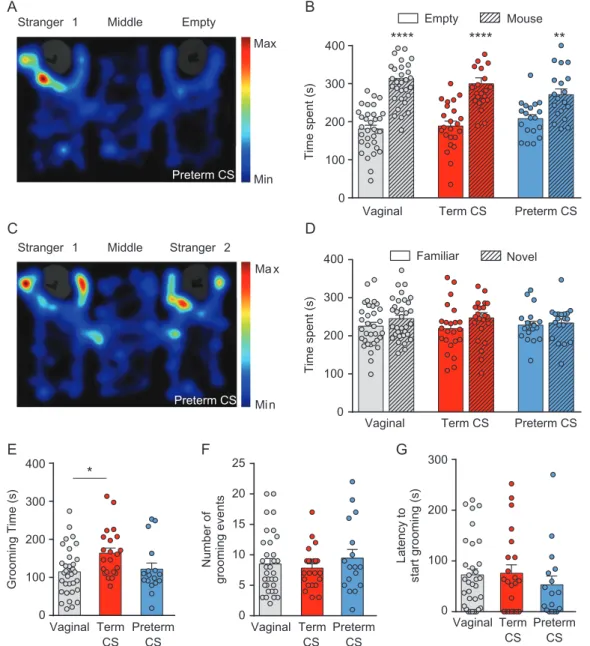
The three-chamber test was used to assess sociability and social novelty in young-adult male mice born vaginally or by C-section delivery. Social behavior, assessed by a higher time spent in the unfamiliar mouse chamber compared with the time spent in the empty chamber, was seen for vaginal (314± 12.25 s vs. 181 ± 9.91 s; P < 0.0001), term C-section (299.9 ± 15.47 s vs. 189± 12.93 s; P < 0.0001), and preterm C-section mice (271.2 ± 14.88 s vs. 208 ± 10.66 s; P = 0.0027; Fig. 1A,B, Supplementary Table 1). In the second part of the test, mice exhibited no social novelty as the time spent in the now-familiar mouse chamber versus the time spent in the novel mouse chamber was similar for vaginal (225.6± 9.87 s vs. 245.2 ± 9.39 s; P = 0.1782), term C-section (219± 13.15 s vs. 247.1 ± 13.63 s; P = 0.0687), and pre-term C-section mice (228.3± 10.09 s vs. 233.9 ± 10.79 s; P = 0.3211; Fig. 1C,D, Supplementary Table 1). Therefore, mice born by C-section delivery are not socially different from vaginal-born mice.
Term Cesarean Section Delivery Produces Minor Alterations of Stereotypic Behaviors in Adulthood
Self-grooming was used to assess stereotypic behaviors by measuring the time spent grooming, the number of grooming events and the latency to start grooming in young-adult male mice. Mice born at term by C-section spent more time groom-ing (163.7 ± 13.39 s) than vaginal mice (114.9 ± 10.95 s; P = 0.0219), whereas preterm C-section mice spent as much time grooming (121.8 ± 15.99 s) than vaginal and term C-section mice (Fig.1E, Supplementary Table 2). However, the number of grooming events was similar for vaginal (8.52 ± 0.88), term C-section (7.82± 0.72) and preterm C-section mice (9.47 ± 1.42; Fig.1F, Supplementary Table 2), and the latency to start groom-ing did not differ between vaginal (72.55± 12.1 s), term C-section (75.77 ± 16.69 s) and preterm C-section mice (53.12 ± 17.22 s; Fig. 1G, Supplementary Table 2). Hence, term C-section mice show a mild increase in stereotypic behavior whereas preterm C-section mice show no alteration of this behavior.
Term and Preterm Cesarean Section Delivery Do Not Alter Locomotor Nor Anxiety-Like Behavior in Adulthood
Open field was used to assess both anxiety-like and locomotor behaviors in young-adult male mice. To assess anxiety, we mea-sured the time mice spent in the center of an openfield arena.
Term and preterm C-section mice spent a similar amount of time in the center (49.29± 3.77 s and 39.04 ± 3.28 s, respectively) as vagi-nal mice (45.71 ± 3.6 s; Fig. 2A,B, Supplementary Table 3). The number of entries in the center (Fig.2C, Supplementary Table 3) was also similar between vaginal (47.64 ± 2.5), term C-section (46.26± 2.88), and preterm C-section mice (42.84 ± 3.34). Finally, the latency to first enter the center of the open field (Fig. 2D, Supplementary Table 3) was not statistically different for vagi-nal (12.76± 2.61 s), term C-section (16.92 ± 4.29 s), and preterm C-section mice (13.64± 3.42 s). Therefore, mice born by C-section do not show anxiety-like behavior.
To characterize locomotor activity, we assessed three para-meters: the velocity, the distance traveled, and the time mice spent moving. The velocity (Fig. 2E, Supplementary Table 4) was similar for vaginal (7.18± 0.3 cm/s), term C-section (6.55 ± 0.2 cm/s), and preterm C-section mice (6.49 ± 0.37 cm/s). The total distance traveled (Fig.2F, Supplementary Table 4) was not statistically different for vaginal (4301 ± 177.5 cm), term C-section (3919± 120.5 cm), and preterm C-section mice (3893 ± 220.5 cm). The same observation was made for the time spent moving (Fig.2G, Supplementary Table 4) as this parameter was not different between vaginal (440.7 ± 8.5 s), term C-section (419.2 ± 9.02 s), and preterm C-section mice (408.6 ± 10.44 s). These results show that C-section delivery does not induce an alteration in the locomotor behavior of mice.
Cesarean Section Delivery Alters Communicative Behaviors of Pups
Isolation-induced USVs were evaluated at P9 to assess early communicative alterations. Preterm C-section pups presented a higher number of calls than vaginal pups (206.7± 29.25 s vs. 89.05± 14.14 s; P = 0.0013; Fig.3A) as well as a higher total call-ing time (8.17± 1.27 s vs. 3.24 ± 0.55 s; P = 0.0011; Fig.3B). No differences were seen between vaginal and term C-section pups for these two parameters (Fig. 3A,B and Supplementary Table 5). The call duration was similar for term C-section (34.48 ± 1.24 ms), preterm C-section (36.15 ± 1.23 ms) and vaginal pups (32.44 ± 1.16 ms; Fig. 3C and Supplementary Table 5). Therefore, preterm C-section delivery is associated with USVs modifications in pre-wean mice.
Cesarean Section Delivery Does Not Affect the GABA Excitatory to Inhibitory Shift
To examine whether the action of GABA was altered after C-section delivery, cell-attached recordings were performed and the driving force of GABAAreceptors (DFGABA) recorded. At
P14–P15, neurons from term and preterm C-section born pups presented a similar DFGABA (4.7 ± 5.8 mV and 2.6 ± 2.9 mV,
respectively), with no difference compared with vaginal CA3 pyramidal neurons (4.1 ± 3.6 mV; P = 0.226 and P = 0.357, respectively; Fig.4A,B, Supplementary Table 6). To confirm the physiological effect of GABA polarity, extracellularfield poten-tial recordings were performed at P15–P16. Isoguvacine was applied on hippocampal slices to determine whether GABA had an excitatory or an inhibitory effect. Isoguvacine significantly decreased the spike frequency in hippocampal slices of vaginal (78.89 ± 2.71% of control; P = 0.0001), term C-section (73.92 ± 5.47%; P = 0.0009) and preterm C-section mice (79.55 ± 3.23%; P = 0.0001; Fig. 4C–F; Supplementary Table 7). Therefore, CA3 pyramidal neurons of pups born vaginally or after C-section exhibit an inhibitory GABA with a similar polarity at P14–P16.
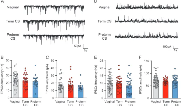
Cesarean Section Delivery Does Not Alter Spontaneous Glutamatergic and GABAergic Activities
Network activities were assessed in CA3 pyramidal neurons with whole-cell patch clamp recordings. At P14–P15, the ampli-tude and frequency of sEPSCs (Fig. 5A–C, Supplementary Table 8) were similar for neurons from vaginal (15.76± 0.94 pA; 24.93± 1.18 Hz), term C-section (17.01 ± 1.37 pA; 22.88 ± 1.45 Hz), and preterm C-section mice (13.85± 1.06 pA; 21.87 ± 1.68 Hz). Furthermore, no difference was seen in the amplitude and fre-quency of sIPSCs (Fig.5D–F, Supplementary Table 8) in neurons from vaginal (72.02 ± 2.81 pA; 9.68 ± 0.63 Hz), term C-section (66.57 ± 1.69 pA; 9.84 ± 0.72 Hz), and preterm C-section mice (68.82± 3.26 pA; 8.39 ± 0.97 Hz). Thus, C-section delivery does
not affect the glutamatergic and GABAergic network activities in CA3 pyramidal neurons at P14–P15.
C-section Delivery Transiently Delays CA3 Pyramidal Neurons Apical Dendrite Arborization
To evaluate if the morphological properties of CA3 pyramidal neurons were altered after birth by C-section delivery, neurons werefilled with biocytin (Fig.6A) and reconstructed for morpho-logical analysis (Fig.6B,C). We observed that at P0.5, the total dendritic length is shorter for pups born by C-section delivery at term (573± 42.04 μm; P = 0.0218) or preterm (537.2 ± 49.43 μm; P = 0.004) compared with vaginally born pups (784.6 ± 58.83 μm;
Vaginal Term CS Preterm CS
Time spent (s)
****
****
**
Vaginal Term CS Preterm CS
Empty Mouse Familiar Novel Empty
A
C
E
B
D
F
G
Middle Stranger 1 Stranger 2 Middle Stranger 1 Preterm CS Preterm CS Grooming Time (s) Vaginal Term CS Preterm CS*
Number of grooming events Vaginal Term CS Preterm CS Latency to start grooming (s) Vaginal Term CS Preterm CS 0 100 200 300 400 Time spent (s) 0 100 200 300 400 0 100 200 300 400 0 5 10 15 20 25 100 200 300 0 Min Max Mi n Ma xFigure 1. Effects of term or preterm cesarean section (CS) delivery on autistic-like behaviors in young-adult male mice. (A) Representative heatmap of a preterm CS mouse movements in the sociability test. (B) Sociability is represented by the time the mouse spent in the chamber with the unfamiliar mouse versus the empty chamber. (C) Representative heatmap of a preterm CS mouse movements in the social novelty test. (D) Social novelty test is represented by the time the mouse spent in the chamber with the now-familiar mouse versus with a novel mouse. Grooming behavior was assessed for vaginal, term CS, and preterm CS mice by (E) the time spent grooming, (F) the grooming frequency, and (G) the latency to start grooming. Data are presented as mean ± SEM, *P < 0.05, **P < 0.01, ****P < 0.0001. (B and D) n = 33 for vaginal, n = 23 for term CS and n = 19 for preterm CS. (E, F, and G) n = 33 for vaginal, n = 22 for term CS and n = 17 for preterm CS.
Fig.6E). The total number of intersections is also lower in neu-rons of pups born by C-section at term or preterm (419.9 ± 30.52μm; P = 0.0265; and 370.8 ± 31.21 μm; P = 0.0017,
respectively) compared with vaginal ones (563.1 ± 43.05 μm; Fig.6H,I). In addition, the primary dendritic length was shorter for preterm neurons (10.39 ± 2.09 μm) compared with term Vaginal Term CS Preterm CS Vaginal Term CS Preterm CS Time in Center (s) Velocity (cm/s) Vaginal Term CS Preterm CS Distance (cm) Number of entries in center Latency to center (s) Vaginal Term CS Preterm CS Vaginal Term CS Preterm CS Moving time (s) Vaginal Term CS Preterm CS Vaginal
A
B
E
F
G
C
D
Term CS Preterm CS 0 20 40 60 80 100 2000 4000 6000 8000 0 10 30 50 70 90 0 200 400 600 0 20 40 60 80 100 0 5 10 15 Min MaxFigure 2. Locomotor and anxiety-like behaviors in young-adult male mice are not affected by term or preterm CS delivery. (A) Representative heatmap of the move-ment of mice during the openfield test. Anxiety-like behaviors were compared between vaginal, term CS and preterm CS mice with (B) the time spent in the center of the arena, (C) the number of entries in the center, and (D) the latency to enter the center for the first time. Locomotor activities were determined in mice by (E) the velocity, (F) the total distance traveled, and (G) the time spent moving. Data are presented as mean ± SEM, (B–G) n = 33 for vaginal, n = 23 for term CS, and n = 19 for preterm CS. Vaginal Term CS Preterm CS 0 200 400 600
A
B
C
Number of calls / 3min
**
0 20 40 60 Vaginal Term CS Preterm CS Call duration (ms) 0 10 20 30 Vaginal Term CS Preterm CS**
Total calling time (s)
Figure 3. Preterm CS delivery alters early communicative behaviors in mice. Effect of CS delivery on isolation-induced ultrasonic vocalizations at P9 for (A) number of calls, (B) total calling time, and (C) call duration. Data are presented as mean ± SEM, **P < 0.01. (A–C) n = 55 for vaginal, n = 20 for term CS, and n = 35 for preterm CS.
C-section (19.04± 1.92 μm; P = 0.0003) and vaginal neurons (19.72 ± 2.88 μm; P = 0.0054; Fig.6D). Finally, the ending radius and criti-cal radius were shorter for preterm C-section delivery compared with vaginal neurons (99.9 ± 4.73 μm vs. 130.4 ± 5.11 μm; P = 0.0004; and 59.51± 4.90 μm vs. 86.61 ± 5.63 μm; P = 0.0022, respec-tively; Fig.6F–G, Supplementary Table 9). However, no difference could be seen between the three groups for all these parameters at P1.5 and P2.5 (Fig. 6D–I, Supplementary Tables 10 and 11). Thus, C-section delivery transiently alters the neuronal growth of CA3 pyramidal neurons at P0.5, a phenotype which is aggra-vated by the gestational age at the time of birth.
Discussion
Over the past couple of decades, the constant rise in rates of birth by C-section has raised concern due to its positive association with an increased incidence of neurodevelopmental disorders (Glasson et al. 2004;Chudal et al. 2014;Curran et al. 2015;Brander et al. 2016,2017;Yip et al. 2017). Yet, epidemiological studies are
limited as they inherently include variable parameters that can hardly be individualized or generalized. In this study, we used a mouse model of birth by C-section under controlled conditions, aiming to determine its possible deleterious sequels at different levels. Our results suggest that birth by C-section induces tran-sient developmental delays dependent on the gestational age at the time of delivery, but no long-term alterations. However, addi-tional early insults might aggravate these transient sequels, lead-ing to more persistent and deleterious consequences.
Preterm C-Section Delivery Alters Early Communicative Behavior But Does Not Lead to Long-Term Behavioral Modifications
ASD are neurodevelopmental disorders characterized by three main core symptoms: impairment in social behavior, difficul-ties in verbal and nonverbal communications, and a narrow range of actions carried out repetitively (WHO 2017a). Even
Spikes (%) Spikes #
****
***
****
0 300 600 900 1200 Preterm CS Vaginal 0 300 600 900 1200 0 800 1600 Term CS Time (s 0 300 600 900 1200 Time (s ) Time (s) Control Isoguvacine 0 400 800 0 800 1600 80µV Spikes # Isoguvacine Isoguvacine Isoguvacine VaginalA
C
D
E
F
B
Term Preterm CS CS DF GABA (mV) 80µV 80µV Spikes # Vaginal Term CS Preterm CS 20 40 60 –20 –40 1,5 1 0,5 –5 0 5 10 15 –0,5 -I, pA -Vp, mV 0 20 40 60 80 100 120Vaginal Term CS Preterm CS
Figure 4. Inhibitory action of GABA in CA3 pyramidal neurons of pups born by term or preterm CS around postnatal age P14. (A) Average values of DFGABAmeasured in hippocampal CA3 pyramidal neurons from P14 to P15. (B) Representative I/V curves from cell-attached recordings for vaginal, term CS, and preterm CS groups. (C– E) Effect of isoguvacine (10 μM; black bar) on representative traces of spontaneous extracellular field potentials from P15 to P16 with corresponding time courses of spike frequency changes for vaginal (C), term CS (D), and preterm CS (E). (F) Histogram of the averaged normalized spike frequency in control and isoguvacine (hatched bars) periods for vaginal, term CS, and preterm CS mice. Data are presented as mean± SEM, ***P < 0.001, ****P < 0.0001. (A) n = 31 for vaginal, n = 19 for term CS, and n = 15 for preterm CS. (F) n = 22 for vaginal, n = 13 for term CS, and n = 13 for preterm CS.
though using animal models to study neuropsychiatric disor-ders (such as ASD) is challenging, behavioral tests have been widely used to evaluate pathological symptoms in rodents (Kazdoba et al. 2016) and remain a useful aid to complement physiological studies (Del Pino et al. 2018). In young-adult male mice, repetitive behavior was mildly increased in mice born by C-section delivery at term but not preterm, whereas social behavior was not affected by C-section delivery, be it at term or preterm. Because social interaction tasks usually solicit explor-atory and anxiety-like behavior as well as social behavior, it was also necessary to assess these parameters separately to avoid confounding the outcome of this test. As a result, we found that birth by C-section delivery did not modify locomotor and anxiety-like behaviors. However, neonatal ultrasonic voca-lizations (USVs) were altered in mice born preterm by C-section compared with vaginally delivered ones. It is noteworthy that USVs are considered an early communicative behavior as well as the sign of an aversive affective state, and as such, their analysis has not only been widely applied to neurodevelopmen-tal studies (Scattoni et al. 2009), but has also been described as an autistic-like feature. In addition, repetitive behaviors seen in ASD are also a component of attention deficit hyperactivity dis-order (ADHD), Tourette syndrome and obsessive–compulsive disorder (Rapanelli et al. 2017;Rizzo et al. 2017). Moreover, a dis-ruption in social interactions has been associated with schizo-phrenia and bipolar disorder (Pappas et al. 2017) whereas an increase in locomotor activity is a parameter seen in pathologies such as schizophrenia, ADHD, or bipolar disorder (Powell and Miyakawa 2006). Altogether, our results suggest that C-section delivery does not lead to long-term behavioral alterations such as ASD, but prematurity associated to C-section delivery does affect developmental behaviors.
C-Section Delivery Does Not Impact GABA and Glutamate Activities in Juvenile Mice
An imbalance between excitation and inhibition has been sug-gested to underlie numerous neurodevelopmental disorders (Janik et al. 2010;Bozzi et al. 2018;Chiu et al. 2018), hence the need to study the effect of C-section delivery on GABAergic and glutamatergic signaling. Indeed, our lab previously showed that these two systems are affected in 2-week old pups of two dif-ferent rodent models of autism with an excitatory action of GABA and an increase in activity of spontaneous glutamatergic postsynaptic currents (Tyzio et al. 2014). Yet, in the current study, mice delivered by C-section did not show any of those changes, as glutamatergic and GABAergic spontaneous activi-ties as well as GABA polarity are similar to age-matched vaginal pups. The slightly depolarizing action of GABA observed is in agreement with our earlier determination of DFGABA (Tyzio
et al. 2008) and might be explained by the permeability of GABAAchannels to bicarbonate ions and the NKCC1-dependant
chloride equilibrium potential. In fact, in bicarbonate-free con-ditions or following the blockade of the chloride importer NKCC1 with its antagonist bumetanide, the previously slightly depolarizing DFGABAbecame hyperpolarizing (Tyzio et al. 2008).
Furthermore, the slight depolarization observed in all our groups is associated with inhibitory actions of GABA suggest-ing, as shown in earlier studies including our own, that this depolarization underlies a shunting inhibition (Monsivais et al. 2000;Vida et al. 2006;Howard et al. 2007;Tyzio et al. 2008;Tang et al. 2011). Altogether, these results suggest that C-section delivery at term or preterm does not lead to long-term altera-tions in GABAergic and glutamatergic hippocampal synaptic activities. Vaginal
A
B
C
E
F
D
Term CS Preterm CS Vaginal Term CS Preterm CS 0 10 20 30 40 50EPSCs amplitude (pA)
0 10 20 30 40 50 Vaginal Term CS Preterm CS Vaginal Term CS Preterm CS Vaginal Term CS Preterm CS Vaginal Term CS Preterm CS EPSCs frequency (Hz) 0 50 100 150
IPSCs amplitude (pA)
0 5 10 15 20 25 IPSCs frequency (Hz) 50pA 1s 100pA 1s
Figure 5. CS delivery at term or preterm does not affect spontaneous glutamatergic and GABAergic network activities in CA3 pyramidal neurons at P14–P15. (A) Representative traces of sEPSCs for vaginal, term CS, and preterm CS pyramidal neurons (holding potential:−70 mV). Histograms of the averaged (B) frequency and (C) amplitude of sEPSCs in vaginal, term CS, and preterm CS mice. (D) Representative traces of sIPSCs for vaginal, term CS, and preterm CS pyramidal neurons (holding potential:+5 mV). Histograms of the averaged (E) frequency and (F) amplitude of sIPSCs in vaginal, term CS, and preterm CS mice. Data are presented as mean ± SEM. (B and C) n = 47 for vaginal, n = 31 for term CS, and n = 21 for preterm CS. (E and F) n = 51 for vaginal, n = 34 for term CS, and n = 31 for preterm CS.
0 20 40 60 80 100 Primary dendritic length (µm)
**
***
0 500 1000 1500 2000Total dendritic length (µm)
0 500 1000 1500 2000
Total dendritic length (µm)
0 500 1000 1500 2000
Total dendritic length (µm)
*
**
***
**
Total number of intersections
0 400 800 1200
Total number of intersections Total number of intersections
0 400 800 1200
*
**
P0.5 0 20 40 60 80 100 Primary dendritic length (µm) 0 100 200 300 Ending radius (µm) 0 100 200 300 Ending radius (µm) 0 50 100 150 200 250 Critical radius (µm) 0 50 100 150 200 250 Critical radius (µm) P1.5 0 20 40 60 80 100 500 1000 1500 0 Primary dendritic length (µm) P2.5 0 50 100 150 200 Critical radius (µm) 0 50 100 150 200 250 Ending radius (µm)Vaginal Term CS Preterm CS
Number of intersections Radius (µm) Radius (µm) 0 50 100 150 200 1 3 5 7 Number of intersections 1 3 5 7 Number of intersections 1 3 5 7 0 100 200 300 0 100 200 300 Radius (µm) Vaginal P0.5 40 µm
C1
40 µm 40µmµC2
C3
B1
A
D
E
F
G
H
I
B2
B3
40 µmFigure 6. CA3 pyramidal cells apical morphology is transiently altered in mice born by CS delivery. (A) Image of biocytin-filled vaginal neurons acquired with a confo-cal microscope. (B) Reconstruction of representative neurons at P0.5 for vaginal (B1), term CS (B2), and preterm CS (B3) mice. (C) Reconstruction of representative neu-rons at P1.5 for vaginal (C1), term CS (C2), and preterm CS (C3) mice. Average values in CA3 pyramidal neuneu-rons from vaginal, term CS and preterm CS mice at P0.5, P1.5, and P2.5 for (D) primary apical dendritic length (E) total apical dendritic length, (F) ending radius, (G) critical radius, (H) total number of intersections in the apical arbor, and (I) Sholl analysis. Data are presented as mean ± SEM, *P < 0.05, **P < 0.01, ***P < 0.001. At P0.5, n = 28 for vaginal, n = 35 for term CS, and n = 29 for preterm CS. At P1.5, n = 23 for vaginal, n = 25 for term CS, and n = 33 for preterm CS. At P2.5, n = 30 for vaginal, n = 30 for term CS, and n = 24 for preterm CS.
CA3 Pyramidal Neurons of Mice Born Preterm by C-Section are Transiently Underdeveloped
Delivery and birth are critical periods associated with major physiological changes (Jaykka and Laakso 1967; Bland et al. 1982;Ward Platt and Deshpande 2005;Tyzio et al. 2006;Hooper et al. 2016), yet little is known on brain operation prior to and right after birth. Rabinowicz and colleagues showed that the late gestational period in humans was accompanied by a rapid decline of neuronal cortical density which stabilizes after birth (Rabinowicz et al. 1996). C-section delivery at term or preterm might affect this sequence. Here, we show that neurons of pups delivered by C-section at term or preterm have shorter total dendritic length and fewer intersections than vaginally delivered ones. Prematurity was also an aggravating factor for the morphological development of these neurons since they present a smaller primary dendritic length and shorter ending and critical radius. These results could be due to a lower expression of the mitochondrial uncoupling protein 2 which at birth is decreased in pups born by C-section delivery and is involved in neuronal size and dendritic arbor in culture (Simon-Areces et al. 2012). Furthermore, our results are in accordance with neuronal and brain growth alterations reported in autistic individuals (Courchesne et al. 2003,Redcay and Courchesne 2005;Wegiel et al. 2010;Petinou and Minaidou 2017), which support the hypothesis of aberrant brain connections impeding functional brain connectivity. Animal models of ASD further support these findings as brain area-dependent morphological alterations have been previously described in such models. In the ANKRD11-deficient mouse model of KBG syndrome, which presents autistic behavior, cortical pyra-midal neurons had morphological alterations which consisted in fewer number of dendrites and shorter arborization than control neurons (Ka and Kim 2018). Similar observations were done in 15 weeks old CD38 deficient mice, an ASD candidate gene (Nelissen et al. 2018). In the dentate gyrus and the visual cortex of these mice, the cell number was lower and the neurons shorter, whereas CA1 was only affected in terms of neuronal morphology, with shorter pyramidal cells (Nelissen et al. 2018). However, in contrast to our results, the morphological alterations were long-lasting and not restricted to 1 day postnatal as here. Altogether, our results suggest that even though developmental delays are observed after being born by C-section delivery, they are not sufficient to underlie long-lasting ASD features.
Limitations of the Use of a Mouse Model and its Correspondence to Humans
Rodent models are essential to understand the fundamental mechanisms underlying the onset of neurodevelopmental dis-orders, but their use comes with intrinsic limitations that are important to address. In these species, as gestational length differs, so does development. The Carnegie and Theiler stages comparison suggest that a mouse in utero is similar to a human fetus during its first and second trimesters (Otis and Brent 1954). The third trimester of gestation in humans corresponds to the early postnatal development in rodents, leading to a human baby brain at birth being comparable to a P7 rat brain (Dobbing and Sands 1979). Even though development takes places at different ages in humans and rodents, developmental patterns are conserved across species. Indeed, the switch in sensory processing from a “bursting” mode to an adult-like “acuity” mode happens at birth in humans and before eye opening (P14–15) in rats (Colonnese et al. 2010). In the hippo-campus, developmental patterns and spontaneous
network-driven endogenous activities are also similar despite occurring mostly in utero in primates (Berger and Alvarez 1996;Khazipov et al. 2001) and humans (Kostovic et al. 1989) but shifting to ex-utero in rodents (Tyzio et al. 1999). Furthermore, in rodents, oxytocin receptor blockers lower the threshold of pain at birth while bumetanide and oxytocin reverse this effect (Mazzuca et al. 2011). Interestingly, human babies born by C-section also have a lower threshold of pain compared with those born vagi-nally (Bergqvist et al. 2009), suggesting that the analgesic effect of oxytocin at birth might be preserved across species. These commonalities between species allowed us to test the hypothesis that birth is a critical period, and that changes in the mode or time of delivery around birth could impact this important devel-opmental milestone.
Relevance of Our Observations to Human Epidemiological Studies
Epidemiological studies aimed at evaluating if C-section deliv-ery might be associated with an increased incidence of disor-ders such as ASD have not yet been conclusive. They remain limited as few of them differentiate the reasons for C-section delivery such as due to an emergency (because of health risks for the mother and/or the baby) or if it was planned. In addi-tion, only one study compared C-section across gestational ages with the probability of developing ASD (Yip et al. 2017). The authors concluded that the odds for developing ASD were similar for children born by C-section delivery between 36 and 42 weeks of gestation, but were highly variable before 36 weeks of gestation, rendering the conclusion at these ages more diffi-cult. Furthermore, in these studies, children born by C-section delivery were neither assessed for their genetic background nor the environment their mother was exposed to during preg-nancy, even though ASD has been associated with those fac-tors. For these reasons, as of now, it is impossible to know if the increased incidence of ASD in children born by C-section delivery is due to one or more external factor(s) or to the surgi-cal act by itself. Our study sheds light on this issue by showing that C-section delivery at term or preterm induces transient developmental delays but does not lead to long-term conse-quences nor to autistic-like features per se. Interestingly, we also show that prematurity could be an aggravating factor, and an earlier prematurity might have led to permanent alterations in our experimental conditions. Unfortunately, we could not test this possibility as we failed to obtain viable pups born ear-lier than E17.75. This might be explained by the lung immatu-rity at earlier gestational ages in mice. In fact, before E17.4, the lymphatic network in lung tissue is not well developed and the surfactant synthesis and secretion, which allow gas exchange, are immature (Warburton et al. 2010). Finally, we cannot exclude the possibility that a double hit combining C-section delivery with an early or postnatal insult might lead to the increased incidence of ASD observed in some epidemiological studies (Glasson et al. 2004;Al-Ansari and Ahmed 2013;Curran et al. 2015;Yip et al. 2017). Indeed, these transient developmen-tal alterations seen in mice born by C-section delivery might be aggravated by an early in utero, neonatal or postnatal insult. In this scheme, future studies should focus on the hypothesis that delivery is a critical period whose alteration may play an aggravating role to a secondary insult.
Supplementary Material
Funding
This work was supported by Neurochlore; Fondation Bettencourt Schueller; France’s Agence Nationale de la Recherche (ANR-14-CE13-0021-01); A*MIDEX project (n° A*M-AAP-TR-14-02-140522-13.02-BURNASHEV-HLS) funded by the“Investissements d’Avenir” French Government program, managed by the French National Research Agency (ANR); Federation pour la Recherche sur le Cerveau (FRC) by the call Rotary-Espoir en Tête; and a Fellowship CIFRE-ANRT (2014/1056) to Morgane Chiesa.
Notes
We wish to thank Dr John Cryan and Dr Yuliya Borre for their help in establishing the cesarean section procedure in mice. We wish to thank Sanaz Eftekhari, Baptiste Riffault, Maxime Billon-Grand, Laurie-Anne Gouty-Colomer, and Amandine Dufour at Neurochlore for their technical assistance. We wish to thank Camille Dumon and Baptiste Riffault for their input on the paper. Conflict of Interest: Y.B.-A., D.C.F., N.B., R.T., and N.L. are shareholders of Neurochlore, a biotech company dedicated to the development of treatments for children with autism. Y.B.-A. is also the CEO of the company.
References
Al-Ansari AM, Ahmed MM. 2013. Epidemiology of autistic disor-der in Bahrain: prevalence and obstetric and familial charac-teristics. East Mediterr Health J. 19(9):769–774.
Ben-Ari Y. 2014. The GABA excitatory/inhibitory developmental sequence: a personal journey. Neuroscience. 279:187–219. doi:10.1016/j.neuroscience.2014.08.001.
Ben-Ari Y, Gaiarsa JL, Tyzio R, Khazipov R. 2007. GABA: a pio-neer transmitter that excites immature neurons and gener-ates primitive oscillations. Physiol Rev. 87(4):1215–1284. doi:10.1152/physrev.00017.2006.
Berger B, Alvarez C. 1996. Neurochemical development of the hippocampal region in the fetal rhesus monkey, III: calbindin-D28K, calretinin and parvalbumin with special mention of cajal-retzius cells and the retrosplenial cortex. J Comp Neurol. 366(4):674–699. doi:10.1002/(SICI)1096-9861 (19960318)366:4<674::AID-CNE8>3.0.CO;2-1.
Bergqvist LL, Katz-Salamon M, Hertegard S, Anand KJ, Lagercrantz H. 2009. Mode of delivery modulates physiologi-cal and behavioral responses to neonatal pain. J Perinatol. 29(1):44–50. doi:10.1038/jp.2008.129.
Betran AP, Ye J, Moller AB, Zhang J, Gulmezoglu AM, Torloni MR. 2016. The increasing trend in caesarean section rates: global, regional and national estimates: 1990–2014. PLoS One. 11(2):e0148343. doi:10.1371/journal.pone.0148343. Bilder D, Pinborough-Zimmerman J, Miller J, McMahon W. 2009.
Prenatal, perinatal, and neonatal factors associated with autism spectrum disorders. Pediatrics. 123(5):1293–1300. doi:10.1542/peds.2008-0927.
Bland RD, Hansen TN, Haberkern CM, Bressack MA, Hazinski TA, Raj JU, Goldberg RB. 1982. Lungfluid balance in lambs before and after birth. J Appl Physiol Respir Environ Exerc Physiol. 53(4):992–1004.
Boksa P, El-Khodor BF. 2003. Birth insult interacts with stress at adulthood to alter dopaminergic function in animal models: possible implications for schizophrenia and other disorders. Neurosci Biobehav Rev. 27(1–2):91–101.
Boksa P, Zhang Y. 2008. Epinephrine administration at birth pre-vents long-term changes in dopaminergic parameters caused
by cesarean section birth in the rat. Psychopharmacology (Berl). 200(3):381–391. doi:10.1007/s00213-008-1213-9.
Bozzi Y, Provenzano G, Casarosa S. 2018. Neurobiological bases of autism-epilepsy comorbidity: a focus on excitation/ inhibition imbalance. Eur J Neurosci. 47(6):534–548. doi:10. 1111/ejn.13595.
Brander G, Rydell M, Kuja-Halkola R, Fernandez de la Cruz L, Lichtenstein P, Serlachius E, Ruck C, Almqvist C, D’Onofrio BM, Larsson H, et al. 2016. Association of perinatal risk fac-tors with obsessive-compulsive disorder: a population-based birth cohort, sibling control study. JAMA Psychiatry. 73(11):1135–1144. doi:10.1001/jamapsychiatry.2016.2095. Brander G, Rydell M, Kuja-Halkola R, Fernandez de la Cruz L,
Lichtenstein P, Serlachius E, Ruck C, Almqvist C, D’Onofrio BM, Larsson H, et al. 2017. Perinatal risk factors in Tourette’s and chronic tic disorders: a total population sibling comparison study. Mol Psychiatry. doi:10.1038/mp.2017.31. Chiu PW, Lui SSY, Hung KSY, Chan RCK, Chan Q, Sham PC,
Cheung EFC, Mak HKF. 2018. In vivo gamma-aminobutyric acid and glutamate levels in people with first-episode schizophrenia: a proton magnetic resonance spectroscopy study. Schizophr Res. 193:295–303. doi:10.1016/j.schres.2017. 07.021.
Chudal R, Sourander A, Polo-Kantola P, Hinkka-Yli-Salomaki S, Lehti V, Sucksdorff D, Gissler M, Brown AS. 2014. Perinatal factors and the risk of bipolar disorder in Finland. J Affect Disord. 155:75–80. doi:10.1016/j.jad.2013.10.026.
Colonnese MT, Kaminska A, Minlebaev M, Milh M, Bloem B, Lescure S, Moriette G, Chiron C, Ben-Ari Y, Khazipov R. 2010. A conserved switch in sensory processing prepares develop-ing neocortex for vision. Neuron. 67(3):480–498. doi:10.1016/ j.neuron.2010.07.015.
Courchesne E, Carper R, Akshoomoff N. 2003. Evidence of brain overgrowth in thefirst year of life in autism. JAMA. 290(3): 337–344. doi:10.1001/jama.290.3.337.
Curran EA, Dalman C, Kearney PM, Kenny LC, Cryan JF, Dinan TG, Khashan AS. 2015. Association between obstetric mode of delivery and autism spectrum disorder: a population-based sibling design study. JAMA Psychiatry. 72(9):935–942. doi:10.1001/jamapsychiatry.2015.0846.
Del Pino I, Rico B, Marin O. 2018. Neural circuit dysfunction in mouse models of neurodevelopmental disorders. Curr Opin Neurobiol. 48:174–182. doi:10.1016/j.conb.2017.12.013. Desbonnet L, Clarke G, Shanahan F, Dinan TG, Cryan JF. 2014.
Microbiota is essential for social development in the mouse. Mol Psychiatry. 19(2):146–148. doi:10.1038/mp.2013.65. Dobbing J, Sands J. 1979. Comparative aspects of the brain
growth spurt. Early Hum Dev. 3(1):79–83.
Eftekhari S, Shahrokhi A, Tsintsadze V, Nardou R, Brouchoud C, Conesa M, Burnashev N, Ferrari DC, Ben-Ari Y. 2014. Response to Comment on“Oxytocin-mediated GABA inhibition during delivery attenuates autism pathogenesis in rodent offspring”. Science. 346(6206):176. doi:10.1126/science.1256009.
El-Khodor BF, Boksa P. 1997. Long-term reciprocal changes in dopamine levels in prefrontal cortex versus nucleus accum-bens in rats born by caesarean section compared to vaginal birth. Exp Neurol. 145(1):118–129. doi:10.1006/exnr.1997.6437. Glasson EJ, Bower C, Petterson B, de Klerk N, Chaney G, Hallmayer JF. 2004. Perinatal factors and the development of autism: a population study. Arch Gen Psychiatry. 61(6): 618–627. doi:10.1001/archpsyc.61.6.618.
Hillman NH, Kallapur SG, Jobe AH. 2012. Physiology of trans-ition from intrauterine to extrauterine life. Clin Perinatol. 39 (4):769–783. doi:10.1016/j.clp.2012.09.009.
Hooper SB, Te Pas AB, Kitchen MJ. 2016. Respiratory transition in the newborn: a three-phase process. Arch Dis Child Fetal Neonatal Ed. 101(3):F266–F271. doi:10.1136/archdischild-2013-305704.
Howard MA, Burger RM, Rubel EW. 2007. A developmental switch to GABAergic inhibition dependent on increases in Kv1-type K+ currents. J Neurosci. 27(8):2112–2123. doi:10. 1523/JNEUROSCI.5266-06.2007.
Janik P, Kalbarczyk A, Gutowicz M, Baranczyk-Kuzma A, Kwiecinski H. 2010. The analysis of selected neurotransmit-ter concentrations in serum of patients with Tourette syn-drome. Neurol Neurochir Pol. 44(3):251–259.
Jaykka S, Laakso L. 1967. Changes in skin temperature during thefirst minute of life as signs of circulatory transition at birth. Acta Obstet Gynecol Scand. 46(3):359–368.
Ka M, Kim WY. 2018. ANKRD11 associated with intellectual dis-ability and autism regulates dendrite differentiation via the BDNF/TrkB signaling pathway. Neurobiol Dis. 111:138–152. doi:10.1016/j.nbd.2017.12.008.
Kazdoba TM, Leach PT, Yang M, Silverman JL, Solomon M, Crawley JN. 2016. Translational mouse models of autism: advancing toward pharmacological therapeutics. Curr Top Behav Neurosci. 28:1–52. doi:10.1007/7854_2015_5003. Khazipov R, Esclapez M, Caillard O, Bernard C, Khalilov I, Tyzio
R, Hirsch J, Dzhala V, Berger B, Ben-Ari Y. 2001. Early devel-opment of neuronal activity in the primate hippocampus in utero. J Neurosci. 21(24):9770–9781.
Khazipov R, Ragozzino D, Bregestovski P. 1995. Kinetics and Mg2+ block of N-methyl-D-aspartate receptor channels dur-ing postnatal development of hippocampal CA3 pyramidal neurons. Neuroscience. 69(4):1057–1065.
Kostovic I, Seress L, Mrzljak L, Judas M. 1989. Early onset of syn-apse formation in the human hippocampus: a correlation with Nissl-Golgi architectonics in 15- and 16.5-week-old fetuses. Neuroscience. 30(1):105–116.
Lagercrantz H, Slotkin TA. 1986. The“stress” of being born. Sci Am. 254(4):100–107.
Mazzuca M, Minlebaev M, Shakirzyanova A, Tyzio R, Taccola G, Janackova S, Gataullina S, Ben-Ari Y, Giniatullin R, Khazipov R. 2011. Newborn analgesia mediated by oxytocin during delivery. Front Cell Neurosci. 5:3. doi:10.3389/fncel.2011. 00003.
Monsivais P, Yang L, Rubel EW. 2000. GABAergic inhibition in nucleus magnocellularis: implications for phase locking in the avian auditory brainstem. J Neurosci. 20(8):2954–2963. Nelissen TP, Bamford RA, Tochitani S, Akkus K, Kudzinskas A,
Yokoi K, Okamoto H, Yamamoto Y, Burbach JPH, Matsuzaki H, et al. 2018. CD38 is required for dendritic organization in visual cortex and hippocampus. Neuroscience. 372:114–125. doi:10.1016/j.neuroscience.2017.12.050.
Otis EM, Brent R. 1954. Equivalent ages in mouse and human embryos. Anat Rec. 120(1):33–63.
Pappas AL, Bey AL, Wang X, Rossi M, Kim YH, Yan H, Porkka F, Duffney LJ, Phillips SM, Cao X, et al. 2017. Deficiency of Shank2 causes mania-like behavior that responds to mood stabilizers. JCI Insight. 2(20). 10.1172/jci.insight.92052. Petinou K, Minaidou D. 2017. Neurobiological bases of autism
spectrum disorders and implications for early intervention: a brief overview. Folia Phoniatr Logop. 69(1–2):38–42. doi:10. 1159/000479181.
Pobbe RL, Pearson BL, Defensor EB, Bolivar VJ, Young WS 3rd, Lee HJ, Blanchard DC, Blanchard RJ. 2012. Oxytocin receptor knockout mice display deficits in the expression of
autism-related behaviors. Horm Behav. 61(3):436–444. doi:10.1016/j. yhbeh.2011.10.010.
Powell CM, Miyakawa T. 2006. Schizophrenia-relevant behav-ioral testing in rodent models: a uniquely human disorder? Biol Psychiatry. 59(12):1198–1207. doi:10.1016/j.biopsych. 2006.05.008.
Rabinowicz T, de Courten-Myers GM, Petetot JM, Xi G, de los Reyes E. 1996. Human cortex development: estimates of neuronal numbers indicate major loss late during gestation. J Neuropathol Exp Neurol. 55(3):320–328.
Rapanelli M, Frick L, Bito H, Pittenger C. 2017. Histamine modu-lation of the basal ganglia circuitry in the development of pathological grooming. Proc Natl Acad Sci U S A. 114(25): 6599–6604. doi:10.1073/pnas.1704547114.
Redcay E, Courchesne E. 2005. When is the brain enlarged in autism? A meta-analysis of all brain size reports. Biol Psychiatry. 58(1):1–9. doi:10.1016/j.biopsych.2005.03.026. Rizzo F, Abaei A, Nespoli E, Fegert JM, Hengerer B, Rasche V,
Boeckers TM. 2017. Aripiprazole and Riluzole treatment alters behavior and neurometabolites in young ADHD rats: a longitudinal (1)H-NMR spectroscopy study at 11.7T. Transl Psychiatry. 7(8):e1189. doi:10.1038/tp.2017.167.
Rubenstein JL, Merzenich MM. 2003. Model of autism: increased ratio of excitation/inhibition in key neural systems. Genes Brain Behav. 2(5):255–267.
Scattoni ML, Crawley J, Ricceri L. 2009. Ultrasonic vocalizations: a tool for behavioural phenotyping of mouse models of neu-rodevelopmental disorders. Neurosci Biobehav Rev. 33(4): 508–515. doi:10.1016/j.neubiorev.2008.08.003.
Schindelin J, Arganda-Carreras I, Frise E, Kaynig V, Longair M, Pietzsch T, Preibisch S, Rueden C, Saalfeld S, Schmid B, et al. 2012. Fiji: an open-source platform for biological-image analysis. Nat Methods. 9(7):676–682. doi:10.1038/nmeth.2019. Simon-Areces J, Dietrich MO, Hermes G, Garcia-Segura LM, Arevalo MA, Horvath TL. 2012. UCP2 induced by natural birth regulates neuronal differentiation of the hippocampus and related adult behavior. PLoS One. 7(8):e42911. doi:10. 1371/journal.pone.0042911.
Tang ZQ, Dinh EH, Shi W, Lu Y. 2011. Ambient GABA-activated tonic inhibition sharpens auditory coincidence detection via a depolarizing shunting mechanism. J Neurosci. 31(16): 6121–6131. doi:10.1523/JNEUROSCI.4733-10.2011.
Toda T, Homma D, Tokuoka H, Hayakawa I, Sugimoto Y, Ichinose H, Kawasaki H. 2013. Birth regulates the initiation of sensory map formation through serotonin signaling. Dev Cell. 27(1):32–46. doi:10.1016/j.devcel.2013.09.002.
Tyzio R, Cossart R, Khalilov I, Minlebaev M, Hubner CA, Represa A, Ben-Ari Y, Khazipov R. 2006. Maternal oxytocin triggers a transient inhibitory switch in GABA signaling in the fetal brain during delivery. Science. 314(5806):1788–1792. doi:10. 1126/science.1133212.
Tyzio R, Ivanov A, Bernard C, Holmes GL, Ben-Ari Y, Khazipov R. 2003. Membrane potential of CA3 hippocampal pyramidal cells during postnatal development. J Neurophysiol. 90(5): 2964–2972. doi:10.1152/jn.00172.2003.
Tyzio R, Minlebaev M, Rheims S, Ivanov A, Jorquera I, Holmes GL, Zilberter Y, Ben-Ari Y, Khazipov R. 2008. Postnatal changes in somatic gamma-aminobutyric acid signalling in the rat hippocampus. Eur J Neurosci. 27(10):2515–2528. doi:10.1111/j.1460-9568.2008.06234.x.
Tyzio R, Nardou R, Ferrari DC, Tsintsadze T, Shahrokhi A, Eftekhari S, Khalilov I, Tsintsadze V, Brouchoud C, Chazal G, et al. 2014. Oxytocin-mediated GABA inhibition during
delivery attenuates autism pathogenesis in rodent offspring. Science. 343(6171):675–679. doi:10.1126/science.1247190. Tyzio R, Represa A, Jorquera I, Ben-Ari Y, Gozlan H, Aniksztejn
L. 1999. The establishment of GABAergic and glutamatergic synapses on CA1 pyramidal neurons is sequential and cor-relates with the development of the apical dendrite. J Neurosci. 19(23):10372–10382.
Usher RH, Allen AC, McLean FH. 1971. Risk of respiratory distress syndrome related to gestational age, route of deliv-ery, and maternal diabetes. Am J Obstet Gynecol. 111(6): 826–832.
Vaillancourt C, Boksa P. 2000. Birth insult alters dopamine-mediated behavior in a precocial species, the guinea pig. Implications for schizophrenia. Neuropsychopharmacology. 23(6):654–666. doi:10.1016/S0893-133×(00)00164-0.
Vida I, Bartos M, Jonas P. 2006. Shunting inhibition improves robustness of gamma oscillations in hippocampal interneu-ron networks by homogenizingfiring rates. Neuron. 49(1): 107–117. doi:10.1016/j.neuron.2005.11.036.
Warburton D, El-Hashash A, Carraro G, Tiozzo C, Sala F, Rogers O, De Langhe S, Kemp PJ, Riccardi D, Torday J, et al. 2010. Lung organogenesis. Curr Top Dev Biol. 90:73–158. doi:10. 1016/S0070-2153(10)90003-3.
Ward Platt M, Deshpande S. 2005. Metabolic adaptation at birth. Semin Fetal Neonatal Med. 10(4):341–350. doi:10.1016/j.siny. 2005.04.001.
Wegiel J, Kuchna I, Nowicki K, Imaki H, Wegiel J, Marchi E, Ma SY, Chauhan A, Chauhan V, Bobrowicz TW, et al. 2010. The neuropathology of autism: defects of neurogenesis and neu-ronal migration, and dysplastic changes. Acta Neuropathol. 119(6):755–770. doi:10.1007/s00401-010-0655-4.
WHO. 2017a. Autism spectrum disorders. Geneva, Switzerland: World Health Organization.http://www.who.int/mediacentre/ factsheets/autism-spectrum-disorders/en/.
WHO. 2017b. Preterm birth. Geneva, Switzerland: World Health Organization. http://www.who.int/mediacentre/factsheets/ fs363/en/.
Yip BHK, Leonard H, Stock S, Stoltenberg C, Francis RW, Gissler M, Gross R, Schendel D, Sandin S. 2017. Caesarean section and risk of autism across gestational age: a multi-national cohort study of 5 million births. Int J Epidemiol. 46(2): 429–439. doi:10.1093/ije/dyw336.
Zhang X, Lv CC, Tian J, Miao RJ, Xi W, Hertz-Picciotto I, Qi L. 2010. Prenatal and perinatal risk factors for autism in China. J Autism Dev Disord. 40(11):1311–1321. doi:10.1007/s10803-010-0992-0.