SHORT ILLUSTRATED REVIEW
Intradural spinal metastasis of renal cell cancer. Report
of a case and review of 26 published cases
Gregory Jost&Stephan Zimmerer&Stephan Frank& Dominik Cordier&Adrian Merlo
Received: 5 June 2008 / Accepted: 3 December 2008 / Published online: 5 May 2009
# Springer-Verlag 2009
Abstract Metastatic disease in the intradural compartment of the spine is a rare manifestation of cancer. We report the case of an 82-year-old patient with an intradural, extra-medullary metastasis of renal cell carcinoma in the cervical spine. A literature search for intradural spinal metastases of renal cell carcinoma yielded a total of 26 further cases. 18 patients had sporadic renal cell carcinoma, and 9 patients had von Hippel-Lindau disease (VHL) in which the metastases of the renal cell carcinoma were embedded within spinal haemangioblastomas. Patients presented with paresis, back pain, altered sensation or, less frequently, bladder dysfunction. Intradural spinal metastases were diagnosed at an earlier age in VHL patients than in sporadic cases (mean 43±5 years vs. 60±14.5 years). The metastasis was surgically removed in 81% of patients. Pain improved in all patients, paresis in 90%, hypaesthesia in 38% and bladder dysfunction in 50%. Death occured as a result of systemic cancer progression. 93% of patients in the sporadic renal cell cancer group died within 1.5 years, whereas two thirds of the VHL patients were alive after 2 years.
Keywords Renal cell carcinoma . Renal cell cancer . Intradural metastasis . Intramedullary metastasis . Spine . VHL . von Hippel-Lindau disease
Introduction
Metastatic disease in the intradural compartment of the spine is a rare manifestation of cancer with an incidence of less than 1% in patients dying from systemic malignancies [9]. The most commonly associated primary tumours are cancers of the lung, breast, kidney and skin [38,44]. Renal cell carcinoma is usually diagnosed at a disseminated stage [35]. Advanced disease predominantly presents as metasta-ses to the lung (50%), bones (49%), lymph nodes (6–32%), liver (8%) and occasionally brain (3–10%) [32]. We present an 82 year old patient with an intradural metastasis of renal cell carcinoma (RCC) in the cervical spine and discuss it in the context of a further 26 previously published examples of this condition.
Literature review
The Medline database (PubMed, http://www.ncbi.nlm.nih. gov/PubMed/) was searched for the keywords intradural renal cell carcinoma and renal spinal metastasis. After screening of titles and abstracts, all suitable reports and important cross-references were obtained as full copies. Where appropriate, their authors were contacted by mail for follow-up information, such as progression of local or systemic disease and survival since publication.
Clinical details
An 82 year old man presented with progressive, moderately severe left hemiparesis and neck pain aggravated by coughing. A year earlier, he had sustained an infarction in the territory of the right middle cerebral artery, from which
DOI 10.1007/s00701-009-0358-6
G. Jost (*)
:
S. Zimmerer:
D. Cordier:
A. Merlo Department of Neurosurgery,University Hospital Basel,
Spitalstrasse 21, 4031 Basel, Switzerland e-mail: gregoryjost@gmx.ch
S. Frank
Department of Neuropathology, Institute of Pathology, University Hospital Basel,
he had completely recovered. Six years earlier, he had undergone a nephrectomy for clear-cell renal carcinoma. He had also undergone orchidectomy for prostatic carcinoma.
Neurological examination revealed a left-sided spastic hemiparesis of grade III–IV on the Medical Research Council Scale [18] and was otherwise unremarkable, as was the general physical examination.
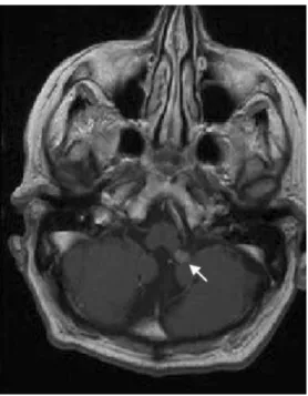
Magnetic resonance imaging (MRI) of the entire spine revealed a solitary, well-circumscribed intradural tumour at C6/7, compressing and displacing the spinal cord to the right. The lesion was iso-intense on T1- and hypo-intense on T2-weighted images, with slight homogeneous contrast enhancement (Fig. 1). MRI of the brain disclosed a small 0.8×0.6 cm extra-axial contrast enhancing lesion at the left foramen of Luschka (Fig. 2). In consideration of the patient’s advanced age and the small size of the cerebral lesion, Gamma Knife therapy was planned. Surgery was recommended for the symptomatic, space-occupying, well-defined tumour in the spine. A C6 laminectomy was performed. When the dura mater was opened, the tumour was found to be attached to the left C6 root. Macroscopi-cally, it displayed a nerve sheath tumour-like aspect with a rubbery consistency and a well demarcated cleavage plane. Histopathology of the completely removed lesion revealed partly haemorrhagic, highly vascularised tissue consisting of clear cells arranged in small lobules (Fig.3). The tumour cells had pronounced cytoplasmic accumulations of PAS-positive granules, reflecting glycogen deposition. Immuno-histochemical staining revealed strong reactivity with the pan-cytokeratin marker CK22 as well as with a renal cell carcinoma marker (NCL-RCC, clone 66.4.C2). Post-operatively, the left hemiparesis improved progressively. Adjuvant local irradiation was administered. One year after
surgery, the patient is doing well and is able to walk unaided.
Results
A Medline literature search yielded 19 articles dating from 1990 to 2007 concerning 32 examples of intradural spinal cord metastases (ISCM) of renal cell carcinoma [1,2,4,5, 11,13,15,19,20,22–24,28,29,33,38,39,41,42]. Six of these dated from the pre-CT and pre-MRI era and were therefore discarded from the analysis [15]. A total of 18 patients, including our patient, had sporadic RCC (Table1) [2,4,5,12,14,19,23,24,28,29,39,41], while 9 patients had von Hippel-Lindau disease (VHL) (Table2) [1,4,13, 19, 20]. In the latter group of patients, RCC was found within haemangioblastomas of the spinal cord as a tumour-to-tumour metastasis. Eleven of the patients with sporadic RCC were male, and 7 were female. In the VHL group, female patients (n=6) predominated over male patients (n=3). Nine ISCM were located in the cervical, 8 in the thoracic, 9 in the lumbar, and one in the sacral spine. All intradural metastases occurring between C1 and L1-2 were intramedullary except for our patient in whom the lesion mimicked an extradural extramedullary nerve sheath tu-mour. All intradural metastases below the conus medullaris were extramedullary, as expected.
Patients presented with limb paresis (n=20, 74%), back pain (n=15, 56%), hypaesthesia or paraesthesia (n=13, 48%) and bladder dysfunction (n=6, 22%). In 4 patients
Fig. 1 T2-weighted sagittal MRI of the cervical spine revealed a sharply circumscribed, intradural/extramedullary, hypointense tumour at C6/7 with dorsal displacement of the spinal cord
Fig. 2 Contrast-enhanced, T1-weighted axial MRI of the brain showed a small, homogeneously enhancing lesion at the left foramen of Luschka
(15%), cervical or upper thoracic ISCM caused at least a partial Brown-Séquard syndrome [11,13, 39]. The lesion was surgically resected in 22 (81%) patients. In four (15%) adjuvant post-operative radiotherapy was administered [11, 15, 28]. Five (19%) patients received external beam radiotherapy without surgery [13,33]. All patients experi-enced pain relief. Muscle strength improved in 18 of 20 patients (90%), hypaesthesia in 5 of 13 (38%) and urinary disturbance in 3 of 6 (50%). Histopathology confirmed metastatic renal cell cancer in all patients; the metastases were admixed with a haemangioblastoma lesion in the patients with VHL [1,4,13,19,20,34].
Sporadic cases
The mean age at diagnosis of RCC and ISCM was 57± 13 years (range, 36 to 78) and 60±14.5 years (range, 36 to 84), respectively. In the majority of sporadic cases (13 of 18, 72%), ISCM occurred after renal cell carcinoma had already been diagnosed, at a mean latency of 4.3 years (range 2 months to 15 years). In the remaining 5 patients
(28%), ISCM was the first manifestation of disease [11,13, 16,19,39]. When ISCM was diagnosed, 6 patients (33%) had no other metastases [2,5,13,23,29,39]. Among the 12 patients (66%) with systemic disease, 9 (50%) had involvement of the lungs [11, 13, 16, 24, 33, 41] and 4 (22%) of the brain [13, 15], while a further 4 patients developed brain metastases later on [13,33,41]. Metastasis to the brain was a frequent event in RCC (44% of all patients). Brain metastases were treated with external beam radiotherapy (n=5) [13,15,33], with corticosteroids (n=2) [13] and with surgery (n=1) [33]. In 5 patients, lung metastases appeared before ISCM was diagnosed [13, 24, 33, 41]. Interestingly, an aggressive strategy consisting of nephrectomy, resection of lung metastases and chemother-apy was associated with late development of ISCM, namely 6, 7 and 15 years after the diagnosis of RCC [13,24,41]. In the patients that had been treated with nephrectomy and chemo- or immunotherapy alone, ISCM was diagnosed within a few months [13,33].
In general, the prognosis was poor. Survival times were available for 14 patients. Seven (50%) died within 6 months
a
b
c
d
Fig. 3 On conventional H&E stains, the clear-cell aspect of the partly haemorrhagic tumour could easily be appreciated (a). The tumour cell cytoplasm featured conspicuous PAS-positive granula, corresponding to glycogen deposits (b). Upon immunohistochemical examination,
tumour cells were found to express cytokeratin (c) as well as a renal cell carcinoma-specific antigen d). See text for details. Scale bar 50 µm, all images same magnification
[5,11,13,23] and 13 (93%) within 1.5 years [5,11,13,15, 16, 23, 28,39]. Only 1 patient survived for 3 years [41]. The overall mean survival time for sporadic RCC was 11 months. Death always manifested as a result of systemic progression of RCC.
VHL patients
In VHL patients, ISCM was diagnosed at a mean age of 43±5 years (range, 36 to 52). All but two of the VHL patients had already been diagnosed with this syndrome; for
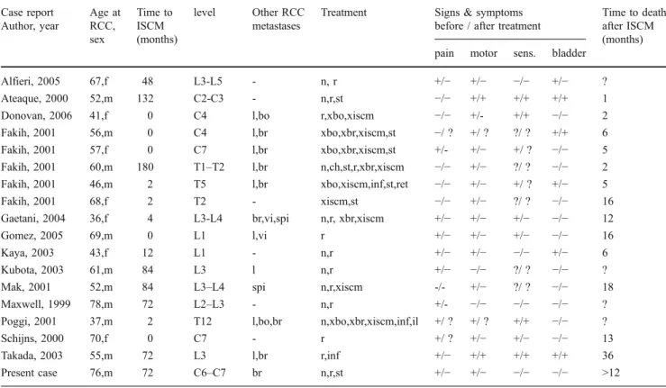
Table 1 Data from patients with intradural spinal cord metastases (ISCM) from sporadic renal cancer Case report Author, year Age at RCC, sex Time to ISCM (months) level Other RCC metastases
Treatment Signs & symptoms before / after treatment
Time to death after ISCM (months) pain motor sens. bladder
Alfieri, 2005 67,f 48 L3-L5 - n, r +/− +/− −/− +/− ? Ateaque, 2000 52,m 132 C2-C3 - n,r,st −/− +/+ +/+ +/+ 1 Donovan, 2006 41,f 0 C4 l,bo r,xbo,xiscm −/− +/- +/+ −/− 2 Fakih, 2001 56,m 0 C4 l,br xbo,xbr,xiscm,st −/ ? +/ ? ?/ ? +/+ 6 Fakih, 2001 57,f 0 C7 l,br xbo,xbr,xiscm,st +/- +/− +/ ? −/− 5 Fakih, 2001 60,m 180 T1–T2 l,br n,ch,st,r,xbr,xiscm −/− +/− ?/ ? −/− 2 Fakih, 2001 46,m 2 T5 l,br xbo,xiscm,inf,st,ret −/− +/− +/ ? +/− 5 Fakih, 2001 68,f 2 T2 - xiscm,st −/− +/− ?/ ? −/− 16 Gaetani, 2004 36,f 4 L3-L4 br,vi,spi n,r, xbr,xiscm +/− +/− +/− −/− 12 Gomez, 2005 69,m 0 L1 l,vi r +/− +/− +/− −/− 16 Kaya, 2003 43,f 12 L1 - n,r +/− +/− −/− +/− 6 Kubota, 2003 61,m 84 L3 l n,r +/− −/− ?/ ? −/− ? Mak, 2001 52,m 84 L3–L4 spi n,r,xiscm -/- +/− ?/ ? −/− 18 Maxwell, 1999 78,m 72 L2–L3 - n,r +/- −/− −/− −/− ? Poggi, 2001 37,m 2 T12 l,bo,br n,xbo,xbr,xiscm,inf,il +/ ? +/ ? +/+ −/− ? Schijns, 2000 70,f 0 C7 - r +/ ? +/− +/− −/− 13 Takada, 2003 55,m 72 L3 l,br r,inf +/− +/+ +/+ +/+ 36 Present case 76,m 72 C6–C7 br n,r,st +/− +/− −/− −/− >12
l lungs, bo bones, br brain, vi visceral, spi vertebra of the spine, n nephrectomy, r resection of the intraspinal metastasis, xbo radiotherapy to bones, xiscm to the ISCM, xbr to the brain, st steroids, ch chemotherapy, inf interferon, ret cisretinoic, il interleukin2, ? not reported, + present, -not present.
Table 2 Data from VHL patients with intradural spinal cord metastases (ISCM) of renal cancer Case report Author, year Age at
RCC, sex Time to ISCM (months) level Other RCC metastases
Treatment Signs & symptoms before / after treatment
Time to death after ISCM (months) pain motor sens. bladder
A-Hamden, 2003 39,m 0 C1-C4 ? n,r −/− +/− +/+ −/− ? Altinoz, 2005 43,m 24 T6-T7 l,br,adr n,il,xbr,r,sut +/− −/− −/− −/− >96 Fakih, 2001 37,f 24 C2 l n,il,r +/− +/− +/− −/− 23 Hamazaki, 2001 47,f 0 T5-6,T9 - n,r −/− +/ ? +/ ? −/− >24 Jarrel, 2006 36a,f ? sacrum - r +/− −/− −/− −/− >36 Jarrel, 2006 44a,f ? thoracic - r +/− −/− −/− −/− >12 Jarrel, 2006 41a,m ? thoracic l r −/− +/− −/− −/− 3 Jarrel, 2006 50af ? lumbar - r ?/ ? ?/ ? ?/ ? ?/ ? ? Polydorides, 2007 46,f 72 cervical ? n,r −/− −/− +/ ? −/− ? a
age at diagnosis of intradural spinal cord metastasis (ISCM), l lungs, br brain, adr adrenals, n neprectomy, r resection of the ISCM, xbr radiotherapy to the brain, il interleukin-2, sut sunitinib, ? not reported, + present, - not present.
two of them, however, it was the spinal tumour that brought the underlying VHL to clinical attention [1,19]. On average, the time from diagnosis of RCC to diagnosis of an intradural spinal metastasis was only 1 year. Most of the VHL patients had known CNS haemangioblastomas. Two of 8 patients (25%) had lung metastases of RCC 2 years before diagnosis of ISCM, treated with immunotherapy [4, 13]. One of these (13%) developed a brain metastasis 3 years after ISCM, which was irradiated and resected. This particular patient also received systemic interleukin-2, chemotherapy and the anti-angiogenic protein kinase inhibitor sunitinib and was still alive 6 years after removal of the ISCM [4,12]. One patient declined resection of the primary RCC and died 3 months later of intestinal obstruction [20]. Two thirds of VHL patients were alive after 2 years.
Discussion
Intradural metastatic disease to the spine is a rare complication of cancer. It accounts for only 0.8–3.9% of all symptomatic spinal metastases [9]. The strategic local-isation within the intradural spinal compartment often leads to irreversible neurological deficits. Patients with ISCM commonly suffer from advanced disease [26]. Therefore, the main goal of therapy is palliation. Most patients analysed in this systematic review of published reports underwent surgical removal of the ISCM, which relieved pain and improved motor function. Sensory and urinary dysfunction were less likely to recover. Five patients received radiotherapy instead of surgery because of advanced disease [13, 33]. In experienced hands, the microsurgical resection of an intradural spinal tumour is a safe procedure [8] and is associated with longer survival than conservative therapy [21], though selection bias is likely to play a part in the latter finding. The resection of metastases provides an effective local treatment and thus plays an important role in the management of radio-resistant tumours such as RCC [30]. In this historical and retrospective series, the male to female distribution in sporadic RCC reflected the same 1.5 :1 predominance of men over women found in other studies [27]. In the VHL group, there were twice as many women as men, but the number of patients was small. The mean age at presentation of ISCM in sporadic RCC, 60 years, matched Kalayci’s and Schiff’s findings in 138 and 40 patients, respectively, with mixed ISCM [21, 38]. Presumably, because of the hereditary factor, ISCM of RCC manifested much earlier in patients with VHL than in sporadic cases. In 40–70% of VHL patients RCC develop at a mean age of 37 years, as opposed to a peak incidence between 60 and 70 years for sporadic RCC [27]. Typically, CNS haemangioblastomas in
VHL cases become symptomatic in the third decade, and RCC is then detected during screening. In this disease, germ-line mutations of the tumour suppressor gene VHL on the short arm of chromosome 3 leads to deficient ubiquitination of hypoxia-inducible factor, which is in-volved in the molecular pathogenesis of CNS haemangio-blastomas, phaeochromocytomas, pancreatic cysts as well as islet cell tumours, papillary cystadenomas of the epididymis, renal cysts and carcinoma [40]. Consequently, individuals with VHL have a reduced average life expec-tancy of less than 50 years [14].
Intradural spinal cord metastasis is a sign of advanced disease and therefore associated with a poor prognosis, irrespective of whether it is intra- or extramedullary. According to the modified Robson staging system for RCC, patients with distant metastasis have stage IV cancer [36], with a 5-year survival from 0 to 10 percent [10]. Indeed, 93% of patients with sporadic RCC were dead 15 months after diagnosis of ISCM (mean survival 11 months), whereas two-thirds of the VHL patients were alive after 2 years. The cause of death was always systemic progression of cancer. The bulk of tumours causing symptomatic ISCM in VHL patients were haemangioblas-tomas that contained nests of renal cell cancer as a minor component. Thus, RCC was detected and treated at an earlier stage. Accordingly, the apparently faster diagnosis of the spinal metastasis after primary renal cell carcinoma and the apparently longer survival after ISCM of RCC is probably due, at least in part, to accelerated diagnosis because of the co-existing haemangioblastoma. In reports of ISCM from multiple sources and with mixed treatment strategies, the mean survival was 3 to 7.3 months, and 9.4 months if only surgically treated cases were considered [21, 38]. At the outset, 41% of patients had concomitant brain metastases [21], and 57.5% developed metastatic brain lesions over the course of their disease [38], a slightly higher figure than was found in the present study. Given this high probability of brain involvement in patients with ISCM, as opposed to only 5% of patients with RCC at initial presentation [30], imaging studies of the head should be performed as part of staging.
Half the patients had metastases in the lungs. In fact, the results of 687 post mortems suggested that RCC follows a metastatic“cascade” in which the lungs are the first site of haematogeneous spread [45]. Arterial embolism of pulmo-nary metastases might seed to other sites but would be an unlikely means of spread to the spinal cord. Nevertheless, the absence of pulmonary metastases in our patient suggests an alternative mechanism of spread via the vertebral venous plexus, as first proposed by Batson [7] and recently reviewed by Tobinick and Vega [43]. In addition to connections with the cranial venous system, the vertebral venous plexus communicates via widespread anastomoses
with systemic veins, including the inferior and superior venae cavae, the left renal and suprarenal veins, and the azygos and portal venous systems [6,17]. As blood flow in the vertebral venous system has been shown to be bi-directional [7, 17], this venous route provides an accepted pathway of metastatic seeding [31, 37]. Moreover, angio-graphic studies have demonstrated retrograde (inward) flow in the radicular veins and hence a communication between extra- and intradural compartments of the spine [25]. We suggest this route as the probable mechanism in our patient, whose metastasis was located close to the dorsal root. An alternative pathway is seeding via the cerebrospinal fluid (CSF), which, given the co-existent brain metastasis, would also have been possible in our patient. However, the CSF route seems less likely in patients with no prior or concomitant brain involvement. Finally, dissemination along the subarachnoid space via lymph vessels of peripheral nerves can lead to leptomeningeal invasion [46]. On the other hand, vertebral bone metastases can directly invade and/or seed to the CSF [28].
Conclusion
Surgery for ISCM from RCC leads to favourable functional results and may, in combination with aggressive therapy of systemic disease progression, prolong survival. Considering the high risk of brain involvement, imaging of the brain is mandatory in the staging of patients with ISCM. The prognosis for patients with ISCM of RCC is as poor as that of patients with ISCM of other primary tumours. Mortality is mainly due to the systemic disease.
Acknowledgments The authors wish to thank Dr. Ethan Taub for kindly reviewing the manuscript.
References
1. Abou-Hamden A, Koszyca B, Carney PG, Sandhu N, Blumbergs PC (2003) Metastasis of renal cell carcinoma to haemangioblas-toma of the spinal cord in von Hippel-Lindau disease: case report and review of the literature. Pathology 35:224–227. doi:10.1080/ 003130203100023191
2. Alfieri A, Mazzoleni G, Schwarz A, Campello M, Broger M, Vitale M, Vigl EE (2005) Renal cell carcinoma and intradural spinal metastasis with cauda equina infiltration: case report. Spine 30:161–163. doi:10.1097/01.brs.0000149180.75701.d1
3. Alfieri A, Mazzoleni G, Schwarz A, Campello M, Broger M, Vitale M, Vigl EE (2005) Renal cell carcinoma and intradural spinal metastasis with cauda equina infiltration: case report–part II. Spine 30:260–262. doi:10.1097/01.brs.0000149180.75701.d1
4. Altinoz MA, Santaguida C, Guiot MC, Del Maestro RF (2005) Spinal haemangioblastoma containing metastatic renal cell carci-noma in von Hippel-Lindau disease. Case report and review of the literature. J Neurosurg Spine 3:495–500
5. Ateaque A, Martin JL, O’Brien C (2000) Intramedullary spinal cord metastases from a hypernephroma 11 years following the diagnosis and treatment of the primary lesion. Br J Neurosurg 14:474–476. doi:10.1080/02688690050175337
6. Batson OV (1957) The vertebral vein system. Caldwell lecture, 1956. Am J Roentgenol Radium Ther Nucl Med 78:195–212 7. Batson OV (1995) The function of the vertebral veins and their
role in the spread of metastases. 1940. Clin Orthop Relat Res 4–9
8. Brotchi J (2002) Intrinsic spinal cord tumour resection. Neuro-surgery 50:1059–1063. doi:10.1097/00006123-200205000-00021
9. Connolly ES Jr, Winfree CJ, McCormick PC, Cruz M, Stein BM (1996) Intramedullary spinal cord metastasis: report of three cases and review of the literature. Surg Neurol 46:329–337. doi:10.1016/S0090-3019(96)00162-0,discussion 37-8
10. Couillard DR, deVere White RW (1993) Surgery of renal cell carcinoma. Urol Clin North Am 20:263–275
11. Donovan DJ, Freeman JH (2006) Solitary intramedullary spinal cord tumour presenting as the initial manifestation of metastatic renal cell carcinoma: case report. Spine 31:E460–E463. doi:10.1097/01.brs.0000222022.67502.4e
12. Escudier B (2007) Advanced renal cell carcinoma : current and emerging management strategies. Drugs 67:1257–1264. doi:10.2165/00003495-200767090-00002
13. Fakih M, Schiff D, Erlich R, Logan TF (2001) Intramedullary spinal cord metastasis (ISCM) in renal cell carcinoma: a series of six cases. Ann Oncol 12:1173–1177. doi:10.1023/A:101169 3212682
14. Friedrich CA (1999) Von Hippel-Lindau syndrome. A pleomor-phic condition. Cancer 86:2478–2482. doi: 10.1002/(SICI)1097-0142(19991201)86:11+<2478::AID-CNCR4>3.0.CO;2-5
15. Gaetani P, Di Ieva A, Colombo P, Tancioni F, Aimar E, Debernardi A, Rodriguez YBR (2004) Intradural spinal metastasis of renal clear cell carcinoma causing cauda equina syndrome. Acta Neurochir (Wien) 146:857–861. doi: 10.1007/s00701-004-0299-z
16. Gomez de la Riva A, Isla A, Perez-Lopez C, Budke M, Gutierrez M, Frutos R (2005) Metastasis intramedular como primera manifestacion de un carcinoma renal. Neurocirugia 16:359–364 17. Groen RJ, Groenewegen HJ, van Alphen HA, Hoogland PV
(1997) Morphology of the human internal vertebral venous plexus: a cadaver study after intravenous Araldite CY 221 injection. Anat Rec 249:285–294. doi:10.1002/(SICI)1097-0185 (199710)249:2<285::AID-AR16>3.0.CO;2-K
18. Guarantors of Brain (2000) Aids to the examination of the peripheral nervous system. Churchill Livingstone, Edinburgh 19. Hamazaki S, Nakashima H, Matsumoto K, Taguchi K, Okada S
(2001) Metastasis of renal cell carcinoma to central nervous system haemangioblastoma in two patients with von Hippel-Lindau disease. Pathol Int 51:948–953. doi: 10.1046/j.1440-1827.2001.01298.x
20. Jarrell ST, Vortmeyer AO, Linehan WM, Oldfield EH, Lonser RR (2006) Metastases to haemangioblastomas in von Hippel-Lindau disease. J Neurosurg 105:256–263. doi:10.3171/jns.2006. 105.2.256
21. Kalayci M, Cagavi F, Gul S, Yenidunya S, Acikgoz B (2004) Intramedullary spinal cord metastases: diagnosis and treatment -an illustrated review. Acta Neurochir (Wien) 146:1347–1354. doi:10.1007/s00701-004-0386-1discussion 54
22. Kawakami Y, Mair WG (1973) Haematomyelia due to secondary renal carcinoma. Acta Neuropathol 26:85–92. doi:10.1007/ BF00685527
23. Kaya RA, Dalkilic T, Ozer F, Aydin Y (2003) Intramedullary spinal cord metastasis: a rare and devastating complication of cancer- two case reports. Neurol Med Chir (Tokyo) 43:612–615. doi:10.2176/nmc.43.612
24. Kubota M, Saeki N, Yamaura A, Iuchi T, Ohga M, Osato K (2004) A rare case of metastatic renal cell carcinoma resembling a nerve sheath tumour of the cauda equina. J Clin Neurosci 11:530– 532. doi:10.1016/j.jocn.2003.09.010
25. Lasjaunias PL, Berenstein A (1987) Surgical Neuro-angiography. Springer-Verlag, Berlin, Germany
26. Lee SS, Kim MK, Sym SJ, Kim SW, Kim WK, Kim SB, Ahn JH (2007) Intramedullary spinal cord metastases: a single-institution experience. J Neurooncol 84:85–89. doi: 10.1007/s11060-007-9345-z
27. Ljungberg B, Hanbury DC, Kuczyk MA, Merseburger AS, Mulders PF, Patard JJ, Sinescu IC (2007) Renal cell carcinoma guideline. Eur Urol 51:1502–1510. doi:10.1016/j.eururo. 2007.03.035
28. Mak KH, Kwok JC (2001) Intradural spinal metastasis from renal cell carcinoma: a case report. J Orthop Surg (Hong Kong) 9:57–61 29. Maxwell M, Borges LF, Zervas NT (1999) Renal cell carcinoma: a rare source of cauda equina metastasis. Case report. J Neurosurg 90:129–132
30. Motzer RJ, Bander NH, Nanus DM (1996) Renal-cell carcinoma. N Engl J Med 335:865–875. doi:10.1056/NEJM199609193351207
31. Nishijima Y, Koiso K, Nemoto R (1995) The role of the vertebral veins in the dissemination of prostate carcinoma. Nippon Hinyokika Gakkai Zasshi 86:927–932
32. Pagano S, Franzoso F, Ruggeri P (1996) Renal cell carcinoma metastases. Review of unusual clinical metastases, metastatic modes and patterns and comparison between clinical and autopsy metastatic series. Scand J Urol Nephrol 30:165–172
33. Poggi MM, Patronas N, Buttman JA, Hewitt SM, Fuller B (2001) Intramedullary spinal cord metastasis from renal cell carcinoma: detection by positron emission tomography. Clin Nucl Med 26:837–839. doi:10.1097/00003072-200110000-00006
34. Polydorides AD, Rosenblum MK, Edgar MA (2007) Metastatic renal cell carcinoma to haemangioblastoma in von Hippel-Lindau disease. Arch Pathol Lab Med 131:641–645
35. Ritchie AW, deKernion JB (1987) The natural history and clinical features of renal carcinoma. Semin Nephrol 7:131–139
36. Robson CJ, Churchill BM, Anderson W (1969) The results of radical nephrectomy for renal cell carcinoma. J Urol 101:297–301 37. Ryan MW, Rassekh CH, Chaljub G (1996) Metastatic breast carcinoma presenting as cavernous sinus syndrome. Ann Otol Rhinol Laryngol 105:666–668
38. Schiff D, O’Neill BP (1996) Intramedullary spinal cord metastases: clinical features and treatment outcome. Neurology 47:906–912 39. Schijns OE, Kurt E, Wessels P, Luijckx GJ, Beuls EA (2000)
Intramedullary spinal cord metastasis as a first manifestation of a renal cell carcinoma: report of a case and review of the literature. Clin Neurol Neurosurg 102:249–254. doi:10.1016/S0303-8467 (00)00106-2
40. Shuin T, Yamasaki I, Tamura K, Okuda H, Furihata M, Ashida S (2006) Von Hippel-Lindau disease: molecular pathological basis, clinical criteria, genetic testing, clinical features of tumours and treatment. Jpn J Clin Oncol 36:337–343. doi:10.1093/jjco/hyl052
41. Takada T, Doita M, Nishida K, Miura J, Yoshiya S, Kurosaka M (2003) Unusual metastasis to the cauda equina from renal cell carcinoma. Spine 28:E114–E117. doi: 10.1097/00007632-200303150-00022
42. Takahashi I, Isu T, Iwasaki Y, Akino M, Takahashi A, Abe H, Kitagawa M, Kojima H, Inoue K, Saitoh H (1990) Metastatic Grawitz tumour to the cauda equina: case report. No Shinkei Geka 18:1157–1160
43. Tobinick E, Vega CP (2006) The cerebrospinal venous system: anatomy, physiology, and clinical implications. Med Gen Med 8:53 44. Traul DE, Shaffrey ME, Schiff D (2007) Part I: spinal-cord neoplasms-intradural neoplasms. Lancet Oncol 8:35–45. doi:10.1016/S1470-2045(06)71009-9
45. Weiss L, Harlos JP, Torhorst J, Gunthard B, Hartveit F, Svendsen E, Huang WL, Grundmann E, Eder M, Zwicknagl M et al (1988) Metastatic patterns of renal carcinoma: an analysis of 687 necropsies. J Cancer Res Clin Oncol 114:605–612. doi:10.1007/ BF00398185
46. Weissman DE, Grossman SA (1986) Simultaneous leptomenin-geal and intramedullary spinal metastases in small cell lung carcinoma. Med Pediatr Oncol 14:54–56. doi:10.1002/ mpo.2950140113
Comment
The article by Dr. Jost and colleagues focuses on intradural spinal metastasis of renal cell cancer. Although intraspinal spread of neoplasms has been repeatedly reported, the occurrence of metastasis in the intradural compartment of the spine caused by renal carcinoma is highly uncommon. The novel and interesting aspects of the present work particularly lie in the detailed current literature review including both cases of metastasis to haemangioblastomas in von Hippel-Lindau disease and sporadic cases of renal cell carcinoma metastasis, and the conclusion that, although optimal treatment after diagnosis of intra-dural spinal metastatic disease in renal cell carcinoma remains controversial, selected patients may have substantially increased survival and acceptable clinical outcome after surgical resection and adequate multidisciplinary management. This clearly written and well arranged review and case presentation of a rare condition is a valuable contribution to our literature on spinal cord metastasis from systemic cancer.
Markus Florian Oertel Germany