NEURO-ONCOLOGY (LE ABREY, SECTION EDITOR)
Clinical Implications of Molecular Neuropathology
and Biomarkers for Malignant Glioma
Ghazaleh Tabatabai&Monika Hegi&Roger Stupp&
Michael Weller
Published online: 20 March 2012
# Springer Science+Business Media, LLC 2012
Abstract Malignant gliomas are currently diagnosed based on morphological criteria and graded according to the World Health Organization classification of primary brain tumors. This algorithm of diagnosis and classification provides clini-cians with an estimated prognosis of the natural course of the disease. It does not reflect the expected response to specific treatments beyond surgery (eg, radiotherapy or alkylating chemotherapy). Clinical experience has revealed that gliomas sharing similar histomorphological criteria might indeed have different clinical courses and exhibit highly heterogenous responses to treatments. This was very impressively demonstrated first for oligodendrogliomas. The presence or lack of combined deletions of the chromo-somal segments 1p/19q was associated with different benefit from radiotherapy and chemotherapy. We review current molecular markers for malignant gliomas and discuss their current and future impact on clinical neuro-oncology. Keywords Glioma . Molecular diagnostics . Biomarker
Introduction
Malignant gliomas are a heterogeneous group of primary brain tumors. The entities are distinguished based on morphological criteria by histological analysis and presumed cell of origin. The World Health Organization (WHO) classification is a
grading system integrating four ascending grades of malignan-cy. It is based on histomorphological criteria [1] but has been shaped by decades of clinical observation. The WHO classifi-cation reflects the anticipated malignancy of the tumor and serves as a criterion to estimate the prognosis of patients. However, clinical experiences derived from prospective ran-domized clinical trials indicate that histomorphological criteria alone might not be sufficient to predict clinical outcome. Gliomas even with identical histopathological features differ considerably regarding clinical course or response to therapy. Investigation of molecular genetics of the tumor may help to overcome some of these limitations. Moreover, the analysis of blood and urine for chemokines or enzymes as well as the monitoring of circulating cellular subtypes in the peripheral blood have emerged especially during the increasing repertoire of targeted therapies, notably antiangiogenic therapies [2].
Clinicians expect these molecular aberrations or changing expression levels in tumor tissue, blood, or urine ideally to serve as diagnostic, prognostic, predictive, or surrogate bio-markers. Molecular tumor characterization should also refine the histopathological WHO classification and hopefully extend it to allow predictions for specific therapeutic strategies. Bio-markers may help for easy and reliable identification of res-ponders to a specific treatment, and allow identification of escape or resistance mechanisms during ongoing therapy. Sev-eral molecular markers have been characterized with respect to these clinical expectations. We review candidate molecules that have been investigated so far for anaplastic gliomas (WHO grade III) and for glioblastomas (WHO grade IV).
O6Methylguanine DNA methyltransferase (MGMT)
The DNA repair protein MGMT rescues the DNA damage induced by alkylating chemotherapy (eg, lomustine or temo-zolomide) by removing the alkyl group from the O6 position of G. Tabatabai (*)
:
M. WellerDepartment of Neurology, University Hospital Zurich, Frauenklinikstrasse 26,
Zurich 8091, Switzerland e-mail: ghazaleh.tabatabai@usz.ch M. Hegi
:
R. StuppDepartment of Neurosurgery and Clinical Neurosciences, Centre Hospitalier Universitaire Vaudois and University of Lausanne, Lausanne, Switzerland
guanine. During this repair process MGMT is irreversibly degraded and needs to be resynthesized de novo. As a conse-quence, the MGMT protein reservoir is potentially exhaustible. Early studies have examined the prognostic value of MGMT protein expression by immunohistochemistry and failed to consistently demonstrate a correlation with outcome. Of note, interobserver variation was also considerable [3]. The gold standard of protein determination in freshly isolated tumor tissue is of limited practical use, as it requires immediate processing of fresh tumor samples after surgery. Most recent studies analyzed the methylation of the MGMT gene promoter rather than protein levels or enzyme activity in glioma cells. Methylation of the gene promoter leads to silencing of tran-scription and thus lack of translation and absence of synthesis of functional protein. MGMT promoter methylation data con-sistently correlated better with clinical outcome than immuno-histochemical evidence of MGMT protein in glioma tissue [3]. This might be due to the differential accuracy of the methods and to the fact that immunohistochemical MGMT detection precludes the precise distinction of MGMT-positive glioma cells from host-derived glioma-infiltrating non-neoplastic cells (eg, microglia) [4, 5]. In contrast, determination of MGMT promoter methylation using methylation-specific assays essen-tially detects an acquired abnormality thought to be derived exclusively from tumor cells.
An association of MGMT gene promoter methylation and the benefit from alkylating chemotherapy was investigated within the randomized pivotal European Organisation of Research and Treatment of Cancer (EORTC)—National Can-cer Institute of Canada Clinical Trials Group (NCIC) phase III trial (EORTC 26981/22981-NCIC CE.3), which demonstrated improved survival when temozolomide was added to radiother-apy in newly diagnosed glioblastomas [6]. Methylation of the MGMT promoter in glioblastomas correlated with patients’ benefit from adding temozolomide to radiotherapy [7] while there was little effect on progression-free survival in patients receiving radiotherapy alone. These data suggested that MGMT promoter methylation is rather a predictive marker of benefit from temozolomide in glioblastomas.
In an attempt to overcome MGMT-mediated resistance by dose-dense (21/28 days) temozolomide administration, the Radiation Therapy Oncology Group (RTOG)—EORTC Intergroup trial 0525 compared standard dosing regimen of temozolomide (5/28 days) dosage with dose-dense temozo-lomide after completion of radiotherapy and concomitant temozolomide. No difference in outcome (ie, progression-free or overall survival) was seen with no hint for a benefit from dose-intensified temozolomide in any subgroup of patients. Nevertheless, patients with MGMT promoter-methylated glioblastomas had a significantly superior over-all survival (median, 23.2 months) compared to patients with unmethylated glioblastomas (median, 16 months) [8]. Recently, a study of the German Glioma Network analyzed
MGMT promoter methylation in 233 elderly patients older than 70 years of age with glioblastomas (median age, 74 years) and correlated the MGMT status with patients’ clinical outcome. Progression-free survival of patients with MGMT-methylated glioblastomas was longer when treated with radiotherapy plus temozolomide or temozolomide alone compared to patients receiving radiotherapy alone. On the other hand, patients with MGMT-unmethylated glio-blastomas did not gain any significant survival benefit from temozolomide [9•]. Confirmation of these findings in
pro-spective trials is needed to make individual treatment deci-sions based on the MGMT promoter methylation status. Specifically, it remains to be clarified whether elderly patients with MGMT-methylated glioblastomas should be treated with alkylating agent chemotherapy and deferred radiotherapy, while radiotherapy alone should be the treat-ment of choice for elderly patients with an MGMT-unme-thylated promoter. Interestingly, treatment with alkylating agent therapy does not select for loss of MGMT methylation in glioblastoma, as has been determined by the German Glioma Network investigating MGMT promoter methyla-tion in paired primary and recurrent glioblastomas [10•].
This finding is in line with the discovery of a mutator phenotype in MGMT-methylated glioblastoma after alkylat-ing agent therapy allowalkylat-ing for selection of treatment-induced genetic alterations resulting in therapy resistance such as mutations in the MSH6 gene, which is part of the mismatch repair pathway, that might also blunt the treatment efficacy of alkylating agents [11,12•].
In the NOA-04 randomized trial, the impact of sequential treatments with chemotherapy or radiotherapy in patients with anaplastic gliomas was explored. Patients received radiotherapy or alkylating chemotherapy (ie, temozolomide or procarbazine, lomustine, and vincristine [PCV]) at initial diagnosis, and after progression chemotherapy or irradia-tion, respectively. There was no difference in overall out-come and the sequence of treatments did not matter. Overall, patients with a methylated MGMT gene promoter had a better outcome.
Interestingly, time to first treatment progression was com-parable for both treatment arms even in the subgroup of patients with a methylated MGMT promoter, thus suggesting that in anaplastic gliomas the MGMT status is of prognostic value without prediction of benefit from alkylating agent chemotherapy [13]. This was confirmed in an EORTC trial demonstrating that MGMT promoter methylation was prog-nostic but not predictive for outcome to PCV in anaplastic oligodendrogliomas [14]. Thus, the predictive role of MGMT for benefit of temozolomide chemotherapy observed in glio-blastomas cannot be extrapolated to other grades of glioma. Of note, methylation of the MGMT promoter is indicative of other associated alterations in anaplastic glioma predicting a superior clinical outcome notably co-deletion of 1p/19q and
mutations in the isocitrate dehydrogenase (IDH)1 gene. This finding has been further clarified by the discovery of a glioma CpG island methylator phenotype (G-CIMP) present in over 45% of anaplastic glioma that was prognostic for outcome, and was highly correlated with IDH1 mutations, 1p/19q co-deletions, and MGMT methylation [15]. Originally, G-CIMP was discovered in glioblastoma when screening 272 glioblas-toma samples from The Cancer Genome Atlas (TCGA) pro-ject for genome-wide DNA methylation by Noushmehr et al. [16]. In glioblastoma, G-CIMP is infrequent, present in less than 10% of the cases, and strongly associated with mutations of IDH1 and with superior clinical outcome. Subsequent studies suggest that IDH1 mutations and, therefore, also G-CIMP, identify secondary glioblastoma that progress from lower-grade lesions. These findings have elucidated the evo-lution of gliomas, pathogenetically clearly separating primary glioblastoma from secondary glioblastoma and lower-grade gliomas. Thus, in future prospective randomized clinical trials, stratifications for the methylator phenotype and/or IDH muta-tions should be considered. Further, studying cancer-relevant pathways affected by the methylator phenotype may reveal novel promising drug targets.
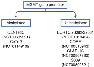
Currently ongoing clinical trials for glioblastomas patients use the MGMT gene promoter methylation status as selection criterion for study inclusion (Fig.1) or stratification factor. Studies selecting MGMT-methylated patients only, expect a synergistic effect of their novel agent with temozolomide treatment. In contrast, selecting MGMT-unmethylated glio-blastoma patients, allows omission of temozolomide. This allows testing of new drugs with a different mode of action and provides the opportunity to develop new treatment
strategies to improve the outcome in these patients who gain less benefit from the current standard of care.
Combined Deletions of Chromosomes 1p and 19q Investigations using microsatellite markers identified a fre-quent combined loss of heterozygosity (LOH) on the short arm of chromosome 1 (1p) and the long arm of chromosome 19 (19q) in malignant gliomas [17]. This combined loss is now commonly referred to as 1p/19q co-deletions and usually affects the whole chromosomal arms that seems to be medi-ated by a t(1;19)(q10;p10). Interestingly, this combination of LOH is almost never found in any non-glial malignancy. Moreover, 1p/19q co-deletions are commonly associated with oligodendroglial differentiation. Histological evidence of an oligodendroglioma or at least an oligodendroglial component in a mixed glioma is virtually always coupled with an IDH1/2 mutation [18]. Patients with oligodendroglioma that harbor 1p/19q deletions have superior outcome after radiotherapy and chemotherapy [19]. In glioblastomas, on the other hand, 1p/19q deletions are rare and do not predict prognosis of patients [20].
The identification of frequent 1p/19q co-deletions in malignant gliomas and the clinical relevance of this finding for predicting response to therapy of oligodendroglioma patients indicated the presence of tumor suppressor genes or other genes with important roles for radiotherapy- or chemotherapy-induced cell death in these chromosomal regions. Recently, candidate genes have been identified by exome sequencing (sequencing of all coding exons).
Fig. 1 Enrollment into clinical glioblastomas trials based on MGMT methylation status. CENTRIC, addition of cilengitide to standard therapy compared with standard treatment in newly diagnosed glioblastomas (recruitment completed). CeTeG, addition of lomustine to standard ther-apy compared with standard treatment in newly diagnosed glioblastomas (recruitment ongoing). EORTC 26082/22081, temsirolimus plus radio-therapy compared with standard treatment in newly diagnosed glioblas-tomas (recruitment ongoing). CORE, standard therapy with cilengitide
either twice weekly or five times per week versus standard therapy in newly diagnosed glioblastomas (recruitment ongoing), GLARIUS, bev-acizumab and irinotecan versus standard therapy in newly diagnosed glioblastomas (recruitment ongoing), S039, enzastaurin before and con-comitant with radiotherapy followed by enzastaurin maintenance in new-ly diagnosed glioblastomas (recruitment completed). The ClinicalTrials. gov identifier numbers are indicated in the figure
Mutations have been identified in CIC, a homolog of the Drosophila gene capicua, on chromosome 19q, and the FUBP1 gene (far upstream element binding protein 1), on chromosome 1p [21]. The functional roles of CIC and FUBP1 in the pathogenesis of these tumors are currently under investigation.
Currently ongoing clinical trials include patients with anaplastic gliomas only after determination of 1p/19q status (eg, CATNON trial [NCT00626990] or the CO-DEL trial [NCT00887146]), further emphasizing the impact of this genetic marker for clinical neuro-oncology.
Isocitrate Dehydrogenase
Somatic mutations of the IDH1 gene were identified by a whole-genome mutational analysis in glioma tissue sam-ples [22]. The mutations occur at codon 132 of the IDH1 gene, and less frequently at corresponding codons of IDH2 [22, 23], and were found in about 60% to 80% of astrocytomas and oligodendrogliomas of grades II and III but in less than 10% of glioblastomas. In glioblasto-mas, the presence of IDH1 mutations likely identifies secondary glioblastomas that originate from a prior lower-grade glioma [24]. This biomarker reliably identi-fies patients with a more favorable prognosis independent of treatment [13, 25, 26].
The neomorphic IDH mutants give rise to the production of D-2-hydroxyglutarate (2-HG) that accumulates to high levels in glioma [27]. The oncometabolite 2-HG is a competitive inhibitor ofα-ketoglutarate-dependent dioxygenases, reducing the activities of the families of histone demethylases and TET 5-methylcytosine hydroxylases, including the tet oncogene family member TET2, resulting in widespread epigenetic changes. Interestingly, in acute myeloid leukemia (AML), IDH1 and IDH2 mutations are also associated with a methyl-ator phenotype, and were shown to be mutually exclusive with loss-of-function mutations in the TET2 gene. These observa-tions suggests a functional link between IDH1/2 mutaobserva-tions, the production of 2-HG, and the development of a methylator phenotype in glioma and AML (ie, metabolism meets epige-netics) [28••,29]. The concentrations of the oncometabolite 2-HG in the blood can be used to monitor disease activity in patients with AML. However, 2-HG levels in the serum of glioma patients are not suitable as surrogate markers. The levels do not correlate with the presence of IDH1 or IDH2 mutations in the glioma tissue nor with tumor size [30]. This might be due to the fact that in contrast to AML, the actual tumor site is not the blood but the brain. This might account for too low concentrations in peripheral blood of glioma patients. Of note, concentrations of 2-HG in the cerebrospinal fluid have not yet been analyzed for 2-HG levels. Currently, efforts aim at
detecting 2-HG by magnetic resonance spectroscopy that would provide a noninvasive diagnostic tool to identify IDH1/2 mutant gliomas [31].
Recently, an antibody has been developed recognizing the mutant IDH1-R132H protein [32], which accounts for over 90% of all mutations in glioma. The development of this antibody has facilitated the widespread use of this marker, the rapid acquisition of data, and correlation with clinical outcome. The survival of patients with IDH1-mutant astrocytomas or oligodendrogliomas of grades II-III and including glioblastoma is longer than that of their IDH wild-type counterparts. Impor-tantly, overall survival of patients with IDH1-mutated glioblas-tomas (WHO grade IV) is better than for patients with IDH1 wild-type anaplastic astrocytomas (WHO grade III) [33], con-firming the potential of molecular markers to improve outcome prediction and clinical utility of the WHO classification.
IDH status reliably identifies patients with a more favorable prognosis but does not predict treatment-specific responses of glioma patients [13, 34, 35••,36–38], at least according to currently available data, and has not yet led to novel treatment strategies for malignant gliomas. In the future, clinical trials with IDH1 status as enrollment criteria might help to further define the impact of this molecular feature on clinical neuro-oncology. Given the fact that the antibody is already available for neuropathological histology diagnostics, it might be even easier to call for an upfront IDH1 status before enrollment into clinical trials than for an MGMT status. For example, a pos-sible strategy might be to exclude glioblastoma patients with IDH mutations from future glioblastoma trials.
Epidermal Growth Factor Receptor
Amplifications of epidermal growth factor receptor (EGFR) are frequent in primary glioblastomas [11,20]; overexpres-sion of EGFR has been associated with shorter survival in glioblastoma. Several compounds targeting EGFR are avail-able for clinical use, and have also been evaluated in malig-nant glioma. Neither of these demonstrated satisfying anti-glioma activity, although bioavailability and activity to de-phosphorylate the EGFR in the tumor tissue could be shown for gefitinib in a phase II trial [39•]. The prospective
ran-domized phase II EORTC 26034 trial on erlotinib in unse-lected recurrent glioblastoma did not demonstrate any evidence for antitumor activity [40]. A recent randomized trial investigated afatinib, a high-affinity irreversible EGFR tyrosine kinase inhibitor, versus dose-dense temozolomide (21/28 days) alone or the combinations of both. Again, no evidence of antitumor activity of EGFR inhibition could be shown [41]. Currently, the EGFR status does not impact clinical practice in neuro-oncology in any direction. The EGFRvIII mutation, a specific subtype of EGFR mutations,
might be used in the future for selecting patients for clinical trial enrollment. This specific mutation encodes for a con-stitutively active receptor and might be a useful target for immunotherapy [42].
Circulating Biomarkers
The investigation of circulating molecules or cells in peripheral blood and urine in patients with malignant gliomas has regained attention by the emergence of sev-eral antiangiogenic compounds. None of these parameters have entered routine clinical practice decision making yet. In a phase II trial investigating cediranib in patients with recurrent glioblastomas, plasma and urine were col-lected at baseline and at several defined time points after start of study medication. Increased levels of teinase 2 in plasma and increased levels of metallopro-teinase 9 in urine were correlated with poor progression-free survival, whereas increased plasma levels of placen-tal growth factor and basic fibroblast factor were corre-lated with longer overall survival. Increased plasma levels of stromal-derived factor 1, soluble vascular growth factor receptor 1, and soluble Tie 2 were corre-lated with radiographic tumor progression [2]. The value of circulating biomarkers still needs to be further defined in future clinical trials.
Conclusions
Biomarkers are an emerging field also for neuro-oncology. Considerable progress has been made in iden-tifying, characterizing, and applying molecular markers. These efforts will certainly refine the current histomor-phologically based WHO classification in the future. In the present daily clinical decision making for the treat-ment of patients with malignant gliomas, however, mo-lecular markers are barely used. The lack of better strategies for MGMT-unmethylated patients and limita-tions of accurate determination of the MGMT status limit its applicability in daily practice outside clinical trials. Deletion of 1p/19q allows identification of a prognostically more favorable subgroup; the ongoing randomized clinical trials will determine whether this allows one to adapt the treatment strategy individually. As outlined above, the identification of the IDH1 status demonstrates that this molecular aberration can be used to amend and refine the existing WHO classification. The recognition of IDH-mutated secondary glioma as a distinct pathogenetic entity will therefore very likely influence the next revision of the WHO classification.
On the other hand, currently ongoing powerful high-throughput analyses will most probably identify novel so-far unknown candidate molecules that could poten-tially serve as biomarkers for malignant gliomas. Yet, the identification of new markers will hopefully be paralleled by the identification of new drugs for the treatment of patients with malignant gliomas.
Disclosure Conflicts of interest: G. Tabatabai: has received honoraria for advisory boards from MSD and Roche.; M. Hegi: is a consultant for Merck Serono, MDxHealth, and MSD; has received honoraria for lectures or advisory boards from Merck Serono, MDxHealth, Shering Plough/ MSD, and Roche; and has received grant support from AstraZeneca; R. Stupp: has served on and received honoraria for advisory boards to Merck Serono (EMD), MSD (Merck & Co), Roche, and MDxHealth; M. Weller: has received research grants from Merck Serono and Roche and honoraria for lectures or advisory boards from Magforce, MSD, Merck Serono, and Roche.
References
Papers of particular interest, published recently, have been highlighted as:
• Of importance •• Of major importance
1. Louis DN, Ohgaki H, Wiestler OD, et al. The 2007 WHO classi-fication of tumours of the central nervous system. Acta Neuro-pathol. 2007;114:97–109.
2. Batchelor TT, Duda DG, di Tomaso E, et al. Phase II study of cediranib, an oral pan-vascular endothelial growth factor receptor tyrosine kinase inhibitor, in patients with recurrent glioblastoma. J Clin Oncol. 2010;28:2817–23.
3. Preusser M, Charles Janzer R, Felsberg J, et al. Anti-O6-methylguanine-methyltransferase (MGMT) immunohistochemis-try in glioblastoma multiforme: observer variability and lack of association with patient survival impede its use as clinical bio-marker. Brain Pathol. 2008;18:520–32.
4. Sciuscio D, Diserens AC, van Dommelen K, et al. Extent and patterns of MGMT promoter methylation in glioblastoma- and respective glioblastoma-derived spheres. Clin Cancer Res. 2011;17:255–66.
5. Weller M, Stupp R, Reifenberger G, et al. MGMT promoter methylation in malignant gliomas: ready for personalized medi-cine? Nat Rev Neurol. 2010;6:39–51.
6. Stupp R, Mason WP, van den Bent MJ, et al. Radiotherapy plus concomitant and adjuvant temozolomide for glioblastoma. N Engl J Med. 2005;352:987–96.
7. Hegi ME, Diserens AC, Gorlia T, et al. MGMT gene silencing and benefit from temozolomide in glioblastoma. N Engl J Med. 2005;352:997–1003.
8. Gilbert MR, Wang M, Aldape KD, et al. RTOG 0525: A random-ized phase III trial comparing standard adjuvant temozolomide (TMZ) with a dose-dense (dd) schedule in newly diagnosed glio-blastoma (GBM). ASCO Meeting Abstracts. 2011;29:2006. 9.• Reifenberger G, Hentschel B, Felsberg J et al. Predictive impact of
MGMT promoter methylation in glioblastoma of the elderly. Int J Canc. Journal international du cancer 2011. This study indicates a
predictive role of MGMT methylation in elderly patients with glio-blastomas. Currently, the role of MGMT in this glioblastoma patient population is further characterized in randomized clinical trials. 10.• Felsberg J, Thon N, Eigenbrod S et al. Promoter methylation and
expression of MGMT and the DNA mismatch repair genes MLH1, MSH2, MSH6 and PMS2 in paired primary and recurrent glio-blastomas. Int J Canc. Journal international du cancer 2011; 129: 659–670. This study demonstrates that the MGMT status is pre-served in recurrent glioblastomas.
11. McLendon R, Friedman A, Bigner D et al. Comprehensive ge-nomic characterization defines human glioblastoma genes and core pathways. Nature 2008; 455: 1061–8.
12.• Yip S, Miao J, Cahill DP et al. MSH6 mutations arise in glio-blastomas during temozolomide therapy and mediate temozolo-mide resistance. Clin Cancer Res. 2009; 15: 4622–9. Overcoming resistance to temozolomide therapy is a critical question. This study defines a role for the mismatch repair gene MSH6. 13. Wick W, Hartmann C, Engel C, et al. NOA-04 randomized phase
III trial of sequential radiochemotherapy of anaplastic glioma with procarbazine, lomustine, and vincristine or temozolomide. J Clin Oncol. 2009;27:5874–80.
14. van den Bent MJ, Dubbink HJ, Sanson M, et al. MGMT promoter methylation is prognostic but not predictive for outcome to adju-vant PCV chemotherapy in anaplastic oligodendroglial tumors: a report from EORTC Brain Tumor Group Study 26951. J Clin Oncol. 2009;27:5881–6.
15. van den Bent MJ, Gravendeel LA, Gorlia T, et al. A hypermethylated phenotype is a better predictor of survival than MGMT methylation in anaplastic oligodendroglial brain tumors: a report from EORTC study 26951. Clin Cancer Res. 2011;17:7148–55.
16. Noushmehr H, Weisenberger DJ, Diefes K, et al. Identification of a CpG island methylator phenotype that defines a distinct subgroup of glioma. Cancer Cell. 2010;17:510–22.
17. Reifenberger J, Reifenberger G, Liu L, et al. Molecular genetic analysis of oligodendroglial tumors shows preferential allelic dele-tions on 19q and 1p. Am J Pathol. 1994;145:1175–90.
18. Labussiere M, Idbaih A, Wang XW, et al. All the 1p19q codeleted gliomas are mutated on IDH1 or IDH2. Neurology. 2010;74:1886–90. 19. Jenkins RB, Blair H, Ballman KV, et al. A t(1;19)(q10;p10) medi-ates the combined deletions of 1p and 19q and predicts a better prognosis of patients with oligodendroglioma. Cancer Res. 2006;66:9852–61.
20. Weller M, Felsberg J, Hartmann C, et al. Molecular predictors of progression-free and overall survival in patients with newly diag-nosed glioblastoma: a prospective translational study of the German Glioma Network. J Clin Oncol. 2009;27:5743–50.
21. Bettegowda C, Agrawal N, Jiao Y, et al. Mutations in CIC and FUBP1 contribute to human oligodendroglioma. Science. 2011;333:1453–5. 22. Parsons DW, Jones S, Zhang X, et al. An integrated genomic analysis
of human glioblastoma multiforme. Science. 2008;321:1807–12. 23. Yan H, Bigner DD, Velculescu V, Parsons DW. Mutant metabolic
enzymes are at the origin of gliomas. Cancer Res. 2009;69:9157–9. 24. Nobusawa S, Watanabe T, Kleihues P, Ohgaki H. IDH1 mutations as molecular signature and predictive factor of secondary glioblas-tomas. Clin Cancer Res. 2009;15:6002–7.
25. Cairncross G, Berkey B, Shaw E, et al. Phase III trial of chemother-apy plus radiotherchemother-apy compared with radiotherchemother-apy alone for pure and mixed anaplastic oligodendroglioma: Intergroup Radiation Therapy Oncology Group Trial 9402. J Clin Oncol. 2006;24:2707–14. 26. van den Bent MJ, Carpentier AF, Brandes AA, et al. Adjuvant
procarbazine, lomustine, and vincristine improves progression-free survival but not overall survival in newly diagnosed anaplastic oli-godendrogliomas and oligoastrocytomas: a randomized European
Organisation for Research and Treatment of Cancer phase III trial. J Clin Oncol. 2006;24:2715–22.
27. Dang L, White DW, Gross S, et al. Cancer-associated IDH1 mutations produce 2-hydroxyglutarate. Nature. 2009;462:739–44. 28.•• Figueroa ME, Abdel-Wahab O, Lu C et al. Leukemic IDH1 and IDH2 mutations result in a hypermethylation phenotype, disrupt TET2 function, and impair hematopoietic differentiation. Cancer cell 2010; 18: 553–67. This is a very important study demonstrat-ing a connection between metabolics and epigenetics (ie, linkdemonstrat-ing the presence of IDH mutations with hypermethylation).
29. Prensner JR, Chinnaiyan AM. Metabolism unhinged: IDH muta-tions in cancer. Nat Med. 2011;17:291–3.
30. Capper D, Simon M, Langhans CD et al. 2-Hydroxyglutarate concentration in serum from patients with gliomas does not corre-late with IDH1/2 mutation status or tumor size. Int J Canc. Journal international du cancer 2011.
31. Pope WB, Prins RM, Albert Thomas M et al. Non-invasive detection of 2-hydroxyglutarate and other metabolites in IDH1 mutant glioma patients using magnetic resonance spectroscopy. J Neuro Oncol 2011. 32. Capper D, Weissert S, Balss J, et al. Characterization of R132H mutation-specific IDH1 antibody binding in brain tumors. Brain Pathol. 2010;20:245–54.
33. Hartmann C, Hentschel B, Wick W, et al. Patients with IDH1 wild type anaplastic astrocytomas exhibit worse prognosis than IDH1-mutated glioblastomas, and IDH1 mutation status accounts for the unfavorable prognostic effect of higher age: implications for clas-sification of gliomas. Acta Neuropathol. 2010;120:707–18. 34. Yan H, Parsons DW, Jin G, et al. IDH1 and IDH2 mutations in
gliomas. N Engl J Med. 2009;360:765–73.
35.•• Sanson M, Marie Y, Paris S et al. Isocitrate dehydrogenase 1 codon 132 mutation is an important prognostic biomarker in gliomas. J Clin Oncol. 2009; 27: 4150–4. This is a very impor-tant study indicating that IDH1 codon 132 mutation is closely linked to the genomic profile of the tumor and constitutes an important prognostic marker in gliomas of different WHO grades. 36. Dubbink HJ, Taal W, van Marion R, et al. IDH1 mutations in low-grade astrocytomas predict survival but not response to temozolo-mide. Neurology. 2009;73:1792–5.
37. Taal W, Dubbink HJ, Zonnenberg CB, et al. First-line temozolo-mide chemotherapy in progressive low-grade astrocytomas after radiotherapy: molecular characteristics in relation to response. Neuro Oncol. 2011;13:235–41.
38. Hartmann C, Hentschel B, Tatagiba M, et al. Molecular markers in low-grade gliomas: predictive or prognostic? Clin Cancer Res. 2011;17:4588–99.
39. • Hegi ME, Diserens AC, Bady P et al. Pathway analysis of glioblastoma tissue after preoperative treatment with the EGFR tyrosine kinase inhibitor gefitinib–a phase II trial. Mol Canc Ther-apeut 2011; 10: 1102–12. Preoperative treatment with targeted therapies enables the assessment of target modulation as demon-strated here for gefitinib.
40. van den Bent MJ, Brandes AA, Rampling R, et al. Randomized phase II trial of erlotinib versus temozolomide or carmustine in recurrent glioblastoma: EORTC brain tumor group study 26034. J Clin Oncol. 2009;27:1268–74.
41. Eisenstat DD, Nabors LB, Mason WP, et al. A phase II study of daily afatinib (BIBW 2992) with or without temozolomide (21/ 28 days) in the treatment of patients with recurrent glioblastoma. ASCO Meeting Abstracts. 2011;29:2010.
42. Sampson JH, Heimberger AB, Archer GE, et al. Immunologic escape after prolonged progression-free survival with epidermal growth factor receptor variant III peptide vaccination in patients with newly diagnosed glioblastoma. J Clin Oncol. 2010;28:4722–9.