ORIGINAL ARTICLE
Recurrent desmoids determine outcome in patients
with Gardner syndrome: a cohort study of three generations
of an APC mutation-positive family across 30 years
Matthias Turina&Caroline Marianne Pavlik&
Karl Heinimann&Frank Behrensmeier&
Hans-Peter Simmen
Accepted: 15 October 2012 / Published online: 1 November 2012 # Springer-Verlag Berlin Heidelberg 2012
Abstract
Purpose Screening of Gardner syndrome (GS) patients is tailored towards prevention of colorectal cancer (CRC). However, many patients suffer from desmoid tumors, which are challenging to treat due to invasive growth and local recurrence. The aims of our study were to determine the effectiveness of screening in GS and analyze outcome of desmoid tumors by treatment modality.
Methods This was a cohort study of a family of 105 descend-ants with GS. All family members who agreed were screened by endoscopy, and colorectal resection was performed upon pending malignancy. Resectable desmoids were excised, whereas large tumors were treated by a combination of bra-chytherapy (BT) and radiotherapy (RT). Main outcome meas-ures were the incidence of CRC and overall and disease-specific mortality (ClinicalTrial.gov ID NCT01286662). Results Thirty-seven of 105 family members have GS. Pre-ventive colorectal resections were performed in 16 patients (15 %), with one death due to gastric cancer. In four patients who denied screening endoscopy, invasive tumors of the colon (three patients) and stomach developed. Of 33 des-moid tumors, 10 (30 %) were located in the mesentery, 17 (52 %) in the abdominal wall, and 6 (18 %) in extra-abdominal sites. Excision of 12 desmoids was performed in eight patients. Four desmoids were treated by BT and RT and showed full or partial remission.
Conclusions Provided adequate screening, good long-term control of colorectal tumors is achievable. However, des-moid tumors determine survival and quality of life in many patients. Our data suggest good local control using a com-bination of brachytherapy/radiotherapy in large desmoids unsuitable for surgical resection.
Keywords Colorectal cancer . Adenomatous polyposis . Colon . Rectum . Familial . Desmoid
Introduction
In 1950, Eldon J. Gardner first described a kindred of patients presenting with familial adenomatous polyposis Parts of the data of the following manuscript were presented during the
Digestive Disease Week 2009 (SSAT poster presentation) as well as on the annual meeting of the German Surgical Society 2006 (Deutsche Gesellschaft für Chirurgie).
M. Turina (*)
Department of Colorectal Surgery, Cleveland Clinic Foundation, 9500 Euclid Ave.,
Cleveland, OH 44195, USA e-mail: [email protected] M. Turina
:
H.-P. SimmenDepartment of Surgery, University of Zurich Hospital, Rämistrasse 100,
8091 Zurich, Switzerland C. M. Pavlik
:
H.-P. Simmen Spital Oberengadin Samedan, Via Nuova 3,7105 Samedan, Switzerland K. Heinimann
Research Group Human Genetics, University of Basel, Mattenstrasse 28,
4058 Basel, Switzerland F. Behrensmeier
Clinic for Radiation Oncology, Inselspital, University of Berne, Freiburgstr.10,
(FAP) in combination with various skin and soft tissue
tumors [1]. Although the term “Gardner syndrome” (GS)
describes more of a historically coined variant of FAP rather
than a distinct subtype of the disease [2–4], the term is
usually reserved for patients in whom extraintestinal mani-festations are especially prominent and relevant during the
course of their disease [5–7].
Since 1978, our group of physicians has been treating a family of GS patients in the remote southeastern part of Switzerland. Similar to ethnic subgroups with little genetic admixture such as the Amish community in Northern Amer-ica, this kindred has been living in an area of alpine Switzer-land with minimal migration of residents, thereby passing on the distinct genetic Gardner subtype in a manner allow-ing population-based genetic analysis. Affected family members present with the phenotype of an attenuated form of FAP (AFAP), distinguished from classic FAP by the presence of fewer colonic adenomas, a lower cancer pene-trance, and an older age distribution of adenomas as well as carcinomas. In most cases, the syndrome is caused by auto-somal mutations in the APC gene. These mutations have been verified since the early 1990s. In this kindred, the APC germ line mutation c.5942delA was identified; this mutation leads to a frameshift at codon 1981 and results in a
prema-ture stop codon (p.Asn1981IlefsX62) [8].
As in most FAP patients, the mainstay in the management is repeat clinical and endoscopic screening to ensure timely colonic resection (usually in the form of subtotal colectomy or proctocolectomy with ileal pouch-anal anastomosis— IPAA) in patients developing malignancy. Experts on hered-itary gastrointestinal cancer recommend initial sigmoidos-copy in individuals with positive genetic testing and a
classical FAP variant at the age of 10–12 years [9]. The
same is recommended in patients with a positive family history and negative genetic testing, whereas in AFAP mem-bers with an identified mutation, initial colonoscopy is in-dicated at the age of 18–20. Thereafter, repeat endoscopy should be performed every 2 years; once adenomas are detected, annual colonoscopy should be performed until
colectomy [9]. In addition, 10–15 % of GS patients develop
desmoid tumors which frequently develop following surgi-cal resection in accessible areas such as the abdominal wall, or in sites unsuitable for resection like the intestinal mesen-tery. Despite their lack of metastatic potential, desmoids cause significant morbidity and mortality due to their ability to surround, compress, and erode adjacent tissues, providing a challenge to surgeons and oncologists alike. Affected patients suffer from prolonged and often lethal courses.
The aim of our present study was twofold. First, we wished to investigate whether early genetic testing, repeat clinical and endoscopic testing, and prophylactic colectomy were associ-ated with acceptable morbidity and mortality in GS. Secondly, we wished to better characterize extraintestinal manifestations
with an emphasis on treatment and outcome of desmoid tumors in our GS population.
Methods
Study design, eligibility criteria, and outcome variables
Prognostic cohort study: Since 1978, our group of physicians has been following a family of 105 members (43 women) from three generations of Gardner syndrome patients living in southeastern Switzerland. They are all direct descendants of a couple who lived at the end of the nineteenth century, and of which we know that five of their eight children died from
gastrointestinal cancer [10]. They all suffer from an identical
mutation of the APC gene that is being passed from one generation to the next. Both family members with confirmed APC mutations and those unwilling to undergo genetic testing but showing typical phenotype (endoscopic presence of mul-tiple adenomas) were eligible for this study and defined our sample size. Operations were performed at the local hospital in cooperation with pathology services from a regional referral center and (radio-) oncology and genetic counseling services from two national university hospitals. Outcome variables included overall survival, incidence of intestinal and extrain-testinal neoplasms, and treatment-specific recurrence rates and survival.
Diagnostic criteria: molecular and genetic testing
All family members at risk for the previously identified APC germ line mutation (c.5942delA) were counseled and
offered genetic testing by direct DNA sequencing [3, 4].
Genetic testing of our patient cohort was started in 1995
[11]. Routine testing was recommended in adolescents by
the age of 16 in order to timely identify patients in need of endoscopic screening and to reassure individuals not carry-ing the mutation. All tests were performed twice from two different samples of whole blood drawn on different days to minimize false-positive testing.
Clinical and endoscopic screening program
In patients with positive APC gene mutations, endoscopic screening was recommended to begin at the age of 16 years. Colonoscopy was indicated in patients with confirmed APC mutation and those who did or could not undergo genetic testing but who were related to known carriers of the muta-tion. Colonoscopy was recommended every 2 years in patients without adenomas; once adenomas were detected, annual colonoscopy was recommended.
Gastroscopy was performed upon detection of colorectal adenomas, or earlier upon onset of any upper gastrointestinal
symptoms. The extent of duodenal polyposis was assessed using the Spigelman classification and patients were
moni-tored accordingly [12]. In case of few, small duodenal
adeno-mas, screening and endoscopic removal was continued, whereas resection was encouraged in multiple or dysplastic adenomas (Spigelman IV).
Treatment of colonic adenoma, carcinoma, and soft tissue tumors
Preventive resections were recommended to patients with confirmed dysplasia and/or endoscopically uncontrollable numbers of adenomas. Subtotal colectomy with ileorectal anastomosis (IRA) was the procedure of choice in our patients due to the clinical phenotype of GS with predomi-nantly right-sided polyposis and no or only moderate in-volvement of the rectum. Despite the risk of late rectal cancer and the need for lifetime follow-up in patients with IRA, we and others believe that this procedure is justified in GS due to its technical simplicity, good functional results,
and lower perioperative morbidity compared to IPAA [13].
Desmoids and other soft tissue tumors
Desmoids of the abdominal wall were treated by wide excision in symptomatic patients or those exhibiting rapid growth and were routinely followed by CT. Recurrent des-moids were treated by radical resection or a combination of percutaneous radiotherapy (RT) and brachytherapy (BT) if unsuitable for resection. Intra-abdominal desmoids were usually considered nonresectable due to invasive growth in the intestinal mesentery and their often multifocal and late clinical appearance.
Statistical analysis
The study is registered at the National Institute of Health’s registry (ClinicalTrial.gov ID NCT01286662). Descriptive analyses and univariate statistical comparisons were per-formed using Sigma Stat 3.11.0, Systat Software, Inc.
Results
Thirty-seven of the 105 members of this family were diag-nosed with Gardner syndrome, either by positive genetic test-ing (27 patients) or by their phenotype (10 patients). The mean age at time of testing was 30 years (range 5–73). Genetic testing was done in 52 family members (27 positive, 25 neg-ative). The APC mutation status of the remaining 43 patients is unknown, either because they declined genetic testing, have not undergone colonoscopy for age reasons, or were lost to
follow-up during the course of their lives (Table1).
Thirty-two of the 37 GS patients were screened using colonoscopy. The five remaining patients denied screening endoscopy. The youngest patient undergoing endoscopy was 12 years, and the youngest patient showing colonic polyposis was 14 years old. The most prevalent endoscopic findings of
the gastrointestinal tract are listed in Table 2. In our series,
patients most frequently presented with attenuated polyposis with a predominantly right-sided affection of the colon, main-ly the cecum and the ascending colon. In 18 patients, few polyps (100 or less) were observed, whereas five patients showed up to 1,000 polyps. The rectum was mildly affected in 14 cases. Altogether, 18 patients (17 % of the entire kindred and 49 % of patients genotypically and/or phenotypically positive for GS) had to undergo colonic resections, and 7 patients had to undergo a total of 10 resections for extraco-lonic tumors. Overall mortality throughout the entire time period was six patients (16 % of all patients with Gardner syndrome), three of whom died from colorectal carcinoma, one from gastric carcinoma, and two from mesenteric des-moids tumors.
A total of 34 adult family members (32 %) either de-clined to undergo genetic testing or screening endoscopies Table 1 Prevalence of Gardner syndrome in the study cohort Established diagnosis of GS Genotype positive—27 (26 %)
Phenotype positive—10 (10 %) Diagnosis of GS ruled out Genotype negative—25 (24 %) Juvenile family members at
risk w/o genetic testing or colonoscopya
9 (8 %)
Adult family members at risk who decline genetic testing and colonoscopy
10 (10 %)
Lost to follow-up 24 (22 %)
Entire cohort 105 family members (100 %) GS Gardner syndrome, w/o without
a
Endoscopy planned at age 16
Table 2 Gastrointestinal tumors and polyps (36 patients, 93 tumors)
Site Tumor No. of patients
Stomach Fundic gland polyposis 9
Adenoma 15 Ulceration/gastritis 9 Dysplasia/adenocarcinoma 3 Duodenum Adenomas 13 Jejunum Adenocarcinoma 1 Ileum Polyps 3 Colon Adenomas/polyps 23 Adenocarcinoma 2 Rectum Adenomas/polyps 14 Adenocarcinoma 1
or were lost to follow-up in the course of the entire
obser-vation period (Table1). In addition, two patients with
his-tologically confirmed colorectal carcinoma refused surgical treatment.
Outcome related to colorectal adenoma and carcinoma
Of the entire cohort, 18 patients underwent colorectal resec-tions. Sixteen underwent preventive resections (15 colecto-mies with ileorectal anastomosis and one left colectomy) due to endoscopic findings of either too numerous or dys-plastic adenoma and two patients because of colorectal cancer (CRC) without previous suspicion of dysplastic ad-enoma (one proctocolectomy and one resection of the trans-verse colon). Of all patients with colorectal resections, 16 remained free of recurrence for a mean 93 months (range 14–387), whereas two patients died from recurrent CRC. Both patients either refused screening endoscopy or surgical resection at a premalignant state. One patient died from gastric cancer and two from mesenteric desmoids after 48 months (range 24–103). Most importantly, however, none of the patients who underwent screening endoscopy followed by preventive colectomy for suspicious endoscop-ic findings died from intestinal neoplasms.
Treatment and outcome of desmoid tumors
Fifteen patients (5 women, 10 men) developed a total of 33 desmoids tumors (14 % of the entire family, 41 % of Gardner
patients at a mean age of 43 years [31–77 years], Table3).
Most desmoids (17 tumors, 52 %) were found in the
abdom-inal wall (Fig.1a). Ten patients suffered from solitary
mesen-teric desmoids (30 %, Fig.1b). Six desmoids (18 %) were
located in extra-abdominal locations (back, groin, thigh, but-tocks). Sixty-one percent occurred in context with pervious surgery (mainly laparotomy) after a median interval of 39 months after surgery (range 6–94 months). Thirteen des-moids developed in patients without prior surgery (one mes-enteric desmoid, six abdominal wall desmoids, six others). Ten abdominal wall desmoids could be treated by wide
exci-sion alone (Fig.2), with three recurrences in one single patient
within 28 months. Complications in patients with progressive mesenteric desmoids were ureteric obstruction in two patients and bowel obstruction in two patients with stage III mesen-teric desmoids, one of whom was treated by small bowel resection and intestinal transplantation.
Recurrences of abdominal wall desmoids were treated by repeat local excision. Six patients stayed recurrence-free for a mean follow-up of 70 months (range 6–120 months). The remaining one patient decided to discontinue follow-up, and his status is unknown. From 2006 onwards, two desmoids were treated by high-dose radiation (HDR) brachytherapy and two others by a combination of HDR brachytherapy and
percutaneous radiotherapy. All were recurrent desmoids which were unsuitable for resection due to their large size at presentation. Desmoid tumors treated in this manner showed complete and partial remission in two cases each, with an average length of follow-up of 19 months (range
13–25 months, Fig.3). Indications for BT were large tumors
requiring plastic coverage if resected and patients denying surgery. In addition, desmoids had to be of adequate size to
be properly exposed to the applicators (Fig.4). Clinoril and
tamoxifen was used in five patients with mesenterial des-moids that were unsuitable for resection, or in patients reluctant to undergo surgical therapy.
Osteoma and other extracolonic manifestations
A disproportionate number of GS patients developed a variety of both benign and malignant extracolonic tumors, including a renal cell carcinoma, pancreatic insular carcino-ma, Hodgkin and non-Hodgkin lymphocarcino-ma, uterus myocarcino-ma, adrenal incidentaloma, cervical carcinoma, and one endo-metrioma. Eight patients developed a total of 12 osteoma which were located in the skull (seven), spine (two), and pelvic bone (three). Two such tumors were located in the mandibula of separate patients and required surgical exci-sion due to progressive growth. No case of thyroid carcino-ma occurred in our patient cohort.
Discussion
Since Elrond J. Gardner’s original description in the 1950s,
a number of case reports and small series have been pub-lished which describe this unique variant of FAP. Most authors agree on the need for early screening endoscopy followed by colectomy upon detection of dysplasia or
en-doscopically uncontrollable adenomatosis [9,14,15]. Less
consent, however, exists on the management of associated desmoid tumors. In our present study, we have been privileged to follow on a large number of patients in a
socially and geographically “contained” environment,
pro-viding us the unique opportunity to study not only problems related to colonic adenomatosis and its consequences, but also the occurrence and management of extracolonic mani-festations in a large number of patients. In summary, we believe to contribute to the following issues related to Gard-ner syndrome patients:
Screening Our data show that early screening endoscopy in patients with identified APC gene mutation, and subsequent prophylactic colectomy in patients with dysplasia or sub-stantial adenomatosis reliably prevents the occurrence of CRC in Gardner patients. In fact, none of the members of this family willing to participate in our surveillance program
T able 3 Overview of desmoid patients Patient no. Gender Cause of death Age at diagnosis Location and number of desmoids Abdominal sur gery before desmoid occurrence Occurrence in context with previous sur gery T ime of
desmoid occurrence after
sur
gery
(month)
Incidence without previous sur
gery Resection of A W D Resection of MD Recurrence n (month postop) Brachytherapy/ radiotherapy Remission after BT 1 w 73 2 (buttocks) Cholecystectomy , hysterectomy , colectomy No 2 2 w CRC 36 1 A WD Resection of pancreatic tumor Y es (1) 82 1 3 (12, 19, 28) 3 w MD 52 MD Colectomy Y es (1) 37 4 m MD 41 1 A WD, MD Colectomy Y es (2) 23, 23 1 1 (6) 5 m 54 MD Colectomy , splenectomy Y es (1) 61 6 w 56 3 A WD, MD, back, groin – No 6 2 2 (29, 69) HDR-BT (14 Gy) + R T (20 Gy) Partial 7 w 53 2 A WD, MD, 1 thigh Colectomy Y es (3) 38, 38, 38 1 8 w 42 4 A WD Colectomy , hysterectomy Y es (4) 13, 37, 48, 48 1 1 (95) HDR-BT (35Gy) Complete 9 m 40 1 A WD Colectomy No 1 1 1 (?) 10 m 4 1 M D Colectomy Y es (1) 22 1 1 (4) 1 1 m 6 3 4 A W D Colectomy Y es (4) 46 ,46, 94, 94 1 1 (50) HDR-BT (35 Gy) Complete HDR BT (35 Gy) Partial 12 w Gastric cancer 63 1 A WD, MD Lipoma excision abdominal Y es (2) 24, 72 2 1 1 (72) 13 w 3 2 2 A W D – No 1 2 2 (120) 14 w 3 1 1 other (back) – No 15 w 3 8 M D Colectomy Y es (1) Unknown CRC colorectal cancer , AW D abdominal wall desmoid, MD mesenterial desmoid, HDR-BT high dose radiation brachytherapy , RT radiotherapy
with genetic testing, screening endoscopy, and prophylactic colectomy developed CRC. These encouraging results sup-port the findings of Heiskanen et al., who found a mortality
rate in FAP patients with family screening approaching that of the normal population up to 18 years after colectomy
[16].
With respect to the surgical management of colorectal polyposis, two main options exist: colectomy with IRA and proctocolectomy with IPAA. Given the phenotypic presen-tation of our cohort, we have consistently utilized IRA in these patients, most of whom have experienced excellent functional outcome and quality of life. Due to this pheno-type’s distinct affection of the right-sided colon, we and others see no need for IPAA due to its higher morbidity
and lower postoperative quality of life [17]. There was also
no need for a secondary conversion to IPAA as none of our patients developed significant rectal polyposis, adenoma, or cancer. In fact, it is our observation that the numbers of rectal polyps appeared to decrease after colectomy even though we are lacking data to confirm this assumption. In addition, life-long repeat endoscopic surveillance and abla-tion of polyps is mandatory following both IRA and IPAA,
since carcinoma may also develop in the pouch [18,19]. In
patients who had undergone colectomy, mortality mainly arose from desmoid tumors and extracolonic gastrointestinal malignancies, which supports previous studies on the causes
of mortality in FAP [16,20,21].
A
B
Fig. 1 Desmoids of the abdominal wall (a) and the intestinal mesen-tery (b). CT scanning; note the epifascial location in (a) and the proximity to the mesenteric root in (b)
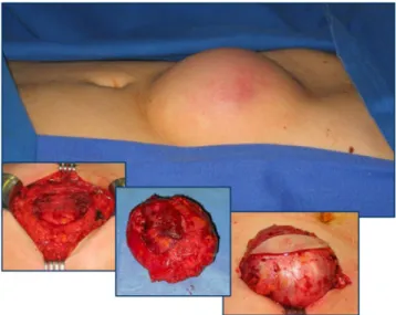
Fig. 2 Surgical treatment of desmoid tumors. En bloc resection of skin, subcutaneous fat, desmoid tumor, and adjacent abdominal wall musculature and fascia due to locally invasive growth and frequent recurrence
Fig. 3 Brachytherapy of desmoid tumors before (left) and 2 years after brachytherapy (right). Good remission with only residual thickening of rectus sheath after brachytherapy
Fig. 4 HDR brachytherapy applicators. Seven applicators in situ dur-ing treatment of a desmoid of the lower abdominal wall considered unsuitable for resection and direct closure
We found that desmoids develop in a significant percent-age of patients. The greatest challenge posed by these tumors is their locally invasive growth in sites precluding radical resection. Several potential risk factors for the de-velopment of desmoids are being discussed in the literature, and previous surgery is believed to promote growth of
desmoids [22]. Two thirds of the desmoids in our patients
occurred after a mean interval of 34 months after colectomy. Partly, these results may be influenced by the fact that the mean age at diagnosis of desmoid tumors is 48 years (range 31–73 years), whereas colectomy is usually performed at a younger age of 44 years (range 30–61 years), thereby maybe implicating a false causative relationship. In addition, no desmoids at all were detected in seven colectomy patients during a mean follow-up of 104 months.
The location of the genetic mutation also seems to influ-ence the development of desmoids (higher risk in mutations beyond codon 1444, or in mutations at the 3′-end of the
codon) [23]. Our results, however, show pronounced
intra-familial variations of the phenotype with a seemingly un-predictable clinical course, indicating the influence of
genetic factors other than the APC mutation itself [11].
Best results were obtained in patients with abdominal wall desmoids at an early stage appropriate for radical resection and direct closure. Resection of desmoids was generally rec-ommended in symptomatic patients and those exhibiting rapid growth, whereas observation with frequent follow-up was done in patients with small, asymptomatic desmoids and those unwilling to undergo resection. Alternatively, we used HDR brachytherapy with good success in four patients with large recurrent tumors unfit for resection. No recurrence was ob-served throughout their follow-up. Despite our limited expe-rience, we believe that HDR brachytherapy may provide an alternative to resection in patients that are unfit for surgery or in whom the size or location of the desmoids precludes radical resection, since HDR brachytherapy is known to improve
local control of desmoid tumors [24].
The greatest therapeutic challenge was desmoids arising within the intestinal mesentery. In these patients, operative treatment is associated with serious morbidity and mortality arising from intestinal ischemia following radical resection, anastomotic failure, or late small bowel obstruction. Fur-thermore, evidence exists that resection of mesenteric des-moids may in fact stimulate tumor growth, which is why treatment is often directed only at relieving specific local consequences such as small bowel obstruction or ischemia
[25, 26]. Chemotherapy has so far not been part of our
treatment rationale in desmoid patients due to its controver-sial benefits; however, two recent trials have shown im-proved progression-free survival rates in patients with nonresectable desmoids treated with doxorubicin or imati-nib, thereby potentially offering a true benefit in this
diffi-cult to treat subgroup of patients [27,28].
A point of interest is the phenotype–genotype correlation in GS: Since 1992 several attempts have been made to correlate specific APC mutations with clinical phenotypes and explain clinical variability on the basis of different mutations. However, there is considerable variability in the expression of specific phenotypes within families and even
among individuals with identical mutations [29,30]. In our
family, the mutation is located on exon 15, codon 1981 which causes an attenuated form of polyposis with fewer adenomas (100 or fewer).
Finally, although sophisticated screening and surveil-lance programs are available, a considerable number of individuals refuse participation or are reluctant to act once their disease has been diagnosed. We have encountered this denial frequently and have sadly had to observe progression into fatal cancer in three patients. While the problem of noncompliance pertains to many problems in medicine, the high percentage of individuals that remain reluctant to un-dergo preventive testing remains of great concern. In our opinion, this phenomenon emphasizes the need for a con-tinuous follow-up of all individuals at risk through the same credible group of physicians and emphasizes the need for a sustainable physician–patient relationship throughout the life of any Gardner patient.
Acknowledgments The authors wish to thank Dr. Werner Wüst (Director Emeritus of the Institute of Pathology at the Kantonsspital Graubünden, Switzerland) and his successors Prof. Thomas Stallmach and Dr. Harald Frick for their valuable support in both the diagnostic work-up and in obtaining macro- and microscopic picture analyses of our Gardner patients.
References
1. Gardner EJ, Stephens FE (1950) Cancer of the lower digestive tract in one family group. Am J Hum Genet 2:41–48
2. Bodmer WF, Bailey CJ, Bodmer J, Bussey HJ, Ellis A, Gorman P, Lucibello FC, Murday VA, Rider SH, Scambler P (1987) Localization of the gene for familial adenomatous polyposis on chromosome 5. Nature 328:614–616
3. Groden J, Thliveris A, Samowitz W, Carlson M, Gelbert L, Albertsen H, Joslyn G, Stevens J, Spirio L, Robertson M (1991) Identification and characterization of the familial adenomatous polyposis coli gene. Cell 66:589–600
4. Kinzler KW, Nilbert MC, Su LK, Vogelstein B, Bryan TM, Levy DB, Smith KJ, Preisinger AC, Hedge P, McKechnie D (1991) Identification of FAP locus genes from chromosome 5q21. Science 253:661–665
5. Galiatsatos P, Foulkes WD (2006) Familial adenomatous polypo-sis. Am J Gastroenterol 101:385–398
6. Gardner EJ, Richards RC (1953) Multiple cutaneous and subcuta-neous lesions occurring simultasubcuta-neously with hereditary polyposis and osteomatosis. Am J Hum Genet 5:139–147
7. Gardner EJ, Plenk HP (1952) Hereditary pattern for multiple osteomas in a family group. Am J Hum Genet 4:31–36
8. Dobbie Z, Spycher M, Mary JL, Haner M, Guldenschuh I, Hurliman R, Amman R, Roth J, Muller H, Scott RJ (1996) Correlation between the development of extracolonic manifesta-tions in FAP patients and mutamanifesta-tions beyond codon 1403 in the APC gene. J Med Genet 33:274–280
9. Vasen HF, Moslein G, Alonso A, Aretz S, Bernstein I, Bertario L, Blanco I, Bulow S, Burn J, Capella G, Colas C, Engel C, Frayling I, Friedl W, Hes FJ, Hodgson S, Jarvinen H, Mecklin JP, Moller P, Myrhoi T, Nagengast FM, Parc Y, Phillips R, Clark SK, de Leon MP, Renkonen-Sinisalo L, Sampson JR, Stormorken A, Tejpar S, Thomas HJ, Wijnen J (2008) Guidelines for the clinical manage-ment of familial adenomatous polyposis (FAP). Gut 57:704–713 10. Simmen HP, Müller G, Roth A, Hasler T (1979) Das
Gardner-Syndrom: Definition, Ausdrucksformen, Frühdiagnostik und Therapie. Dtsch med Wochenschr 104:799–803
11. Plasilova M, Russell AM, Wanner A, Wolf A, Dobbie Z, Muller HJ, Heinimann K (2004) Exclusion of an extracolonic disease modifier locus on chromosome 1p33-36 in a large Swiss familial adenomatous polyposis kindred. Eur J Hum Genet 12:365–371 12. Spigelman AD, Williams CB, Talbot IC, Domizio P, Phillips RK
(1989) Upper gastrointestinal cancer in patients with familial ade-nomatous polyposis. Lancet 2:783–785
13. Bulow C, Vasen H, Jarvinen H, Bjork J, Bisgaard ML, Bulow S (2000) Ileorectal anastomosis is appropriate for a subset of patients with familial adenomatous polyposis. Gastroenterology 119:1454–1460 14. Heiskanen I, Luostarinen T, Jarvinen HJ (2000) Impact of
screen-ing examinations on survival in familial adenomatous polyposis. Scand J Gastroenterol 35:1284–1287
15. Winawer S, Fletcher R, Rex D, Bond J, Burt R, Ferrucci J, Ganiats T, Levin T, Woolf S, Johnson D, Kirk L, Litin S, Simmang C (2003) Colorectal cancer screening and surveillance: clinical guidelines and rationale—update based on new evidence. Gastroenterology 124:544– 560
16. Belchetz LA, Berk T, Bapat BV, Cohen Z, Gallinger S (1996) Changing causes of mortality in patients with familial adenoma-tous polyposis. Dis Colon Rectum 39:384–387
17. da Luz MA, Church JM, Burke CA (2009) The evolution of prophylactic colorectal surgery for familial adenomatous polypo-sis. Dis Colon Rectum 52:1481–1486
18. Parc YR, Olschwang S, Desaint B, Schmitt G, Parc RG, Tiret E (2001) Familial adenomatous polyposis: prevalence of adenomas in the ileal pouch after restorative proctocolectomy. Ann Surg 233:360–364
19. Vrouenraets BC, Van Duijvendijk P, Bemelman WA, Offerhaus GJ, Slors JF (2004) Adenocarcinoma in the anal canal after ileal
pouch-anal anastomosis for familial adenomatous polyposis using a double-stapled technique: report of two cases. Dis Colon Rectum 47:530–534
20. Arvanitis ML, Jagelman DG, Fazio VW, Lavery IC, McGannon E (1990) Mortality in patients with familial adenomatous polyposis. Dis Colon Rectum 33:639–642
21. Nugent KP, Spigelman AD, Phillips RK (1993) Life expectancy after colectomy and ileorectal anastomosis for familial adenoma-tous polyposis. Dis Colon Rectum 36:1059–1062
22. Gurbuz AK, Giardiello FM, Petersen GM, Krush AJ, Offerhaus GJ, Booker SV, Kerr MC, Hamilton SR (1994) Desmoid tumours in familial adenomatous polyposis. Gut 35:377–381
23. Eccles DM, van der LR, Breukel C, Bullman H, Bunyan D, Fisher A, Barber J, du BC, Primrose J, Burn J, Fodde R (1996) Hereditary desmoid disease due to a frameshift mutation at codon 1924 of the APC gene. Am J Hum Genet 59:1193–1201
24. Assad WA, Nori D, Hilaris BS, Shiu MH, Hajdu SI (1986) Role of brachytherapy in the management of desmoid tumors. Int J Radiat Oncol Biol Phys 12:901–906
25. Jones IT, Jagelman DG, Fazio VW, Lavery IC, Weakley FL, McGannon E (1986) Desmoid tumors in familial polyposis coli. Ann Surg 204:94–97
26. Lotfi AM, Dozois RR, Gordon H, Hruska LS, Weiland LH, Carryer PW, Hurt RD (1989) Mesenteric fibromatosis complicat-ing familial adenomatous polyposis: predisposcomplicat-ing factors and results of treatment. Int J Color Dis 4:30–36
27. Chugh R, Wathen JK, Patel SR, Maki RG, Meyers PA, Schuetze SM, Priebat DA, Thomas DG, Jacobson JA, Samuels BL, Benjamin RS, Baker LH (2010) Efficacy of imatinib in aggressive fibromatosis: results of a phase II multicenter Sarcoma Alliance for Research through Collaboration (SARC) trial. Clin Cancer Res 16:4884–4891 28. Nieuwenhuis MH, Mathus-Vliegen EM, Baeten CG, Nagengast FM, van der BJ, van Dalsen AD, Kleibeuker JH, Dekker E, Langers AM, Vecht J, Peters FT, van Dam R, van Gemert WG, Stuifbergen WN, Schouten WR, Gelderblom H, Vasen HF (2011) Evaluation of man-agement of desmoid tumours associated with familial adenomatous polyposis in Dutch patients. Br J Cancer 104:37–42
29. Giardiello FM, Krush AJ, Petersen GM, Booker SV, Kerr M, Tong LL, Hamilton SR (1994) Phenotypic variability of familial adeno-matous polyposis in 11 unrelated families with identical APC gene mutation. Gastroenterology 106:1542–1547
30. Leppert M, Burt R, Hughes JP, Samowitz W, Nakamura Y, Woodward S, Gardner E, Lalouel JM, White R (1990) Genetic analysis of an inherited predisposition to colon cancer in a family with a variable number of adenomatous polyps. N Engl J Med 322:904–908