SCIENTIFIC ARTICLE
Imaging findings of avalanche victims
Alexandra B. Grosse&Claudia A. Grosse&
Lynne S. Steinbach&Heinz Zimmermann&
Suzanne Anderson
Received: 21 May 2006 / Revised: 4 November 2006 / Accepted: 15 January 2007 / Published online: 5 April 2007 # ISS 2007
Abstract
Objective Skiing and hiking outside the boundaries remains an attractive wilderness activity despite the danger of avalanches. Avalanches occur on a relatively frequent basis and may be devastating. Musculoskeletal radiologists should be acquainted with these injuries.
Design and patients Fourteen avalanche victims (11 men and 3 women; age range 17–59 years, mean age 37.4 years) were air transported to a high-grade trauma centre over a period of 2 years.
Results Radiographs, CT and MR images were prospective-ly evaluated by two observers in consensus. Musculoskeletal findings (61%) were more frequent than extraskeletal findings (39%). Fractures were most commonly seen (36.6%), involving the spine (14.6%) more frequently than the extremities (9.8%). Blunt abdominal and thoracic trauma were the most frequent extraskeletal findings.
Conclusion A wide spectrum of injuries can be found in avalanche victims, ranging from extremity fractures to massive polytrauma. Asphyxia remains the main cause of death along with hypoxic brain injury and hypothermia.
Keywords Avalanche victims . Injury patterns . Asphyxia . Hypothermia
Introduction
Sparse literature and imaging concerning the types of injuries of avalanche victims have been published [1, 2]. Rapid and careful radiological work-up is vital for adequate prompt patient management.
Back country skiing and snowboarding are particularly dangerous. The overall mortality rate of avalanche victims has been reported to be as high as 23% in a Swiss study [3,
4]. The mortality rate of partly buried (the head and upper part of the body are not buried) or non-buried victims is approximately 4.0%, the mortality rate of completely buried victims is significantly higher at 52.4%. Avalanche victims buried by snow masses on roads or in buildings have a higher survival probability than those buried in open areas, due to the unevenly packed snow masses with air pocket formation delaying the onset of asphyxia.
The aim of the present study was to evaluate the types and frequency of injuries in avalanche victims over a period of 2 years with a focus on musculoskeletal findings.
Materials and methods
This study was prospective in design. Over a 2-year period 14 avalanche victims (11 men and 3 women; age range 17– 59 years, mean age 37.4 years) underwent tertiary referral to the emergency unit of a major level I referral trauma centre for severely injured avalanche victims in the Swiss Alps. We defined avalanche victims as people caught by avalanches and carried some distance or partially or completely buried
A. B. Grosse (*)
:
C. A. Grosse:
S. AndersonDepartment of Diagnostic, Pediatric and Interventional Radiology, University Hospital of Berne, Inselspital,
Freiburg Strasse, 3010 Berne, Switzerland e-mail: alexandragrosse@gmx.at
L. S. Steinbach
Department of Radiology, University of California San Francisco, San Francisco, CA 94143-0628, USA
H. Zimmermann
Department of Trauma and Emergency Medicine, University Hospital of Berne, Inselspital, Berne, Switzerland
by snow slides. Partial burial included no burial of the head and upper body. Twelve victims had been completely buried (n=12) and 2 (n=2) had been partially buried by the snow. Seven victims had been engaged in skiing or snowboarding (n=7), 2 had been engaged in mountain climbing (n=2) and 1 in snowshoeing (n=1). The type of sports activity of 4 victims (n=4) remained unknown.
Twelve (n=12) victims survived, and 2 (n=2) were declared dead after hospitalisation. Autopsy was denied in 1 of the latter and no injury-related data could be obtained. In both fatal cases asphyxiation and hypothermia were listed as the main causes of death. None of the survivors had any late complications, except for 1 young woman who was subjected to a fore-foot amputation due to severe
frostbite and vascular injury. Burial time varied. The 2 patients later pronounced dead had been buried for 40 min and 60–90 min, respectively, both without an air pocket around mouth and nose, and at a depth of approximately 1.5 m. Both had been reanimated and intubated, 1 recovered heart circulation, but was later pronounced brain dead on angiography, and the other victim did not recover. All survivors had been buried for half an hour or less, except for 1 young man who had been buried for almost an hour with a large air pocket around his head.
Three victims (n=3) had been able to rescue themselves (2 partially buried, 1 completely buried with a shovel). The majority arrived at the hospital directly from the rescue site (n=10). Four patients (n=4) had been initially briefly evaluated at smaller hospitals before they were referred to the level I trauma centre. The patients had been transported by helicopter (Air Zermatt, Rega), using trained transport teams. They had been provided with emergency care during the transport time. Four victims (n=4) had a GCS (Glasgow Coma Scale) score of less than 8 on admission, indicating severe injury, 1 victim had a GSC score of 11 (n=1),
Table 1 Frequency of injuries in avalanche victims
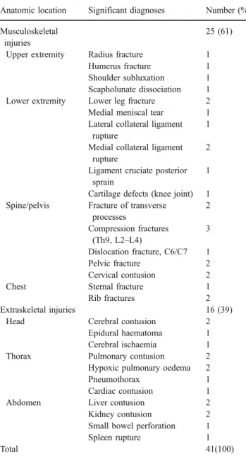
Anatomic location Significant diagnoses Number (%)
Musculoskeletal injuries
25 (61)
Upper extremity Radius fracture 1
Humerus fracture 1
Shoulder subluxation 1
Scapholunate dissociation 1
Lower extremity Lower leg fracture 2
Medial meniscal tear 1
Lateral collateral ligament rupture
1 Medial collateral ligament
rupture
2 Ligament cruciate posterior
sprain
1
Cartilage defects (knee joint) 1
Spine/pelvis Fracture of transverse
processes 2 Compression fractures (Th9, L2–L4) 3 Dislocation fracture, C6/C7 1 Pelvic fracture 2 Cervical contusion 2
Chest Sternal fracture 1
Rib fractures 2
Extraskeletal injuries 16 (39)
Head Cerebral contusion 2
Epidural haematoma 1
Cerebral ischaemia 1
Thorax Pulmonary contusion 2
Hypoxic pulmonary oedema 2
Pneumothorax 1
Cardiac contusion 1
Abdomen Liver contusion 2
Kidney contusion 2
Small bowel perforation 1
Spleen rupture 1
Total 41(100)
Fig. 1 A 53-year-old man thrown into a rocky area by an avalanche. He suffered multiple fractures, a deep skull wound and a large flank haematoma. a CT of the lumbar spine demonstrates an unstable fracture of the L3 vertebral body (arrow), b stabilised with vertebroplasty
indicating moderate injury, and 9 victims (n=9) had a GCS score of 13–15, suggesting minor injury [5].
The patients were prospectively recruited from the emergency database, and the radiological data were obtained from the radiology record system and analysed by two radiologists. The site and type of injuries were recorded. All structures were reviewed with emphasis on the musculoskeletal system.
Results
A total of 133 imaging studies were evaluated, including 68 radiographs (16 chest, 52 peripheral and axial skeleton), 50 computerised tomography scans (110 head, 10 chest, 6 abdomen, 7 cervical spine, 8 thoracic spine, 7 lumbar spine, 1 neck), 3 peripheral MR images (2 knee [1 bilateral, 1 unilateral], 1 hand) and 9 sonographic studies (abdomen and pelvis). Three digital peripheral subtraction angio-graphic (DSA) examinations were performed.
The overall number of diagnoses attributed to seven areas of the body (upper and lower extremities, spine/pelvis, chest, head, thorax, abdomen) is presented in Table1. Musculo-skeletal findings (61%) were more frequent than extra-skeletal findings (39%). Fractures were most commonly seen (36.6%). Fifteen fractures (n=15) were found in 8 patients. Fractures of the lumbar spine included compres-sion fractures of the L3, (Fig. 1), and L2–L3 vertebral
bodies and fractures of the transverse processes of L1–L2 (Fig. 2). Fractures of the thoracic spine included a compression fracture of the Th9 vertebral body and fractures of the transverse processes of Th1–Th4 on the left and Th1– Th3 on the right. One patient had a sternal fracture associated
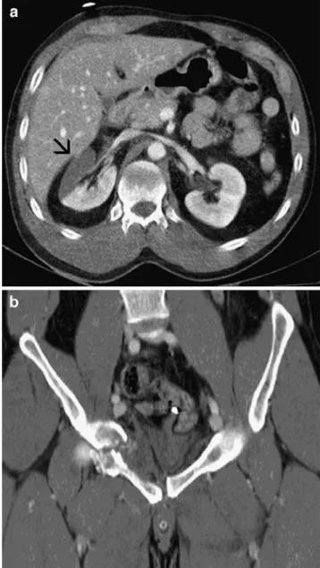
with cardiac contusion and mediastinal hematoma. Pelvic fractures were evident in 2 patients (Fig.3b).
Fractures of the axial skeleton (26.8%) were more frequent than extremity fractures (9.8%). The lower and upper extremities were equally affected (4.9% each). One patient with a dislocated lower leg fracture and soft tissue contusion with frostbite required a lower extremity DSA (Fig. 4a–f). In this patient reconstruction was no
longer possible and amputation of the foot had to be performed.
Severe knee disruption evident in 2 patients was bilateral in 1 and unilateral in the second patient. The first patient had suffered a complete medial meniscal tear, strain grade
Fig. 3 A 46-year-old man with a kidney contusion of the right kidney (arrow) and b pelvic fracture, reflecting the forceful body compression due to the snow mass
Fig. 2 A 17-year-old mountain climber dragged down a 600-m snow field. He had been buried under 1.5-m-thick snow for almost an hour with a large air pocket around his head. Axial CT scan of the abdomen reveals a fracture of the transverse process of the L2 vertebral body (arrow); multiple contusions and skin abrasions were evident, but there was no life-threatening injury (GSC initially 11, then 15)
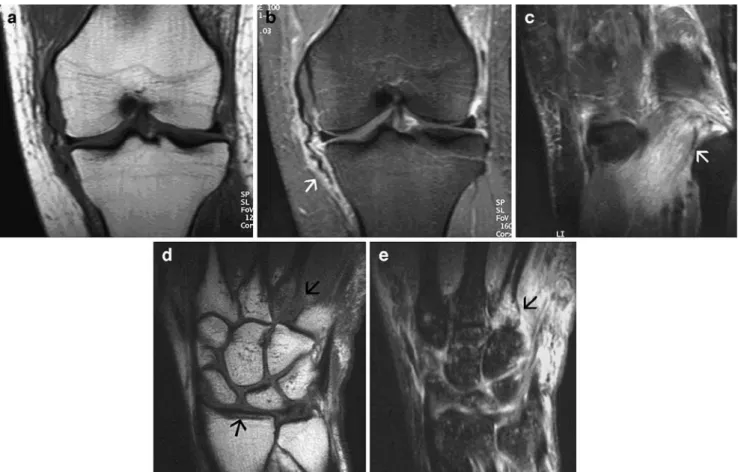
II of the lateral collateral ligament and rupture of the medial collateral ligament of the left knee (Fig.5), and a discrete medial meniscal tear of the right knee with patellofemoral cartilage defects. The second patient had suffered a rupture of the medial collateral ligament, partial sprain of the posterior cruciate ligament and a discrete medial meniscal tear of the left knee (Fig.6a–c). The same patient had also
suffered a distortion of the left ankle joint, however, without evidence of fracture or ligament tears.
Subluxation of the right shoulder with hypoesthesia in the second and third finger of the right hand and diverse contusions of the upper extremities were found in 1 patient. Rupture of the scapholunar ligament of the left hand was evident in 1 patient with scapholunate dissociation, bone contusion in the base of the fourth metacarpal (Fig. 6d,e) and rupture of the flexor tendon of the third finger with soft tissue laceration. Prominent soft tissue swelling of the thenar musculature in 1 patient was considered due to haematoma as there was no evidence of fracture.
Extraskeletal findings (39%) included intrathoracic (14.6%), intra-abdominal (14.6%) and cerebral findings (9.8%). Cerebral contusion was diagnosed on CT in 2 patients and brain death was diagnosed on angiography in 1 patient with massive hypoxic cerebral oedema evident on CT (Fig.7a). Superficial skin lacerations and abrasions to the face and nose and extensive, deep skin wounds were common in the scalp and facial region including one deep scalp wound, 30 cm long and transversed by a second laceration, 10 cm long.
Intrathoracic pulmonary findings included pulmonary contusions in 2 patients (Fig. 7b), 1 of them requiring mechanical ventilation, and hypoxic pulmonary oedema in 2 patients (Fig. 8). Abdominal injuries included small intestine perforation requiring partial bowel resection in 1 patient, focal kidney contusion in 2 patients (Fig. 3a),
spleen rupture in 1 patient and liver contusions in 2 patients, with free intra-abdominal fluid in all 6 patients.
Discussion
Most accidents occur in high altitude back country skiers and snowboarders. The main cause of death is asphyxia, and in only one-third is blunt trauma the primary or contributory cause of death [6,7]. Other data from autopsy reports of avalanche victims state that mechanical trauma is the cause of death in only 13% [1]. According to different data, mechanical injuries are the second most frequent cause of death (43%) after asphyxia (46%) [3].
Asphyxiation occurs in trapped victims without an air pocket usually within the first 15 min of burial. Humidified
Fig. 5 A 59-year-old avalanche victim with intestine perforation and knee distortion. a Coronal T1-weighted (TR/TE, 472/14) and b coronal T2-weighted fat-saturated (TR/TE, 4,000/30) MR images demonstrate a rupture of the medial collateral ligament (arrow) and medial meniscal tear
Fig. 4 A 38-year-old avalanche victim, carried down a 400-m snow field. She had been completely buried under snow, but managed to dig herself free with her own shovel. She had lost both her boots, suffering frostbite on her feet. Anteroposterior and lateral radiographs demon-strate a, b a dislocated fracture of the lower leg, c stabilised with
osteotomy. d On digital subtraction angiography (DSA), abrupt termination of the contrast media is seen in the distal arteries above
the ankle joint, due to intimal lesions, spasm or thromboses—
air condenses around the nose and mouth of the victim forming an ice mask that further prevents gas exchange. Avalanche victims with air pockets survive longer (the longest reported survival time for a completely buried person in an open area was 44 h [8]); however, they may die of late asphyxiation. One victim in our series with the longest burial time of approximately 1 h did have a large air
pocket around his head, and the two victims later pronounced dead had no air pockets.
Mechanical injuries are common in victims who are dragged across forested or rocky slopes or thrown against ice blocks or blocks of dense snow. The risk of mechanical injury depends on the type of snow and increases with the snow density. Wet and dense snow tends to form big rolling
Fig. 6 A 28-year-old female avalanche victim with knee and hand injury. a Coronal T1-weighted (TR/TE, 600/12) and b coronal T2-weighted fat-saturated (TR/TE, 5,100/30) MR images of the left knee demonstrate a rupture of the medial collateral ligament (arrow) with c soft tissue oedema in the popliteal muscle (arrow). d Coronal T1-weighted (TR/TE, 600/17) and e post-contrast coronal
fat-saturated T1-weighted (TR/TE, 600/17) MR images of the left hand demonstrate bone marrow signal alterations with enhancement in the base of the fourth metacarpal (upper arrow) consistent with bone contusion and a scapholunate dissociation (lower arrow) corresponding to the force of energy the victim had been exposed to
Fig. 7 A 46-year-old man, caught by an avalanche on a mountain tour with his son. He had been completely buried without an air pocket for 40 min. Clinically, he had wide fixed pupils and his core tem-perature was initially 27°C. a Findings on CT of the brain performed as part of the formal trauma evaluation are consistent with cerebral hypoxia and oe-dema. He was later pronounced dead on angiography. b CT of the chest reveals pulmonary contusions
snow balls causing stronger mechanical forces, which carry a risk of blunt injury, even with burial at only some feet below the surface. Oxygen transfer in the densely packed snow masses is also reduced and limits the chances of survival.
Upper extremity injuries are common in trapped victims, making swimming movements or putting their hands up in an attempt to free themselves very difficult, and lower extremity injuries are frequently found in victims getting entangled with their skis. A study by Brugger et al. [9] demonstrated that only 18% of skiers are able to free themselves from their skis and 8% from their skis and poles during the descent of the avalanche, which increases the probability of fractures and knee injuries. Failure to release the bindings of skis and snowboard, further traps the victim at the bottom of the debris, the so-called anchor effect. Thus severe open extremity fractures are a common finding and complex knee injuries reflecting the strong forces of distortion of the knee joint. In patients with fractures and knee luxation it is important to check the foot pulses after hospitalisation in order to recognise intimal arterial lesions of the lower leg, which subsequently may occur.
Closed head injuries (CHI) are often severe, although none of the victims in our series had a skull fracture or evidence of intracranial bleeding. Johnson et al. [10] reviewed the records of 28 killed avalanche victims and found that up to 61% had some degree of CHI and 21% had severe head injuries, noted as either the primary or contributory cause of death. On viewing fatal cases, it appears that the incidence of minor CHI is rather high, although CHI less commonly causes death itself. In our series only 4 patients had abnormal findings on brain CT, but the majority had minor or multiple large lacerations and abrasions in the scalp and facial region. Even minor degree
CHI may impair the victim’s consciousness, rendering rescue techniques useless (e.g. swimming to the surface, putting the hands up to create an air pocket, releasing the bindings of skis and snowboard).
Burial at great depth increases the risk of blunt trauma. It may also lead to immediate death through general body compression with acute respiratory and circulatory failure. The deeper the burial depth, the greater the body compres-sion and the more the chances of survival are reduced. Also, extrication time is usually longer, and there is a direct correlation between burial time and burial depth [11].
Lung contusions are depicted more accurately and at an earlier stage with CT than with radiographs. If about one-third of the pulmonary air space is involved, the patient usually requires mechanical ventilation [12], which was the case in 1 patient in our series. Pleural effusions in avalanche victims may be particularly large, although this was not the case in any of the patients in our series. Stalsberg et al. [1] found a haematothorax of 500 ml at autopsy in an avalanche victim with a costal fracture and superficial tear of the lung. He related this finding to the centralisation of blood circulation, which occurs with extreme body compression and sympa-thetic stimulation. Hypoxic pulmonary oedema was recog-nised in 2 victims in our series. Schmid [2] described this phenomenon and stated that circulatory disturbances and respiratory mechanisms with hypoxic constriction of the pulmonary arterioles and pulmonary hypertension are re-sponsible for its pathogenesis.
In avalanche victims mechanical injuries and blunt trauma are frequently seen. Our study provides an overview of the spectrum of injuries. Despite modern warning systems, avalanches happen on a relatively frequent basis. The mortality rate is high and survivor collectives are relatively rare. On clinical assessment, avalanche victims should be evaluated for polytrauma with CT being the method of choice for diagnostic speed. Radiographs provide answers to most questions relating to fractures. MRI performed for joint and soft tissue injuries, particular-ly of the knee, ankle and hand region, is useful. Angiography may be required for complex fractures or in a setting of extensive frostbite.
References
1. Stalsberg H, Albretsen C, Gilbert M, et al. Mechanism of death in avalanche victims. Virchows Archiv A Pathol Anat 1989;414:
415–422.
2. Schmid F. Zur Pathogenese des Lungenödems nach
Lawinenver-schüttung. Schweiz Med Wochenschr 1981;111: 1441–1445.
3. Strohm PC, Köstler W, Hammer T, Südkamp NP. Lawinennotfall
und akzidentelle Hypothermie. Unfallchirurg 2003;106: 343–347.
4. Eidgenössisches Institut für Schnee- und Lawinenforschung.
Winterberichte. Davos (Switzerland): 1981–1998;46–62.
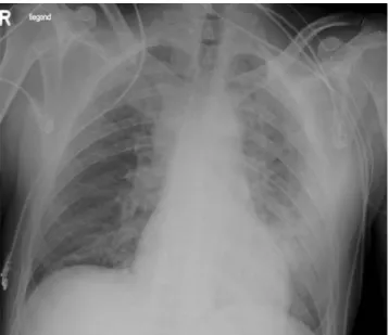
Fig. 8 Chest radiography of a 51-year-old man with hypoxic pulmonary oedema
5. Teasdale G, Jennett B. Assessment of coma and impaired
consciousness: a practical scale. Lancet 1974;2: 81–84.
6. Rostrup M, Gilbert M. Avalanche accidents. Tidsskr Nor
Laegeforen 1993;113: 1100–1102.
7. Williams K, Armstrong BR, Armstrong RL. Avalanches. In: Auerbach PS, ed. Wilderness medicine: management of wilder-ness and environmental emergencies, 3rd edn. St. Louis: Mosby;
1995: 617–643.
8. Eidgenössisches Institut für Schnee- und Lawinenforschung.
Winterberichte. Davos (Switzerland): 1971–1972;36: 129–131.
9. Brugger H, Flora G, Falk M. Möglichkeiten der Selbstrettung und posttraumatische Belastungsstörungen beim Lawinenunfall.
Notarzt 2002;18: 1–4.
10. Johnson SM, Johnson AC, Barton RG. Avalanche trauma and closed head injury: adding insult to injury. Wilderness Environ
Med 2001;12: 244–247.
11. Burtscher M. Avalanche survival chances. Nature 1994;371: 482. 12. Lomoschitz FM, Eisenhuber E, Linnau KF, Peloschek P, Schoder M, Bankier A. Imaging of chest trauma: radiological patterns of injury and diagnostic algorithms. Eur Radiol 2003;48: 61–70.