Results from percutaneous drainage of Hinchey stage II
diverticulitis guided by computed tomography scan
Y. Durmishi,1P. Gervaz,1D. Brandt,1P. Bucher,1A. Platon,2P. Morel,1P. A. Poletti2
1Department of Surgery, University Hospital Geneva, Rue Micheli-du-Crest 24, 1211, Gene`ve, Switzerland 2
Department of Radiology, University Hospital Geneva, Rue Micheli-du-Crest 24, 1211, Gene`ve, Switzerland Received: 19 August 2005/Accepted: 15 February 2006/Online publication: 3 June 2006
Abstract
Background: Percutaneous abscess drainage guided by computed tomography scan is considered the initial step in the management of patients presenting with Hinchey II diverticulitis. The rationale behind this approach is to manage the septic complication conservatively and to follow this later using elective sigmoidectomy with pri-mary anastomosis.
Methods: The clinical outcomes for Hinchey II patients who underwent percutaneous abscess drainage in our institution were reviewed. Drainage was considered a failure when signs of continuing sepsis developed, ab-scess or fistula recurred within 4weeks of drainage, and emergency surgical resection with or without a colos-tomy had to be performed.
Results: A total of 34patients (17 men and 17 women; median age, 71 years; range, 34–90 years) were consid-ered for analysis. The median abscess size was 6 cm (range, 3–18 cm), and the median duration of drainage was 8 days (range, 1–18 days). Drainage was considered successful for 23 patients (67%). The causes of failure for the remaining 11 patients included continuing sepsis (n = 5), abscess recurrence (n = 5), and fistula forma-tion (n = 1). Ten patients who failed percutaneous ab-scess drainage underwent an emergency Hartmann procedure, with a median delay of 14days (range, 1–65 days) between drainage and surgery. Three patients in this group (33%) died in the immediate postoperative period. Among the 23 patients successfully drained, 12 underwent elective sigmoid resection with a primary anastomosis. The median delay between drainage and surgery was 101 days (range, 40–420 days). In this group, there were no anastomotic leaks and no mortality. Conclusion: Drainage of Hinchey II diverticulitis guided by computed scan was successful in two-thirds of the cases, and 35% of the patients eventually underwent a safe elective sigmoid resection with primary anastomosis. By contrast, failure of percutaneous abscess drainage to
control sepsis is associated with a high mortality rate when an emergency resection is performed. The current results demonstrate that percutaneous abscess drainage is an effective initial therapeutic approach for patients with Hinchey II diverticulitis, and that emergency surgery should be avoided whenever possible.
Key words: Abscess — CT scan — Diverticulitis — Drainage — Outcome
Sigmoid diverticulitis complicated with a pelvic or abdominal (extramesocolic) abscess (Hinchey stage II) [1] is currently considered by most review committees of surgical or gastroenterologic societies as an indication for percutaneous abscess drainage (PAD) associated with intravenous antibiotics [2–4]. The rationale behind this strategy is twofold: (1) initial conservative treatment of sepsis, and (2) subsequent elective sigmoidectomy with primary colorectal anastomosis [5]. This widely adopted approach has benefited from the rapid devel-opment of radiologic interventional procedures during the past two decades [6], as well as by recognition of the role that computed tomography (CT) scan has as the initial imaging method for suspected diverticulitis [7, 8]. Some authors, however, have questioned this atti-tude, observing that PAD for complicated diverticulitis is a typical example of how a new therapeutic approach enters surgical practice without much scientific evidence [9]. There are indeed several limitations to percutaneous drainage of diverticular abscesses. First, accessibility is not always possible because of small bowel loops in contiguity with the fluid collection [10]. Second, drain-age, when feasible, is not always successful. Reported failure rates for diverticular abscesses range from 15% to 30% [11–13], and when PAD fails, the mortality rate may be as high as 75% [14]. Third, when drainage is feasible and initially successful, abscess recurrence or fistula may occur, compromising the performance of an elective surgical resection [15].
Correspondence to:P. Gervaz DOI: 10.1007/s00464-005-0574-y
Finally, and most importantly, the debate whether to perform a Hartmann procedure systematically for a Hinchey II diverticulitis remains unresolved. However, some surgeons have reported good results for selected series of patients who underwent semi-urgent sigmoi-dectomy with a colorectal anastomosis [16–18]. Thus, the question is to determine whether PAD for Hinchey stage II diverticulitis actually is delaying definitive treatment rather than downstaging the initial disease.
The response to this question is likely to require a prospective multicentric trial. In the meantime, it is important to assess the results of PAD for Hinchey stage II diverticulitis critically. To our knowledge, these data are lacking for several reasons. Most series report mixed cases of abdominal fluid collections (i.e., postoperative collections, appendiceal abscesses, and the like) [19–21]. Furthermore, the final diagnosis of diverticulitis has not been confirmed by subsequent colonoscopy or at the time of surgery [22, 23]. Finally, end points considered for evaluation of the technique have been inappropriate [24]. This study aimed to assess the results of PAD for Hinchey stage II diverticulitis critically in terms of initial abscess resolution, complication of drainage, abscess or fistula recurrence, and eventual performance of elective sigmoid resection with a primary anastomosis.
Methods Charts review
All patients who underwent percutaneous drainage of diverticular abscesses guided by CT scan at University Hospital Geneva from January 1, 1991, to December 31, 2004were identified by a search of the interventional radiology database. The study protocol was sub-mitted to the ethical committee at our institution, and permission was obtained from the patients for our review of their medical records.
Each patientÕs medical record was reviewed separately by a sur-geon (Y.D., D.B., P.G.) to determine the demographics, the size and numbers of abscesses, the date and duration of drainage, and the outcome of PAD. Drainage was considered a failure when the general condition of the patient did not improve and signs of worsening sepsis developed, abscess or fistula recurred within 4weeks of drainage, and emergency surgical resection with or without a colostomy had to be performed. The medical record also was reviewed to document the occurrence of an eventual surgical resection, the type of surgery (Hartmann vs sigmoidectomy with primary anastomosis), the timing of the procedure (emergent vs elective), and the outcome of surgery.
Abscess drainage
Since 1991, our institution has considered CT scan with oral, rectal, and intravenous contrast as the standard imaging technique for pa-tients with suspected diverticulitis. When the initial CT scan demon-strates complicated diverticulitis with abscess, the indication for drainage and its feasibility are discussed jointly between interventional radiologists and colorectal surgeons. All drainage procedures are performed by experienced interventional radiologists under CT scan guidance (Figs. 1 and 2). Depending on the location of the abscess, drainage of fluid collection is achieved through a transgluteal or lateral transabdominal approach. Self-retaining pigtail catheters with a distal hydrophilic tip and ranging from 8 to 14Fr in size are used in all cases. Catheters are placed using either a tandem trocar technique or a Seldinger technique.
Immediately after catheter placement, a syringe is inserted, and the fluid collection is aspirated until the flow ceases. The pus then is sent for bacteriologic analysis. After aspiration, the catheter is left in place
to allow gravity drainage, and its adequate position subsequently is verified with additional imaging. The interventional radiology team and the surgical staff jointly perform patient follow-up evaluation.
The outcome of PAD is assessed postprocedurally by CT scan imaging of the lower abdomen to determine abscess resolution or recurrence, and by observation of improvement or deterioration of patientÕs clinical condition. All patients who underwent a simple fluid evacuation without permanent catheter placement were excluded from the study.
Definition and medical management of Hinchey II diverticulitis
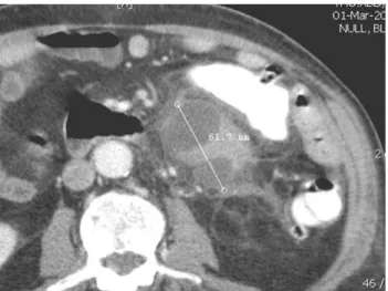
The staging of complicated diverticulitis was established according to the original Hinchey classification. In this classification, stage II is Fig. 1. Computed tomography scan of diverticulitis stage Hinchey II. A 6· 4-cm pus collection of diverticular origin is demonstrated in a 56-year-old man who presented with fever, leucocytosis, and pain in the left iliac fossa.
Fig. 2. Percutaneous drainage of diverticular abscess guided by com-puted tomography (CT) scan. A large pigtail catheter was placed under CT scan guidance inside the fluid collection, and 20 ml of pus was aspirated (below). Control 3 days later shows complete resolution of the abscess. The drain was removed the next day.
related to a large (>3–4cm) pus collection, which is at a distance (in the pelvis, abdomen, or retroperitoneum) from the sigmoid colon. Unlike stage I abscesses, Hinchey stage II abscesses are not confined within the mesocolon, and are considered amenable to percutaneous drainage.
All patients had their final diagnosis of complicated diverticulitis confirmed, either at the time of surgery or by colonoscopy. In addition to drainage, the initial medical management included broad-spectrum intravenous antibiotherapy, either with a combination of metronida-zole and ceftriaxone or with monotherapy comprising imipenem or tazobactam.
Three patients with an initial diagnosis of complicated diverticu-litis eventually underwent surgery with a final diagnosis of sigmoid adenocarcinoma, and thus were excluded from the study. Similarly, all patients who had Hinchey II diverticulitis, but who were considered for technical reasons not amenable to percutaneous drainage, were excluded from the study.
Statistical analysis
Statistical analyses were undertaken using the software package STATGRAPH 3.0 software for Windows (Statgraph Software Inc., San Diego, CA, USA). Quantitative data were expressed as mean ± standard deviation. Group comparisons were made using a two-sided FisherÕs exact test for categorical variables and a two-sided StudentÕs t-test for continuous variables. All p values of 0.05 or less were considered statistically significant.
Results
A total of 34patients (17 men and 17 women; median age, 71 years; range, 34–90 years) were considered for analysis. The median abscess size was 6 cm (range, 3–18 cm), and the median duration of drainage was 8 days (range, 1–18 days). Drainage was considered successful for 23 (67%) of the patients. The causes of failure for 11 patients included aggravating sepsis (n = 5), abscess recurrence (n = 5), and fistula formation (n = 1). Ten patients who failed PAD underwent an emergency Hartmann procedure with a median delay of 14days (range, 1–65 days) between drainage and surgery. Three patients in this group (33%) died in the immediate postoperative period. Thus, the overall mortality rate in this series was 8.8%. The mortality rate was higher for the group of patients who failed to respond to PAD (27% vs 0%; p = 0.02). The patients who failed PAD also were older than those treated successfully with this approach (76 ± 15 vs 65 ± 13 years; p = 0.03). Table 1 summarizes a comparison of clinical features between the patients who failed PAD and those who were successfully treated.
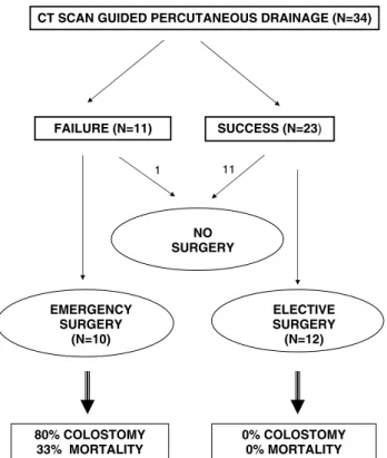
Of 23 patients successfully drained, 12 eventually underwent an elective sigmoid resection with a pri-mary anastomosis. The median delay between drain-age and surgery was 101 days (range, 40–420 days). In this group, there were no anastomotic leaks and no mortality. Among the 11 patients who for various reasons did not undergo sigmoid resection after their initial episode of complicated diverticulitis, 2 elderly women eventually presented with a perforated diver-ticulitis and underwent emergency surgery more than 7 years after the initial diagnosis. Figure 3 summarizes the therapeutic outcomes experienced by this series of patients.
Discussion
The data presented in this report indicate that for 67% of the Hinchey II patients who underwent CT scan-guided drainage, surgery was successfully delayed or avoided. There were no deaths in the group of patients who eventually underwent an elective sigmoid resection with primary anastomosis. By contrast, PAD failure and subsequent emergency surgery were associated a mor-tality rate of 30% and a colostomy rate of 80%.
During the past two decades, PAD has emerged as the standard treatment for peridiverticular abscesses larger than 3 to 4cm in diameter. Using a statewide hospital discharge database, Salem et al. [25] demon-strated that the odds of percutaneous abscess drainage for complicated diverticulitis increased 7% per year
be-Table 1. Comparison of patients who failed or who responded to percutaneous abscess drainage (PAD)
Variable Failure (n = 11) Success (n = 23) p Value Age (years): n (range) 76 (34–90) 65 (41–86) 0.03
Gender 1.00 Male 5 12 Female 6 11 Abscess size (cm): n(range) 6.8 (4–13) 6.9 (3–18) 0.99 Number of abscesses 1 9 22 0.23 2 or more 2 1 Duration of PAD (days): n (range) 9.3 (2–18) 8 (1–18) 0.40 Mortality 3/11 0/22 0.02
CT SCAN GUIDED PERCUTANEOUS DRAINAGE (N=34)
FAILURE (N=11) SUCCESS (N=23) EMERGENCY SURGERY (N=10) ELECTIVE SURGERY (N=12) NO SURGERY 1 11 80% COLOSTOMY 33% MORTALITY 0% COLOSTOMY 0% MORTALITY
Fig. 3. Outcome of percutaneous abscess drainage for Hinchey stage II diverticulitis.
tween 1987 and 2001, and that emergency colectomies concomitantly decreased 2% per year. The two series most often quoted regarding CT scan–guided drainage of diverticular abscesses report success rates of 74% 80% [26, 27]. However, in the series reported by Schechter et al. [26], only 10 of the 133 patients had a proven complicated diverticulitis. In the seminal paper by Neff et al. [27], 16 patients with diverticular abscesses were included, and it is unclear exactly how many were true Hinchey II patients. Therefore, the current standard for percutaneous drainage of Hinchey II diverticulitis has little, if any, evidence-based support, although its use is obviously increasing, in parallel with the development of interventional radiology techniques.
Our data are in accordance with series from the UK and Germany, which have demonstrated an overall mortality rate of 12% for diverticular abscess and 23% after the Hartmann procedure used for Hinchey II diverticulitis [28, 29]. Thus, the mortality rate after emergency surgery for Hinchey stage II diverticulitis remains excessive, and probably is related to associated comorbid conditions in an elderly population. There-fore, it is reassuring to consider that PAD was successful in avoiding emergency surgery for two-thirds of our patients. This success rate is somewhat inferior to that of other series that included drainage of postoperative fluid collections. Technically, PAD is more difficult (and probably less successful) in abdominal septic conditions, such as complicated diverticulitis or pancreatitis. How-ever, PAD has its limitations, and it is significant that advanced age seems to be associated with a higher risk for PAD failure in our series and others as well [17].
This study considered only Hinchey stage II patients who were good candidates for PAD, but in certain cases, abscess location or sentinel small bowel loops make this approach unfeasible. Such patients should be treated with intravenous antibiotics initially, and emergency surgery should be reserved for those who do not respond to the conservative management. It would be interesting to determine whether systemic antibiotics alone may represent an alternative to PAD for complicated diver-ticulitis [30]. Of 37 patients initially thought to have Hinchey stage II diverticulitis, three (8%) eventually received a diagnosis of sigmoid adenocarcinoma. Per-cutaneous drainage failed in all patients with perforated cancer, who subsequently underwent emergency surgery with a median delay of 28 days after drainage. There frequently is a diagnostic dilemma in this clinical pre-sentation, and ruling out a perforated cancer may prove difficult. Colonoscopy is therefore mandatory before surgery for patients with perforated diverticulitis.
In summary, Hinchey stage II diverticulitis, a severe condition often affecting elderly patients, carries a mortality rate of nearly 10%. Emergency surgery in this situation, with a mortality rate exceeding 25%, should be avoided whenever possible. The clinical outcome is dependent on the patientÕs age and response to percu-taneous drainage guided by CT scan. In up to two-thirds of patients treated in collaboration with interventional radiologists, this initial approach is successful in avoiding emergency surgery. Elective sigmoidectomy, usually performed 3 months after the initial episode of
complicated diverticulitis, is a safe procedure that carries a null mortality rate.
References
1. Hinchey EJ, Schaal PH, Richards MB (1978) Treatment of per-forated diverticular disease of the colon. Adv Surg 12: 85–109 2. The standards task force and the American Society of Colon and
Rectal Surgeons (2000) Practice parameters for the treatment of sigmoid diverticulitis. Dis Colon Rectum 43: 289–297
3. European Association for Endoscopic Surgery Consensus state-ment (1999) Diagnosis and treatstate-ment of diverticular disease. Surg Endosc 13: 430–436
4. Stollmann NH, Raskin JB (1999) Ad Hoc Practice Parameters Committee of the American College of Gastroenterology: Diag-nosis and management of diverticular disease of the colon in adults. Am J Gastroenterol 94: 3110–3121
5. Parks TG (1969) Natural history of diverticular disease of the colon: a review of 521 cases. BMJ 4: 639–642
6. Ferzoco LB, Raptopoulos V, Silen W (1998) Acute diverticulitis. New Engl J Med 338: 1521–1526
7. vanSonnenberg E, Wittich GR, Goodacre BW, Casola G, DÕAg-ostino HB (2001) Percutaneous abscess drainage: update. World J Surg 25: 362–369
8. Stinner B (2001) Invited commentary. World J Surg 25: 370–372 9. Ambrosetti P, Jenny A, Becker C, Terrier F, Morel Ph (2000)
Acute left colonic diverticulitis: compared performance of com-puted tomography and water-soluble contrast enema: prospec-tive evaluation of 420 patients. Dis Colon Rectum 43: 1363– 1367
10. Kaiser AM, Jiang JK, Lake JP, Ault G, Artinyan A, Gonzalez-Ruiz C, Essani R, Beart RW Jr (2005) The management of com-plicated diverticulitis and the role of computed tomography. Am J Gastroenterol 100: 910–917
11. Labs JD, Sarr MG, Fishman EK, Siegelman SS, Cameron JL (1988) Complications of acute diverticulitis of the colon: improved early diagnosis with computerized tomography. Am J Surg 155: 331–336
12. Aydin HN, Remzi FH (2004) Diverticulitis: when and how to operate? Dig Liver Dis 36: 435–445
13. Belmonte C, Klas JV, Perez JJ, Wong WD, Rothenberger DA, Goldberg SM, Madoff RD (1996) The Hartmann procedure: first choice or last resort in diverticular disease. Arch Surg 131: 612–617
14. Wedell J, Banzhaf G, Chaoui R, Fischer R, Reichmann J (1997) Surgical management of complicated colonic diverticulitis. Br J Surg 84: 380–383
15. Kockerling F, Schneider C, Reymond MA, Scheidbach H, Sche-uerlein H, Konradt J, et al. (1999) Laparoscopic resection of sig-moid diverticulitis: results of a multicenter study. Surg Endosc 13: 567–571
16. Meyer C, Rohr S, Iderne A, Tiberio G, Bourtoul C (1997) The value of preoperative colonic lavage in urgent colonic sugery: a propos of 54patients. J Chir 134: 271
17. Regenet N, Pessaux P, Hennekinne S, Lermite E, Tuech JJ, Brehant O, Arnaud JP (2003) Primary anastomosis after intra-operative colonic lavage vs HartmannÕs procedure in generalized peritonitis complicating diverticular disease of the colon. Int J Colorectal Dis 18: 503–507
18. Brolin RE, Flancbaum L, Ercoli FR, Milgrim LM, Bocage JP, Blum A, Needell GS, Nosher JL (1991) Limitations of percuta-neous catheter drainage of abdominal abscesses. Surg Gynecol Obstet 173: 203–210
19. Cinat ME, Wilson SE, Din AM (2002) Determinants for successful percutaneous image-guided drainage of intraabdominal abscess. Arch Surg 137: 845–849
20. Mueller PR, Saini S, Wittenburg J, Simeone J, Hahn PF, Steiner E, et al. (1987) Sigmoid diverticular abscesses: percutaneous drainage as an adjunct to surgical resection in 24cases. Radiology 164: 321–325
21. Farmakis N, Tudor RG, Keighley MRB (1994) The 5-year nat-ural history of complicated diverticular disease. Br J Surg 81: 733–735
22. Bahadursingh AM, Virgo KS, Kaminski DL, Longo WE (2003) Spectrum of disease and outcome of complicated diverticular disease. Am J Surg 186: 696–701
23. Bernini A, Spencer MP, Wong WD, Rothenberger DA, Madoff RD (1997) Computed tomography-guided percutaneous abscess drainage in intestinal disease: factors associated with outcome. Dis Colon Rectum 40: 1009–1013
24. Lee EC, Murray JJ, Coller JA, Roberts PL, Schoetz DJ Jr (1997) Intraoperative colonic lavage in nonelective surgery for divertic-ular disease. Dis Colon Rectum 40: 669–674
25. Salem L, Anaya DA, Flum DR (2005) Temporal changes in the management of diverticulitis. J Surg Res 124: 318–323
26. Schechter S, Eisenstat TE, Oliver GC, Rubin RJ, Salvati EP (1994) Computerized tomographic scan-guided drainage of
intraabdom-inal abscesses: preoperative and postoperative modalities in colon and rectal surgery. Dis Colon Rectum 37: 984–988
27. Neff CC, vanSonnenberg E, Casola G, Wittich GR, Hoyt DB, Halasz NA, Martini DJ (1987) Diverticular abscesses: percutane-ous drainage. Radiology 163: 15–18
28. Tudor RG, Farmakis N, Keighley MR (1994) National audit of complicated diverticular disease: analysis of index cases. Br J Surg 81: 730–732
29. Wedell J, Banzhaf G, Chaoui R, Fischer R, Reichmann J (1997) Surgical management of complicated colonic diverticulitis. Br J Surg 84: 380–383
30. Janes S, Meagher A, Frizelle FA (2005) Elective surgery after acute diverticulitis. Br J Surg 92: 133–142