Hans Scheffel Thomas Pfammatter Stefan Wildi Peter Bauerfeind Borut Marincek Hatem Alkadhi Received: 5 September 2006 Accepted: 17 October 2006
Published online: 15 December 2006 # Springer-Verlag 2006
Acute gastrointestinal bleeding: detection
of source and etiology with multi-detector-row
CT
Abstract This study was conducted to determine the ability of multi-detector-row computed tomography (CT) to identify the source and etiol-ogy of acute gastrointestinal bleeding. Eighteen patients with acute upper (n=10) and lower (n=8) gastro-intestinal bleeding underwent
4-detector-row CT (n=6), 16-detector-row CT (n=11), and 64-slice CT (n=1) with an arterial and portal venous phase of contrast enhance-ment. Unenhanced scans were performed in nine patients. CT scans were reviewed to determine conspi-cuity of bleeding source, underlying etiology, and for potential causes of false-negative prospective interpre-tations. Bleeding sources were prospectively identified with CT in 15 (83%) patients, and three (17%) bleeding sources were visualized in
retrospect, allowing the characteriza-tion of all sources of bleeding with CT. Contrast extravasation was demon-strated with CT in all 11 patients with severe bleeding, but only in 1 of 7 patients with mild bleeding. The eti-ology could not be identified on unenhanced CT scans in any patient, whereas arterial-phase and portal ve-nous-phase CT depicted etiology in 15 (83%) patients. Underlying etiology was correctly identified in all eight patients with mild GI bleeding. Multi-detector-row CT enables the identifi-cation of bleeding source and precise etiology in patients with acute gastro-intestinal bleeding.
Keywords Gastrointestinal hemorrhage . Multi-detector-row computed tomography
Introduction
Acute gastrointestinal (GI) bleeding represents a common
medical emergency with an annual incidence of 40–150
episodes per 100,000 persons for upper GI hemorrhage and
20–27 episodes per 100,000 persons for lower GI
hemor-rhage [1]. GI bleeding is usually classified as upper or
lower based on whether the bleeding source is proximal or
distal to the ligament of Treitz [2]. Depending on the
amount of blood loss and location, the clinical presentation varies considerably: hematemesis represents vomiting of
fresh blood,“coffee-ground” emesis is vomiting of altered
black blood, whereas melena is characterized by black tarry
stools. In contrast, hemochezia is defined as passing of
fresh blood via the rectum [3].
Before the initiation of diagnostic and therapeutic procedures, patients with acute GI bleeding should undergo resuscitation, including stabilization of blood pressure and
restoration of intravascular volume [2]. Depending on the
hemodynamic stability of the patient and the amount of bleeding, various diagnostic procedures are conducted. The goal of any diagnostic tool is to identify and, if possible, initiate treatment of bleeding. Diagnostic methods that have been used for the localization of acute GI bleeding include barium examinations, upper and lower GI endos-copy, capsule endosendos-copy, technetium-labeled red blood cell
H. Scheffel . T. Pfammatter . B. Marincek . H. Alkadhi (*) Institute of Diagnostic Radiology, University Hospital Zurich, Raemistrasse 100, 8091 Zurich, Switzerland e-mail: [email protected] Tel.: +41-1-2551111 Fax: +41-1-2554443 S. Wildi
Department of Visceral and Transplant Surgery, University Hospital Zurich, Zurich, Switzerland
P. Bauerfeind
Division of Gastroenterology, University Hospital Zurich, Zurich, Switzerland
scintigraphy, angiography, and computed tomography (CT).
The introduction of helical CT with multiple detectors and thin collimation have led to increased image resolution and decreased scanning time, and therefore to an improve-ment in the quality of abdominal CT. These attributes enable acquisition of separated arterial- and portal venous-phase images, and thus identification of extravasation of contrast medium into the bowel lumen before dilution has
occurred [4]. In fact, recent studies have suggested a good
performance for the detection of sources of GI bleeding using CT. However, reports about the usefulness of CT in evaluating lower GI bleeding so far have been limited in
number and scope [5–9], and studies on the utility of CT
for the evaluation of patients with upper GI bleeding are
mostly restricted to a few anecdotal reports [10, 11]. In
addition, CT protocols vary considerably across the different studies. Some investigators used water as oral
contrast media (CM) [6] whereas other administered oral
iodinated CM [7]. Some authors performed unenhanced
and arterial phase CT imaging [4–6] whereas others have
shown good conspicuity for bleeding source detection with CT after intra-arterial injection of CM with an angiography
catheter placed in the celiac trunc [9]. Moreover, most of
the above-mentioned studies [4,5,7,8] did not assess if CT
is a method that can provide a comprehensive workup of the disease by detecting both the bleeding source and the underlying etiology, and did not investigate the role of CT in the diagnostic process of patients with GI bleeding.
The aims of this study were to review our experience in the detection of bleeding sources and underlying etiology in patients with acute upper and lower GI bleeding by using multi-detector-row CT. We determined the conspicuity of bleeding sources on the different phases of enhancement, evaluated the ability of CT to detect the underlying pathology, analyzed causes of false-negative findings on prospective CT interpretations, and investigated the impact of CT findings on the diagnostic process of patients with GI bleeding.
Materials and methods Patient identification
Institutional databases were reviewed to identify patients who had a diagnosis of acute upper or lower GI bleeding and who were referred for CT over a 5-year period from January 2001 to May 2006. Acute GI bleeding was defined as hematemesis (both fresh and altered black blood), melena, or hemochezia that occurred within 24 h before CT. Surgical, angiography, endoscopy (complemented by endosonography), and pathology reports were reviewed to determine the location and etiology of GI bleeding.
CT technique
All scans were performed using multi-detector-row CT scanners. Six patients were scanned on 4-detector-row helical CT (Volume Zoom, Siemens Medical Solutions, Forchheim, Germany), 11 patients were scanned on 16-detector-row CT (Sensation 16, Siemens), and one patient was scanned on a 64-slice CT scanner (Sensation 64, Siemens). All scans were performed as biphasic examina-tion covering the abdomen from the diaphragm to the level of the lesser trochanter. The protocol included imaging in all patients an arterial and portal venous phase with intravenous iodinated CM (iodixanol, Visipaque 270, 270 mg/ml, GE Healthcare, Buckinghamshire, UK). Additional unenhanced scans were performed in nine patients. No oral CM was administered.
The start of arterial phase imaging was controlled by bolus-tracking with a region of interest in the descending aorta at the level of the first lumbar vertebra. With the 4-detector-row CT scanner, image acquisition started after a predefined threshold of 100 Hounsfield units (HU) had been reached; with the 16-detector-row and 64-slice CT scanner, the threshold was set at 140 HU. The portal venous phase was performed 65 s after initiation of CM injection. Images were reconstructed using a soft-medium
tissue kernel (B30f). Table 1 summarizes CT techniques
and CM regimens that were used for the examinations in this study. Automated tube current modulation was routinely used in all patients. Images for diagnostic evaluation were reconstructed with a slice thickness of
1–2 mm, depending on the scanner type (see Table 1).
Additional thick slices (5 mm) were reconstructed for archiving purposes and for hard-copy print-outs.
CT data analysis
All preoperative imaging reports were reviewed to determine the prospective sensitivity of CT. The radiolo-gists prospectively interpreting the examinations in the 18 patients were not blinded to the results of the other methods, which included endoscopy (complemented by endosonography) (n = 15), digital subtraction catheter angiography (n =2), technetium-labeled red blood cell scintigraphy (n =1), and capsule endoscopy (n =1).
CT scans were then retrospectively reviewed by two radiologists in consensus who were not involved in the initial reading and who were blinded to the results of the prospective image interpretation. The two radiologists assessed possible sources of hemorrhage and bleeding source conspicuity in each phase, and tried to assess the underlying pathology. The following two features were considered diagnostic of acute GI bleeding: first, presence of extravasation of contrast medium into the bowel lumen
that progressed from one phase to the other (i.e., that was present in the arterial but not in the native phase, or that progressed from the arterial to the portal venous phase) and second, extravasated contrast medium with attenuation
levels greater than 90 HU [4].
Retrospective CT data analysis was performed on a Wizard workstation (Siemens Medical Solutions) by using the multiplanar reformation tool. With this tool, the readers first interactively assessed the axial source images in scroll-through mode in order to make the diagnosis. If considered necessary, additional secondary reconstructions such as multiplanar reformations (MPR) or maximum intensity
projections (MIP) were made as previously described [12].
Finally, the original prospective interpretations of the radiologists were compared with the retrospective review to determine potential causes of initial false-negative examinations.
Results
Database searches revealed 18 patients (16 men, 2 women,
mean age 57.7 ±12.6, range 38–78 years) with upper and
lower GI bleeding who underwent CT imaging in the acute phase of hemorrhage. Bleeding sources were located in the duodenum (n =7), jejunum (n =3), ileum (n =3), colon (n =3) or in the rectum (n =2). Ten patients suffered from upper and 8 patients from lower GI bleeding. Two patients with biliodigestive anastomosis and one patient with cysto-jejunal anastomosis with pancreatitis suffering from acute GI hemorrhage were classified as having upper GI bleeding. Bleeding source detection was established by catheter angiography in 6 of 10 patients with upper GI bleeding and in 3 of 8 patients with lower GI bleeding, by endoscopy in 2, and with surgery in 5 patients. Bleeding source in two
patients was not detected by catheter angiography, endosco-py, or surgery, but diagnosis was established with CT.
The underlying pathologies causing hemorrhage were aortoduodenal fistula (n =4), pseudoaneurysm of the hepatic artery after biliodigestive anastomosis (n =2), pseudoaneu-rysm of the gastroduodenal artery (n =1), pseudoaneupseudoaneu-rysm of the splenal artery (n =1), jejunal mucositis (n =1), arteriobiliary fistula after liver biopsy (n =1), ischemic anastomotic ulcer of the duodenum (n =1), GI stromal tumor of the ileum (n =1), neuroendocrine carcinoma of the ileum (n =1), nonocclusive ischemic ulcer of the caecum (n =2), nonocclusive ischemic ulcer of the colon transversum (n =1), varices in the caecum due to portal hypertension (n =1), and diverticula of the sigmoid colon (n =1).
The mean time to performance of multi-detector-row CT after onset of symptoms for the 18 patients was 12.0 ±6.2 h
(range 1.3–21.0 h).
Bleeding-source detection
A total of 22 different radiologists (mostly two at a time) prospectively evaluated the CT examinations. The sensi-tivity of the prospective interpretation for identifying bleeding sources was 83% (15/18). Three additional bleeding sources were detected by retrospective consensus reading. The retrospective read-out identified all bleeding sources that were also prospectively detected.
Imaging characteristics of bleeding sources
The two readers made the diagnoses by interactively scrolling through the axial CT source images in all patients. Additional 2D reconstructions were considered as being
Table 1 Summary of CT scanning parameters and contrast media protocols
Scannersa Native phase Arterial phase Portal venous phase
Slice thickness kV Collimation Slice thickness kV Collimation CM amount Slice thickness kV Collimation CM amount Increment mAsa Increment mAsa Delay Increment mAsa Delay 4-detector-row CT (n=6) 3.0 120 4×2.5 2.0 120 4×1.0 150 mL 3.0 120 4×1.0 150 mL 2.0 180 1.0 180 Bolus tracking 2.0 180 65 s 16-detector-row CT (n=11) 2.0 120 16×1.5 1.5 120 16×0.75 120 mL 2.0 120 16×0.75 120 mL 1.0 180 1.0 180 Bolus tracking 1.0 180 65 s 64-slice CT (n=1) 2.0 120 64×0.6b 1.0 120 64×0.6b 120 mL 2.0 120 64×0.6b 120 mL 1.5 180 0.8 180 Bolus tracking 1.5 180 65 s
kV Kilo voltage, mAs tube current time product, CM contrast media aAutomated tube current modulation was routinely used in all patients b
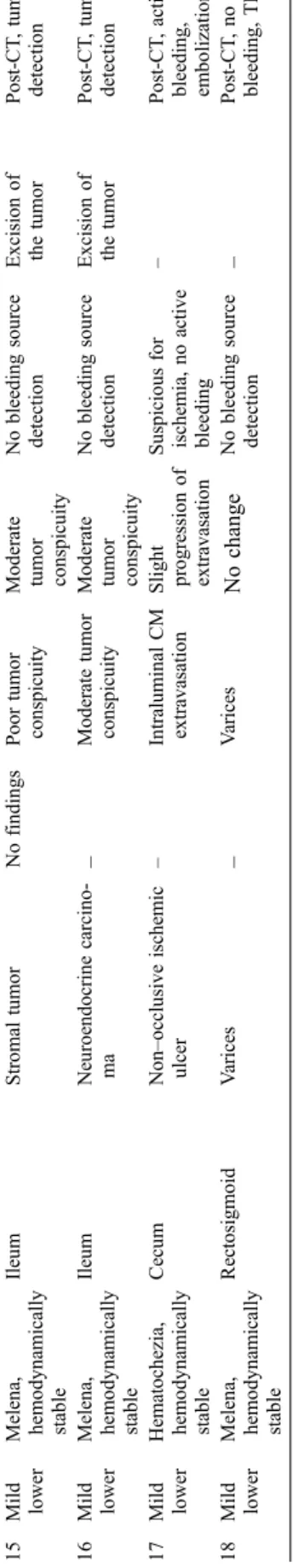
T able 2 Major clinical and CT findings, endoscopic findings, and interventional procedures in all 18 patients with acute GI bleeding No. GI bleeding Clinical findings Bleeding source Pathology Phase Endoscopy Sur gery Angiography Native Arterial Portal venous 1 Severe upper Hematemesis, hemodynamic instability
Duodenum Aortoduodenal fistula – Intraluminal CM extravasation Progression of extravasation – Excision of the fistula – 2 Severe upper Melena, hematochezia, hemodynamic instability
Duodenum Aortoduodenal fistula – Intraluminal CM extravasation Progression of extravasation Inconclusive – Pre-CT no finding, stentgraft post-CT 3 Severe upper Hematochezia, hemodynamic instability
Duodenum Aortoduodenal fistula – Intraluminal CM extravasation Progression of extravasation Ulcera, no active bleeding Resection of pars III duodeni Stentgraft post-CT 4 Severe upper Hematemesis, melena, hemodynamic instability
Duodenum Aortoduodenal fistula – Intraluminal CM extravasation Progression of extravasation Suspicious for aortoduodenal fistula Excision of the fistula Stentgraft post-CT 5 Severe upper Hematemesis, hemodynamic instability Biliodigestive anastomosis Pseudoaneurysm hepatic artery No findings Intraluminal CM extravasation Progression of extravasation –– Coiling post-CT 6 Severe upper Hematochezia, hemodynamic instability
Duodenum Arteriobiliary fistula – CM in common bile duct, GB, and duodenum Progression of extravasation Suspicious for hemo-bilia, no bleeding source detection – Pre-/post-CT no finding 7 Severe upper Blood in stoma, hemodynamic instability Duodenum Ischemic anastomotic ulcer Intraluminal hyperdensity Intraluminal CM extravasation Progression of extravasation Jejunal blood, no bleeding source detection – Embolization post-CT 8 Mild upper Hematemesis, melena, hemodynamically stable
Biliodigestive anasto-mosis Pseudoaneurysm hepatic artery No findings Pseudoaneurysm, no extravasation No change Inconclusive – Coiling post-CT 9 Mild upper Melena, hemodynamically stable
Duodenum
Pseudoaneurysm gastroduodenal artery
– Pseudoaneurysm, air collections duodenal wall No change Ulcera with bleeding – Coiling post-CT 10 Mild upper Hematemesis Cystojejunal anastomosis Pseudoaneurysm lienal artery Intraluminal hyperdensity Pseudoaneurysm, no extravasation No change –– Coiling post-CT 11 Severe lower Melena, hemodynamic instability
Jejunum Mucositis – Intraluminal CM extravasation Progression of extravasation Mucositis – Coiling post-CT 12 Severe lower Hematochezia, hemodynamic instability
Right colonic flexure Non –occlusive ischemic ulcer Intraluminal hyperdensity Intraluminal CM extravasation Progression of extravasation Bleeding, clipping –– 13 Severe lower Melena, hemodynamic instability
Sigmoid Diverticulum Intraluminal hyperdensity Intraluminal CM extravasation Progression of extravasation No bleeding source detection – Post-CT active bleeding, embolization 14 Severe lower Hematochezia, hemodynamically unstable
T ransverse colon Ischemic ulcer No findings Intraluminal CM extravasation Progression of extravasation No bleeding source detection – Post-CT , active bleeding, embolization
not necessary for diagnostic purposes, MPR and MIP reconstructions were only made for illustration purposes (see figures). Similarly, 3D reconstructions such as volume rendering or surface-shaded display were not performed.
Table 2 summarizes findings of the retrospective
con-sensus reading of the two radiologists in all 18 patients with upper and lower GI bleeding in each CT imaging phase.
Un–enhanced CT revealed hyperdensity within the
bowel lumen indicating fresh blood in four of eight (50%) patients. In two patients with biliodigestive anasto-mosis, no abnormality indicating GI bleeding could be identified, and the GI stromal tumor in one patient could not be identified on the unenhanced scan.
Arterial-phase CT demonstrated intraluminal CM extra-vasation indicating active GI bleeding in 11 of 18 (61%) patients, whereas no active CM extravasation was found in 6 (33%) patients. Portal venous CT demonstrated progres-sion of CM extravasation compared to the arterial phase in all 11 (100%) patients.
In the 12 patients in whom multi-detector-row CT depicted intraluminal blood (n =5) and/or CM extravasa-tion (n =7), the mean attenuaextravasa-tion level of blood in the
bowel lumen on unenhanced CT was 47 HU (range 29–
58 HU). The mean attenuation level of extravasated blood
(i.e., CM) was 73 HU (range 50–114 HU) in the arterial
phase and 114 HU (range 52–177 HU) in the portal venous
phase. Significant differences regarding attenuation values were found between the three phases (P <0.001, Wilcoxon signed rank test).
Underlying pathology causing GI bleeding could not be identified on unenhanced CT scans in any patient. Arterial-and portal venous-phase CT allowed identification of underlying pathology in 14 of 18 (78%) patients. One of these patients had a GI stromal tumor which was depicted with a better conspicuity on the portal venous-phase images than on the arterial-phase CT images. The diagnosis of underlying pathology in the other four patients was made by endoscopy (ischemic anastomotic ulcer, mucositis, and nonocclusive ulcer in two patients). In these patients, CT demonstrated unspecific bowel wall abnormalities adjacent to intraluminal CM extravasation; however, the definite diagnosis causing GI hemorrhage could not be made.
Typical examples of upper and lower GI bleeding source detection with multi-detector-row CT are demonstrated in
Figs.1,2,3,4,5and6.
Following international guidelines [3, 13], we divided
the blood loss into acute severe (defined as hemodynamic instability with hypotension and systolic blood pressure <100 mm Hg, pulse greater than 100 beats/min, hemoglo-bin concentration less than 100 g/l or required transfusion
of more than 4 U of packed red blood cells per 24 h [13])
and acute mild GI bleeding (defined as a bleeding without hemodynamic instability and no need for red blood cell transfusions). Eleven (61%) of our patients suffered from acute severe upper (n =7) and lower (n =4) GI bleeding
No. GI bleeding Clinical findings Bleeding source Pathology Phase Endoscopy Sur gery Angiography Native Arterial Portal venous 15 Mild lower Melena, hemodynamically stable
Ileum Stromal tumor No findings Poor tumor conspicuity
Moderate tumor conspicuity
No bleeding source detection Excision of the tumor Post-CT , tumor detection 16 Mild lower Melena, hemodynamically stable
Ileum Neuroendocrine carcino-ma – Moderate tumor conspicuity
Moderate tumor conspicuity
No bleeding source detection Excision of the tumor Post-CT , tumor detection 17 Mild lower Hematochezia, hemodynamically stable
Cecum Non –occlusive ischemic ulcer – Intraluminal CM extravasation Slight progression of extravasation Suspicious for ischemia, no active bleeding – Post-CT , active bleeding, embolization 18 Mild lower Melena, hemodynamically stable
Rectosigmoid V arices – V arices No change No bleeding source detection – Post-CT , n o bleeding, TIPS CM Contrast media, GB gallbladder , TIPS transjugular intrahepatic portosystemic shunt
and 7 (39%) from acute mild upper (n =3) and lower (n = 4) GI bleeding.
In the 11 patients with acute severe GI bleeding, multi-detector-row CT demonstrated CM extravasation in all (100%) patients, whereas this finding was encountered in only 1 of the 7 (14%) patients with acute mild GI bleeding. In the remaining 6 (86%) patients from this group, multi-detector-row CT showed no CM extravasation but under-lying disease, such as pseudoaneurysms of the hepatic, splenal, and gastroduodenal artery, intestinal tumors (n = 3), and submucosal varices (n =1).
False-negative judgments and retrospective bleeding source detection
There were three CT examinations in which the prospective interpretation by radiologists of our institute failed to identify the GI bleeding source, despite its presence being clinically documented. Our retrospective consensus read-ing could definitely identify the bleedread-ing source in all of these three patients. The bleeding source was most likely misinterpreted as being part of a vessel in one patient, a small bowel tumor was missed in one, and ischemic bowel wall disease was not identified in one patient. All three of these patients suffered from mild lower GI bleeding.
Role of CT within the diagnostic process
From the 10 patients with upper GI bleeding, CT provided a diagnosis in 6 patients after negative findings at angiography (n =2) and endoscopy (n =4). In the remaining four patients, CT was the initial imaging method providing a diagnosis in all four, and no further diagnostic work-up was performed.
From the eight patients with lower GI bleeding, CT provided a diagnosis in three patients after negative findings at endoscopy and was the initial imaging method providing a diagnosis in another two. CT in three patients from this group was performed after inconclusive endos-copy, prospectively failed to provide a diagnosis, and was followed by catheter angiography that revealed the under-lying bleeding source.
Discussion
GI bleeding often poses a frustrating clinical problem because even extensive and repetitive diagnostic work-up may fail to reveal the source of hemorrhage. Common diagnostic procedures are either of low diagnostic accuracy (such as barium examinations and scintigraphy), invasive (such as catheter angiography) or insensitive in localizing
Fig. 1a–c A 55-year-old man with acute severe upper GI bleeding due to aortoduodenal fistula 8 years after aortobife-moral Y-grafting of an infrarenal aortic aneurysm. a Contrast-en-hanced axial CT image during the arterial phase shows the aortoduodenal fistulous tract (arrow) with contrast media ex-travasation into the duodenum. b Contrast-enhanced axial CT image during the portal venous phase at the same level. The dynamic process of ongoing bleeding fed by the fistulous tract results in a net increase in intraluminal contrast agent vol-ume and attenuation (arrow) as compared to the arterial phase image. c Midsagittal multiplanar reformation of arterial-phase CT demonstrates the aorto-duodenal fistula and the previously ex-cluded infrarenal aortic aneurysm
small bowel lesions (such as endoscopy and capsule
endoscopy). Our study is—to the best of our knowledge—
the first to investigate the role of CT in patients with both upper and lower GI hemorrhage and to incorporate the whole diagnostic pathway including bleeding source and underlying pathology detection with recent multi-detector-row CT scanner technology. We found in the 18 patients a high sensitivity of CT for prospective bleeding source detection, which was even more improved in the retro-spective image analysis. When only patients with acute
severe GI bleeding were included, multi-detector-row CT allowed the direct visualization of active bleeding sources in all patients.
In an animal model of colonic hemorrhage, Kuhle and
Sheiman [14] reported that single detector helical CT
angiography can depict active hemorrhage with a rate of 0.3 mL/min, thus exceeding the sensitivity of mesenteric angiography of 0.5 mL/min and approaching values of red
Fig. 3a, b A 58-year-old man with acute severe upper GI bleeding due to arteriobiliary fistula 1 week after transjugular liver biopsy. a Contrast-enhanced axial CT image during the portal venous phase demonstrates contrast media extravasation into the distal common bile duct (arrow) and into the duodenum (arrowhead). Note extensive intraperitoneal fluid collections. b Oblique coronal multiplanar reformation of portal venous-phase CT image demon-strates contrast media in the gallbladder, common bile duct, and duodenum
Fig. 2a, b A 48-year-old man with acute mild upper GI bleeding from a pseudoaneurysm of the gastroduodenal artery following chronic tuberculous ulceration of the duodenum. a Contrast-enhanced axial CT image during the portal venous phase demonstrates the pseudoaneurysm (arrow) surrounded by centrally hypodense peripancreatic lymphnodes (arrowhead, inlay). No active contrast media extravasation was seen in either the arterial (not shown) or portal venous phase. b Selective celiacography demonstrates the pseudoaneurysm of the gastroduodenal artery. Subsequent coil embolization was performed
blood cell scintigraphy of 0.2 mL/min. The value of CT in patients with lower GI bleeding in the clinical setting has been reported only infrequently, and studies investigating the ability of CT to detect the source of upper GI bleeding
are limited to a few case reports. Ettorre [9] reported their
experience in 18 patients with acute lower GI bleeding after intra-arterial injection of CM through an angiography
catheter positioned in the celiac trunk. With this invasive technique, the authors found the bleeding source in 72% of
the patients. Ernst and coworkers [8] reported their
experience with single-detector helical CT with intrave-nously administered CM and found the bleeding source in 79% of their patients. However, CM extravasation as the only reliable sign of acute bleeding has been documented
Fig. 4a–c A 61-year-old man with acute severe lower GI bleeding due to ischemic mucositis of the jejunum. a Contrast-enhanced axial CT image during the arterial phase demonstrates contrast media extravasation into the jejunal lumen. b Coronal multiplanar CT reformation in the arterial phase illustrates the bleeding source
(arrow) in the proximal jejunum just distal to the ligament of Treitz. c Catheter angiography of the superior mesenteric artery with carbon dioxide confirms the source of bleeding (arrow). The angiogram with iodinated contrast agent had been negative. Subsequent microcoil embolization was performed
Fig. 5a, b A 38-year-old man with acute mild lower GI bleeding due to gastrointestinal stromal tumor in the ileum. a Unenhanced (left), arterial-phase (middle), and portal venous-phase (right) axial CT images through the tumor (arrowheads) demonstrate a poor conspicuity of the lesion in the arterial phase and a better demarcation in the portal venous phase. The tumor cannot be depicted on unen-hanced CT and was prospec-tively missed on all three phases. b Digital subtraction catheter angiography of the su-perior mesenteric artery depicts the homogeneously hypervas-cular tumor
in only three patients. Tew et al. [5] found lower GI bleeding sources using a 4-detector-row CT scanner in 54%
of 13 patients. Miller et al. [6] performed a triphasic CT
imaging protocol with oral water in 19 patients and showed helical CT to identify a wide variety of causes of lower GI
hemorrhage. Rajan et al. [7] evaluated seven patients with
acute lower GI bleeding using a biphasic protocol with positive oral contrast and showed CT to be helpful to localize lower GI bleeding sources in patients where endoscopy failed to find a bleeding source. In a recent
study, Yoon et al. [4] demonstrated extravasation of CM
with 4-detector-row CT in 21 of 26 patients with acute severe lower GI bleeding with bleeding source localization exactly matching results from catheter angiography. Our study confirms these conclusions drawn in the literature with regard to bleeding source detection in patients with acute lower GI bleeding and extends the results to patients with GI bleeding originating proximal to the ligament of Treitz. In addition, multi-detector-row CT has been shown to allow the diagnosis of underlying etiology for GI bleeding in 78% and demonstrated unspecific bowel wall abnormalities adjacent to intraluminal CM extravasation in the other 22% of the patients.
When CT is performed in patients with GI bleeding, negative or positive oral contrast material should not be administered because positive contrast obscures the
presence of extravasation of contrast material that was given intravenously and excessive water may dilute
extravasated CM in the intestinal lumen [15]. Unenhanced
CT scans usually are recommended to identify and differentiate hyperattenuating material in the bowel lumen such as metallic clips, suture materials, and foreign bodies including tablets. In this study, unenhanced CT scans were not helpful for bleeding source detection, but only demonstrated hyperdense material within the bowel lumen indicating fresh blood in 50% of the patients. Arterial phase imaging usually is aimed at the detection of extravasation of contrast material into the bowel lumen, a finding that is diagnostic of ongoing GI hemorrhage. Portal venous-phase imaging is considered useful for determining the cause of acute GI bleeding, particularly in patients with
intestinal tumors [15]. Furthermore, it can help to better
delineate the source of bleeding by demonstrating an increase in the amount of extravasated CM compared to the arterial phase, a finding that was encountered in 92% patients of our series. This increase in amount was accompanied by an increase in attenuation of the extra-vasated CM from 73 HU in the arterial phase to 114 HU on portal venous-phase images. The fact that bleeding source conspicuity was comparable with arterial-phase and portal venous-phase CT images in the patients from this study suggests that future studies should prospectively
investi-Fig. 6a–d 74-year-old man with acute severe lower GI bleeding due to ischemic ulcer in the transverse colon. a Con-trast-enhanced axial CT image in the arterial phase demon-strates the bleeding source in the transverse colon (arrow). b Contrast-enhanced axial CT image during the portal venous phase at the same level demon-strates the dynamic process of ongoing bleeding (arrow). c Coronal maximum intensity projection in the arterial phase demonstrates the possible feed-ing artery (arrowhead), the bleeding source (white arrow), and the capsule endoscope (black arrow) in the small bowel that did not provide a diagnosis. d Selective injection of the middle colic artery confirms the bleeding source (arrow). Sub-sequent coil embolization of the middle colic artery led to ces-sation of hemorrhage
gate the use of only one contrast-enhanced phase. Although our study designs allowed no comparison of the sensitivity for bleeding source detection of multi-detector-row CT as compared to endoscopy, it seems nevertheless noteworthy that CT provided a diagnosis in 6 of 11 patients after negative or inconclusive findings at endoscopy.
A general advantage of CT over strictly endoluminal procedures such as endoscopy and capsule endoscopy is the ability to evaluate the pathology precisely with regard to extraluminal abnormalities, feeding and draining vessels, and the anatomical region with its relationship to surrounding structures. In addition, CT is readily available in most hospitals around the clock and imaging is accomplished fast and is not operator or patient dependent. On the other hand, CT may also reveal findings that are unrelated to the actual disease of the patient, which may in turn lead to further cost-extensive diagnostic workup with possible harm to the individual. It is obvious that hemostatic interventions are not feasible in the CT unit, but CT findings may guide operators towards the best-suited treatment for the individual patient, whether it is surgical, endoscopical, or catheter-directed.
Study limitations
Several limitations to our study have to be acknowledged. Owing to the nature of the retrospective study design, our scanning technique was not uniform among patients, with the use of three different helical CT scanner generations. In addition, there was a variation with different radiologists performing the prospective interpretations. Therefore, the results are subject to interobserver variability. However, this variation mirrors general clinical practice because patients are scanned on different CT scanners with different capabilities and interpreted by a spectrum of radiologists.
The retrospective review of CT images was not performed in a blinded fashion, nor did a control population assist in
estimating specificity. These were not intended aims of our study because we analyzed the ability of CT to localize and characterize bleeding sources. If the CT images had been reviewed in a blinded fashion, the results for bleeding source conspicuity might have been different.
Our results may be a reflection of selection bias or of the small number of included cases. False-positive and false-negative findings would be anticipated in a larger study because of the intermittent nature of GI bleeding.
A randomized study to test detection of GI bleeding with CT compared with scintigraphy or invasive angiography may not be ethically justified because of the absence of a relevant control group. Furthermore, the limited availability of after-hours scintigraphy and the wide-spread accessi-bility of CT would also make a randomized trial comparing the two techniques for initial diagnosis difficult to perform. Finally, CT may not be feasible in patients with renal failure and allergies to iodinated contrast agents.
Conclusion
Multi-detector-row CT enables the identification of bleed-ing source and etiology in patients with acute upper and lower GI bleeding. CT allows the depiction of indirect signs suggesting the origin and cause of hemorrhage in patients with acute non-severe GI bleeding, whereas the bleeding source can be directly demonstrated in patients with acute severe hemorrhage. Further prospective studies including clinical decision-making based on CT findings are necessary to confirm these retrospective results and to define the exact role of the different imaging phases and CM protocols for a comprehensive diagnosis of the disease.
Acknowledgements This research has been supported by the National Center of Competence in Research, Computer Aided and Image Guided Medical Interventions (NCCR CO-ME) of the Swiss National Science Foundation.
References
1. Manning-Dimmitt LL, Dimmitt SG, Wilson GR (2005) Diagnosis of gas-trointestinal bleeding in adults. Am Fam Physician 71:1339–1346 2. Barkun A, Bardou M, Marshall JK
(2003) Consensus recommendations for managing patients with nonvariceal upper gastrointestinal bleeding. Ann Intern Med 139:843–857
3. Palmer KR (2002) Non-variceal upper gastrointestinal haemorrhage: guide-lines. Gut 51 Suppl 4:iv1–iv6 4. Yoon W et al (2006) Acute massive
gastrointestinal bleeding: detection and localization with arterial phase multi-detector row helical CT. Radiology 239:160–167
5. Tew K, Davies RP, Jadun CK, Kew J (2004) MDCT of acute lower gastroin-testinal bleeding. AJR Am J
Roentgenol 182:427–430
6. Miller FH, Hwang CM (2004) An initial experience: using helical CT imaging to detect obscure gastrointes-tinal bleeding. Clin Imaging 28:245– 251
7. Rajan R, Dhar P, Praseedom RK, Sudhindran S, Moorthy S (2004) Role of contrast CT in acute lower gastroin-testinal bleeding. Dig Surg 21:293–296 8. Ernst O, Bulois P, Saint-Drenant S,
Leroy C, Paris JC, Sergent G (2003) Helical CT in acute lower gastrointes-tinal bleeding. Eur Radiol 13:114–117 9. Ettorre GC, Francioso G, Garribba AP,
Fracella MR, Greco A, Farchi G (1997) Helical CT angiography in gastrointes-tinal bleeding of obscure origin. AJR Am J Roentgenol 168:727–731
10. Frauenfelder T, Wildermuth S, Marincek B, Boehm T (2004) Non-traumatic emergent abdominal vascular conditions: advantages of multi-detector row CT and three-dimensional imaging. Radiographics 24:481–496
11. Roos JE, Willmann JK, Hilfiker PR (2002) Secondary aortoenteric fistula: active bleeding detected with multi-detector-row CT. Eur Radiol 12 Suppl 3:S196–200
12. Leschka S, Alkadhi H, Wildermuth S, Marincek B (2005) Multi-detector computed tomography of acute abdo-men. Eur Radiol 15:2435–2447 13. Leitman IM, Paull DE, Shires GT 3rd
(1989) Evaluation and management of massive lower gastrointestinal hemor-rhage. Ann Surg 209:175–180
14. Kuhle WG, Sheiman RG (2003) De-tection of active colonic hemorrhage with use of helical CT: findings in a swine model. Radiology 228:743–752 15. Yoon W, Jeong YY, Kim JK (2006)
Acute gastrointestinal bleeding: con-trast-enhanced MDCT. Abdom Imaging 31:1–8