Myc and Mammary Cancer: Myc is a Downstream Effector
of the ErbB2 Receptor Tyrosine Kinase
Nancy E. Hynes
1,3and Heidi A. Lane
2The proto-oncogene c-myc encodes a transcription factor which plays a major role in the regu-lation of normal cellular proliferation and is aberrantly expressed in many breast tumors. In a normal cell Myc expression levels are tightly regulated being subject to many layers of control. Errantly expressed Myc collaborates with other oncogenes to promote transformation. In this review we will focus on the association between abnormal Myc expression and mammary cancer. In particular, we will discuss the role of Myc as a downstream effector of the ErbB2 receptor tyrosine kinase which is overexpressed and constitutively activate in many mammary tumors. The cooperation between Myc and ErbB2 in transformation will be discussed in rela-tion to clinical studies on Myc in human cancer and with considerarela-tion of transgenic models of Myc-induced mammary cancer. Data from our laboratory will be presented showing that deregulated ErbB2 activity strongly stimulates cytoplasmic signaling pathways which in turn impinge on Myc at multiple levels causing its deregulated expression.
KEY WORDS: EGF receptor family; transgenic mice; p27Kip1; apoptosis; transcription factor.
INTRODUCTION
Cancer arises as a result of cumulative alter-ations in the genetic make-up of somatic cells. Such alterations principally lead to the aberrant expres-sion, mutation or deletion of proteins involved in the modulation of intracellular pathways governing nor-mal cellular proliferation and differentiation. These defects allow cancer cells to evade signals eminat-ing from their external environment or from internal checkpoint controls through deregulation of signaling pathways which normally keep cell proliferation and survival tightly controlled. In this context, the proto-oncogene c-myc, which encodes a transcription fac-tor playing a major role in the regulation of normal cell proliferation, is abberantly expressed in many hu-man cancers. Indeed, taking breast cancer as an ex-ample, c-myc amplification was one of the first consis-1Friedrich Miescher Institute, PO Box 2543, CH-4002 Basel,
Switzerland.
2Novartis Pharma, K-125.1317, CH-4002 Basel, Switzerland. 3To whom correspondence should be addressed. e-mail: hynes@
fmi.ch
tent genetic alterations discovered in primary human tumors (1).
Myc is a member of the bHLHZip4family of
tran-scription factors which, when dimerized with its part-ner Max, binds to specific DNA sequences resulting in the transcriptional regulation of target genes in-volved in the control of cellular growth/proliferation [reviewed (2 – 8)]. All known biological effects of Myc, including the ability to transform cells, result from its activity as a transcription factor. It has been proposed that Myc acts as a sensor of the cellular environment (9), able to trigger proliferation and, in stress situa-tions, apoptosis. Furthermore, when deregulated, Myc 4Abbreviations: epidermal growth factor (EGF); receptor
tyro-sine kinase (RTK); phosphatidylinositol-30kinase (PI3K); endo-plasmic reticulum (ER); mitogen-activated protein kinase (MAP kinase); basic helix-loop-helix zipper (bHLHZip); Myc Box II (MbII); transactivation/transformation-domain associated pro-tein (TRRAP); histone acetyltransferase (HAT); histone deacety-lase (HDAC); human mammary epithelial cells (HMEC); human telomerase catalytic subunit (hTERT); cyclin-dependent kinase inhibitor (CKI); monoclonal antibody (mAb); mouse mammary tumor virus (MMTV); whey acidic protein (WAP); transforming growth factorα (TGFα).
141
P1: VENDOR/GCQ/lmd/GEE P2: GCR
Journal of Mammary Gland Biology and Neoplasia (JMGBN) PP067-295943 February 7, 2001 9:8 Style file version Nov. 07, 2000
142 Hynes and Lane
has characteristics which would clearly contribute to its transforming ability; including its ability to drive cells through the G1/S-transition of the cell cycle as well as connections between Myc and the induction of genomic instability.
In this short review we have focused on the connection between aberrant Myc expression and mammary tumorigenesis. Emphasis is placed on the biological activities of Myc, clinical studies address-ing Myc deregulation in human breast cancer and the application of transgenic models of Myc-induced mammary cancer. Additionally, based on our own ex-perimentation, the role of Myc as a downstream ef-fector of the ErbB2 receptor tyrosine kinase (RTK) will be discussed (10,11). ErbB2 is activated due to gene amplification and overexpression in a high per-centage of human breast tumors. Through antibody-mediated downregulation of ErbB2 receptor signal-ing pathways in ErbB2-overexpresssignal-ing breast tumor cell lines, we suggest that a major downstream effector contributing to the potentiation of ErbB2-dependent tumor cell proliferation is Myc. This model will be discussed in relation to clinical aspects of the impact of Myc and ErbB2 deregulation in mammary carcino-genesis, and the application of ErbB2-directed tumor-specific therapies.
BIOLOGICAL ACTIVITIES OF Myc Myc, A Key Regulator of Proliferation and Apoptosis
Myc has a central role in normal cellular growth/proliferation control. This is most dramati-cally demonstrated by the observation that expression of exogenous Myc in quiescent fibroblasts induces S-phase entry in the absence of growth factors (12). Furthermore, Myc-negative fibroblasts proliferate at a slower rate and exhibit reduced expression of G1cell
cycle regulators (9). Myc has been suggested to act as an environmental sensor, coordinating adjustment of cell cycle progression to the external milieu by affect-ing cell cycle regulators at multiple levels (9). Con-sistent with a role in proliferation c-myc transcription is low in resting or differentiated cells, is rapidly in-duced following mitogenic stimulation and is required for continuous cellular proliferation (4,5). In the case of the mammary gland, similar fluctuations in c-myc RNA levels are observed; reflecting the proliferation index of the gland. Indeed, the c-myc promoter is a
tar-get for peptide growth factors and for estrogen (4,13), both of which contribute to proliferation of the gland. Myc mRNA levels increase during pregnancy-related proliferation (14). In addition to elevated expression during pregnancy, increases in Myc expression may also be important during the proliferative changes which occur in the breast during the menstrual cycle (15). In contrast, Myc is not detected during lactation (14), reflecting the differentiated state of the gland at this time.
A second distinctive characteristic of Myc is its ability to induce apoptosis. This was discovered fol-lowing growth factor-withdrawal from cells ectopi-cally expressing Myc. In this case, DNA synthesis oc-curred, but cells failed to multiply and apoptosis was accelerated (16). It is now known that deregulated Myc expression sensitizes cells to diverse proapop-totic stimuli [reviewed (17)]. Indeed, it has been sug-gested that a cell is constantly poised to enter apop-tosis and requires stimuli, either intracellular or from the external environment, in order to survive and pro-liferate (17). In order for tumor cells to propro-liferate in the presence of deregulated Myc expression, there-fore, additional genetic alterations must occur, which would allow the malignant cells to circumvent pro-apoptotic stimuli. It should also be mentioned here that Myc probably plays a role in regulated apopto-sis occurring under normal physiological conditions. In this context, during murine mammary gland invo-lution following pup withdrawal, c-myc RNA levels have been observed to increase (13). It is tempting to speculate, therefore, that this upregulation is linked to the massive apoptosis which occurs during this de-velopmental stage.
Myc the Transcription Factor
Myc is a member of the bHLHZip family of tran-scription factors, possessing a C-terminal DNA bind-ing domain and a N-terminal transactivation domain. Myc dimerization with its partner Max is essential for DNA binding to a consensus site, the so-called E-box, and transcriptional regulation of target genes. The N-terminus of Myc contains two highly conserved regions, the Myc box (Mb) I and II domains. All biological properties of Myc, including those asso-ciated with transformation, require its DNA bind-ing domain and dependent upon the cellular context, the N-terminal MbI and/or MbII domains, making it probable that Myc exercises its biological effects
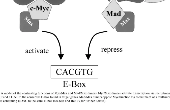
Fig. 1. A model of the contrasting functions of Myc/Max and Mad/Max dimers. Myc/Max dimers activate transcription via recruitment of TRRAP and a HAT to the consensus E-box found in target genes. Mad/Max dimers oppose Myc function via recruitment of a multisubunit complex containing HDAC to the same E-box (see text and Ref. 19 for further details).
through modulation of target gene expression. The MbII domain binds the nuclear cofactor TRRAP (18), which in turn recruits the HAT hGCN5 (19). Recruit-ment of this chromatin remodeling complex to the DNA by Myc provides an explanation for Myc/Max induced transcriptional activation. Myc/Max activ-ity is opposed by Max, when dimerized with one of the Mad family members. Mad proteins bind a complex containing co-repressors and HDACs (2,3). Since Mad/Max dimers and Myc/Max dimers rec-ognize the same DNA sequence, a very simplistic model proposes that Mad-Max dimers, via recruit-ment of HDACs, antagonize the transcriptional ac-tivity of HAT-associated Myc-Max complexes (19) (Fig. 1). A final point to consider is that the balance of the respective dimers may determine Myc target gene expression, particularly as Myc is expressed in prolif-erating cells and is down-regulated in differentiated cells, while the opposite tends to be true for the Mad family members (5). Myc and Mad1 protein expres-sion have been examined in normal human breast and
in progressive breast disease, where this correlation was observed. Mad1 expression was high in differen-tiated cells and decreased in high-grade tumors, while the inverse was observed for Myc levels (20).
A comprehensive understanding of the mecha-nism underlying Myc’s biological activities will arise from the identification of Myc target genes. These have remained rather elusive, despite intense study, for various reasons. However, the general consensus is that the identified targets reflect Myc’s functions and can be catagorized as targets involved in growth con-trol and apoptosis, including among others: hEST2, the catalytic subunit of telomerase; p19Arf, the tumor suppressor; thymidine kinase, involved with DNA metabolism; and lactate dehydrogenase-A, a partici-pant in anaerobic glycolysis. For a detailed discussion of Myc targets and their identification, the reader is referred to recent reviews (2,3). Meaningful for the discussion of our results (see later), is the fact that important cell cycle regulators, such as the D-type cy-clins, are transcriptionally regulated by Myc (21,22).
P1: VENDOR/GCQ/lmd/GEE P2: GCR
Journal of Mammary Gland Biology and Neoplasia (JMGBN) PP067-295943 February 7, 2001 9:8 Style file version Nov. 07, 2000
144 Hynes and Lane
Myc AND MAMMARY CANCER Mechanisms Leading to Deregulated Myc Expression in Cancer
In a normal cell c-myc transcription is tightly con-trolled and dependent upon proliferative stimuli. Fur-thermore, c-myc mRNA and Myc protein are both short-lived [reviewed (4)]. Thus, in a normal cell Myc expression is highly regulated, at all possible levels. In sharp contrast to this, deregulated Myc expres-sion is quite prevalent in human tumors. Deregu-lation arises through diverse mechanisms, including
c-myc amplification in solid tumors, and
transloca-tions in leukemias (23). One of the first genetic al-terations reported in human breast tumors was c-myc amplification (1). Since then, c-myc amplification has been extensively studied and the current consen-sus is that it occurs in approximately 20% of all breast cancer cases [reviewed (24)]. However, exam-ination of c-myc mRNA in a relatively small num-ber of breast tumors by RT-PCR has suggested that
c-myc overexpression does not necessarily arise from
gene amplification (25). Although the mechanism was not further examined, the results are not supris-ing considersupris-ing that Myc expression is controlled by transcriptional as well as post-transcriptional mechanisms.
Myc protein levels are also tightly regulated. Mu-tations within c-myc occur in a high percentage of Burkitt’s and other lymphomas (26). It has recently been shown that some of these mutations lead to sta-bilization of the Myc protein, preventing its destruc-tion by ubiquitin-mediated proteolysis (27). Stabiliz-ing Myc mutations have not been reported in breast cancers. However, Myc protein levels are also con-trolled by cytoplasmic signaling pathways which are often deregulated in breast tumor cells. In this con-text, two pathways which are highly activated due to overexpression of the RTK ErbB2 in breast tu-mors are the PI3K and MAP kinase pathways. In-triguingly, the PI3K pathway has been implicated in the translational induction of Myc (28), and high MAP kinase activity promotes an increase in the stabil-ity of Myc protein (29). Considering that the ulti-mate level of Myc protein is subject to so many lay-ers of control and that tumor cells have alterations in many signaling pathways which impact on this, it would not be suprising to find that Myc expression is deregulated in the vast majority of human breast cancers.
Myc Expression in Primary Breast Tumors
Clinical studies on primary human breast cancer are valuable not only for predicting patient progno-sis but also provide clues about tumor biology. It is generally agreed that there is an association between
c-myc amplification and poor prognosis in breast
can-cer [discussed (23,24,30)]. An immunohistochemical study on Myc expression in breast tumors revealed that during tumor progression from well differenti-ated, low-grade to poorly differentidifferenti-ated, high-grade, Myc expression increased significantly (20). Very in-teresting in light of our results, described later, is the recent report that coamplification of erbB2 and
c-myc strongly correlates with a reduction in patient
survival in a series of breast cancer patients (31). This suggests a certain level of cooperation between dereg-ulation of these two proteins in breast tumor devel-opment.
How Does Myc Contribute to Breast Cancer Development?
Myc deregulation induces inappropriate prolifer-ation, as evidenced by the ability of Myc to drive some quiescent cell lines into S-phase (12). This character-istic might contribute to cancer, not only by keep-ing cells cyclkeep-ing, i.e., preventkeep-ing differentiation, but also by contributing to genomic instability. Deregu-lated Myc expression has been associated with gene amplification and karyotypic abnormalities in cul-tured cells (32,33) and with abnormal ploidy in in
vivo models (34). Altered Myc expression
acceler-ates the passage of cells through G1 into S-phase and has also been shown to allow cells to pass through mitotic checkpoints (35). Indeed, a comparison be-tween A1N4 immortalized human mammary epithe-lial cells and A1N4 cells expressing exogenous Myc revealed that, while EGF-withdrawal caused a G1 ar-rest in both cell lines, EGF readdition induced pre-mature entry into S-phase in the Myc-expressing cells (36). Additionally, expression of hTERT, a transcrip-tional target of Myc (37) has been demonstrated to be induced following ectopic expression of Myc in non-immortalized HMEC cultures. This is associated with increased telomerase activity in these HMECs, which may in part be responsible for the reported extended life-span of these cultures (37).
These characteristics of Myc have the potential to contribute to tumorigenesis by influencing cellular senescence and/or allowing cells with chromosomal
abnormalities or damaged DNA to replicate. Whether these effects reflect expression of very specific target genes, such as hTERT, or are due to overall effects on chromatin structure and ensuing gene expression, leading for example to chromosomal instability, re-mains to be elucidated (2).
Mouse Models for Myc-Induced Mammary Cancer
Transgenic mouse models have been developed in order to study the role of Myc in mammary trans-formation. Myc expression has been targeted to the mammary gland through the use of several specific promoters, including WAP and MMTV [reviewed (38,39)]. These mouse models have clearly demon-strated that deregulated Myc expression does indeed induce tumor formation in the mammary gland. How-ever, in each Myc transgenic strain so far examined, the females display a long latency and a dependency upon passage through pregnancy for mammary tu-mor development. This characteristic is shared with most mammary-specific tumor models, reflecting the fact that a single oncogene is generally unable to in-duce a full tumor phenotype. The necessity for mice to pass through pregnancy before tumor development might be explained by the upregulation of the mam-mary specific promoters driving the transgene, as well as by the enhanced proliferation at this developmen-tal stage, which may potentiate selection of additional mutations.
In order to test for oncogenic cooperativity, Myc transgenics have been crossed with mice express-ing other oncogenes, includexpress-ing TGFα, Ha-Ras and Bcl-2. TGFα, a member of the EGF-related family of peptide growth factors, binds the ErbB1/EGF recep-tor and induces activation of EGF receprecep-tor dimers and ErbB2-containing heterodimers (40). In human breast cancer there is often coexpression of the EGF receptor and TGFα or one of the other EGF-related ligands, which leads to autocrine receptor activation (41,42). As observed for Myc transgenics, there was also a requirement for pregnancy and an extended tu-mor latency in the transgenic strains expressing TGFα in the mammary gland. In striking contrast, how-ever, tumorigenesis in dual transgene carriers (coex-pressing TGFα and Myc in the mammary gland) was dramatically enhanced (38,39,43–45). Furthermore, the double transgenic mice developed mammary tu-mors rapidly in all glands without a requirement for pregnancy or ovarian hormone stimulation (44). These results suggest that there is strong cooperativity
between Myc and the ligand-activated EGF receptor in promoting mammary cancer.
Offspring arising from the cross of MMTV-Myc and MMTV-Ras transgenic strains also developed mammary tumors more rapidly than either of the sin-gle transgenic strains (46). Interestingly, tumor cells arising from MMTV-Myc mice showed a different cell cycle distribution in comparison to cells from tumors of the MMTV-Ras mice or the dual transgene carriers. In the Myc tumor cells there was a significant reduc-tion in the G1 fraction, compared to the cells taken
from the Ras or dual transgene-induced tumors, and this was accompanied by an increase in the S-phase fraction (47). These observations accurately reflect the results seen in the A1N4-Myc cells discussed ear-lier (36), suggesting that mammary cells with deregu-lated Myc generally progress rapidly through G1.
As previously mentioned, Myc expression in the absence of growth factors often leads to apopto-sis. Taking this in mind, the rapid tumor formation observed in the dual transgenics might in part be due to enhanced survival of Myc-expressing cells in glands with elevated levels of TGFα. In support of this proposition, examination of mammary tumors arising in double and single transgenics for apop-totic nuclei revealed that there was less apoptosis in the dual transgene carriers, coexpressing TGFα and Myc, than in Myc transgenics (43). Furthermore, when cells from the tumors coexpressing TGFα and Myc were cultured in the presence of an EGF receptor in-hibitor, apoptosis was elevated (43). Tumors arising from transgenics coexpressing Myc and Ras also dis-played a reduced rate of apoptosis when compared to Myc transgenics (47). These results suggest that deregulated Myc expression in the mammary gland promotes a high level of apoptosis. Only when path-ways emanating from activated growth factor recep-tors are stimulated, is apoptosis reduced, allowing for the rapid onset of tumors.
Bcl-2, a pro-survival member of the Bcl-2 fam-ily, is able to inhibit apoptosis arising from a wide variety of insults, including deregulated Myc expres-sion (48,49). Bcl-2 overexpresexpres-sion in the mammary gland, driven from a WAP-Bcl-2 transgene, was in-sufficient to induce mammary tumors (50). However, when these mice were crossed with Myc transgenics, accelerated development of Myc-induced tumor for-mation was observed. This was accompanied by a re-duction in the fraction of apoptotic cells in the dual transgenic glands, when compared to glands express-ing Myc alone (50). These results, as well as those discussed before, suggest that Myc’s ability to induce
P1: VENDOR/GCQ/lmd/GEE P2: GCR
Journal of Mammary Gland Biology and Neoplasia (JMGBN) PP067-295943 February 7, 2001 9:8 Style file version Nov. 07, 2000
146 Hynes and Lane
mammary cancer is enhanced upon suppression of its apoptotic activity. This can be achieved by various mechanisms, including increased expression of pro-survival members of the Bcl-2 family or increased activity of ErbB RTKs. However, it is quite likely that the cooperativity between Myc and TGFα goes beyond the ability of this peptide growth factor to suppress Myc-induced apoptosis. This supposition is based on the fact that tumor latency was shorter in the Myc/TGFα dual transgenics as compared to the Myc/Bcl-2 transgenics (44,50). This situation possibly relates to the observation that ErbB receptor activa-tion impinges on multiple cellular pathways, whereas in comparison Bcl-2 has a relatively limited biological activity.
Equally relevant for this discussion are the phe-notypes of dual transgenics coexpressing TGFα and ErbB2 in the mammary epithelium. These mice also display an accelerated rate of tumorigenesis in com-parison to mice expressing a single transgene (51). Thus, co-expression of TGFα with either Myc or ErbB2 leads to accelerated tumorigenesis. It is, there-fore, of great interest to examine the tumors arising in the TGFα-ErbB2 coexpressors. Based upon the re-sults discussed later, showing that Myc is an effector of oncogenic ErbB2, one prediction would be that the mammary tumors arising in the dual TGFα-ErbB2 transgenics would have elevated levels of Myc expres-sion. Taken together, these results from transgenic mice suggest that the mammary epithelium is particu-larly sensitive to deregulated expression of both ErbB RTKs and Myc; both of which are overexpressed in human breast cancer.
Myc as an Effector of Oncogenic ErbB2
Together with c-myc, amplification of the c-erbB2 gene, leading to overexpression of the receptor, was one of the initial genetic alterations found in breast tu-mors (52,53). Today there is a wealth of clinical data demonstrating the importance of ErbB2, and other members of the ErbB family, in breast cancer. It is now clear that ErbB2 overexpression correlates with more aggressive tumor types and a worse patient prognosis (for reviews on this subject, the interested reader is re-ferred to: 41,42,54,55). However, despite the obvious involvement of ErbB2 in breast tumor malignancy, the underlying mechanisms by which overexpression of this receptor potentiates tumor cell proliferation have remained poorly understood. In order to ad-dress the question of how ErbB2 overexpression
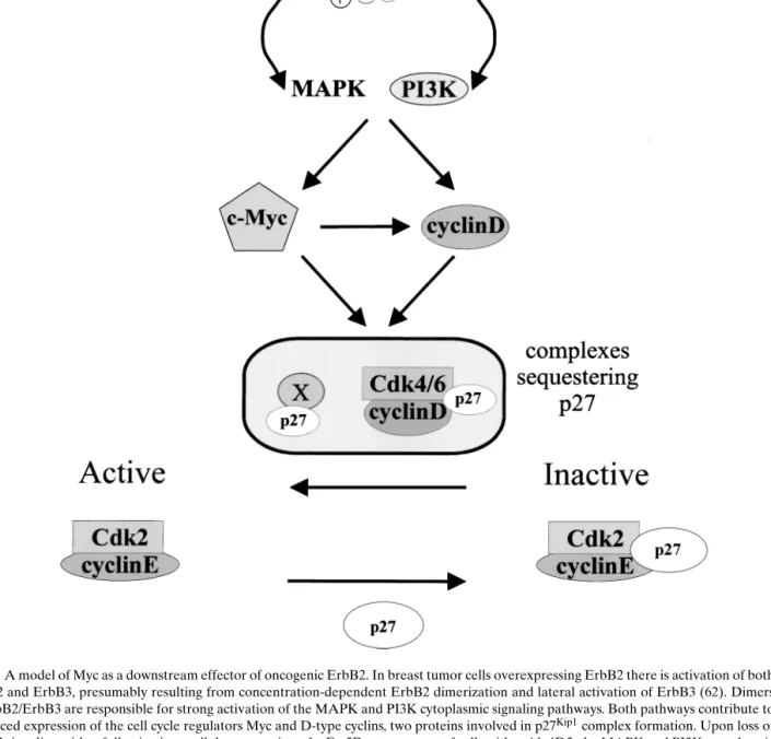
con-tributes to the deregulated proliferation characteris-tics of tumors, we have used a strategy which allows efficient down-regulation of the receptor: intracellu-lar expression of an ErbB2-specific single chain anti-body (scFv-5R). Specific targeting of scFv-5R to the ER results in retention of ErbB2 in this compartment, leading to loss of receptor function (56). Using ErbB2-overexpressing SKBr3 breast tumor cells, inducible expression of scFv-5R was demonstrated to result in the loss of plasma membrane-localized ErbB2. Con-comitant with this loss of functionally active ErbB2, there was a dramatic downregulation of ErbB3 activ-ity and the MAP kinase and PI3K pathways, culmi-nating in accumulation of the cells in the G1 phase of the cell cycle (11). Notably, the expression of Myc pro-tein and mRNA was also decreased in the absence of ErbB2 signaling. Furthermore, ectopic expression of Myc partially overcame the scFv-5R-imposed G1 ac-cumulation, indicating that Myc is a primary effector of ErbB2-mediated oncogenicity. A closer look at the mechanisms underlying reductions in Myc expression following ErbB2 downregulation, revealed that both Myc mRNA levels and protein stability were affected (11). As mentioned previously, the PI3K pathway is involved in the translational induction of Myc (28) and MAP kinase activation stabilizes the Myc pro-tein (29). Taken together, therefore, our results sug-gest that enhanced ErbB2 signaling acts at multiple points to ensure that Myc levels remain elevated in breast tumor cells (Fig. 2).
Detailed analyses of cell cycle regulators in scFv-5R expressing SKBr3 cells revealed an important role for Myc in ErbB2-overexpressing cells. Specifically, the G1 block induced by ErbB2 downregulation was
demonstrated to be a consequence of redistribution of the CKI p27Kip1from sequestering complexes to cy-clin E/Cdk2, resulting in cycy-clin E/Cdk2 inactivation. This redistribution was found to parallel the decrease in Myc and cyclin D protein levels (11). It is well es-tablished that the D-type cyclins as well as Myc play major roles in the regulation of p27Kip1 complex
for-mation (57). On the one hand, Myc stimulates D-type cyclin expression at the transcriptional level (21,22) and, on the other hand, the stability/activation of cy-clin D-dependent Cdks is in turn facilitated by inter-action with p27Kip1 (57). It has also been proposed
that other, as yet unknown, p27Kip1sequestration
pro-teins may be downstream of Myc (58) (“x” in Fig. 2). Our results revealed that ectopic expression of Myc in scFv-5R expressing SKBr3 cells delayed the accu-mulation of these cells in G1. This phenomenon
Fig. 2. A model of Myc as a downstream effector of oncogenic ErbB2. In breast tumor cells overexpressing ErbB2 there is activation of both ErbB2 and ErbB3, presumably resulting from concentration-dependent ErbB2 dimerization and lateral activation of ErbB3 (62). Dimers of ErbB2/ErbB3 are responsible for strong activation of the MAPK and PI3K cytoplasmic signaling pathways. Both pathways contribute to enhanced expression of the cell cycle regulators Myc and D-type cyclins, two proteins involved in p27Kip1complex formation. Upon loss of
ErbB2 signaling, either following intracellular expression of scFv-5R or treatment of cells with mAb 4D5, the MAPK and PI3K cytoplasmic signaling pathways are down-regulated. This leads to a decrease in the level of Myc and the D-type cyclins. Myc stimulates D-type cyclin expression transcriptionally (21,22), however, the level of D-type cyclins is also controlled by the PI3K cytoplasmic signaling pathway (63). It has also been proposed that other, as yet unknown, p27Kip1sequestration proteins may be downstream of Myc (58) (indicated by “x”).
Taken together, the drop in Myc and D-cyclin levels allows the redistribution of p27Kip1onto cyclinE/cdk2 and concomitant loss of kinase
P1: VENDOR/GCQ/lmd/GEE P2: GCR
Journal of Mammary Gland Biology and Neoplasia (JMGBN) PP067-295943 February 7, 2001 9:8 Style file version Nov. 07, 2000
148 Hynes and Lane
p27Kip1redistribution to cyclin E/Cdk2 complexes and
maintenance of cyclin E/Cdk2 activity. These obser-vations, therefore, add further weight to the proposal that elevated Myc activity plays a major role in ErbB2-dependent breast tumor proliferation, through the maintenance of p27Kip1sequestration proteins.
The clear importance of ErbB2 overexpression in the potentiation of breast tumor proliferation has resulted in intense scrutiny of ErbB2 as a target for tumor-directed therapies. In this respect, a mAb that targets the extracellular domain of ErbB2 (known as 4D5) specifically inhibits the in vitro growth of ErbB2-overexpressing breast tumor cells (59,60). In-deed, the humanized version of 4D5 (HerceptinTM)
has been validated in the clinic as an ErbB2-directed therapeutic approach (61). In order to understand the mechanism(s) underlying the effects of 4D5 on tumor cell growth, we set out to examine specific ef-fects on cell cycle regulators in ErbB2-overexpressing breast tumor cells treated with this mAb (10). 4D5-mediated down-regulation of ErbB2 signaling led to a G1 accumulation in two ErbB2-overexpressing breast tumor cell lines (BT474 and SKBr3). Accumulation in G1 was preceded by a reduction in Cdk2 activ-ity. Importantly, this correlated with a reduction in the expression of Myc and cyclin D proteins, as well as with an increase in p27Kip1 association with cy-clin E/Cdk2 complexes; events which were not ob-served with a noninhibitory control antibody (10). In BT474 cells, both Myc and D-type cyclin protein levels dropped rapidly (1–2 hrs) after 4D5 addition, with ki-netics which slightly preceded the accumulation of the p27Kip1protein on cyclin E/Cdk2 complexes and
cy-clin E/Cdk2 inactivation. These data not only strongly support the previous observations outlined (11), but also point to the importance of ErbB2-dependent maintenance of Myc and cyclin D protein levels in ErbB2-overexpressing tumor cells in the clinical set-ting, particularly since coamplification of c-erbB2 and
c-myc correlates with poorer survival in breast
can-cer patients (31). Future experiments will be aimed at establishing the role of Myc in the deregulation of tumor cell proliferation; specifically as a downstream effector of elevated ErbB2 receptor activity.
ACKNOWLEDGMENTS
The work in this laboratory is supported by the Novartis Research Foundation. H. A. Lane was sup-ported in part by a grant from the Swiss Cancer League. We would like to thank R. Neve, I. Beuvink
and K. Horsch for help with the illustrations. We also thank T. Holbro and other members of the lab for stimulating discussions and suggestions.
REFERENCES
1. C. Escot, C. Theillet, R. Lidereau, F. Spyratos, M. H. Champeme, J. Gest, and R. Callahan (1986). Genetic alteration of the c-myc proto-oncogene (MYC) in human primary breast carcinomas. Proc. Natl. Acad. Sci. U.S.A. 83:4834–4838. 2. M. D. Cole and S. B. McMahon (1999). The Myc oncoprotein: A
critical evaluation of transactivation and target gene regulation.
Oncogene 18:2916–2924.
3. C. V. Dang (1999). c-Myc target genes involved in cell growth, apoptosis and metabolism. Mol. Cell. Biol. 19:1–11.
4. M. Henriksson and B. L ¨uscher (1996). Proteins of the Myc net-work: Essential regulators of cell growth and differentiation.
Adv. Cancer Res. 68:109–182.
5. B. Luscher and L.-G. Larsson (1999). The basic region/helix-loop-helix/leucine zipper domain of Myc proto-oncoproteins: Function and regulation. Oncogene 18:2955–2966.
6. C. Bouchard, P. Staller, and M. Eilers (1998). Control of cell proliferation by myc. Trends Cell Biol. 8:202–206.
7. B. Amati, K. Alevizopoulos, and J. Vlach (1998). Myc and the cell cycle. Frontiers in Bioscience 3:250–268.
8. G. C. Prendergast (1999). Mechanisms of apoptosis. Oncogene 18:2967–2987.
9. M. K. Mateyak, A. J. Obaya, and J. M. Sedivy (1999). c-Myc regulates cyclin D-Cdk4 and-cdk6 activity but affects cell cycle progression at multiple independent points. Mol. Cell. Biol. 19:4672–4683.
10. H. A. Lane, I. Beuvink, A. B. Motoyama, J. M. Daly, R. M. Neve, and N. E. Hynes (2000). ErbB2 potentiates breast tumor proliferation through modulation of p27Kip1-cdk2 complex
for-mation: receptor overexpression does not determine growht dependency. Mol. Cell. Biol. 20:3210–3223.
11. R. M. Neve, H. Sutterl ¨uty, N. Pullen, H. A. Lane, J. M. Daly, W. Krek, and N. E. Hynes (2000). Effects of oncogenic ErbB2 on G1 cell cycle regulators in breast tumor cells. Oncogene 19: 1647–1656.
12. M. Eilers, S. Schirm, and J. M. Bishop (1991). The MYC protein activates transcription of theα-prothymosin gene. EMBO J. 10:133–141.
13. D. Dubik and R. P. C. Shiu (1992). Mechanism of estrogen activation of c-myc oncogene expression. Oncogene 7:1587– 1594.
14. R. Strange, F. Li, S. Saurer, A. Burkhardt, and R. R. Friis (1992). Apoptotic cell death and tissue remodeling during mouse mam-mary gland involution. Development 115:49–68.
15. L. D. Odom, J. M. Barrett, C. G. Pantazis, L. D. Stoddard, and P. G. McDonough (1989). Immunohistochemical study of
ras and myc proto-oncogene polypeptide expression in the
human menstrual cycle. Am. J. Obstet. Gynecol. 161:1663– 1668.
16. D. Askew, R. Ashmun, B. Simmons, and J. Cleveland (1991). Constitutive c-myc expression in IL-3-dependent myeloid cell lines suppresses cycle arrest and accelerates apoptosis.
Onco-gene 6:1915–1922.
17. G. Evan and T. Littlewood (1998). A matter of life and cell death. Science 281:1317–1322.
18. S. B. McMahon, H. A. VanBuskirk, K. A. Dugan, T. D. Copeland, and M. D. Cole (1998). The novel ATM-related pro-tein TRRAP is an essential cofactor for the c-Myc and E2F oncoproteins. Cell 94:363–374.
19. S. B. McMahon, M. A. Wood, and M. D. Cole (2000). The es-sential cofactor TRRAP recruits the histone acetyltransferase hGCN5 to c-Myc. Mol. Cell. Biol. 20:556–562.
20. S. Han, K. Park, H.-Y. Kim, M.-S. Lee, H.-J. Kim, Y.-D. Kim, Y. J. Yuh, S. R. Kim, and H. S. Suh (2000). Clin-ical impliction of altered expression of Mad1 protein in human breast carcinoma. Cancer 88:1623–1632.
21. I. Perez-Roger, S.-H. Kim, B. Griffiths, A. Sewing, and H. Land (1999). Cyclins D1 and D2 mediate Myc-induced proliferation via sequestration of p27Kip1and p21Cip1. EMBO J. 18:5310–
5320.
22. C. Bouchard, K. Thieke, A. Maier, R. Saffrich, J. Hanley-Hyde, W. Ansorge, S. Reed, P. Sicinski, J. Bartek, and M. Eilers (1999). Direct induction of cyclin D2 by Myc contributes to cell cycle progression and sequestration of p27. EMBO J. 18:5321–5333. 23. C. E. Nesbit, J. M. Tersak, and E. V. Prochownik (1999). MYC oncogenes and human neoplastic disease. Oncogene 18:3004– 3016.
24. S. J. Nass and R. B. Dickson (1997). Defining a role for c-myc in breast tumorigenesis. Breast Cancer Res. Treat. 44:1–22. 25. I. Bieche, I. Laurendeau, S. Tozlu, M. Olivi, D. Vidaud, R.
Lidereau, and M. Vidaus (1999). Quantitation of MYC gene expression in sporadic breast tumors with a real-time reverse transcriptase-PCR assay. Cancer Res. 59:2759–2765.
26. K. Bhatia, K. Huppi, G. Spangler, D. Siwarski, R. Iyer, and I. Magrath (1993). Point mutations in the c-Myc transactivation domain are common in Burkitt’s lymphoma and mouse plas-macytomas. Nature Genet. 5:56–61.
27. S. E. Salghetti, S. Y. Kim, and W. P. Tansey (1999). Destruction of Myc by ubiquitin-mediated proteolysis: Cancer-associated and transforming mutations stabilize Myc. EMBO J. 18:717– 726.
28. M. J. West, M. Stoneley, and A. E. Willis (1998). Transla-tional induction of the c-myc oncogene via activation of the FRAP/TOR signaling pathway. Oncogene 17:769–780. 29. R. Sears, G. Lone, J. DeGregori, and J. R. Nevins (1999). Ras
enhances Myc protein stability. Mol. Cell. 3:169–179. 30. A. Scorilas, T. Trangas, J. Yotis, C. Pateras, and M. Talieri
(1999). Determination of c-myc amplification and overexpres-sion in breast cancer patients: Evaluation of its prognostic value against c-erbB-2, cathepsin-D and clinicopathological charac-teristics using univariate and multivariate analysis. Brit. J.
Can-cer 81:1385–1391.
31. M. Cuny, A. Kramar, F. Courjal, V. Johannsdottir, B. Iacopetta, H. Fontaine, J. Grenier, S. Culine, and C. Theillet (2000). Re-lating genotype and phenotype in breast cancer: An analysis of the prognostic significance of amplification at eight differ-ent genes or loci and of p53 mutations. Cancer Res. 60:1077– 1083.
32. D. W. Felsher and J. M. Bishop (1999). Transient excess of MYC activity can elicit genomic instability. Proc. Natl. Acad.
Sci. U.S.A. 96:3940–3944.
33. S. Mai, J. Hanley-Hyde, and M. Fluri (1996). C-myc overex-pression addociated DHFR gene amplification in hamster, rat, mouse and human cell lines. Oncogene 12:277–288.
34. L. M. Sargent, N. D. Sanderson, and S. S. Thorgiersson (1996). Ploidy and karyotypic alterations associated with early events in the development of hepatocarcinomas in transgenic mice
harboring c-myc and transforming growth factorα. Cancer Res. 56:2137–2142.
35. Q. Li and C. V. Dang (1999). c-Myc overexpression uncouples DNA replication from mitosis. Mol. Cell. Biol. 19:5339–5351. 36. S. J. Nass and R. B. Dickson (1998). Epidermal growth
factor-dependent cell cycle progression is altered in mammary epithe-lial cells that overexpress c-myc. Clin. Cancer Res. 4:1813–1822. 37. J. Wang, L. Y. Xie, S. Allan, D. Beach, and G. H. Hannon (1998).
Myc activates telomerase. Genes Dev. 12:1769–1774. 38. M. H. Jamerson, M. D. Johnson, and R. B. Dickson (2000). Dual
regulation of proliferation and apoptosis: c-myc in bitransgenic murine mammary tumor models. Oncogene 19:1065–1071. 39. T. A. Rose-Hellekant and E. P. Sandgren (2000). Transforming
growth factorα- and c-myc-induced mammary carcinogenesis in transgenic mice. Oncogene 19:1092–1096.
40. D. J. Reise, II and D. F. Stern (1998). Specificity within the EGF family/ErbB receptor family signaling network.
BioEs-says 20:41–48.
41. D. S. Salomon, R. Brandt, F. Ciardiello and N. Normanno (1995). Epidermal growth factor-related peptides and their receptors in human malignancies. Crit. Rev. Oncol. Hematol. 19:183–232.
42. C. K. Tang and M. E. Lippman (1998). EGF family receptors and their ligands in human cancer. In B. W. O’Malley (ed.),
Hormones and Signaling, Vol. I., Academic Press, San Diego,
California, pp. 113–165.
43. L. T. Amundadottir, S. J. Nass, G. J. Berchem, M. D. Johnson, and R. B. Dickson (1996). Cooperation of TGFα and c-Myc in mouse mammary tumorigenesis: Coordinated stimulation of growth and suppression of apoptosis. Oncogene 13:757–765. 44. L. T. Amundadottir, M. D. Johnson, G. Merlino, G. H. Smith,
and R. B. Dickson (1995). Synergistic interaction of transform-ing growth factorα and c-myc in mouse mammary and salivary gland tumorigenesis. Cell Growth Differ. 6:737–748.
45. E. P. Sandgren, N. C. Luetteke, R. D. Palmiter, R. L. Brinster, and D. C. Lee (1990). Overexpression of TGFα in transgenic mice: Induction of epithelial hyperplasia, pancreatic metaplasia and carcinoma of the breast. Cell 61:1121–1135.
46. E. Sinn, W. Muller, P. Pattengale, I. Tepler, R. Wallace, and P. Leder (1987). Coexpression of MMTV/v-Ha-ras and
MMTV/c-myc genes in transgenic mice: Synergistic action of oncogenes in vivo. Cell 49:465–475.
47. J. E. Hundley, S. K. Koester, D. A. Troyer, S. G. Hilsenbeck, and R. E. Barrington (1997). Differential regulation of cell cycle characteristics and apoptosis in MMTV-myc and MMTV-ras mouse mammary tumors. Cancer Res. 57:600–603.
48. R. P. Bissonnette, F. Echeverri, A. Mahboubi, and D. R. Green (1992). Apoptotic cell death induced by c-myc is inhibited by
bcl-2. Nature 359:552–554.
49. A. F. Fanidi, E. A. Harrington, and G. I. Evan (1992). Coop-erative interaction between c-myc and bcl-2 proto-oncogenes.
Nature 359:554–557.
50. R. J ¨ager, U. Herzer, J. Schenkel, and H. Weiher (1997). Over-expression of Bcl-2 inhibits alveolar cell apoptosis during in-volution and accelerates c-myc-induced tumorigenesis of the mammary gland in transgenic mice. Oncogene 15:1787–1795. 51. W. J. Muller, C. L. Arteaga, S. K. Muthuswamy, P. M. Siegel,
M. A. Webster, R. D. Cardiff, K. S. Meise, F. Li, S.A. Halter, and R. J. Coffey (1996). Synergistic interaction of the neu proto-oncogene product and transforming growth factorα in the mammary epithelium of transgenic mice. Mol. Cell. Biol. 16:5726–5736.
P1: VENDOR/GCQ/lmd/GEE P2: GCR
Journal of Mammary Gland Biology and Neoplasia (JMGBN) PP067-295943 February 7, 2001 9:8 Style file version Nov. 07, 2000
150 Hynes and Lane
52. D. J. Slamon, G. M. Clark, S. G. Wong, W. J. Levin, A. Ullrich, and W. L. McGuire (1987). Human breast cancer: Correlation of relapse and survival with the amplification of the HER-2/neu oncogene. Science 235:177–182.
53. M. S. Berger, G. W. Locher, S. Saurer, W. J. Gullick, M. D. Waterfield, B. Groner, and N. E. Hynes (1988). Corre-lation of c-erbB-2 gene amplification and protein expression in human breast cancer with nodal status and nuclear grading.
Cancer Res. 48:1238–1243.
54. N. E. Hynes and D. F. Stern (1994). The biology of erbB-2/neu/HER-2 and its role in cancer. Biochim. Biophys. Acta 1198:165–184.
55. M. A. Olayioye, R. M. Neve, H. A. Lane, and N. E. Hynes (2000). The ErbB signaling network: Receptor heterodimer-ization in development and cancer. EMBO J. 19:1–9. 56. R. R. Beerli, W. Wels, and N. E. Hynes (1994). Intracellular
expression of single chain antibodies reverts ErbB-2 transfor-mation. J. Biol. Chem. 269:23931–23936.
57. C. J. Sherr and J. M. Roberts (1999). CDK inhibitors: Positive and negative regulators of G1-phase progression. Genes Dev.
13:1501–1512.
58. J. Vlach, S. Hennecke, K. Alevizopoulos, D. Conti, and B. Amati (1996). Growth arrest by the cyclin-dependent inase inhibitor p27Kip1 is abrogated by c-Myc. EMBO J. 15:6595– 6604.
59. R. M. Hudziak, G. D. Lewis, M. Winget, B. M. Fendly, H. M. Shepard, and A. Ullrich (1989). P185HER2 monoclonal
anti-body has antiproliferative effects in vitro and sensitizes human breast tumor cells to tumor necrosis factor. Mol. Cell. Biol. 9:1165–1172.
60. G. D. Lewis, J. A. Lofgren, A. E. McMurtrey, A. Huijens, B. M. Fendly, K. D. Bauer, and M. X. Sliwkowski (1996). Growth reg-ulation of human breast and ovarian tumor cells by heterulin: Evidence for the requirement of ErbB2 as a critical component in mediating heregulin responsiveness. Cancer Res. 56:1457– 1465.
61. M. A. Cobleigh, C. L. Vogel, D. Tripathy, N. J. Robert, S. Scholl, L. Fehrenbacher, J. Wolter, V. Paton, S. Shak, G. Lieberman, and D. J. Slamon (1999). Multinational study of the efficacy and safety of humanized anti-HER2 monoclonal antibody in women who have HER2-overexpressing metastatic breast can-cer that has progressed after chemotherapy for metastatic dis-ease. J. Clin. Oncol. 17:2639–2648.
62. D. Graus-Porta, R. R. Beerli, J. M. Daly, and N. E. Hynes (1997). ErbB-2, the preferred heterodimerization partner of all ErbB receptors, is a mediator of lateral signaling. EMBO J. 16:1647– 1655.
63. J. A. Diehl, M. Cheng, M. F. Roussel, and C. J. Sherr (1998). Glycogen synthase kinase-3β regulates cyclin D1 proteolysis and subcellular localization. Genes Dev. 12:3499–3511.